HAL Id: hal-03168365
https://hal.inria.fr/hal-03168365
Submitted on 12 Mar 2021
HAL is a multi-disciplinary open access
archive for the deposit and dissemination of sci-entific research documents, whether they are pub-lished or not. The documents may come from teaching and research institutions in France or abroad, or from public or private research centers.
L’archive ouverte pluridisciplinaire HAL, est destinée au dépôt et à la diffusion de documents scientifiques de niveau recherche, publiés ou non, émanant des établissements d’enseignement et de recherche français ou étrangers, des laboratoires publics ou privés.
Comprehensive Map of the Regulated Cell Death
Signaling Network: A Powerful Analytical Tool for
Studying Diseases
Jean-Marie Ravel, Luis Cristobal Monraz Gomez, Nicolas Sompairac,
Laurence Calzone, Boris Zhivotovsky, Guido Kroemer, Emmanuel Barillot,
Andrei Zinovyev, Inna Kuperstein
To cite this version:
Jean-Marie Ravel, Luis Cristobal Monraz Gomez, Nicolas Sompairac, Laurence Calzone, Boris Zhivo-tovsky, et al.. Comprehensive Map of the Regulated Cell Death Signaling Network: A Powerful Ana-lytical Tool for Studying Diseases. Cancers, MDPI, 2020, 12 (4), pp.990. �10.3390/cancers12040990�. �hal-03168365�
https://github.com/sysbio-curie/rRoma https://github.com/sysbio-curie/rRomaDash
1. Kang K, Lee S-R, Piao X, Hur GM. Post-translational modification of the death receptor complex as a potential therapeutic target in cancer. Arch Pharm Res. 2019;42(1):76-87. doi:10.1007/s12272-018-01107-8
death pathways and the immune system for cancer therapy. Genes Immun. December 2018. doi:10.1038/s41435-018-0052-x
3. Ouyang L, Shi Z, Zhao S, et al. Programmed cell death pathways in cancer: a review of apoptosis, autophagy and programmed necrosis. Cell Prolif. 2012;45(6):487-498. doi:10.1111/j.1365-2184.2012.00845.x
4. Stevens JB, Abdallah BY, Liu G, et al. Heterogeneity of cell death. Cytogenet Genome Res. 2013;139(3):164-173. doi:000348679 [pii]10.1159/000348679
5. Kaczmarek A, Vandenabeele P, Krysko D V. Necroptosis: the release of damage-associated molecular patterns and its physiological relevance. Immunity. 2013;38(2):209-223.
doi:S1074-7613(13)00058-7 [pii]10.1016/j.immuni.2013.02.003
6. Lalaoui N, Lindqvist LM, Sandow JJ, Ekert PG. The molecular relationships between apoptosis, autophagy and necroptosis. Semin Cell Dev Biol. 2015.
doi:10.1016/j.semcdb.2015.02.003
7. Galluzzi L, Vitale I, Aaronson SA, et al. Molecular mechanisms of cell death:
recommendations of the Nomenclature Committee on Cell Death 2018. Cell Death Differ. 2018;25(3):486-541. doi:10.1038/s41418-017-0012-4
8. Yatim N, Cullen S, Albert ML. Dying cells actively regulate adaptive immune responses. Nat Rev Immunol. 2017;17(4):262-275. doi:10.1038/nri.2017.9
9. Ellis RE, Yuan JY, Horvitz HR. Mechanisms and functions of cell death. Annu Rev Cell Biol. 1991;7:663-698. doi:10.1146/annurev.cb.07.110191.003311
10. Russell RC, Yuan H-X, Guan K-L. Autophagy regulation by nutrient signalling. Cell Res. 2014;24(1):42-57. doi:10.1038/cr.2013.166
11. Xie Y, Hou W, Song X, et al. Ferroptosis: process and function. Cell Death Differ. January 2016. doi:10.1038/cdd.2015.158
12. Fatokun AA, Dawson VL, Dawson TM. Parthanatos: Mitochondrial-linked mechanisms and therapeutic opportunities. Br J Pharmacol. 2014;171(8):2000-2016. doi:10.1111/bph.12416 13. Kanehisa M. The KEGG database. Novartis Found Symp. 2002;247:91-93,119-128,244-252.
http://www.ncbi.nlm.nih.gov/entrez/query.fcgi?cmd=Retrieve&db=PubMed&dopt=Citation& list_uids=12539951.
14. Kanehisa M, Goto S, Sato Y, Furumichi M, Tanabe M. KEGG for integration and
interpretation of large-scale molecular data sets. Nucleic Acids Res. 2012;40(D1):D109-D114. doi:10.1093/nar/gkr988
15. Croft D, Mundo AF, Haw R, et al. The Reactome pathway knowledgebase. Nucleic Acids Res. 2014;42(D1):D472-D477. doi:10.1093/nar/gkt1102
16. Hoffmann R. A wiki for the life sciences where authorship matters. Nat Genet. 2008;40(9):1047-1051. doi:ng.f.217 [pii]10.1038/ng.f.217
17. Perfetto L, Briganti L, Calderone A, et al. SIGNOR: a database of causal relationships between biological entities. Nucleic Acids Res. 2016;44(D1):D548-54.
doi:10.1093/nar/gkv1048
18. Calzone L, Gelay A, Zinovyev A, Radvanyi F, Barillot E. A comprehensive modular map of molecular interactions in RB/E2F pathway. Mol Syst Biol. 2008;4:173.
doi:10.1038/msb.2008.7
19. Caron E, Ghosh S, Matsuoka Y, et al. A comprehensive map of the mTOR signalling network. Mol Syst Biol. 2010;6:453. doi:10.1038/msb.2010.108
20. Mizuno S, Iijima R, Ogishima S, et al. AlzPathway: a comprehensive map of signalling BMC Syst Biol. 2012;6:52. doi:1752-0509-6-52 [pii]10.1186/1752-0509-6-52
21. Matsuoka Y, Matsumae H, Katoh M, et al. A comprehensive map of the influenza A virus replication cycle. BMC Syst Biol. 2013;7:97. doi:1752-0509-7-97 [pii]10.1186/1752-0509-7-97
22. Kuperstein I, Bonnet E, Nguyen H-A, et al. Atlas of Cancer Signalling Network: a systems biology resource for integrative analysis of cancer data with Google Maps. Oncogenesis. 2015;4(7):e160-e160. doi:10.1038/oncsis.2015.19
23. Mazein A, Ostaszewski M, Kuperstein I, et al. Systems medicine disease maps: community-driven comprehensive representation of disease mechanisms. NPJ Syst Biol Appl.
2018;4(1):21. doi:10.1038/s41540-018-0059-y
24. Dorel M, Viara E, Barillot E, Zinovyev A, Kuperstein I. NaviCom: a web application to create interactive molecular network portraits using multi-level omics data. Database. 2017;2017(1). doi:10.1093/database/bax026
25. Gawron P, Ostaszewski M, Satagopam V, et al. MINERVA-a platform for visualization and curation of molecular interaction networks. NPJ Syst Biol Appl. 2016;2(1):16020.
doi:10.1038/npjsba.2016.20
26. Pratt D, Chen J, Pillich R, et al. NDEx 2.0: A Clearinghouse for Research on Cancer Pathways. Cancer Res. 2017;77(21):e58-e61. doi:10.1158/0008-5472.CAN-17-0606
27. Kondratova M, Sompairac N, Barillot E, Zinovyev A, Kuperstein I. Signalling maps in cancer research: construction and data analysis. Database. 2018;2018. doi:10.1093/database/bay036 28. Le Novère N, Hucka M, Mi H, et al. The Systems Biology Graphical Notation. Nat
Biotechnol. 2009;27(8):735-741. doi:10.1038/nbt0909-864d
29. Kitano H, Funahashi A, Matsuoka Y, Oda K. Using process diagrams for the graphical representation of biological networks. Nat Biotechnol. 2005;23(8):961-966. doi:nbt1111 [pii]10.1038/nbt1111
30. Ashkenazi A, Dixit VM. Death receptors: signalling and modulation. Science.
1998;281(5381):1305-1308. http://www.ncbi.nlm.nih.gov/pubmed/9721089. Accessed March 6, 2019.
31.
doi:10.1016/j.yexcr.2012.04.005
32. Czabotar PE, Lessene G, Strasser A, Adams JM. Control of apoptosis by the BCL-2 protein family: implications for physiology and therapy. Nat Rev Mol Cell Biol. 2014;15(1):49-63. doi:10.1038/nrm3722
33. Vandenabeele P, Galluzzi L, Vanden Berghe T, Kroemer G. Molecular mechanisms of necroptosis: an ordered cellular explosion. Nat Rev Mol Cell Biol. 2010;11(10):700-714. doi:nrm2970 [pii] 10.1038/nrm2970
34. Galluzzi L, Kepp O, Trojel-Hansen C, Kroemer G. Mitochondrial control of cellular life, stress, and death. Circ Res. 2012;111(9):1198-1207. doi:10.1161/CIRCRESAHA.112.268946 35. David KK, Andrabi SA, Dawson TM, Dawson VL. Parthanatos, a messenger of death. Front Biosci (Landmark Ed. 2009;14:1116-1128. http://www.ncbi.nlm.nih.gov/pubmed/19273119. Accessed October 10, 2017.
36. Shi J, Gao W, Shao F. Pyroptosis: Gasdermin-Mediated Programmed Necrotic Cell Death. Trends Biochem Sci. 2017;42(4):245-254. doi:10.1016/j.tibs.2016.10.004
37. Mamik MK, Power C. Inflammasomes in neurological diseases: Emerging pathogenic and therapeutic concepts. Brain. 2017;140(9):2273-2285. doi:10.1093/brain/awx133
38. Magtanong L, Ko PJ, Dixon SJ. Emerging roles for lipids in non-apoptotic cell death. Cell Death Differ. 2016;23(7):1099-1109. doi:10.1038/cdd.2016.25
39. Latunde-Dada GO. Ferroptosis: Role of lipid peroxidation, iron and ferritinophagy. Biochim Biophys Acta - Gen Subj. 2017;1861(8):1893-1900. doi:10.1016/j.bbagen.2017.05.019 40. Geng J, Ito Y, Shi L, et al. Regulation of RIPK1 activation by TAK1-mediated
phosphorylation dictates apoptosis and necroptosis. Nat Commun. 2017;8(1):359. doi:10.1038/s41467-017-00406-w
41. Silke J, Vucic D. IAP Family of Cell Death and Signalling Regulators. Vol 545. 1st ed. Elsevier Inc.; 2014. doi:10.1016/B978-0-12-801430-1.00002-0
42. Witt A, Vucic D. Diverse ubiquitin linkages regulate RIP kinases-mediated inflammatory and cell death signalling. Cell Death Differ. 2017;24(7):1160-1171. doi:10.1038/cdd.2017.33 43. Hitomi J, Christofferson DE, Ng A, et al. Identification of a Molecular Signalling Network
that Regulates a Cellular Necrotic Cell Death Pathway. Cell. 2008;135(7):1311-1323. doi:10.1016/j.cell.2008.10.044
44. Chaabane W, User SD, El-Gazzah M, et al. Autophagy, apoptosis, mitoptosis and necrosis: interdependence between those pathways and effects on cancer. Arch Immunol Ther Exp. 2013;61(1):43-58. doi:10.1007/s00005-012-0205-y
45. Galluzzi L, Kepp O, Krautwald S, Kroemer G, Linkermann A. Molecular mechanisms of regulated necrosis. Semin Cell Dev Biol. 2014;35:24-32. doi:10.1016/j.semcdb.2014.02.006 46. Tsuchihashi NA, Hayashi K, Dan K, et al. Autophagy through 4EBP1 and AMPK regulates oxidative stress-induced premature senescence in auditory cells. Oncotarget. 2015;6(6):3644-3655. doi:2874 [pii]
47. Choi K, Ryu S-W, Song S, Choi H, Kang SW, Choi C. Caspase-dependent generation of reactive oxygen species in human astrocytoma cells contributes to resistance to TRAIL-mediated apoptosis. Cell Death Differ. 2010;17(5):833-845. doi:10.1038/cdd.2009.154 48. Bartel DP. MicroRNAs: target recognition and regulatory functions. Cell.
2009;136(2):215-233. doi:10.1016/j.cell.2009.01.002
49. Thomas MP, Lieberman J. Live or let die: posttranscriptional gene regulation in cell stress and cell death. Immunol Rev. 2013;253(1):237-252. doi:10.1111/imr.12052
50. Lima RT, Busacca S, Almeida GM, Gaudino G, Fennell DA, Vasconcelos MH. MicroRNA regulation of core apoptosis pathways in cancer. Eur J Cancer. 2011;47(2):163-174. doi:S0959-8049(10)01073-7 [pii]10.1016/j.ejca.2010.11.005
51. Bienertova-Vasku J, Sana J, Slaby O. The role of microRNAs in mitochondria in cancer. Cancer Lett. 2013;336(1):1-7. doi:10.1016/j.canlet.2013.05.001
52. Frankel LB, Lund AH. MicroRNA regulation of autophagy. Carcinogenesis. 2012;33(11):2018-2025. doi:bgs266 [pii] 10.1093/carcin/bgs266
53. Tufekci KU, Meuwissen RL, Genc S. The Role of MicroRNAs in Biological Processes. Methods Mol Biol. 2014;1107:15-31. doi:10.1007/978-1-62703-748-8_2
54. Farazi TA, Hoell JI, Morozov P, Tuschl T. MicroRNAs in human cancer. Adv Exp Med Biol. 2013;774:1-20. doi:10.1007/978-94-007-5590-1_1
55. Serpico D, Molino L, Di Cosimo S. microRNAs in breast cancer development and treatment. Cancer Treat Rev. 2013. doi:S0305-7372(13)00230-2 [pii]10.1016/j.ctrv.2013.11.002 56. Wang G, Wang W, Gao W, Lv J, Fang J. Two functional polymorphisms in microRNAs and
lung cancer risk: a meta-analysis. Tumor Biol. 2013. doi:10.1007/s13277-013-1355-1
57. Czochor JR, Glazer PM. microRNAs in Cancer Cell Response to Ionizing Radiation. Antioxid Redox Signal. 2013. doi:10.1089/ars.2013.5718
58. Metheetrairut C, Slack FJ. MicroRNAs in the ionizing radiation response and in radiotherapy. Curr Opin Genet Dev. 2013;23(1):12-19. doi:S0959-437X(13)00010-5
[pii]10.1016/j.gde.2013.01.002
59. Hata A, Lieberman J. Dysregulation of microRNA biogenesis and gene silencing in cancer. 2015;8(368):1-12.
60. Lu J, Getz G, Miska EA, et al. MicroRNA expression profiles classify human cancers. Nature. 2005;435(7043):834-838. doi:10.1038/nature03702
61. Zhang C, Peng G. Non-coding RNAs: An emerging player in DNA damage response. Mutat Res Mutat Res. 2015;763:202-211. doi:10.1016/j.mrrev.2014.11.003
62. Kuperstein I, Cohen DP, Pook S, et al. NaviCell: a web-based environment for navigation, curation and maintenance of large molecular interaction maps. BMC Syst Biol. 2013;7(1):100. doi:10.1186/1752-0509-7-100
63. Sperling RA, Aisen PS, Beckett LA, et al. Toward defining the preclinical stages of
-Dement. 2011;7(3):280-292. doi:10.1016/j.jalz.2011.03.003 64. Raskin J, Cummings J, Hardy
Integrated Molecular, Physiological, Anatomical, Biomarker, and Cognitive Dimensions. Curr Alzheimer Res. 2015;12:712-722. doi:1875-5828/1
65. Martignetti L, Calzone L, Bonnet E, Barillot E, Zinovyev A. ROMA: Representation and quantification of module activity from target expression data. Front Genet. 2016;7(FEB):1-12. doi:10.3389/fgene.2016.00018
66. Bonnet E, Calzone L, Rovera D, Stoll G, Barillot E, Zinovyev A. BiNoM 2.0, a Cytoscape plugin for accessing and analyzing pathways using standard systems biology formats. BMC Syst Biol. 2013;7(1):18. doi:10.1186/1752-0509-7-18
67. Competition L, Affects D, In I. NIH Public Access. 2008;86(12):3279-3288. doi:10.1007/s11103-011-9767-z.Plastid
68. Liang WS, Reiman EM, Valla J, et al.
expression of energy metabolism genes in posterior cingulate neurons. Proc Natl Acad Sci U S A. 2008;105(11):4441-4446. doi:10.1073/pnas.0709259105
69. Sánchez-Valle J, Tejero H, Ibáñez K, et al. A molecular hypothesis to explain direct and
inverse co- Sci Rep.
2017. doi:10.1038/s41598-017-04400-6
70. Musicco M, Adorni F, Di Santo S, et al. Inverse occurrence of cancer and Alzheimer disease: A population-based incidence study. Neurology. 2013;81(4):322-328.
doi:10.1212/WNL.0b013e31829c5ec1 71. Ou SM, Lee YJ, Hu YW, et al.
nationwide population-based study. Neuroepidemiology. 2012;40(1):42-49. doi:10.1159/000341411
72. Tabarés-Seisdedos R, Baudot A. Editorial: Direct and Inverse Comorbidities Between Complex Disorders. Front Physiol. 2016;7:117. doi:10.3389/fphys.2016.00117
73. Patel MN, Carroll RG, Galván-Peña S, et al. Inflammasome Priming in Sterile Inflammatory Disease. Trends Mol Med. 2017;23(2):165-180. doi:10.1016/j.molmed.2016.12.007
74. Kaushal V, Dye R, Pakavathkumar P, et al. Neuronal NLRP1 inflammasome activation of Caspase-1 coordinately regulates inflammatory interleukin-1-beta production and axonal degeneration-associated Caspase-6 activation. Cell Death Differ. 2015;22(10):1676-1686. doi:10.1038/cdd.2015.16
75. Schmid-Burgk JL, Chauhan D, Schmidt T, et al. A genome-wide CRISPR (clustered regularly interspaced short palindromic repeats) screen identifies NEK7 as an essential component of NLRP3 inflammasome activation. J Biol Chem. 2016;291(1):103-109.
doi:10.1074/jbc.C115.700492
interleukin- Neurobiol Aging. 2009;30(2):198-209. doi:10.1016/j.neurobiolaging.2007.06.006
77. Kajiwara Y, McKenzie A, Dorr N, et al. The human-specific CASP4 gene product contributes to Alzheimer-related synaptic and behavioural deficits. Hum Mol Genet. 2016;25(19):4315-4327. doi:10.1093/hmg/ddw265
78. PN P, Yemul S, Xiang Z, Al E. CAspase gene expression in the brain as a function of the clinical progression of alzheimer disease. Arch Neurol. 2003;60(3):369-376.
http://dx.doi.org/10.1001/archneur.60.3.369.
79. Saresella M, La Rosa F, Piancone F, et al. The NLRP3 and NLRP1 inflammasomes are activated in Mol Neurodegener. 2016;11(1):1-14. doi:10.1186/s13024-016-0088-1
80. Giampietri C, Petrungaro S, Conti S, Facchiano A, Filippini A, Ziparo E. Cancer microenvironment and endoplasmic reticulum stress response. Mediators Inflamm. 2015;2015. doi:10.1155/2015/417281
81. Avril T, Vauléon E, Chevet E. Endoplasmic reticulum stress signalling and chemotherapy resistance in solid cancers. Oncogenesis. 2017;6(8):e373. doi:10.1038/oncsis.2017.72 82. Salaroglio IC, Panada E, Moiso E, et al. PERK induces resistance to cell death elicited by
endoplasmic reticulum stress and chemotherapy. Mol Cancer. 2017;16(1):1-13. doi:10.1186/s12943-017-0657-0
83. Tufo G, Jones AWE, Wang Z, et al. The protein disulfide isomerases PDIA4 and PDIA6 mediate resistance to cisplatin-induced cell death in lung adenocarcinoma. Cell Death Differ. 2014;21(5):685-695. doi:10.1038/cdd.2013.193
84. Xia Y, Shen S, Verma IM. NF- B, an Active Player in Human Cancers. Cancer Immunol Res. 2014;2(9):823-830. doi:10.1158/2326-6066.CIR-14-0112
85. Logue SE, Cleary P, Saveljeva S, Samali A. New directions in ER stress-induced cell death. Apoptosis. 2013;18(5):537-546. doi:10.1007/s10495-013-0818-6
86. Välk K, Vooder T, Kolde R, et al. Gene expression profiles of non-small cell lung cancer: survival prediction and new biomarkers. Oncology. 2010;79(3-4):283-292.
doi:10.1159/000322116
87. McCluggage WG. Morphological subtypes of ovarian carcinoma: a review with emphasis on new developments and pathogenesis. Pathology. 2011;43(5):420-432.
doi:10.1097/PAT.0b013e328348a6e7
88. Gilks CB, Prat J. Ovarian carcinoma pathology and genetics: recent advances. Hum Pathol. 2009;40(9):1213-1223. doi:10.1016/j.humpath.2009.04.017
89. M. The Cancer Genome Atlas (TCGA): an
immeasurable source of knowledge. Contemp Oncol (Poznan, Poland). 2015;19(1A):A68-77. doi:10.5114/wo.2014.47136
Nature. 2011;474(7353):609-615. doi:10.1038/nature10166
91. Konecny GE, Wang C, Hamidi H, et al. Prognostic and Therapeutic Relevance of Molecular Subtypes in High-Grade Serous Ovarian Cancer. JNCI J Natl Cancer Inst. 2014;106(10). doi:10.1093/jnci/dju249
92. Bell D, Berchuck A, Birrer M, et al. Integrated genomic analyses of ovarian carcinoma. Nature. 2011;474(7353):609-615. doi:10.1038/nature10166
93. Etemadmoghadam D, deFazio A, Beroukhim R, et al. Integrated Genome-Wide DNA Copy Number and Expression Analysis Identifies Distinct Mechanisms of Primary
Chemoresistance in Ovarian Carcinomas. Clin Cancer Res. 2009;15(4):1417-1427. doi:10.1158/1078-0432.CCR-08-1564
94. Shi J, Ye J, Fei H, et al. YWHAZ promotes ovarian cancer metastasis by modulating glycolysis. Oncol Rep. 2018;41(2):1101-1112. doi:10.3892/or.2018.6920
95. Tang Q, Jiang X, Li H, et al. Expression and prognostic value of WISP-1 in patients with endometrial endometrioid adenocarcinoma. J Obstet Gynaecol Res. 2011;37(6):606-612. doi:10.1111/j.1447-0756.2011.01631.x
96. WANG B, LI J, YE Z, LI Z, WU X. N-myc downstream regulated gene 1 acts as a tumor suppressor in ovarian cancer. Oncol Rep. 2014;31(5):2279-2285. doi:10.3892/or.2014.3072 97. Zhang D, Chen P, Zheng C-H, Xia J. Identification of ovarian cancer subtype-specific
network modules and candidate drivers through an integrative genomics approach. Oncotarget. 2016;7(4):4298-4309. doi:10.18632/oncotarget.6774
98. Kuperstein I, Bonnet E, Nguyen H-A, et al. Atlas of Cancer Signalling Network: a systems biology resource for integrative analysis of cancer data with Google Maps. Oncogenesis. 2015;4(7):e160-e160. doi:10.1038/oncsis.2015.19
99. Zinovyev A, Viara E, Calzone L, Barillot E. BiNoM: A Cytoscape plugin for manipulating and analyzing biological networks. Bioinformatics. 2008;24(6):876-877.
doi:10.1093/bioinformatics/btm553
100. Subramanian A, Tamayo P, Mootha VK, et al. Gene set enrichment analysis: a knowledge-based approach for interpreting genome-wide expression profiles. Proc Natl Acad Sci U S A. 2005;102(43):15545-15550. doi:10.1073/pnas.0506580102
Initiation (reversible) STRESS RESPONSE
ANTIOXIDANT RESPONSE 213 151 4 9 12 95 DNA DAMAGE RESPONSE 128 59 3 3 5 80 ER STRESS 382 161 45 44 4 291 STARVATION-AUTOPHAGY 438 165 19 31 31 290 LIGAND RECEPTOR
DEATH RECEPTOR PATHWAYS 640 287 23 27 32 469 TRAIL RESPONSE 36 25 0 0 0 18 FAS RESPONSE 29 24 0 0 0 14 TNF RESPONSE 87 39 3 3 0 57 DEPENDENCE RECEPTORS 78 52 0 0 0 36 METABOLISM CELL METABOLISM
FATTY ACID BIOSYNTHESIS 78 34 1 3 2 33 GLUCOSE METABOLISM 190 112 0 0 0 104 GLUTAMINE METABOLISM 42 20 0 0 0 20 PENTOSE PHOSPHATE PATHWAY 64 12 2 2 2 39 PORPHYRIN METABOLISM 51 17 0 0 0 26 MITOCHONDRIAL METABOLISM 608 349 1 35 0 360 OXIDATIVE PHOSPHORYLATION AND TCA CYCLE 232 179 0 0 0 109 MITOCHONDRIAL GENES 114 116 2 30 0 36 Signaling (rewirable) APOPTOSIS 584 246 34 40 57 401 NECROPTOSIS 242 118 0 0 0 188 FERROPTOSIS 193 65 17 19 0 110 PARTHANATOS 80 17 1 0 0 64 PYROPTOSIS 112 36 1 0 0 79 Execution (irreversible) MOMP REGULATION 630 319 19 48 17 396 MITOCHONDRIAL PERMEABILITY TRANSITION 57 36 0 0 0 33
CASPASES 318 158 5 5 12 221
RCD GENES 633 135 168 179 90 365
Table 2: Top contributing genes per module for Alzheimer and lung cancer datasets. Green: positively contributing genes, red: negatively contributing genes.
Literature curation review papers
Defining the map content biological processes boundaries
Map backbone structure main players and phenotypes
Extensive literature curation Original papers
Map construction using process diagram mode of representation Biochemical reactions and molecular mechanisms
Comparison with existing databases
Validation by the field experts
Hallmarks of Cancer
Modular map in open source Web-based platforms NaviCell, MINERVA, NDEx
Figure 6
A
C
D
B
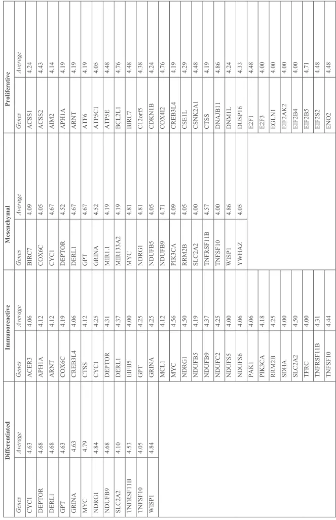
T ab le 3 .G en es c or re sp on di ng to c op y nu m be r av er ag e eq ua l o r la rg er th an 4 in th e gr ou ps o f ov ar ia n ca nc er c as es . D if fe re nt ia te d I m m un or ea ct iv e M es en ch ym al P ro li fe ra ti ve G en es A ve ra ge G en es A ve ra ge G en es A ve ra ge G en es A ve ra ge C Y C 1 4. 63 A C E R 3 4. 06 B IR C 7 4. 09 A C SS 1 4. 24 D E P T O R 4. 68 A P H 1A 4. 12 C O X 6C 4. 05 A C SS 2 4. 43 D E R L 1 4. 68 A R N T 4. 12 C Y C 1 4. 67 A IM 2 4. 14 G P T 4. 63 C O X 6C 4. 19 D E P T O R 4. 52 A P H 1A 4. 19 G R IN A 4. 63 C R E B 3L 4 4. 06 D E R L 1 4. 67 A R N T 4. 19 M Y C 4. 79 C T S S 4. 12 G P T 4. 67 A T F 6 4. 19 N D R G 1 4. 84 C Y C 1 4. 25 G R IN A 4. 52 A T P 5C 1 4. 05 N D U FB 9 4. 68 D E P T O R 4. 31 M IR 1. 1 4. 19 A T P 5E 4. 48 SL C 2A 2 4. 10 D E R L 1 4. 37 M IR 13 3A 2 4. 19 B C L 2L 1 4. 76 T N FR SF 11 B 4. 53 E IF B 5 4. 00 M Y C 4. 81 B IR C 7 4. 48 T N FS F 10 4. 05 G P T 4. 25 N D R G 1 4. 81 C 12 or f5 4. 38 W IS P 1 4. 84 G R IN A 4. 25 N D U FB 5 4. 05 C D K N 1B 4. 24 M C L 1 4. 12 N D U FB 9 4. 71 C O X 4I 2 4. 76 M Y C 4. 56 P IK 3C A 4. 09 C R E B 3L 4 4. 19 N D R G 1 4. 50 R R M 2B 4. 05 C SE 1L 4. 29 N D U FB 5 4. 19 SL C 2A 2 4. 00 C SN K 2A 1 4. 48 N D U FB 9 4. 37 T N FR SF 11 B 4. 57 C T S S 4. 19 N D U FC 2 4. 25 T N FS F 10 4. 00 D N A JB 11 4. 86 N D U FS 5 4. 00 W IS P 1 4. 86 D N M 1L 4. 24 N D U FS 6 4. 06 Y W H A Z 4. 05 D U SP 16 4. 33 P A K 1 4. 06 E 2F 1 4. 48 P IK 3C A 4. 18 E 2F 3 4. 00 R R M 2B 4. 25 E G L N 1 4. 00 SD H A 4. 00 E IF 2A K 2 4. 00 SL C 2A 2 4. 50 E IF 2B 4 4. 00 T F R C 4. 00 E IF 2B 5 4. 71 T N FR SF 11 B 4. 31 E IF 2S 2 4. 48 T N FS F 10 4. 44 E N O 2 4. 48
T R IT 1 4. 06 FA IM 4. 05 W IS P 1 4. 50 FA S L G 4. 05 Y W H A Z 4. 12 FB X O 45 4. 48 FH 4. 14 FL A D 1 4. 19 G A B A R A P L 1 4. 33 G A P D H 4. 43 G L U L 4. 09 G SS 4. 43 H 2A F J 4. 29 H A X 1 4. 19 H IS T 2H 2A C 4. 19 ID H 3B 4. 38 IN G 4 4. 43 IT C H 4. 48 IT P R 2 4. 48 K IA A 02 26 4. 47 8 L D H B 4. 19 L M N A 4. 24 M A P 1L C 3A 4. 48 M C L 1 4. 19 M IR 1. 1 4. 48 M IR 13 3A 2 4. 48 M IR 16 .2 4. 43 M IR 19 9A 2 4. 09 M IR 21 4 4. 09 M IR 23 A 4. 09 M Y C 4. 14 N C F2 4. 09 N D U FA 9 4. 38 N D U FB 5 4. 81 N D U FB 7 4. 33
N D U FB 9 4. 09 N D U FS 2 4. 19 N FE 2L 2 4. 00 N L R P 3 4. 33 N M E 7 4. 09 PF K FB 3 4. 09 P IK 3C A 4. 86 P K L R 4. 24 P L A 2G 4A 4. 00 PP O X 4. 19 P R K A B 2 4. 29 P R K A C A 4. 14 R N F1 68 4. 48 R Y R 2 4. 24 SD H C 4. 19 SH C 1 4. 19 SL C 2A 2 4. 86 SL C 2A 3 4. 38 SR X N 1 4. 43 T F B 2M 4. 29 T F R C 4. 48 T N FR SF 1A 4. 29 T N FS F 10 4. 86 T P I1 4. 43 T R IB 3 4. 48 T R P C 1 4. 14 T X N IP 4. 14 U Q C R FS 1 4. 00 Y W H A B 4. 24