Calcitic nanofibres in soils and caves: a putative fungal
contribution to carbonatogenesis
SASKIA BINDSCHEDLER
1, L. MILLIE
` RE
1, G. CAILLEAU
1, D. JOB
2&
E. P. VERRECCHIA
1*
1
Institut de Ge´ologie et de Pale´ontologie, Universite´ de Lausanne, Anthropole,
1015 Lausanne, Switzerland
2
Institut de Biologie, Universite´ de Neuchaˆtel, Rue Emile Argand 11,
2007 Neuchaˆtel, Switzerland
*Corresponding author (e-mail: eric.verrecchia@unil.ch)
Abstract: The origin of soil mineralized nanofibres remains controversial. It is attributed to either biogenic factors or physicochemical processes. Scanning electron microscope and transmission electron microscope observations show that nanofibres could originate from the breakdown of fungal hyphae, especially its cell wall. It is hypothesized that during the decay of organic matter, cell wall microfibrils are released in the soil where they are exposed to mineralizing pore fluids, leading to their calcitic pseudomorphosis and/or are used as a template for calcite pre-cipitation. When associated with needle fibre calcite bundles, nanofibres could indicate the relict of an organic sheath in which calcite has precipitated. This paper emphasizes the important roles of both organic matter and fungi in carbonatogenesis, and consequently in the soil carbon cycle.
Natural nanofibres have been observed in various environments such as subtropical and temperate soils (Verrecchia & Verrecchia 1994; Cailleau et al. 2005) and cave deposits such as moonmilk (Borsato et al. 2000; Can˜averas et al. 2006). They are often associated with Needle Fibre Calcite (NFC; Verrecchia & Verrecchia 1994; Borsato et al. 2000). The aim of this study is to provide new insight into the processes at the origin of nanofibres. In order to differentiate between organic and mineral nanofibres, the term ‘organic nanofibre’ will be used for nanofibres whose organic nature has been determined by analytical methods. The term ‘mineral nanofibre’ will be used for: (i) for nanofibres observed in scanning electron micro-scopy (SEM), in absence of specific labelling (see ‘Materials and methods’ section); and (ii) for nano-fibres diffracting the electron beam under trans-mission electron microscopy (TEM). The term nanofibre alone refers only to a shape or an object and therefore used for morphological descriptions.
Previous work on mineral nanofibres
Since 1980, many authors have reported mineral nanofibres from various environments (Table 1). The four following authors have specifically observed organized structures related to mineral nanofibres: (i) filamentous, ramified, microscopic
structure composed by a dense nanofibre scaffolding (Borsato et al. 2000; Richter et al. 2008); (ii) a straight macro-structural alignment (3 – 5 mm wide and .70 mm long) of unordered nanofibres obser-ved close to an organic filament (possibly actino-mycetes, cyanobacteria, or fungi; Benzerara et al. 2003); and (iii) 3 mm wide and .50 mm long fila-ments interpreted as ‘calcified filafila-ments with needles in grain-coating needle mat’ have also been observed by Jones & Ng (1988).
Filamentous organisms and structures
in soils and caves
Filamentous organisms living within the soil or in caves must be heterotrophic organisms. Algae and cyanobacteria are photosynthetic organisms and thus are present only at the soil surface or in rock fractures near a light source. Indeed, in mineral sub-strates that are far away from any light, these organ-isms are absent due to the lack of their energy source. Accordingly, filamentous fabrics present in these environments could be fungi, filamentous bacteria (in soils and caves mostly actinomycetes), and roots (Paul & Clark 1996; Gobat et al. 2003). Taking into account their sizes and morphologies summarized in Table 2, fungi are the most suitable organisms associated with nanofibres and NFC.
Fungal presence and activity
in soil and caves
Fungi are present in large amounts in soils. As an example, one metre square of fertile soil can contain a 10 000-km long fungal network (Gobat et al. 2003). About 80% of land plant species are colonized by arbuscular mycorrhizal fungi (endo-mycorrhiza), and around 3% of phanerogam species are colonized by ectomycorrhizal fungi (EcM), especially plants with a large distribution at a global scale (Pinaceae, Fagaceae). In soils, a vertical distribution can be distinguished regarding fungal type in terms of their ecology. Organic layers are mostly colonized by saprophytic fungi, whereas mineral layers are colonized by EcM fungi (van Scho¨ll et al. 2008). The latter has been demonstrated as being a significant agent of mineral weathering of ecosystem-wide importance (van Scho¨ll et al. 2008).
The mycelial network is able to efficiently trans-locate nutrients in solution from one place to another (Gobat et al. 2003). Basidiomycetes, and among them EcM fungi, are able to build structures
named fungal strands that can extend meters away from the roots (Finlay & So¨derstro¨m 1992; van Scho¨ll et al. 2008). Thus, the presence of mycor-rhized roots, fungal hyphae and strands in deep mineral layers or in caves is not surprising. Canadell et al. (1996) showed an average rooting depth of 4.6 m þ/20.5 m, with a maximum depth of 7.0 m þ/21.2 m for trees. In their review, only the root itself is taken into account. Considering the mycor-rhiza, it can considerably extend the root network (Timonen & Marschner 2006). Observations of roots at depths below 2 – 3 m in caves have also been observed (Canadell et al. 1996). Jasinska et al. (1996) demonstrated that root mats could be the sole source of food for faunal communities in an Australian cave.
Cave geomicrobiology
Caves are nutrient-limited environments due to the absence of light that prevents primary production through photosynthesis, contrary to other common environments on Earth. Thus, in terms of presence of life, this kind of environment can be considered
Table 1. Review of nanofibres in the literature
Authors Nomenclature Geological setting Interpretation/context
Pouget & Rambaud (1980)
Calcite ‘en baˆtonnets’
Soil with calcareous crust Mesh of monocrystaline calcite crystals Verge`s et al. (1982) Small needle-shaped
crystals
Calcareous soils Tangled crystals
Ducloux et al. (1984) Calcite ‘en baˆtonnets’
Developed on screeslope Covering larger needle-fibre calcite crystals
Phillips & Self (1987) Micro-rods Pedogenic calcrete Interpreted as calcified
rod-shaped bacteria Phillips et al. (1987) Submicron size
rods
Pedogenic calcrete Interpreted as calcified
rod-shaped bacteria
Jones & Ng (1988) Needles Rhizolith from the Pleistocene
Ironshore Formation
Calcified filaments coated withneedles (i.e. nanofibres) Verrecchia &
Verrecchia (1994)
Micro-rods Quaternary calcretes, Israel Disordered mesh
Loisy et al. (1999) Micro-rods Carbonate paleosol in scree
deposits
Mineralized threadlike and bacilliform bacteria
Borsato et al. (2000) Nanofibres Moonmilk (cave deposits) Probably abiogenic
precipitation Benzerara et al.
(2003)
Nanobacteria-like rods
At the surface of the Tataouine meteorite
Straight micro-alignment of nanofibres; possible organic origin
Cailleau et al. (2005) Micro-rods Orthox soils Observed on burnt oxalate
crystals embedded in tree tissues
Jeong & Chun (2006) Nanofibre calcite Aerosols coming from loess plateau and desert
–
Richter et al. (2008) Nanofibres Moonmilk (cave deposits) –
as ‘extreme’. On the other hand, physicochemical parameters tend to be buffered and constant throughout the year (e.g. mild temperature normally equals MAST (Mean Annual Surface Temperature) and fairly high humidity). These extreme but con-stant conditions allow the presence of underground ecosystems, which may or may not be connected to aboveground energy-sources (Jasinska et al. 1996; Sarbu et al. 1996). Prokaryotes and fungi are the most common organisms that can be encoun-tered in caves, and their link in speleothem for-mation is often proposed and debated. Moonmilk is a common speleothem mineral, and its biological or physico-chemical origin has long been discussed. Today most of the theories involve microbial mediation in its formation, but the exact role that microorganisms play, whether it is bacterial or fungal, is still being discussed (Gradzinski et al. 1997; Northup & Lavoie 2001; Can˜averas et al. 2006; Barton & Northup 2007). In caves, moonmilk is more likely to be present in the vicinity of soils (Gradzinski, pers. comm.). This increases the prob-ability of cave access for roots and their fungal associates, and consequently their involvement in the genesis of moonmilk.
Fungal strands and hyphae ultrastructure
Fungal hyphae size
Single fungal hyphae diameter is highly variable, depending on the taxonomic position, environ-mental conditions, age, and function of hyphae. Nevertheless, for functional hyphae, an average diameter of 3 – 6 mm is found in the literature (Dix & Webster 1995; Carlile et al. 2001). Non-functional hyphae, such as those from the cortex of fungal strands, can have a diameter of 1 mm, with an inner diameter of ,0.5 mm.
Fungal strands
Basically, a fungal strand is a bundle of juxtaposed linear hyphae. They are organs produced by fungi to explore their environment and to translocate nutri-ents from one place to another. They have the ability to extend over long distances, that is up to 30 m. In addition, macromorphologically, they exhibit a variable diameter, depending mostly on their age and remoteness from nutrient sources. This diameter ranges from a few mm up to 4 mm
Table 2. Characteristics of filamentous organisms in soils and caves
Diameters Morphologies/
structures
Cell wall morphology
Cell wall biochemistry
Fungi 3 – 6 mm on average 1 mm (min) – 30 mm (max) Strands: 0.02 2. 1 mm Hyphae with or without septum, more or less ramified. Bundles of differentiated hyphae forming linear organs, fungal strands Thick-walled (up to 1 mm) and thin-walled (100 – 200 nm) hyphae
Two layers: a fibrillar component with chitin and b-glucans and an amorphous component with glycoproteins specific to taxonomic groups Actinomycetes 0.5 – 1 mm in average, max 2 mm in some genus Ramified mycelium, sometimes fragmented Wall 20 – 80 nm thick
Gram positives bacteria with one
homogenous layer of peptidoglycan (murein). Four types of peptidoglycans depending on genus Fine roots ,2 – 0.2 mm Single conducting vessel 10 – 30 mm Ramified structure composed, at a micromorphological level, of complex arrangements of linear vessels Highly variable in thickness, from 0.1 mm in young cells to 100 mm in mature cells
Primary wall: network of fibrous cellulose and hemicellulose embedded in a matrix of pectin.
Secondary wall: only in mature cells, can contain lignin Note: Review of the different filamentous organisms in soils synthesising characteristics such as filament diameter and morphology and their cell wall morphology and biochemistry. References from Jones 1970; Dix & Webster 1995; Paul & Clark 1996; Bouma et al. 2001; Carlile et al. 2001; Pregitzer et al. 2002; Prescott et al. 2003; Coleman et al. 2004; Pessoni et al. 2005; Hishi 2007.
in some wood decaying species (Thompson 1984). The structure of the fungal strand is composed of: (i) an outer layer (the cortex) composed of a thick layer of narrow thick-walled multiseptate dead hyphae (average diameter of 1 mm); and (ii) an inner layer (the medulla) composed of a few linear wide thin-walled sparsely septate liv-ing hyphae (average diameter of 6 – 10 mm). The latter seems to be less resistant to hydrostatic pressure (Watkinson 1979) and thus is more rapidly exposed to decay processes than hyphae with a thick melanized wall in the outer layer. The inner part often collapses, leaving fungal strands composed of a thick layer of narrow hyphae with empty wide channels in the middle (Watkinson 1984). The cortex of the fungal strands makes it an impermeable organ where fluids can be bidirec-tionally translocated (Watkinson 1979; Dix & Webster 1995).
Composition and structure of the
fungal cell wall
The thickness of the cell wall also shows great varia-bility depending on physiological processes and the function of hyphae. Single hyphae have an average cell wall thickness of 150 nm (Jones 1970; Beckett
et al. 1974; Farkas 1979; Ruiz-Herrera et al. 1996; Pessoni et al. 2005). On the other hand, hyphae from the cortex of a fungal strand can exhibit very thick cell walls, up to 500 nm. The wall thickening can even occlude the hypha lumen (Watkinson 1984). Moreover, the walls of these hyphae often present a high degree of melanization, which is also a factor in impermeabilization (Paul & Clark 1996).
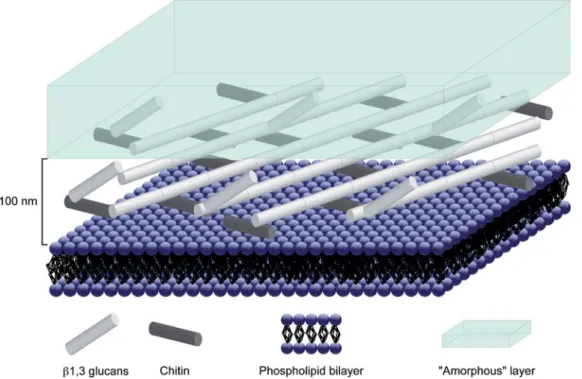
The fungal cell wall can be described by two main types of materials, an outer layer composed of amorphous material (mainly mannoproteins), and an inner layer composed of fibrous material, chitin and b-glucans (Burnett 1979; Ruiz-Herrera 1992; Bowman & Free 2006). Chitin is a polymer of a polysaccharide, N-acetyl-glucosamine. It is present in the form of long microfibrils, sometimes over 1 mm, with a diameter of 10 – 25 nm. It is located in the innermost part of the wall, arranged as an intertwined mesh embedded in an amorphous matrix (Aronson & Preston 1960; Carlile et al. 2001). b-glucans are homopolysaccharides of glu-cose. In the fungal wall, it is present either as b (1-3) glucan or in a lesser amount as b (1-6) glucan. They are found in greater amounts than chitin (Carlile et al. 2001; Farkas 2003). Figure 1 shows a sketch of the fungal cell wall.
Fig. 1. Sketch of the fungal cell wall (modified from Latge´ 2003). Note the fibrous layer composed by chitin and b-glucan fibres and the amorphous layer. In order to give an orientation to this sketch, the plasma membrane of the fungal cell has been represented by the phospholipids bilayer.
Cell wall material can be a significant part of the resistant organic matter in soils. Depending on physico-chemical parameters, enzymatic degra-dation of polymerous substances from the cell wall may or may not be possible (Paul & Clark 1996; Coleman et al. 2004).
Materials and methods
Secondary carbonate accumulations have been sampled at four sites: (i) in the mineral layers (calcic horizon of a calcisol; IUSS Working Group WRB 2006) at a quarry near Villiers (Swiss Jura Mountains, 478040N, 68590E), as well as (ii) at an
outcrop near Aı¨nsa (Spain, 428210N; 08040E); (iii)
in the narrow entrance of a cave near les Cornettes de Bises (Swiss Alps, 468190N, 168480E); and
(iv) in the vers chez le Brandt cave, inside a wide chamber at 100 metres from the entrance (Swiss Jura Mountains, 468560N, 68280E). At the first two
sites (soils developed on scree slopes), samples exhibit two different morphologies directly visible in the outcroup: (i) cotton-ball-like NFC that accumulates in the soil pores; and (ii) coatings on grains and centimetric to decimetric cryoclasts. When wet, these coatings constitute a plastic paste, which becomes pulverulent when dry. At the third and fourth sites (caves), only moonmilk deposits were sampled in the form of a wet plastic wall coating, up to 30 cm thick. In addition to crys-tals, fungal strands associated with different macro-scopic morphologies of NFC have been sampled for electron microscope observations. Strands have been taken from cotton-ball-like NFC, associated with rock fragment coatings, or free in the soil pores. All samples were collected using polypropy-lene tweezers and stored in sterilized 50 mL tubes at low temperature.
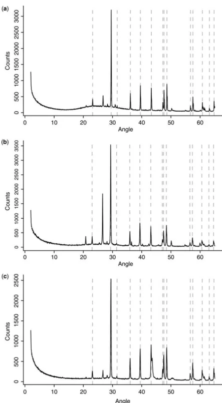
Bulk samples were analysed by X-ray diffrac-tion (XRD) using a Scintag diffractometer in order to determine their mineralogical nature. In each sample, quartz powder was added in order to nor-malize and compare samples with each other.
In order to detect organic from mineral material, prior to observations, each macromorphological sample was stained using a 4% osmium aqueous solution from a modified Pearson et al. (2004) protocol. However, using this labelling method, the presence of the organic matter can be deter-mined but not its nature. Samples were gold-coated (10 nm) and observed using a Phillips ESEM-FEG XL30 Field Emission Gun Scanning Electron Microscope (FEG-SEM). Osmium staining was detected with an EDAX Energy Dispersive Spectrometer (EDS) coupled to the electron micro-scope. With natural non-flat samples and absence of a standard, EDS spectra only give qualitative information.
In order to check possible artifacts due to high-vacuum, some representative samples were obser-ved using XL30 SEM in its LTSEM cryo mode (Low-temperature SEM). Some fungal strands and coatings were embedded in an epoxy resin, and ultrathin sections were performed using a Reichert Ultracut S (Leica) microtome with a diamond knife. Ultrathin sections (200 – 220 nm thick) were observed using a Phillips CM-200 Transmission Electron Microscope (TEM) with a voltage of 200 kV. Crystal properties were determined using microdiffraction.
Results
XRD analysis of the three types of samples (cotton-ball-like structures, coatings on grains, and moonmilk) shows that their mineralogy is calcitic in nature (Fig. 2a – c). Moreover, the absence of shift (expected in presence of Mg in the crystal lattice) after normalization with quartz powder characterizes the presence of low magnesium calcite (LMC) at these sites.
SEM observations of soil samples show recur-sive associations between NFC, unidentified nanofi-bres, and fungal strands. NFC is characterized by a width between 0.5 – 2.0 mm and a length ,100 mm. Some epitactic growths are present but no important development is observed (no big euhedral crystals due to epitactic cementation). The NFC is present either as random meshes, or as bundles, 3 – 30 mm in diameter (average 10 mm; Fig. 3a). This microscopic feature constitutes the macroscopic cotton-ball-like structures. Bundles contain some nanofibres, occasionally associated with amorphous matter assumed to be an organic veil (Fig. 3b). These nanofibres are rarely observed on NFC when the latter are randomly oriented and/or strongly modified by epitactic growth. At a microscopic scale, coatings from soil grains exhi-bit various amounts of NFC and nanofibres, in which needles are less represented than in the cotton-ball-like morphology. NFC often shows ran-dom orientations, whereas nanofibres are packed in clusters. This morphology shows great similarity with microstructures of moonmilk samples, in which NFC is even less represented.
SEM measurements show that nanofibres are up to 6 mm long (the shortest is 0.2 mm long) with an average width of 78.6 nm (standard deviation of 22.5 nm, based on 106 measurements; Fig. 3c). They are characterized by a high flexibility (as mentioned by Borsato et al. 2000), leading to spec-tacular curvature (Fig. 3c). They appear smooth under TEM. Two kinds of structures are observed: (i) a randomly-oriented framework of nanofibres, in which widespread putative organic veils and cal-citic micro-aggregates are present; these meshes are
Fig. 2. X-ray diffractogramme showing the low-magnesium calcite nature of (a) cotton-ball like NFC from the Swiss Jura Mountains, (b) coatings on blocks from Swiss Jura Mountains and (c) moonmilk from the Vers chez le Brandt cave in the Swiss Jura Mountains samples. Dotted lines correspond to the main peaks of low-magnesium calcite (CaCO3), the other peaks mainly correspond to quartz (quartz added prior to analysis to allow peak correction).
often associated with other components (i.e. NFC, fungal strands, hyphae, etc.) as observed in moon-milk deposits (Fig. 3d); and (ii) organized structures of nanofibres (Fig. 4), either as small pieces that have apparently undergone a breakdown, or as a tubular/circular microscopic network (Fig. 4a, c). The term organized refers to a non-random distri-bution of nanofibres, whatever their nature. These networks are composed by intertwined nanofibres oriented in two main directions (Fig. 4b, d).
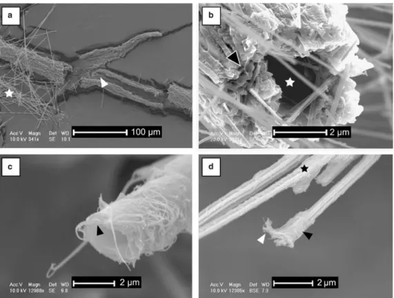
Another main component is frequently observed associated with soil samples: macroscopic, brown organic filaments, identified as fungal strands. Their average diameter can reach 100 mm and they are composed of the two typical mycelial strand structures, an external part made of several narrow fungal hyphae with a thick cell wall and an inner part characterized by a few wide thin-walled hyphae, which often lack in our obser-vations due to their ability to be rapidly decayed
(Fig. 5a, b). Nanofibres are abundant all along the macroscopic filament where fungal strands seem to break down.
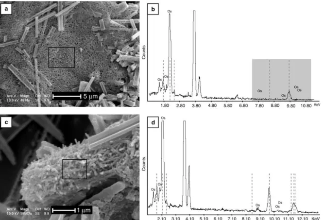
A cross-section of a fungal hypha shows that the fungal wall is composed of two layers, an inner part composed of fibrous material and an external part composed of an amorphous material (Fig. 5c, d). From these observations, it is obvious that there is an intimate relationship between the hyphae and the nanofibres (Fig. 5c, d). Optical observations, hydrochloric acid tests on moonmilk, as well as TEM microdiffractions (Borsato et al. 2000) indi-cate that the nanofibres are mineral in nature. In order to test this hypothesis, in-situ analyses were performed to distinguish organic from mineral matter using osmium labelling with EDS control on samples. The osmium stains only organic matter and not mineral material (Pearson et al. 2004). Osmium peaks indicate that non-organized frameworks composed of only nanofibres do not
Fig. 3. (a – d) SEM photomicrographs. (a) Bundle of NFC present in a sample from the Spanish site of cotton-ball-like NFC associated with a fungal strand (observed macroscopically). (b) Bundle of NFC covered by putative organic veils. Some organic nanofibres are also present. Sample from a grain coating associated with a fungal strand and some cotton-ball like NFC. (c) Close-up of an organized mesh composed by nanofibres (moonmilk, Swiss Alps). Preferential orientations of nanofibres are shown by the crossed double-headed arrows. Some nanofibres are curved (arrows). This characteristic indicates a contact-deformation. (d) Mesh composed by randomly-oriented nanofibres associated with NFC (white arrows) and putative organic veils (black arrows). Swiss Jura Mountains, coatings on block.
contain organic material (Fig. 6a, b) and organized meshes do contain organic matter (Fig. 6c, d).
Discussion
The presence of nanofibres in various vadose environments has been widely observed. Their origin is either attributed to a biogenic factor (‘probably rod-shaped calcified bacteria’ Phillips & Self 1987; Ould Mohamed & Bruand 1994; ‘microrod attributed to bacteria or nuclei in gel’ Verrecchia & Verrecchia 1994; Loisy et al. 1999) or physicochemical processes [‘precipitation from pore filling fluids’, sometimes assoicated with
organic filaments, that is, nanofibres ‘cannot be fully attributed to direct organic activity’ (Jones & Ng 1988); ‘nanofibres show microstructures that are typical of inorganic, crystalline material’ (Borsato et al. 2000)]. Therefore, the origin of terrestrial accumulations of nanofibres remains controversial.
The nanofibres discussed in this paper are similar to those described in Phillips & Self (1987), based on their size and flexibility. It is important to note that they identified the organic filaments associated with nanofibres as fungi, and the same conclusion is drawn in this study, based on their size and mor-phology, as well as presence of macrostructures
Fig. 4. Samples from the Swiss Jura Mountains site. (a) SEM photograph showing a tubular-like structure resembling an organic filament (e.g. a fungal hypha) and composed by nanofibres (cotton-ball-like NFC associated with decaying organic matter). (b – d) Sample from coatings on a cryoclast associated with decaying fungal strands. (b) SEM photograph showing a network of intertwined nanofibres oriented in two main directions (white rectangle). White star indicates a NFC crystal. (c) TEM photomicrograph showing a section of a circular structure interpreted as a possible organic filament (e.g. a fungal hypha) and composed by nanofibres. (d) TEM photomicrograph showing a network of intertwined nanofibres oriented in two main directions. Also note the presence of a circular section similar to the one shown in Figure 2c (stars).
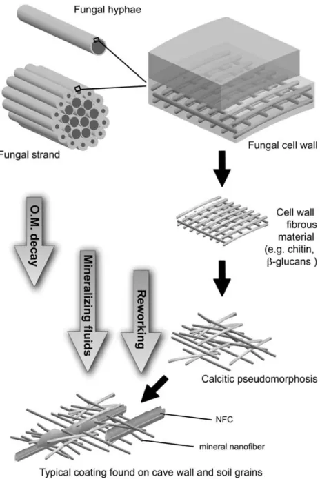
such as mycelial strands (Fig. 5a, b). Two layers are visible during the breakdown of the hyphal cell wall (Fig. 5d). It is known that the inner wall layer is composed of a hard microfibril framework (theoretically 10 – 25 nm in diameter, Carlile et al. 2001) made of chitin and b (1-3), b (1-6) glucan. Based on this fact and the recognition of the organic nature of some nanofibres (Fig. 6c, d), the organized meshes of nanofibres are considered as the result of a slightly destructive decay of the fungal fibrous cell wall material. At this stage, the organized meshes are interpreted as the first step in the breakdown of the fungal hyphae cell wall, whereas the non-organized mats represent the ulti-mate state of decay and reworking of this organic matter. In other words, the nanofibres could be inter-preted as organic in origin and being the result of an incomplete decaying of fungal matter. During early
diagenesis, their calcitic pseudomorphosis (Cailleau 2005; Cailleau et al. 2005) and/or their role as a template for calcitic precipitation results from the release of the nanofibres in the soil environment, followed by their exposure to mineralizing pore filling fluids. This could explain why non-organized meshes (interpreted as the oldest decay product) are often composed of nanofibres of calcite, due to the longer exposure time to soil fluids. To con-clude, the release of nanofibres may represent a partly destructive decay of the fibrous cell wall material.
Moreover, this interpretation has an important implication for NFC origin. The observation of NFC inside organic sleeves and the presence of small mats of nanofibres on bundles of NFC (Fig. 5d (star); Cailleau et al. 2009) suggest a large contribution of organic matter for their genesis.
Fig. 5. SEM photomicrographs. (a, b) Samples from Aı¨nsa (Spain). (a) Fungal strand associated with cotton-ball-like NFC (star). Note the absence of the inner hyphae (arrow), which seem to have already undergone breakdown. (b) Close-up of another fungal strand showing the narrow and thick-walled hyphae from the outer part (black arrow). Note that the internal wide thin-walled hyphae are absent (white star). (c, d) Swiss Jura Mountains samples. (c) Close-up of a decaying hypha from a grain coating sample showing the release of cell wall fibrous material (e.g. chitin, b-glucan) from its cell wall (arrow). (d) Remains of a putative fungal hyphae on needle fibre calcite. Two textures can be distinguished, an outer one that appears to be smooth (black arrow) and an inner one that appears to be composed of nanofibres (white arrow). Note also the presence of a mesh of nanofibres along the other calcite needles in this picture (star). Sample from cotton-ball like NFC associated with decaying organic matter.
Fig. 6. (a) and (c) SEM view of samples. The black window shows the area analysed. (b) and (d): EDS spectra, dotted lines correspond to Au peaks (samples coated with gold for SEM observations) and Os is the osmium labelling. (a, b) Analysis performed on a dense unorganized mesh of nanofibres (sample from coatings on a block). Note the absence of an osmium peak on the spectrum in the grey box due to the purely mineral nature of nanofibres. (c, d) Analysis performed on a dense non-random mesh of nanofibres (sample from a coating on a block associated with a decaying fungal strand). Note the presence of osmium peaks on the spectrum due to the organic nature of nanofibres.
Fig. 7. Hypothetical sketch recapitulating the potential processes of fungal organic matter decay and mineralization of cell wall fibrous material. This model is based on observations, analysis, and interpretations given in this paper. The first stage starts with a fungal hyphae or a group of hyphae forming a fungal strand. The cell wall is constituted by two main layers, an amorphous one (on the top) and a fibrous one (at the bottom; for more details see Fig. 1). It is assumed that the cell wall fibrous material is not decayed at the same rate than the amorphous material. When released in the soil microenvironment, these nanofibres could act as template for calcite nucleation, eventually leading to calcitic pseudomorphosis. The ultimate step is represented by possible reworking due to various processes (bioturbation, water movements, cryoturbation, etc.). Nanofibres are often associated with other calcitic features, such as NFC, leading to the complex microfabric observed in soil and cave deposits. O.M., organic matter.
As first noted by Phillips & Self (1987), NFC bundles could be the first step in the distribution of NFC in soil pores. Indeed, with time, the collapse of bundles due to various processes, such as weath-ering and bioturbation, would lead to a random distribution of NFC (mesh). Nanofibres on bundles would then be a relict of the organic sheath (assumed to be fungal in origin). The pres-ence of mycelial strands is critical to understand the origin of the bundles. Strands and bundles are both organized as a tubular structure (Figs 3a & 5b) composed of sub-parallel components. They have similar diameters: 2 – 30 mm on average for the bundle and 8 – 80 mm for the whole fungal strand. But the outer layer of the mycelial strand is usually wide and often represents between a third to a half of the strand section. Thus, only the inner diameter should be considered in this case. The outer layer is composed of hyphae with thick cell walls. Their central hole is probably too small to contain any NFC, whereas the inner part of the strand contains wider hyphae that would have enough room to allow the formation of a crystal such as a needle.
One of the most important elements for fungal growth is calcium. Indeed, it is implicated in the apical growth control. Nevertheless, Ca2þ is con-sidered as toxic when present in high concentrations (Gadd 1993). Consequently, its concentration within the fungal cell, and especially in the apex, must be under strict control of the organism in order to allow proper growth (Jackson & Heath 1993). Under hydrous stress conditions, the con-centration of calcium could reach a high level, close to saturation. As it has been suggested for metal-oxalate (Whitney 1989; Gadd 1999), fungi could induce the precipitation of carbonate, possibly leading to a decrease of their internal calcium content (Gadd 2007). This process is documented for bacteria (Simkiss 1986; Schultze-Lam et al. 1996; Barton & Northup 2007). The inner layer of the fungal cell wall is composed by a large amount of chitin known to be a good template for calcite precipitation (Manoli et al. 1997). Conse-quently, nucleation of calcite crystals inside the inner functional hyphae from mycelial strands constitutes a serious hypothesis. The role of fungal hyphae as a crystal nucleation enhancer has already been suggested in the past (Went 1969; Northup & Lavoie 2001; Gadd 2007). Any other cell wall fibrous material or polymeric substance, for example b (1-3) glucan or a glycoprotein, may have the same effect (Burazerovic et al. 2007; Shen et al. 2007). To conclude, all our observations are recapitulated in a step-by-step hypothetical model (Fig. 7), showing the potential relationships between fungal organic matter and calcium carbon-ate precipitation.
Conclusion
Considering previous hypotheses on the origin of nanofibres (i.e. biogenic or purely physico-chemical), the results presented here indicate that nanofibres could also originate from the breakdown of fungal hyphae, especially their cell walls. During the decay of organic matter, microfibrils such as chitin or b (1-3) glucan, are released from the inner layer of the fungal cell wall. When these organic nanofibres are exposed to mineralizing pore fluids, they could undergo calcitic pseudomor-phosis and/or be used as templates for calcitic pre-cipitation. In the case of NFC bundles, which have an intimate relationship with nanofibres, these nano-features could indicate the relict of an organic sheath. As interpreted by Phillips & Self (1987), the implication of fungal strands in the genesis of NFC is now better supported. In other words: bundles could be the ultimate remains of the pres-ence of a fungal strand. This hypothesis emphasizes the important role of organic matter in carbonato-genesis as well as the fundamental role of fungi in the terrestrial carbon cycle.
The authors would like to thank Andre´ Villard and Miche`le Vlimant for their technical assistance for sample prep-aration, especially for TEM purposes; Dr Massoud Dadras, Dr Vladislav Spassov, and Mireille Leboeuf from CSEM for their assistance in using electron microscopy, and Dr. Thierry Adatte from UNIL for X-ray diffraction analysis. This work is supported by the Swiss National Science Foundation, Grant No. FN 205320-109497/1 and FN 205320-122171.
References
ARONSON, J. M. & PRESTON, R. D. 1960. An electron microscopic and X-ray analysis of the walls of selected lower phycomycetes. Proceedings of the Royal Society of London. Series B, Biological Sciences, 152(948), 346 – 352.
BARTON, H. A. & NORTHUP, D. E. 2007. Geomicro-biology in cave environments: past, current and future perspectives. Journal of Cave and Karst Studies, 69(1), 163 – 178.
BECKETT, A., HEATH, I. B. & MCLAUGHLIN, D. J. 1974. An atlas of fungal ultrastructure. Longman Group Limited, London.
BENZERARA, K., MENGUY, N., GUYOT, F., DOMINICI, D. & GILLET, P. 2003. Nannobacteria-like calcites at surface of the Tatahouine meteorite. Proceedings of the National Academy of Sciences of the United States of America, 100, 7438 – 7442.
BORSATO, A., FRISIA, S., JONES, B. & VAN DERBORG, K. 2000. Calcite moonmilk: crystal morphology and environment of formation in caves in the Italian Alps. Journal of Sedimentary Research, 70, 1179 – 1190.
BOUMA, T. J., NIELSEN, K. L., VANHAL, J. & K OUT-STAAL, B. 2001. Root system topology and diameter
distribution of species from habitats differing in inun-dation frequency. Functional Ecology, 15, 360 – 369. BOWMAN, S. M. & FREE, S. J. 2006. The structure and
synthesis of the fungal cell wall. BioEssays, 28, 799 – 808.
BURAZEROVIC, S., GRADINARU, J., PIERRON, J. & WARD, R. 2007. Hierarchical self-assembly of one-dimensional streptavidin bundles as a collagen mimetic for the biomineralization of calcite. Ange-wandte Chemie, 46, 1 – 5.
BURNETT, J. H. 1979. Aspects of the structure and growth of hyphal walls. In: BURNETT, J. H. & TRINCI, A. P. J. (eds) Fungal Walls and Hyphal Growth. British Myco-logical Society Symposium. Cambridge University Press, Cambridge, 1 – 25.
CAILLEAU, G. 2005. Carbon cycle and carbonate biomi-neralization in terrestrial environments: diagenesis of oxalate – carbonate phases. PhD thesis, Neuchaˆtel University, Switzerland.
CAILLEAU, G., BRAISSANT, O., DUPRAZ, C., ARAGNO, M. & VERRECCHIA, E. P. 2005. Biologically induced accumulations of CaCO3 in orthox soils of
Biga, Ivory Coast. Catena, 59, 1 – 17.
CAILLEAU, G., VERRECCHIA, E. P., BRAISSANT, O. & EMMANUEL, L. 2009. The biogenic origin of needle fibre calcite. Sedimentology, 56, 1858 – 1875. CANADELL, J., JACKSON, R. B., EHLERINGER, J. R.,
MOONEY, H. A., SALA, O. E. & SCHULZE, E. D. 1996. Maximum rooting depth of vegetation types at the global scale. Oecologia, 108(4), 583 – 595. CAN˜ AVERAS, J. C., CUEVZA, S., SANCHEZ-MORAL, S.,
LARIO, J., LAIZ, L., GONZALEZ, J. M. & SAIZ -JIMENEZ, C. 2006. On the origin of fibre calcite crys-tals in moonmilk deposits. Naturwissenschaften, 93, 27 – 32.
CARLILE, M. J., WATKINSON, S. C. & GOODAY, G. W. 2001. The Fungi, 2nd edn. Elsevier Academic Press, Amsterdam.
COLEMAN, D. C., CROSSLEY, D. A., JR. & HENDRIX, P. F. 2004. Fundamentals of Soil Ecology. Elsevier Academic Press, Amsterdam.
DIX, N. J. & WEBSTER, J. 1995. Fungal Ecology. Chapman & Hall, London.
DUCLOUX, J., BUTEL, P. & DUPUIS, T. 1984. Micro-se´quence mine´ralogique des carbonates de calcium dans une accumulation carbonate´e sous galets calcaires, dans l’ouest de la France. Pedologie, 34, 161 – 177. FARKAS, V. 1979. Biosynthesis of cell-walls of fungi.
Microbiological Reviews, 43(2), 117 – 144.
FARKAS, V. 2003. Structure and biosynthesis of fungal cell walls: methodological approaches. Folia Microbiologica, 48(4), 469 – 478.
FINLAY, R. D. & SO¨ DERSTRO¨M, B. 1992. Mycorrhiza and carbon flow to the soil. In: ALLEN, M. (ed.) Mycorrhi-zal Functioning: an Integrative Plant – Fungal Process. Chapman and Hall, London, 134 – 160. GADD, G. M. 1993. Interactions of fungi with toxic
metals. New Phytologist, 124, 25 – 60.
GADD, G. M. 1999. Fungal production of citric and oxalic acid: importance in metal speciation, physiology and biogeochemical processes. Advances in Microbial Physiology, 41, 47 – 92.
GADD, G. M. 2007. Geomycology: biogeochemical transformations of rocks, minerals, metals and
radionuclides by fungi, bioweathering and bioremedia-tion. Mycological Research, 111, 3 – 49.
GOBAT, J.-M., ARAGNO, M. & MATTHEY, W. 2003. Le Sol Vivant. 2nd edn. Presses polytechniques et uni-versitaires romandes, Lausanne.
GRADZINSKI, M., SZULC, J. & SMYK, B. 1997. Microbial agents of moonmilk calcification. Proceedings of the 12th International Congress of Speleology, 1, 275 – 278.
HISHI, T. 2007. Heterogeneity of individual roots within the fine root architecture: causal links between physio-logical and ecosystem functions. Journal of Forest Research, 12, 126 – 133.
IUSS WORKING GROUP WORLD REFERENCE BASE. 2006. World Reference Base for Resources 2006. 2nd edn. World Soil Resources Reports No. 103. FAO, Rome.
JACKSON, S. L. & HEATH, I. B. 1993. Roles of calcium ions in hyphal tip growth. Microbiological Reviews, 57(2), 367 – 382.
JASINSKA, E. J., KNOTT, B. & MCCOMB, A. J. 1996. Root mats in ground water: a fauna-rich cave habitat. Journal of the North American Benthological Society, 15(4), 508 – 519.
JEONG, G. Y. & CHUN, Y. 2006. Nanofibre calcite in Asian dust and its atmospheric roles. Geophysical Research Letters, 33, L24802, doi: 10.1029/ 2006GL028280.
JONES, B. & NG, K.-C. 1988. The structure and diagenesis of rhizoliths from Cayman Brac, British West Indies. Journal of Sedimentary Petrology, 58, 457 – 467. JONES, D. 1970. Ultrastructure and composition of cell
walls of Sclerotinia – Sclerotiorum. Transactions of the British Mycological Society, 54, 351 – 360. LATGE´, J.-P. 2003. Activity Reports, Institute Pasteur,
Paris. World Wide Web Address: http://www. pasteur.fr/recherche/RAR/RAR2003/print/Aspergil-en.html.
LOISY, C., VERRECCHIA, E. P. & DUFOUR, P. 1999. Microbial origin for pedogenic micrite associated with a carbonate paleosol (Champagne, France). Sedi-mentary Geology, 126, 193 – 204.
MANOLI, F., KOUTSOPOULOS, S. & DALAS, E. 1997. Crystallization of calcite on chitin. Journal of Crystal Growth, 182, 116 – 124.
NORTHUP, D. E. & LAVOIE, K. H. 2001. Geomicrobio-logy of caves: a review. GeomicrobioGeomicrobio-logy Journal, 18, 199 – 222.
OULDMOHAMED, S. & BRUAND, A. 1994. Morphology and origin of secondary calcite in soils from Beauce, France. In: RINGROSE-VOASE, A. J. & HUMPHREYS, G. S. (eds) Soil Micromorphology: Studies in Manage-ment and Genesis. Proceedings of the IX International working Meeting on Soil Micromorphology, Towns-ville, Australia, July 1992. Developments in Soil Science, Elsevier, Amsterdam, 22, 27 – 36.
PAUL, E. A. & CLARK, F. E. 1996. Soil Microbiology and Biochemistry. 2nd edn. Academic Press, San Diego. PEARSON, V. K., KEARSLEY, A. T. & SEPHTON, M. A.
2004. The in-situ detection of organic material in extraterrestrial samples. Microscopy and Analysis, 18, 5 – 8.
PESSONI, R. A. B., FRESHOUR, G., FIGUEIREDO -RIBEIRO, R. L., HAHN, M. G. & BRAGE, M. R.
2005. Cell-wall structure and composition of Penicil-lium janczewskii as affected by inulin. Mycologia, 97(2), 304 – 311.
PHILLIPS, S. E. & SELF, P. G. 1987. Morphology, crystal-lography and origin of needle-fibre calcite in Quatern-ary pedogenic calcretes of South Australia. Australian Journal of Soil Research, 25, 429 – 444.
PHILLIPS, S. E., MILNES, A. R. & FOSTER, R. C. 1987. Calcified filaments: an example of biological influ-ences in the formation of calcrete in South Australia. Australian Journal of Soil Research, 25, 405 – 428. POUGET, M. & RAMBAUD, D. 1980. Quelques types de
cristallisation de calcite dans les sols a` crouˆte calcaire (steppes alge´riennes). Apport de la microscopie e´lec-tronique. In: HUMBERT, L. (ed.) Cristallisation – De´formation – Dissolution des Carbonates. Universite´ de Bordeaux III, Institut de Ge´odynamique, 371 – 380. PREGITZER, K. S., DE FOREST, J. L., BURTON, A. J., ALLEN, M. F., RUESS, R. W. & HENDRICK, R. L. 2002. Fine root architecture of nine North American trees. Ecological Monographs, 72(2), 293 – 309. PRESCOTT, L. M., HARLEY, J. P. & KLEIN, D. A. 2003.
Microbiologie. 2nd edn. De Boeck Universite´, Bruxelles.
RICHTER, D. K., IMMENHAUSER, A. & NEUSER, R. D. 2008. Electron backscatter diffraction documents randomly orientated c-axes in moonmilk calcite fibres: evidence for biologically induced precipitation. Sedimentology, 55, 487 – 497.
RUIZ-HERRERA, J. 1992. Fungal Cell Wall: Structure, Synthesis and Assembly. CRC Press, Boca Raton, Florida.
RUIZ-HERRERA, J., LEON, C. G., CARABEZ-TREJO, A. & REYES-SALINAS, E. 1996. Structure and chemical composition of the cell walls from the haploid yeast and mycelial forms of Ustilago maydis. Fungal Gen-etics and Biology, 20(2), 133 – 142.
SARBU, S. M., KANE, T. C. & KINKLE, B. K. 1996. A chemoautotrophically based cave ecosystem. Science, 272, 1953 – 1955.
SCHULTZE-LAM, S., FORTIN, D., DAVIS, B. S. & BEVERIDGE, T. J. 1996. Mineralization of bacterial surfaces. Chemical Geology, 132, 171 – 181. SHEN, Y. H., XIE, A. J., YANG, Y. M., HUANG, F. Z.,
CHEN, L. & HUANG, J. T. 2007. Growth of calcium bilirubinate crystal controlled by functional polymer. Reactive and Functional Polymers, 67(3), 241 – 246.
SIMKISS, K. 1986. The process of biomineralization in lower plants and animals - an overview. In: L ANDBEA-TER, B. S. C. & RIDING, R. (eds) Biomineralization in Lower Plants and Animals. Systematics Association Special Volume, Oxford, Clarendon, 30, 19 – 37. THOMPSON, W. 1984. Distribution, development and
fuctionning of mycelial cord systems of decomposer basidiomycetes of the deciduous woodland floor. In: JENNINGS, D. H. & RAYNER, A. D. M. (eds) The Ecology and Physiology of the Fungal Mycelium. British Mycological Society Symposium, Cambridge University Press, Cambridge, 165 – 184.
TIMONEN, S. & MARSCHNER, P. 2006. Mycorrhizosphere Concept. In: MUKERJI, K. G., MANOHARACHARY, C. & SINGH, J. (eds) Microbial Activity in the Rhizo-sphere. Soil Biology, Volume 7. Springer-Verlag, Berlin, Heidelberg, 155 – 172.
VANSCHO¨ LL, L., KUYPER, T. W., SMITS, M. M., L AND-EWEERT, R., HOFFLAND, E. &VANBREEMEN, N. 2008. Rock-eating mycorrhizas: their role in plant nutrition and biogeochemical cycles. Plant and Soil, 303(1 – 2), 35 – 47.
VERGE` S, V., MADON, M., BRUAND, A. & BOQUIER, G. 1982. Morphologie et crystallogene`se de monocristaux superge`nes de calcite en aiguilles. Bulletin of Mineral-ogy, 105, 351 – 356.
VERRECCHIA, E. P. & VERRECCHIA, K. E. 1994. Needle fibre calcite: a critical review and a proposed classifi-cation. Journal of Sedimentary Research, 64, 650 – 664.
WATKINSON, S. C. 1979. Growth of rizhomorphs, myce-lial strands, coremia and sclerotia. In: BURNETT, J. H. & TRINCI, A. P. J. (eds) Fungal Walls and Hyphal Growth. British Mycological Society Symposium, Cambridge University Press, Cambridge, 93 – 113. WATKINSON, S. C. 1984. Morphogenesis of the Serpula
lacrimans colony in relation to its function in nature. In: JENNINGS, D. H. & RAYNER, A. D. M. (eds) The Ecology and Physiology of the Fungal Mycelium. British Mycological Society Symposium, Cambridge University Press, Cambridge, 165 – 184.
WENT, F. W. 1969. Fungi associated with stalactite growth. Science, 166, 385 – 386.
WHITNEY, K. D. 1989. Systems of biomineralization in the Fungi. In: CRICK, R. E. (ed.) Origin, Evolution and Modern Aspects of Biomineralization in Plants and Animals. Plenum Press, New York, 433 – 441.