HAL Id: hal-02935575
https://hal.sorbonne-universite.fr/hal-02935575
Submitted on 10 Sep 2020
HAL is a multi-disciplinary open access archive for the deposit and dissemination of sci-entific research documents, whether they are pub-lished or not. The documents may come from teaching and research institutions in France or abroad, or from public or private research centers.
L’archive ouverte pluridisciplinaire HAL, est destinée au dépôt et à la diffusion de documents scientifiques de niveau recherche, publiés ou non, émanant des établissements d’enseignement et de recherche français ou étrangers, des laboratoires publics ou privés.
HLA-D and PLA2R1 risk alleles associate with
recurrent primary membranous nephropathy in kidney
transplant recipients.
Lena Berchtold, Eric Letouzé, Mariam Priya Alexander, Guillaume Canaud,
Anne-Els van De Logt, Patrick Hamilton, Christiane Mousson, Vincent
Vuiblet, Ann Moyer, Sylvain Guibert, et al.
To cite this version:
Lena Berchtold, Eric Letouzé, Mariam Priya Alexander, Guillaume Canaud, Anne-Els van De Logt, et al.. HLA-D and PLA2R1 risk alleles associate with recurrent primary membranous nephropathy in kidney transplant recipients.. Kidney International, Nature Publishing Group, 2020, �10.1016/j.kint.2020.08.007�. �hal-02935575�
Journal Pre-proof
HLA-D and PLA2R1 risk alleles associate with recurrent primary membranous nephropathy in kidney transplant recipients.
Lena Berchtold, M.D., Eric Letouzé, Ph.D., Mariam Priya Alexander, M.D., Guillaume Canaud, M.D., Ph.D., Anne-Els van de Logt, M.D., Patrick Hamilton, M.D., Christiane Mousson, M.D., Ph.D., Vincent Vuiblet, M.D., Ph.D., Ann M. Moyer, M.D. Ph.D., Sylvain Guibert, Ph.D, Petra Mrázová, M.D., Charlène Levi, M.D., Valérie Dubois, Josep Maria Cruzado, M.D., Ph.D., Armando Torres, M.D., Ph.D., Manish J. Gandhi, M.D., Nadhir Yousfi, Ph.D., Vladimir Tesar, M.D., Ondrej Viklický, M.D., Maryvonne Hourmant, M.D., Bruno Moulin, M.D., Ph.D., Philippe Rieu, M.D., Ph.D., Gabriel Choukroun, M.D., Ph.D., Christophe Legendre, M.D., Jack Wetzels, M.D., Ph.D, Paul Brenchley, Ph.D., José Aurelio Ballarín Castan, M.D., Hanna Debiec, Ph.D., Pierre Ronco, M.D., Ph.D.
PII: S0085-2538(20)30967-4
DOI: https://doi.org/10.1016/j.kint.2020.08.007 Reference: KINT 2270
To appear in: Kidney International
Received Date: 27 April 2020 Revised Date: 30 July 2020 Accepted Date: 13 August 2020
Please cite this article as: Berchtold L, Letouzé E, Alexander MP, Canaud G, Logt AEvd, Hamilton P, Mousson C, Vuiblet V, Moyer AM, Guibert S, Mrázová P, Levi C, Dubois V, Cruzado JM, Torres A, Gandhi MJ, Yousfi N, Tesar V, Viklický O, Hourmant M, Moulin B, Rieu P, Choukroun G, Legendre C, Wetzels J, Brenchley P, Ballarín Castan JA, Debiec H, Ronco P, HLA-D and PLA2R1 risk alleles associate with recurrent primary membranous nephropathy in kidney transplant recipients., Kidney International (2020), doi: https://doi.org/10.1016/j.kint.2020.08.007.
This is a PDF file of an article that has undergone enhancements after acceptance, such as the addition of a cover page and metadata, and formatting for readability, but it is not yet the definitive version of record. This version will undergo additional copyediting, typesetting and review before it is published in its final form, but we are providing this version to give early visibility of the article. Please note that,
during the production process, errors may be discovered which could affect the content, and all legal disclaimers that apply to the journal pertain.
CONCLUSION:
HLA-D and PLA2R1 risk alleles associate with recurrent primary
membranous nephropathy in kidney transplant recipients.
Recurrent MN is driven by donor variants
in HLA-D and PLA2R1 loci, irrespective of
PLA2R status
Berchtold et al, 2020248 Patients with MN
NGS sequencing
HLA-D locus
PLA2R1 locus
MN associated
SNPs
allotypes
haplotypes
Are they associated
with MN recurrence
after
transplantation?
105 pairs recipient /donor
NGS sequenced and
PLA2R status
45 pairs with
recurrent MN
60 pairs with non
recurrent MN
Results
Aims and Cohorts
Methods
We identified
- We identified MN associated SNPs and HLA allotypes
- Seven SNPs located between HLA-DRB1 and HLA-DQA1
and 3 SNPs in the PLA2R1 region when present
in the donor
were associated with disease recurrence after transplantation
Genetic risk score (GRS)
The combination of the two SNPs most predictive of
recurrence at each locus identified a group of patients with
high risk of recurrence
ROC curve analysis
The addition of the GRS to the clinical variables and
PLA2R status increased the prediction performance
Journal Pre-proof
[QUERY TO AUTHOR: title and abstract rewritten by Editorial Office – not subject to change]
HLA-D and PLA2R1 risk alleles associate with recurrent primary membranous nephropathy in kidney transplant recipients.
Lena Berchtold, M.D.1, 2, Eric Letouzé, Ph.D.3, 4, Mariam Priya Alexander, M.D.5,Guillaume
Canaud, M.D., Ph.D.6, Anne-Els van de Logt, M.D.7, Patrick Hamilton, M.D.8, Christiane
Mousson, M.D., Ph.D.9, Vincent Vuiblet, M.D., Ph.D.10, Ann M. Moyer, M.D. Ph.D.11,
Sylvain Guibert, Ph.D12, Petra Mrázová, M.D.13, Charlène Levi, M.D.14,Valérie Dubois15,
Josep Maria Cruzado, M.D., Ph.D.16, Armando Torres M.D., Ph.D.17,Manish J. Gandhi,
M.D.18, Nadhir Yousfi, Ph.D.1, Vladimir Tesar, M.D.19,OndrejViklický, M.D.20, Maryvonne
Hourmant, M.D.21, Bruno Moulin, M.D., Ph.D.22-24, Philippe Rieu, M.D., Ph.D.25,26, Gabriel
Choukroun, M.D., Ph.D.27, Christophe Legendre, M.D.28, Jack Wetzels, M.D., Ph.D7, Paul
Brenchley, Ph.D.8, José Aurelio Ballarín Castan, M.D.29, Hanna Debiec, Ph.D.*1, Pierre
Ronco, M.D., Ph.D.1, 30.*
*These authors contributed equally to this work.
1. Sorbonne Université, Université Pierre et Marie Curie Paris 06, and Institut National de la
Santé et de la Recherche Médicale, Unité Mixte de Recherche S1155, Paris, France
2. Service of Nephrology, Department of Internal Medicine specialties, University Hospital of
Geneva, Geneva, Switzerland
3. Centre de Recherche des Cordeliers, Sorbonne Universités, INSERM, Paris, Île-de-France,
France.
4. Functional Genomics of Solid Tumor, Labex Immuno-Oncology, équipe labellisée Ligue
Contre le Cancer, Université de Paris, Université Paris 13, Paris, Île-de-France, France.
5. Department of Laboratory Medicine and Pathology, Mayo Clinic, Rochester, MN, USA.
6. Inserm U1151, Necker-Enfants malades hospital; University Paris Descartes, Sorbonne
Paris Cité, Department of adults Nephrology and Transplantation, Necker-Enfants malades
hospital, 149, rue de Sèvres, 75015 Paris, France
7. Radboud Institute for Health Sciences, Department of Nephrology, Radboud University
Medical Center, Nijmegen, The Netherlands
8. Division of Cardiovascular Sciences, School of Medical Sciences, Faculty of Biology,
Medicine and Health, The University of Manchester, Manchester, United Kingdom.
9. Department of Nephrology and Transplantation, University hospital, F-21000 Dijon,
France
10. BioSpec T Laboratory, EA 7506 URCA ; Nephropathology Department of Biopathology
Laboratory, Reims University Hospital ; Division of Nephrology, Reims University Hospital,
Reims, France
11. Department of Laboratory Medicine and Pathology, Personalized Genomics Laboratory,
Mayo Clinic, Rochester, MN, USA
12. IntegraGen SA, Evry, France
13. Transplant Laboratory, Institute for Clinical and Experimental Medicine, Prague, Czech
Republic.
14. Department of Transplantation, Nephrology and Clinical Immunology, Edouard Herriot
Hospital, Hospices Civils de Lyon, France
15. Laboratoire HLA, Etablissement Français du Sang Auvergne Rhone-Alpes, Lyon, France
16. Nephrology Department, Hospital Universitari Bellvitge, Bellvitge Research Institute
(IDIBELL), and RedInRen, RD16/0009/0031, University of Barcelona, L'Hospitalet de
Llobregat, Barcelona, Spain
17. Hospital Universitario de Canarias, Instituto de Tecnologías Biomédicas
(ITB)-Universidad de La Laguna, Tenerife, Spain.
18. Division of Transfusion Medicine, Mayo Clinic, Rochester, MN, USA.
19. Department of Nephrology, First Faculty of Medicine, Charles University and General
University Hospital, Prague, Czech Republic.
20. Department of Nephrology, Transplant Center, Institute for Clinical and Experimental
Medicine, Prague, Czech Republic.
21. Department of Nephrology, University Hospital, Nantes, France.
22. Strasbourg University, Inserm UMR-S 1109, F-67000 Strasbourg, France.
23. Fédération de Médecine Translationnelle de Strasbourg (FMTS).
24. Department of Nephrology, Strasbourg University Hospital, Strasbourg, France.
25. Université de Reims Champagne-Ardenne, CNRS UMR 7369 (Matrice Extracellulaire et
Dynamique Cellulaire, MEDyC), Reims, France.
26. CHU Reims, Division of Nephrology, Reims, France.
27. Department of Nephrology, University Hospital, Amiens, France.
28. Necker hospital, Department of Nephrology, Dialysis, and Renal Transplantation,
Université Paris Descartes, F-75743, Paris, France.
29. Department of Nephrology, Fundación Puigvert, Barcelona, Spain
30. AP-HP, Tenon hospital, Department of Nephrology (Day Hospital), F-75020, Paris,
France
Address all correspondence to: P. Ronco, Inserm unit UMR-S 1155, Tenon hospital, 4 rue de
la Chine, F-75020, Paris, France. Tel: +33 1 56 01 65 08; E-mail: pierre.ronco@upmc.fr
Sources of support: This study was funded by grants from the European Research Council
ERC-2012-ADG_20120314 (Grant Agreement 322947), from 7th Framework Programme of
the European Community, contract 2012-305608 « European Consortium for
High-Throughput Research in Rare Kidney Diseases (EURenOmics) », and the Agence Nationale
pour la Recherche ("MN Progress", Programme Blanc SVSE1 (2012) Decision
ANR-12-BSE1-0002-01 ; "Genetransnephrose", ANR- 16-CE17-0004-01). PB and PH (Manchester)
acknowledge support from Medical Research Council, UK, Project Grant MR/J010847/1 for
collection of donor recipient transplant cases. LB was supported by a grant from the Swiss
National Foundation (P2GEP3_162179).
Running headline: Recurrence of membranous nephropathy on the graft
Word count: abstract, 259 words; main text, 4095 words
Glossary and abbreviations
PLA2R1 vs PLA2R: PLA2R stands for receptor of phospholipase A2. PLA2R1 is the name of
the gene, PLA2R that of the antigen encoded by PLA2R1 gene
Locus: In genetics, a locus is a specific, fixed position on a chromosome where a
particular gene or genetic marker is located. Here we have sequenced PLA2R1 and HLA-D
loci
SNP: Single Nucleotide Polymorphism; it is a substitution of a single nucleotide at a specific
position in the genome, that is present in a sufficiently large fraction of the population (e.g.
1% or more)
Variant: A single-nucleotide variant (SNV) is a variation in a single nucleotide
Alleles: When there is a SNP at a specific position, the two possible nucleotide variations – C
or A – are said to be the alleles for this specific position
Allotypes: here we use the term allelotype to design the classical alleles of the HLA-D locus
that code for HLA class-2 molecules responsible for antigen presentation
GWAS: Genome-Wide Association Studies search the genome for SNPs that occur more
frequently in people with a particular disease than in people without the disease. GWAS only
investigate part of the genome. Here, we sequenced the PLA2R1 and HLA-D loci to provide a
detailed analysis of all the SNPs asoociated with membranous nephropathy in those 2 loci.
eQTL : Expression quantitative trait loci (eQTLs) are genomic loci or SNPs that explain
variation in expression levels of mRNAs, sometime very distantly in the genome.
ABSTRACT 259 words
Recurrence of primary membranous nephropathy after transplantation occurs in up to 44% of patients and is driven by PLA2R antibody. Here, we asked whether genetic determinants could improve risk prediction. First, we sequenced PLA2R1 and HLA-D loci in 248 patients with primary membranous nephropathy and identified two independent single nucleotide polymorphisms (SNPs) at risk for primary membranous nephropathy at each locus. These were rs9271188 (intergenic between HLA-DRB1 and HLA-DQA1,) and rs9275086 (intergenic between HLA-DQB1 and HLA-DQA2) at the HLA-D locus along with rs6726925 and
rs13018963 at the PLA2R1 locus. Then, we investigated whether primary membranous nephropathy at-risk variants were associated with recurrence in a retrospective cohort of 105 donor-recipient pairs and a replication cohort of 40 pairs. Seven SNPs located between
HLA-DRB1 and HLA-DQA1 in linkage disequilibrium with rs9271188, and three SNPs in the PLA2R1 region predicted recurrence when presented by the donor, but not when presented by
the recipient. The two SNPs in the HLA-D region most strongly associated with recurrence (rs9271705 and rs9271550) were confirmed in the replication cohort. A genetic risk score based on the two best predictors at each locus (rs9271705, rs9271550, rs17830558, and rs3828323) identified a group of patients with high risk of recurrence. Thus, our results suggest that the graft contributes to recurrence of primary membranous nephropathy through the disease susceptibility HLA-D and PLA2R1 SNPs in an autoimmune milieu. Further studies are needed before implementation of the genetic testing in donor selection.
Key words: membranous nephropathy, recurrence, transplantation, genetics, HLA-D,
PLA2R1, genetic risk score, next generation sequencing
INTRODUCTION
Considerable progress has occurred in the pathomechanisms of membranous nephropathy
(MN) with the identification of phospholipase A2 receptor antibodies (PLA2R-Ab) in ~70%
of patients with primary MN (pMN)1 . Development of serological tests has induced a
paradigm shift in patient care. Yet, many patients still reach advanced kidney failure2. A
major threat is recurrence in the graft. Recurrence may occur in the first weeks3 or later when
immunosuppression is tapered4. Recurrence rate varies from 7% to 44% depending on biopsy
policy5, 6. Half of the patients with nephrotic recurrence will lose their graft although
prognosis is now improved with anti-CD20 antibody rituximab6. Living donor transplantation
was the only known but controversial risk factor for recurrence until PLA2R-Ab discovery7, 8.
Screening for PLA2R-Ab has improved risk prediction and patient monitoring9, 10. Recurrence
rate is~70% if PLA2R-Ab titers are high at transplantation, while it is~30% in the PLA2R-Ab
and PLA2R-antigen negative patients10. The positive predictive value of pre-transplantation
PLA2R-Ab for recurrence is 83%, while the negative predictive value is 42%11. These results
indicate there are many outliers12. Furthermore, in the largest series from the Mayo Clinic4,
PLA2R-Abs were positive in only 58 % of patients (18/31) pre-transplant. This relatively low
percentage of positivity is explained by the fact that pMN is often immunologically inactive at
the time of transplantation and that other specificities like THSD7A may be involved13. Since
PLA2R status is unknown in about 40% of patients at transplantation, additional biomarkers
are highly desired.
PLA2R-related pMN is a genetically determined auto-immune disease. Two series of single
nucleotide polymorphisms (SNPs) in HLA-DQA1and PLA2R1 loci are strongly associated
with pMN through all ethnicities14, 15. In addition to those mostly non-coding SNPs, classical
HLA-D allotypes that code for histocompatibility class II molecules are at risk for pMN. A
recent trans-ethnic GWAS meta-analysis of 3,782 cases of pMN and 9,038 controls reported
ancestry-specific effects of three classical HLA alleles: DRB1*15:01 in East Asians, DQA1*
05:01 in Europeans, and DRB1*03:01 in both ethnicities16. Of note, PLA2R-Ab titers were
correlated both with the lead SNPs of HLA-DQA117 and with the HLA-D allotypes DQA1*
05:01 and DQB1* 02:01 in Caucasians9, 18, and DRB1*15:01 and DRB3*02:02 in Han
Chinese19. Furthermore, strong gene-gene interaction was noted between the lead SNP in
PLA2R1 and the allotypes DRB1*15:01/ DRB1*03:01, which was suggestive of a role in
PLA2R epitope presentation 20. Structural models further suggested that amino acids at
positions 13 and 71 in the MHC-DR beta1 chain might confer susceptibility to pMN by
facilitating presentation of T cell epitopes on PLA2R20 .
Therefore, we hypothesized that genetic variants in PLA2R1 and HLA-D loci could contribute
to recurrent MN when expressed by the donor or the recipient.. Because in all previous
studies, risk alleles were identified by genome-wide meta-analysis(GWAS), we sought to
finely map the HLA-D and PLA2R1 loci by next-generation sequencing (NGS), then to test
whether the identified risk alleles for pMN would be able to predict recurrence when present
in the recipient and/or the donor kidney.
RESULTS
Identification of pMN-associated SNPs and HLA-D allotypes
The first step of our study was to identify pMN-associated variants. Genotyping was
performed according to established procedure (Supplementary material) in 248 Caucasian
pMN cases recruited at Tenon hospital21 or referred for the current study on recurrence (i.e.
the 105 recipients of the discovery cohort), and in 192 ethnically matched controls from the
Etablissement Français du Sang Auvergne Rhone-Alpes, Lyon, France (Fig 1A). Written
consent was obtained before sample collection. Diagnosis of pMN relied on a kidney biopsy.
Secondary MN was excluded in the absence of malignancy, auto-immune disorder, infectious
disease and a toxic cause.
At the HLA-D locus, the SNP most significantly associated with disease was rs9271188
(P=8.1×10⁻¹⁸, odds ratio [OR], 3.45; 95% confidence interval [CI], 2.6-4.6), localized between
HLA-DRB1 and HLA-DQA1 genes. This SNP is in low LD (r2 = 0.33) with the lead SNP
rs2187668 located in intron 1 of HLA-DQA1 previously identified as disease-associated in the
first GWAS14, and in high LD (r2 = 0.71) with the lead SNP rs9271541 located between
HLA-DRB1 and HLA-DQA1 genes identified in Europeans by the latest GWAS16, (Table S1).
Logistic regression analysis revealed a second SNP, rs9275086, upstream from HLA-DQB1
that was also associated with disease risk (P=2.1×10⁻⁸, OR 2.43; CI 1.8-3.3). No additional
SNP was significant when conditioning on the first 2 SNPs (Fig.2A).
At the PLA2R1 locus, the SNP most significantly associated with disease risk was rs6726925
(P=5.7×10⁻¹¹, OR, 2.48; CI, 1.9-3.3), localized in intron 4. This lead SNP is in low LD (r2 =
0.30) with the lead SNP rs4664308, located in intron 1, previously identified as
disease-associated in the first GWAS14 and with the lead SNP rs17831251, also located in intron 1
identified in both East Asian and European ethnicities by the latest GWAS16, (Table S2).
Logistic regression analysis revealed a second SNP, rs13018963, intergenic between PLA2R1
and ITGB6 that was independently associated with disease risk (P=3.4×10⁻¹⁰, OR, 1.84; CI,
1.3-2.5). No additional SNP was significant when conditioning on the first 2 SNPs (Fig.
2B).These two lead SNPs are located in different haplotype blocks, separated by a
recombination hotspot in intron 1. These results obtained by NGS of the PLA2R1 locus are
suggestive of a second risk haplotype that has not been revealed by large GWAS studies16.
Because the cohort of 248 patients included 105 patients that had to have renal transplantation
and represented a higher proportion with very advanced renal failure than in the usual cohorts,
we asked whether the top signals in these 105 patients compared to the remaining patients
who did not develop severe renal failure. To answer this question, we redid the association
studies considering only the 105 transplanted patients, or only the 143 other cases, compared
with the same control series used for the whole cohort. We obtained globally consistent
results in both comparisons, with top HLA signals in the DQA1/DQB1 region, and top
PLA2R1 signals in the 5' or upstream region of PLA2R1 gene (not shown).
These results were in agreement with the observation that all lead SNPs identified by GWAS
in previous studies popped up in our NGS with highly significant P values (Table 1)22-24. They
indicated that the profiling of genetic at-risk variants was not skewed by enrolling more
severe patients and thus could be used in recurrence studies.
The PHLAT software was used to predict the classical HLA-D allotypes from the raw
sequence data. We found that the allotypes HLA-DQA1*05:01, DQB1*02:01 and
DRB1*03:01, defining a common haplotype HLA-DR3-DQ2, were significantly enriched in
cases (Table S3). None of the risk allotypes remained significant after controlling for the 2
HLA-D lead SNPs (not shown). These results are consistent with the recent analysis of
classical HLA-D allotypes showing that HLA-DQA1*05:01 was the most strongly associated
risk allotypes in Europeans, followed by DRB1*03:01 that remained genome-wide significant
after conditioning on HLA-DQA1*05:0116.
Recurrence of MN in the discovery and the replication cohorts
The second step of our study was to identify recurrence associated variants. To this end, we
collated a discovery cohort and a replication cohort. For the discovery cohort, 138
donor-recipient pairs were retrieved from archival records from 1982 through 2015, 105 were
available for the present study (Fig 1B). Fifteen centers in Europe and one in Canada
participated (See Supplementary material). The replication cohort enrolled all 40
donor-recipient pairs with biopsy-proven primary MN and available DNA, recruited at the Mayo
Clinic, Rochester, MN and in Manchester, UK, from 1998 through 2017 (Fig. S1). Baseline
characteristics of the discovery and the validation cohorts are shown in Table 2 and Table S4,
respectively. The discovery cohort was characterized by an age at transplantation of 50.4±
12.8 years, a large male (81.0%) and Caucasian (95.0%) preponderance, a high rate of
PLA2R- status positivity (76.9%), and of Caucasian (97.7%) and deceased (82.7%) donors.
The replication cohort had similar age at transplantation of 53.7± 11.5 years, large male
(80%) and Caucasian (82.5%) preponderance, but compared to the discovery cohort, a lower
rate of PLA2R-status positivity (46.7%) and deceased (40 %) donors. The PLA2R1 status
overall and the PLA2R-Ab status at transplantation are detailed in the legend of Table 2.
In the discovery cohort, median follow-up time from transplantation was 72 months (IQR:
20-118). Diagnosis of recurrence was established by biopsy (44 patients) or suspected on
undetermined nephrotic syndrome (1patient). Recurrence occurred after a median time of 7
months (IQR: 3-14). No recurrence was established by protocol biopsy, usually performed at
3 and 12 months (38 patients), or was suspected if proteinuria was <0.5g/day on repeated
measurements after >1 year (22 patients). Median follow-up in these non-recurrent patients
was 75.5 months (IQR: 25.5-121). Because of missing data of proteinuria and long-term
outcome, we opted for a histological definition of recurrence, while being aware that weakly
proteinuric recurrence cases were likely missed.
In the replication cohort, median follow-up time was 81 months (IQR: 57-227). Diagnosis of
recurrence was established by biopsy in 9/9 patients after a median time of 9 months (IQR:
5-34) ; no recurrence was established by protocol biopsy in 14/31 patients or on the same
clinical criteria as above (17 patients) after a median follow-up of 137 months (IQR: 69-243).
Clinical predictors of recurrence for the discovery cohort are shown in Table S5 and Fig. S2.
By univariable analysis, PLA2R1 positive status at any time was associated with recurrence
(Table S5). By multivariable analysis in a model without PLA2R status, the recipient gender
(male) was associated with recurrence. When PLA2R status was added (40 missing values),
PLA2R status was the only variable associated with recurrence (P=0.009; HR, 4.5; CI,
1.5-13.9).
We also looked at the correlations with PLA2R-Ab positivity rate at the time of
transplantation. These results show that the level of implementation of the PLA2R-Ab assay
developed since 2011 was low until 2015. Among the 45 recurrent patients, 12 received a
transplant between 2011 and 2015: 4 were tested at transplantation (all positive), 5 were tested
after (all positive), 3 have not been tested. Among the 60 non-recurrent patients, 16 received a
transplant between 2011 and 2015: 5 were tested at transplantation (all negative), 1was tested
after (negative), 10 have not been tested.
Predicting recurrence from genotypes
The 105 recipients were part of the cohort of 248 patients with pMN that had been genotyped
in the case control study to determine pMN associated risk alleles (Fig. 1). HLA-D and
PLA2R1 loci were genotyped by NGS in the 105 donor samples on the same platform as for
the recipients ("Integragen Genomics", Evry, France). We reasoned that the allele variants
that were the most at risk for the development of pMN would also be at risk for disease
recurrence on the graft. We tested 15 SNPs of the HLA-D region and 9 SNPs of the PLA2R1
region. For the HLA-D region, we focused on the lead SNP (rs9271188) and the second lead
SNP (rs9275086) identified by logistic regression in our study, the lead SNPs identified by
Stanescu et al (rs2187668)14 and by Xie et al (rs9271541)16, and SNPs with highly significant
P values representative of each haplotype group (Fig.3, Tables 1&S1). For the PLA2R1
region, we selected the lead SNP (rs6726925) and the second lead SNP (rs13018963)
identified by logistic regression in our study, and all lead SNPs previously reported in the
literature (Fig. 4, Tables 1&S2).
The classical HLA-D allotypes HLA-DQA1*05:01, DQB1*02:01 and DRB1*03:01 were also
tested for association with recurrence. We only tested those 3 allotypes because they were the
only ones identified at the HLA-D locus as at-risk for pMN in Caucasians in previous
reports16, 22 as well as in the current study.
All the selected SNPs and classical HLA-D allotypes were tested both in the donor and the
recipient kidneys.
Whole discovery cohort
Among the 15 SNPs tested, our lead SNP rs9271188 and 7 SNPs in LD (0.57 ≤ r2 ≤ 0.89) in
the haplotype block 1 (Fig. 3) were associated with recurrence when present in the donor (
2×10⁻⁴ <P<0.025), (Fig. 5A) but they did not predict recurrence when present in the recipient
(Fig. S3&S4). Fig. 6 shows Kaplan-Meier curve for SNPs rs9271550 and rs9271705 that were
the best predictors of recurrence in the discovery cohort (rs9271550, P=2.5x10⁻⁴; rs9271705,
P=1.1×10⁻³) and were confirmed as at risk for recurrence in the replication cohort (see below).
By contrast, the other SNPs (including the second lead SNP rs9275086) in the haplotype
block 2 that did not show significant LD with rs9271188 (Fig. 3), were not associated with
recurrence (Fig.5A). None of the risk HLA-D allotypes conferred risk of recurrence when
present in the donor or the recipient (Fig.5A & S5).
To confirm that the donor SNPs at risk for pMN conferred risk of recurrence irrespective of
whether the donor has the classical haplotype
(HLA-DQA1*05:01_HLA-DQB1*02:01_HLA-DRB1*03:01), we redid the analyses of disease recurrence for the most predictive SNPs after
restricting the cohort to (1) pairs for which both the donor and recipient share the classical
at-risk haplotype (n=35), (2) pairs for which neither the donor nor the recipient has the classical
at-risk haplotype (n=45) and (3) pairs for which only the donor or the recipient has the
classical at-risk haplotype (n=25). Results suggest that the SNPs we identified are associated
with relapse risk in absence of the at-risk haplotype, and at least for the top HLA-D SNP
rs9271550, when the donor and the recipient share the at-risk haplotype (not shown). Because
this stratified analysis is underpowered, larger studies will be needed to unravel with
confidence the potential interactions between predictive SNPs and at-risk haplotype.
Nine PLA2R1 SNPs most associated with pMN, including the 2 lead SNPs rs6726925 and
rs13018963, were tested for association with recurrence. Three PLA2R1 SNPs (rs3828323,
rs17830558, rs3749117) out of the 9 tested were associated with recurrence only when
present in the donor kidney (0.012 <P< 0.035 (Fig.5C & S3). These SNPs were in low to
moderate LD (0.24 ≤ r2 ≤ 0.49) with the pMN risk-associated lead SNP rs6726925 (Fig.4). No
other SNP, including the second lead SNP rs13018963, conferred risk of recurrence in the
donor or the recipient (Fig.5C, S3&S4). Fig. 6 shows Kaplan-Meier curves for SNPs
rs17830558 and rs3828323 that were the best predictors of recurrence in the donors of the
discovery cohort (rs17830558, P=0.012; rs3828323, P=0.015).
PLA2R-positive population
PLA2R status could be established in 65 recipients, based on identification of PLA2R-Ab in
serum and/or PLA2R antigen in immune deposits at any time. Fifty patients were positive, 15
were negative. In PLA2R-positive patients, no HLA-D SNP was associated with recurrence,
while more PLA2R1 SNPs (n=5) than in the discovery cohort (n=3) were associated with risk,
including rs1684475, rs4664308 and rs3749119 (Fig.5 B&D). Those 3 SNPs are in strong
LD, while they are in low or no LD with the 3 SNPs (rs3828323, rs17830558, rs3749117)
identified as predictors in the whole cohort (Fig. 4). None of the SNP in the recipient
predicted recurrence (Fig. S3). Fig.6 shows Kaplan-Meier curves for the two SNPs that were
the most significantly associated with recurrence in the donors (rs17830558, P= 0.016;
rs3828323, P=0.05) but were not predictors in the recipients (Fig. S4).
Overall, these results suggest that recurrence is driven by the donor HLA-D and PLA2R1
variants in the whole cohort, and only by the donor PLA2R1 variants in the PLA2R-positive
population. However, interpretation of the data should be cautious given the small size of the
PLA2R-positive population.
Replication cohort
In the replication cohort, we genotyped both in the recipients and the donors, the SNPs most
associated with recurrence in the discovery cohort (Table S6). Fifteen out of the 16 SNPs
tested for recurrence in the discovery cohort were successfully genotyped by PCR in the
replication cohort (Table S6). We first verified that the pMN-associated SNPs tested for at
risk of recurrence in the discovery cohort were risk alleles for pMN in the replication cohort
(Table S6). Then we tested whether they predicted recurrence. The 2 non-coding HLA-D
SNPs that were the best predictors in the discovery cohort were also associated with
recurrence, albeit with large CI, when present in the donor (rs9271550, P=0.038; rs9271705
P=0.042), (Fig. S6). None of the PLA2R1 SNPs was at risk of recurrence most likely because
of small size of the cohort and low number of recurrence events. None of the
disease-associated HLA-D allotypes predicted recurrence, in the donor or the recipient.
Genetic Risk Score (GRS)
We built a genetic risk score based on the combination of the two SNPs most predictive of
recurrence at the HLA-D locus (rs9271550 and rs9271705) and at the PLA2R1 locus
(rs17830558 and rs3828323). When donors were divided into low (GRS 0 = 0-2 alleles, n=41
patients), intermediate (GRS 1 = 3-5 alleles, n=40 patients) and high (GRS 2 = 6-8 alleles,
n=24 patients) genetic risk categories, the risk of recurrence was strongly associated with the
score both in the discovery (GRS1, HR, 4.6; GRS 2, HR, 5.3, P=6.4×10⁻⁵ ) (Fig. 7A) and in
the replication (GRS 1, HR, 1.9; GRS 2, HR, 10.4, P= 0.005) (Fig. 7B) cohorts.
In the 65 patients with informed PLA2R status, the GRS score remained predictive (GRS1,
HR, 2.9; GRS 2, HR, 3.0, P=0.016), (Fig. 7C). The risk of recurrence at 24 months was 25.3%
(3.4-42.3%), 62.4% (39.7-76.5%) and 62.5% (29.4-80.1%) for GRS of 0 (n=20), 1 (n=29) and
2 (n=16), respectively. By comparison, the risk was 62.9% in PLA2R positive patients (n=50)
and 13.8% in the negative ones (n=15), (Fig.7D). By multivariable analysis, the high genetic
risk category and PLA2R status independently predicted recurrence (GRS 0, reference; GRS1,
HR, 2.2 (0.91-5.24), P=0.081; GRS2, HR, 3.1 (1.22-8.04), P=0.017; PLA2R positive status,
HR, 4.05 (1.4-11.8), P=0.010). To see if the genetic risk score increased performance, we
plotted receiver operating characteristic (ROC) curves that include models with clinical,
laboratory and genetic predictors, and compared the areas under the curve (AUC). Results are
shown in Fig. 7E. The ROC curve based on clinical variables previously reported to predict
recurrence (including recipient gender, donor gender, type of donor, number of transplants
and PLA2R status), gave an AUC of 0.72, the genetic ROC curve based on GRS gave a
similar AUC (0.71), while the addition of the GRS to the clinical variables increased the AUC
to 0.81.
This finding was corroborated in PLA2R positive patients (n=50) in whom recurrence was
still driven by the GRS (GRS1, HR, 2.1; GRS 2, HR, 3.1, P=0.022); the risk of recurrence at
24 months being 39.4% (5.3-61.3%), 65.8% (41.4-80.0%) and 81.8% (36.3-94.8%) for GRS
of 0 (n=13), 1 (n=26 ) and 2 (n=11), respectively (Fig. 7F).
DISCUSSION
As recently suggested by a group of expert investigators, genetic factors such as PLA2R1 or
HLA polymorphism might contribute to recurrent MN, and this genetic susceptibility could
enhance the risk of recurrence in the case of living-related donors5. Our findings first provide
strong clues to the unexpected implication of the HLA-D and PLA2R1 loci of the donor,
which suggests that the donor kidney plays a major role in antigen presentation.. Furthermore,
our results suggest that recurrence is driven by SNPs localized to non-coding HLA-D region
of the donor but not by the HLA-D allotypes typically associated with MN in native kidneys.
Third, we provide arguments that recurrence prediction can be improved by the GRS both in
recipients with unknown PLA2R status and in those with PLA2R-related MN.
A prerequisite to this study was the fine mapping by NGS of the HLA-D and PLA2R1 loci
identified by GWAS14, 16, 22. At the HLA-D locus, stepwise conditional analysis revealed 2
independent pMN- associated SNPs (rs9271188and rs9275086) which accounted for the
entire signal at this locus. We confirmed a strong association with HLA-DQA1*05:01,
DQB1*02:01 and DRB1*03:01allotypes that define the common haplotype HLA-DR3-DQ222.
These findings are consistent with the most recent GWAS data in Europeans in whom the lead
SNP rs9271541was in strong LD (r2 = 0.71) with our lead SNP rs9271188 (and with a group
of SNPs located in the same haplotype block (Fig.3), and DQA1*05:01 and DRB1*03:01were
at risk for the development of MN16. Of note, the fact that none of the 3 classical HLA-D
alleles remained significant after controlling for rs9271188 and rs9275086 (the second lead
SNP after logistic regression analysis) suggests that non-coding HLA-D regulatory variants
may play an important functional role. As shown by eQTL analyses in the human
glomerulus25, these variants may regulate HLA-D transcripts (haploblocks 1 and 2), and the
complement component C4A and RNF5 (haploblock 2) which codes for a membrane bound
ubiquitin ligase involved in autophagy, a process that plays a key role in podocyte
biology26,27. These findings are in keeping with the potential role of podocytes as
antigen-presenting cells28. Thus although we do not provide evidence for a direct implication of
HLA-D allotypes in recurrence of pMN, our results suggest that their level of expression might be
regulated by SNPs located in the non-coding HLA-D region of the donor.
Apart from the amount of class II expression, other differences between recurrent and
non-recurrent disease may regard the way relevant PLA2R peptides are loaded into class II, or
transported/expressed on the cell surface of the antigen presenting cells, or there is a non-HLA
related mechanism (see below the discussion on Ly75).
At the PLA2R1 locus, we identified 2 independent pMN-associated SNPs, rs6726925
localized in intron 4, and rs13018963 in PLA2R1 regulatory region that both explained the
entire signal at this locus. These 2 SNPs are located in different haplotype blocks, separated
by a recombination hotspot in intron1. Although these results differ from the findings of Xie
et al16 who identified a unique common haplotype in both East Asians and Europeans, it must
be noted that their lead SNP rs1783251 was in complete LD (r2 = 1) with a group of SNPs
located in the same intron 1 (rs16844715, rs4664308), (Fig.4). Intron 1, particularly
rs1783251, plays an important role in PLA2R1 gene expression16, which is probably a key to
the pathogenesis of the disease since under normal conditions expression of PLA2R at the
podocyte surface is low. This intron might be involved in epigenetic regulation of PLA2R1.
On the other hand, eQTL analysis showed that the SNP rs3828323, associated with pMN
recurrence, increases Ly75 transcripts in the glomerulus25 . Ly75 (DEC-205) belongs to the
mannose receptor family (like PLA2R) and functions in dendritic cells as an antigen uptake
receptor targeting its cargo to intracellular compartments where it is processed for
presentation to T cells. We suggest that by analogy with the immune system29, Ly75 might
play a role in antigen presentation in the podocyte28. Thus rs3828323 might act in concert
with intron-1 SNPs to enhance immune response to PLA2R.
It is, however, conceivable that in the post-transplantation state, host antigen presenting cells
may continue to present self-antigen to host T cells and provide help to host B cells to
produce anti-PLA2R antibodies. At-risk SNPs in the donor, in some indirect way besides
antigen presentation, might render the donor kidney more liable to disease recurrence from
deposition of host-derived anti-PLA2R antibodies. SNPs recently identified in other loci such
as NFKB1 and IRF416 may render the podocyte more susceptible to immunological attack.
Because pMN has a slow evolution and is often immunologically inactive in patients with
advanced CKD, PLA2R status is unknown in about 40% of the recipients at transplantation4.
We anticipated that PLA2R1 and HLA risk variants might help with donor selection. We
found that recurrence was driven by the donor SNPs only. Unexpectedly, none of the HLA-D
classical allele was associated with recurrence, while individual SNPs located in the HLA-D
region were. These results suggest that recurrence might be fostered by the level of expression
of transcripts regulated by a group of HLA-D and PLA2R1 SNPs acting in concert to enhance
antigen presentation. However, considering the relatively small size of the cohort, one cannot
exclude that associations with HLA-D alleles might be revealed by larger size studies. Of
note, when analysis was restricted to PLA2R-positive patients, the predictive value of HLA-D
SNPs lost significance although we cannot exclude a size effect.
Our study was limited to HLA-D and PLA2R1loci. We cannot exclude that SNPs in other
genetic regions in the recipient can contribute to recurrence. This might be the case for the
newly identified genetic risk loci, including NFKB1 and IRF4 (interferon pathway), reported
in the recent trans-ethnic GWAS16. These two pro-inflammatory pathways might contribute to
clinical expression of the recurrence rather than to the recurrence risk. A recent study by Batal
et al30 based on serological typing showed that the only potential predictor of recurrence was
the recipient HLA-A3 antigen, but failed to identify classical risk HLA-DR/-DQ allotypes in
keeping with our data. Future studies should analyze more loci in the donor as well as in the
recipient.
We built a 4-SNP (rs9271705, rs9271550, rs17830558, and rs3828323) GRS that predicted
recurrence independently of PLA2R status. In all populations including the replication cohort,
recurrence was associated with increased burden of risk alleles. By ROC curve analysis, we
showed that GRS increased the prediction performance. Even in the PLA2R-positive
population, the recurrence rate was dependent on allele dosage and the GRS defined a
subgroup of patients with high risk of recurrence. These results indicate that the GRS might
provide an added value to serology. These data may be particularly helpful when more than
one donor is available. The finding that the graft contributes to recurrence of pMN, possibly
through antigen presentation involving HLA-D and PLA2R1 loci and associated SNPs in an
autoimmune milieu, is of great conceptual interest. This concept may apply to transplantation
in other situations and to other organs, and thus may be important for transplantation in other
autoimmune diseases.
This paper has several limitations. The relatively small size of the cohorts is explained by the
rarity of the disease, with only 30% of the patients going to advanced CKD. However, there
are still many patients who require a transplant, sometimes in the context of a living donation,
for whom one must decrease the risk of recurrence. We are aware that the study was under
powered, particularly the replication cohort, and we consider our prediction data as
exploratory, but they have the merit to show the direction in future studies. A kidney biopsy
was not mandatory for the diagnosis of no recurrence. Despite very strict criteria (daily
proteinuria repeatedly <0.5 g/day, follow-up >12months), a few cases of latent recurrence
might have been misclassified as "no recurrence". However, the rate of recurrence in our
series of 105 patients (42.8%) was close to that reported in the Mayo Clinic series with
protocol biopsies (48% of 63 patients)4, which suggests that we have missed only a few per
cent of recurrent cases. This is quite reassuring because due to the retrospective nature of the
study over 3 decades, protocols were not harmonized. In an ideal world, that is in a
prospective study, where all patients benefit from a protocol biopsy at 3 months and one year
and are systematically followed from a clinical point of view, it would be possible to have a
histological end-point and a clinical end-point for the diagnosis of recurrence. Serologic status
was missing in 40/105 patients but the study started in 1982, that is 27 years before the
discovery of PLA2R and a majority of biopsies was not accessible. Another limitation is the
small number of PLA2R-negative patients among those with informed PLA2R status but their
prevalence (23%) is the same as in our general population.
In conclusion, this study provides the first analysis of the HLA-D and PLA2R1 loci by NGS. It
strongly suggests that recurrence of pMN is driven by the genotype of the donor, and it
provides an exploratory GRS that should prompt further studies aimed to choose the best
donor.
METHODS
We declare that the manuscript adheres to the Declaration of Istanbul. The source of donor
kidneys was accidental or natural death. Informed consent was obtained from the families or
the living donors according to the national legislations.
Genotyping of pMN-associated PLA2R1 and HLA-D SNP variants and HLA-D allotypes in the discovery cohort (see Supplementary material)
Genotyping of SNPs in the replication cohort (see Supplementary material)
Statistical analyses
Clinical data (see Supplementary material)
Association with recurrence and genetic risk score
To test the hypothesis that pMN-associated risk alleles could predict recurrence, we
performed log-rank tests for trend comparing for each SNP the risk of recurrence after
transplantation according to the number of risk alleles (0, 1, or 2) in the donor and the
recipient. We defined a 4-SNP genetic risk score (GRS) based on the combination of the two
SNPs most associated with recurrence at each HLA-D and PLA2R1 locus. The scores were "0"
for zero to two risk alleles, "1" for 3 to 5 risk alleles, and "2" for 6 to 8 risk alleles. A Cox
proportional-hazards regression model that included GRS and PLA2R status was used to
assess adjusted associations with recurrence.
The discriminative performance of clinical parameters (PLA2R status, recipient gender, donor
gender, type of donor, number of transplant) and GRS to predict recurrence of GEM was
assessed by plotting receiver operating characteristic (ROC) curves and calculating area under
the curve (AUC) values.
Data availability statement
The data supporting the findings of this study will be openly available under restricted
conditions in EGA: The European Genome-Phenome Archive
ACKNOWLEDGEMENTS
We would like to thank Sophie Caillard (Nephrology, CHU de Strasbourg, France),
Marie-Noelle Péraldi (St Louis hospital, Paris, France), Cécile Vigneau (Nephrology, CHU de
Rennes, France), Dominique Guerrot (Nephrology, CHU de Rouen, France), Tewfik Nawar
(University of Sherbrooke, Canada), Jean-Pierre Venetz (Nephrology, CHUV de Lausanne,
Suisse), Eric Daugas (Nephrology, Bichat hospital, Paris, France), François-Xavier Glowacki
(Nephrology, CHU de Lille, France), Nassim Kamar (Nephrology, CHU de Toulouse,
France), Emmanuel Morelon (Nephrology, CHU de Lyon, France), Antonij Slavcev
(Department of Immunogenetics, IKEM, Prague, Czech Republic), Will Allebes (Laboratory
of Immunology, Radboud UMC, Nijmegen, The Netherlands), and Julia Hofstra (Nephrology,
Radboud UMC, Nijmegen, the Netherlands) for providing samples and medical records of
their patients. We also thank Christophe Combescure (Clinical Research Centre and Division
of Clinical Epidemiology, Geneva University Hospitals, Geneva, Switzerland), Fernando
Fervenza (Mayo Clinic, Rochester, MN) for helpful advice.
This study was funded by grants from the European Research Council
ERC-2012-ADG_20120314 (Grant Agreement 322947), from 7th Framework Programme of the
European Community, contract 2012-305608 « European Consortium for High-Throughput
Research in Rare Kidney Diseases (EURenOmics) », and the Agence Nationale pour la
Recherche ("MN Progress", Programme Blanc SVSE1 (2012) Decision
ANR-12-BSE1-0002-01 ; "Genetransnephrose", ANR- 16-CE17-0004-ANR-12-BSE1-0002-01). PB and PH (Manchester) acknowledge
support from Medical Research Council, UK, Project Grant MR/J010847/1 for collection of
donor recipient transplant cases. LB was supported by a grant from the Swiss National
Foundation (P2GEP3_162179).
SUPPLEMENTARY MATERIAL
Supplementary Text
Supplementary Figures
Fig S1 Flow chart of the replication cohort
Fig S2 Clinical determinants of outcome in the discovery cohort
Fig S3 Hazard ratio and 95% CI of SNPs of the recipients associated with recurrence in the discovery cohort
Fig S4 Kaplan-Meier curves of recurrence-free survival by HLA and PLA2R1 SNPs oftherecipients in the discovery cohort
Fig S5 Kaplan-Meier curves of recurrence-free survival by HLA-Dallotypes in the discovery cohort
Fig S6 Hazard ratio and 95% CI of SNPs associated with recurrence in replication cohort
Supplementary Tables
Table S1 Significant SNPs at HLA-D locus Table S2 Significant SNPs at PLA2R1 locus
Table S3 Top allelotypes and haplotypes at the HLA DQ-DR locus Table S4 Baseline characteristics of the patients in the replication cohort
Table S5 Clinical predictors of recurrence by univariable and multivariable analysis Table S6 List of pMN-associated SNPs genotyped in the replication cohort
Supplementary References
REFERENCES
1. Beck LH Jr, Bonegio RG, Lambeau G, et al. M-type phospholipase A2 receptor as target
antigen in idiopathic MN. N Engl J Med 2009;361:11-21.
2. Ronco P, Debiec H. Pathophysiological advances in membranous nephropathy: time for a
shift in patient’s care. The Lancet 2015; 385:1983-92.
3. Stahl R, Hoxha E, Fechner K. PLA2R1 autoantibodies and recurrent membranous
nephropathy after transplantation. N Engl J Med 2010;363:496-8.
4. Grupper A, Cornell LD, Fervenza FC, Beck LH Jr, Lorenz E, Cosio FG. Recurrent
Membranous nephropathy after kidney transplantation: Treatment and long-term
implications. Transplantation 2016; 100: 2710-6.
5. Leon J, Pérez-Sáez MJ, Batal I, etal. Membranous Nephropathy Post-Transplantation: An
Update of the Pathophysiology and Management.Transplantation. 2019; 103: 1990-2002.
6. El-Zoghby ZM, Grande JP, Fraile MG, Norby SM, Fervenza FC, Cosio FG. Recurrent
idiopathic membranous nephropathy: early diagnosis by protocol biopsies and treatment
with anti-CD20 monoclonal antibodies. Am J Transplant 2009;9:2800-7.
7. Andrésdóttir MB, Wetzels JF. Increased risk of recurrence of membranous nephropathy
after related donor kidney transplantation.Am J Transplant 2012;12:265-6.
8. Kennard AL, Jiang SH, Walters GD. Increased glomerulonephritis recurrence after living
related donation.BMCNephrol 2017;18:25.
9. Quintana LF, Blasco M, Seras M, et al. Antiphospholipase A2 Receptor Antibody Levels
Predict the Risk of Posttransplantation Recurrence of Membranous Nephropathy.
Transplantation 2015;99:1709-14.
10.Cosio FG, Cattran DC. Recent advances in our understanding of recurrent primary
glomerulonephritis after kidney transplantation. Kidney Int 2017;91:304-314.
11.Kattah A, Ayalon R, Beck LH Jr, et al. Anti-phospholipase A₂ receptor antibodies in
recurrent membranous nephropathy. Am J Transplant. 2015;15:1349-59.
12.Debiec H, Martin L, Jouanneau C, et al. Autoantibodies specific for the phospholipase A2
receptor in recurrent and De Novo membranous nephropathy. Am J Transplant
2011;11:2144-52
13.Tomas NM, Beck LH Jr, Meyer-Schwesinger C, et al. Thrombospondin type-1
domain-containing 7A in idiopathic membranous nephropathy. N Engl J Med. 2014
;371:2277-87.
14.Stanescu HC, Arcos-Burgos M, Medlar A, et al. Risk HLA-DQA1 and PLA(2)R1 alleles
in idiopathic membranous nephropathy. N Engl J Med 2011;364:616-26.
15.Gupta S, Köttgen A, Hoxha E, et al. Genetics of membranous nephropathy. Nephrol Dial
Transplant. 2018;33:1493-1502.
16.Xie J, Liu L, Mladkova N, et al. The genetic architecture of membranous nephropathy
and its potential to improve non-invasive diagnosis. Nat Commun. 2020 Mar
30;11(1):1600
17.Lv J, Hou W, Zhou X, et al. Interaction between PLA2R1 and HLA-DQA1 variants
associates with anti-PLA2R1 antibodies and membranous nephropathy. J Am
SocNephrol 2013;24:1323-9.
18.Kanigicherla D, Gummadova J, McKenzie EA, et al. Anti-PLA2R1 antibodies measured
by ELISA predict long-term outcome in a prevalent population of patients with idiopathic
membranous nephropathy. Kidney Int 2013;83:940-8.
19.Le WB, Shi JS, Zhang T, et al.HLA-DRB1*15:01 and HLA-DRB3*02:02 in
PLA2R1-Related Membranous Nephropathy. J Am Soc Nephrol2017;28:1642-50.
20.Cui Z, Xie LJ, Chen FJ, et al. MHC Class II Risk Alleles and Amino Acid Residues in
Idiopathic Membranous Nephropathy. J Am Soc Nephrol. 2017 ;28:1651-64.
21.Pourcine F, Dahan K, Mihout F, et al. Prognostic value of PLA2R1 autoimmunity
detected by measurement of anti-PLA2R1 antibodies combined with detection of
PLA2R1 antigen in membranous nephropathy: A single-centre study over 14 years. PLoS
One 2017;12:e0173201.
22.Sekula P, Li Y, Stanescu HC, Wuttke M, et al. Genetic risk variants for membranous
nephropathy: extension of and association with other chronic kidney disease aetiologies.
Nephrol Dial Transplant. 2017;32:325-32.
23.Kim S, Chin HJ, Na KY, et al.Single nucleotide polymorphisms in the phospholipase A2
receptor gene are associated with genetic susceptibility to idiopathic membranous
nephropathy. Nephron Clin Pract 2011, 117:253-8
24.Thiri M, Honda K, Kashiwase K et al. High-density Association Mapping and Interaction
Analysis of PLA2R1 and HLA Regions with Idiopathic Membranous Nephropathy in
Japanese. Sci Rep. 2016, 6 : 38189
25.Qiu C, Huang S, Park J, et al. Renal compartment-specific genetic variation analyses
identify new pathways in chronic kidney disease.Nat Med 2018;24:1721-31.
26.Kuang E, Okumura CY, Sheffy-Levin S, et al. Regulation of ATG4B stability
by RNF5 limits basal levels of autophagy and influences susceptibility to bacterial
infection.PLoS Genet 2012;8:e1003007.
27.Beeken M, Lindenmeyer MT, Blattner SM, et al. Alterations in the ubiquitin proteasome
system in persistent but not reversible proteinuricdiseases. J Am SocNephrol. 2014
;25:2511-25
28.Goldwich A, Burkard M, Ölke M, et al. Podocytes Are Nonhematopoietic
Professional Antigen-Presenting Cells. J Am Soc Nephrol. 2013 ; 24: 906-16.
29.Jiang W, Swiggard WJ, Heufler C, et al. The receptor DEC-205 expressed by dendritic
cells and thymic epithelial cells is involved in antigen processing. Nature.
1995;375:151-5.
30.Batal I, Vasilescu ER, Dadhania DM, et al. Association of HLA Typing and
Alloimmunity With Posttransplantation Membranous Nephropathy: A Multicenter Case
Series. Am J Kidney Dis. 2020 ;S0272-6386(20)30534-5.
Figure Legends
Figure 1: Flowchart of the discovery cohort and genetic workflow
A. Determination of pMN associated variants: To identify SNPs and classical HLA
allotypes associated with pMN, NGS sequencing of HLA-D and PLA2R1 loci was performed
in a cohort of 248 patients and 192 ethnically matched controls. Patients with pMN were
recruited at Tenon hospital for clinical care, or referred for PLA2R-Ab or glomerular antigen
determination, or for recurrence studies. pMN-associated SNPs, and HLA-D allotypes and
haplotypes were identified. One-hundred five patients with end-stage kidney disease were
recipients of kidney grafts in the discovery cohort (B).
B. Discovery cohort for the study of variants-associated recurrence: All patients, 18 years
of age or older, with biopsy-proven pMN, who received a kidney graft between 1982 and
2015, were eligible. Thirty-three pairs were excluded for the reasons indicated in the boxes..
Finally, 105 patients were included in the study: 45 of themrecurred (42.9%).Donor samples
corresponding to the 105 recipients were sequenced on the same platform as the recipients.
The PLA2R1 and HLA-D SNPs and the HLA-D allotypes most associated with pMN (Fig.
3&4) were tested in donor and recipient samples to identify recurrence risk-associated alleles
and build a genetic risk score.
Figure 2: Locus zoom with logistic regression analysis and haploblock reconstruction of the HLA-D (A) and PLA2R1 (B) regions in patients with pMN: identification of pMN-associated variants
Results are from 248 Caucasian patients with pMN and 192 ethnically matched controls
sequenced and analyzed on the same platform. They show the SNPs associated with pMN.
The upper panels show the Manhattan plots with the log transformed p-value on the y-axis
and the genomic position (in megabases) on the x-axis. The top SNP is indicated in purple,
and SNPs in linkage disequilibrium with the top SNP are represented with color code, as
indicated in box. The recombination rate across the region is also represented as a red line,
and the different HLA and PLA2R1 region genes are indicated below the respective plots. The
red horizontal, dotted lines indicate the locus-wide significance level (2.4×10⁻⁴). The most
significant SNP identifications are indicated in each panel. The corresponding lower panels
represent the haploblock reconstructions of the HLA-D and PLA2R1 regions, respectively. A.
HLA-D region: B; PLA2R1 region.
Figure 3: Summary of SNPs associated with pMN disease considered for recurrence prediction at HLA-D locus: The upper panel presents SNPs and HLA-D allotypes most
significantly associated with pMN (see Table S1for detail of P values and OR). rs in red bold
characters are the lead SNPs associated with pMN before (rs927118) and after (rs9275086)
logistic regression analysis. rs2187668 and rs9271541 are the lead SNPs identified by
Stanescu et al (13) and Xie et al (18), respectively. LD values are shown by color codes. Note
2 groups of SNPs in LD within each haplotype block group but showing no or weak LD with
the other group. HLA-D allotypes are in strong LD with the second group defined by
rs2187668 rs9272729. rs3129716. rs2647004. rs2856674 and rs1794280. The second lead
SNP rs9275086 associated with pMN shows no LD with the other SNPs and HLA-D
allotypes. The lower panel shows the position of the 15 SNPs in the HLA-D locus.
Figure 4: Summary of SNPs associated with pMN disease considered for recurrence prediction at PLA2R1 locus: The upper panel presents the SNPs most significantly
associated with pMN (see Table S2 for detail of P values and OR). rs in bold red characters
are the lead SNPs before (rs6726925) and after (rs13018963) logistic regression analysis.
rs4664308 and rs17831251 are the lead SNPs identified by Stanescu et al (13) and Xie et al
(18), respectively. LD values are shown by color codes. Note 2 groups of SNPs in LD within
each group but showing no or weak LD with the other group. rs13018963 in low LD with the
second group is located in a different haploblock. The lower panel shows the position of the 9
SNPs in the PLA2R1 locus.
Figure 5: Hazard ratio and 95% CI of HLA-D and PLA2R1SNPs and HLA-D classical allotypes of the donor kidneys associated with recurrence in the discovery cohort
The panels show the data for the donor HLA-D SNPs and allotypes (A, B) and for PLA2R1
SNPs (C, D) in the whole cohort (A, C) and in the PLA2R positive population (B, D).The
heterozygous (HR1) and homozygous (HR2) state are shown for each SNP and HLA-D
allotype. Red indicates the risk alleles associated with recurrence. Note the logarithmic scale
used for HR.
Figure 6 Kaplan-Meier curves of recurrence-free survival by HLA-D and PLA2R1 SNPs of the donor kidney in the discovery cohort
The upper panel shows the recurrence-free survival in the whole cohort, and the lower panel
in the PLA2R positive population for the patients harboring the SNPs of HLA-D (rs9271550,
rs9271705) and PLA2R1 (rs17830558, rs3828323) that are the most associated with
recurrence. Color codes refer to 0, 1 (heterozygous), or 2 (homozygous) risk alleles.
Figure 7: Prediction of recurrence by genetic risk score (GRS) in the discovery and replication cohorts
We defined a 4-SNP GRS based on the combination of the two SNPs most associated with
recurrence at each locus (HLA: rs9271550, rs9271705; PLA2R1: rs17830558 and
rs3828323). The scores are "0" for zero to 2 risk alleles, "1" for 3 to 5 risk alleles, and "2" for
6 to 8 risk alleles. Panels A-C show the Kaplan-Meier curves of survival without recurrence
("relapse free") in the whole discovery cohort (A, n=105), in the replication cohort (B, n=40),
and in the patients from the discovery cohort with an informed PLA2R status (C, n=65). The
Kaplan-Meier curves according to the PLA2R status (D, n=65) are also shown for comparison
(compare with C). Panel E shows the ROC curves of multivariable models of MN recurrence
prediction: blue, clinical ROC curve (PLA2R status, recipient gender, donor gender, type of
donor, number of transplants); red, genetic ROC curve (GRS); green, combined clinical and
genetic ROC curve. Note that the AUC is increased by adding GRS to the clinical variables.
Panel F shows the Kaplan-Meier curves of survival without recurrence in the PLA2R-positive
restricted population where a high GRS=2 defines a population at high risk of recurrence.
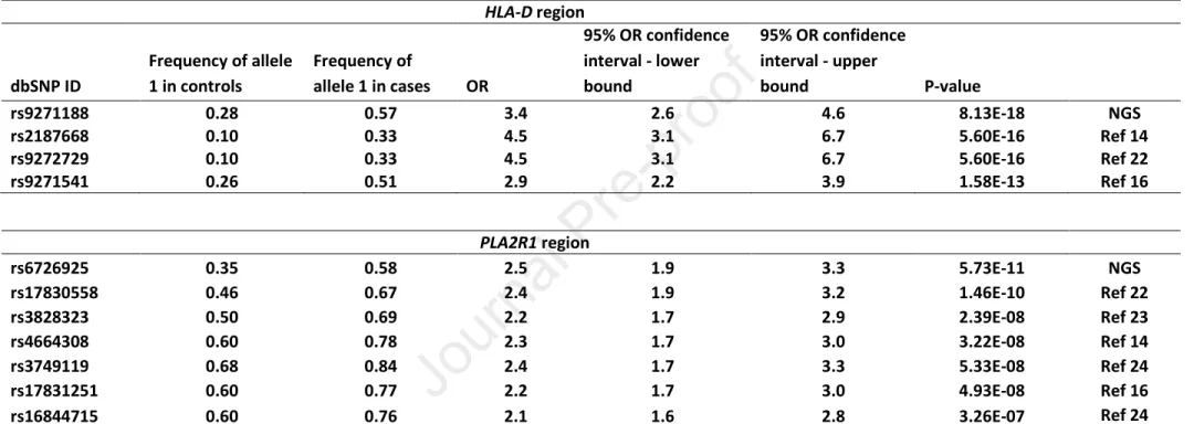
Table 1. Association of previously reported lead SNPs with risk of pMN in the present study HLA-D region dbSNP ID Frequency of allele 1 in controls Frequency of allele 1 in cases OR 95% OR confidence interval - lower bound 95% OR confidence interval - upper bound P-value rs9271188 0.28 0.57 3.4 2.6 4.6 8.13E-18 NGS rs2187668 0.10 0.33 4.5 3.1 6.7 5.60E-16 Ref 14 rs9272729 0.10 0.33 4.5 3.1 6.7 5.60E-16 Ref 22 rs9271541 0.26 0.51 2.9 2.2 3.9 1.58E-13 Ref 16 PLA2R1 region rs6726925 0.35 0.58 2.5 1.9 3.3 5.73E-11 NGS rs17830558 0.46 0.67 2.4 1.9 3.2 1.46E-10 Ref 22 rs3828323 0.50 0.69 2.2 1.7 2.9 2.39E-08 Ref 23 rs4664308 0.60 0.78 2.3 1.7 3.0 3.22E-08 Ref 14 rs3749119 0.68 0.84 2.4 1.7 3.3 5.33E-08 Ref 24 rs17831251 0.60 0.77 2.2 1.7 3.0 4.93E-08 Ref 16
Table 2 : Baseline characteristics of the patients in the discovery cohort
Characteristics Total (n=105) Recurrent (n=45) Non recurrent (n=60) CLINICAL PARAMETERS
Recipients
Age at transplantation. years 50.4 ± 12.8 51.6 ± 12.1 49.5 ± 13.3
Male. n (%) 85/105 (81.0%) 41/45 (91.1%) 44/60 (73.3%)
Caucasian. n (%)* 95/100 (95.0%) 42/44 (95.5%) 53/56 (94.6%) First transplantation. n(%) 93/105 (88.6%) 42/45 (93.3%) 51/60 (85.0%) Dialysis prior to transplantation* 83/94 (88.3%) 39/42 (92.9%) 44/52 (84.6%) Lengths of time on dialysis. months** 16.5 (7-31) 20.5 (7-46) 14.5 (7.5-29.5) Nbr of HLA mismatch (A-B-DR)***
- 0 to 2 mismatch 17/83 (20.5%) 9/36 (25.0%) 8/47 (17.0%)
- 3 mismatch 20/83 (24.1%) 6/36 (16.7%) 14/47 (29.8%)
- 4 mismatch 21/83 (25.3%) 6/36 (16.7%) 15/47 (31.9%)
- 5 to 6 mismatch 25/83 (30.1%) 15/36 (41.7%) 10/47 (21.3%) HLA-antibodies at transplantation*** 27/81 (33.3%) 8/37 (21.6%) 19/44 (43.2%) Anti-HLA donor-specific antibodies*** 3/75 (4.0%) 0/37 (0%) 3/38 (7.9%)
PLA2R positive§ 50/65 (76.9%) 36/40 (90.0%) 14/25 (56.0%)
Time to recurrence/last follow up. months 24 (12-94) 7 (3-14) 75.5 (25.5-121)
Donors
Age. years* 49.7 ± 15.1 48.1 ± 15.4 50.9 ± 14.9
Male. n (%)* 57/103 (55.3%) 27/44 (61.4%) 30/59 (50.9%)
Caucasian. n (%)∆ 43/44 (97.7%) 17/18 (94.4%) 26/26 (100%) Deceased donor transplant. n(%)* 86/104 (82.7%) 35/45 (77.8%) 51/59 (86.4%) Cold ischemia time. min* 1115 (726-1410) 970 (432-1350) 1170 (900-1436)
TREATMENT at the time of transplantation. n (%) Plasma exchange* 2/104 (1.9%) 0/45 (0%) 2/59 (3.4%) IVIG* 4/103 (3.9%) 0/45 (0%) 4/58 (6.9%) ATG* 41/104 (39.4%) 18/45 (40.0%) 23/59 (39.0%) Tacrolimus* 51/104 (49.0%) 24/45 (53.3%) 27/59 (45.8%) Ciclosporin* 53/104 (51.0%) 21/45 (46.7%) 30/59 (50.9%) Mycophenolate mofetil* 89/104 (85.6%) 37/45 (82.2%) 52/59 (88.1%)
Journal Pre-proof
Corticosteroids* 102/104 (98.1%) 43/45 (95.6%) 59/59 (100%)
Anti-IL-2-R* 42/104 (40.4%) 18/45 (40.0%) 24/59 (40.7%)
Rituximab* 2/104 (1.9%) 0/45 (0%) 2/59 (3.4%)
Values reported as numbers and %. mean±SD. or median (interquartile ranges) as appropriate.*between 1 and 10 missing values;
**11-20 missing values;
***21 - 30 missing values;
§31-40 missing values; eGFR (estimated Glomerular Filtration Rate) was calculated according to the Chronic Kidney Disease Epidemiology Collaboration equation.
§Among the 45 recurrent patients, 12 received a transplant between 2011 and 2015: 4 were tested at transplantation (all positive), 5 were tested after (all positive), 3 have not been tested. Among the 60 non-recurrent patients, 16 received a transplant between 2011 and 2015: 5 were tested at transplantation (all negative), 1was tested after (negative), 10 have not been tested.