Design of a Debridement Device Using Impinging Jets
The MIT Faculty has made this article openly available.
Please share
how this access benefits you. Your story matters.
Citation
Raynal, Ashley B., N. Cathy Hogan, and Ian W. Hunter. “Design of
a Debridement Device Using Impinging Jets 1.” Journal of Medical
Devices 10.3 (2016): 030938. © 2016 by ASME
As Published
http://dx.doi.org/10.1115/1.4033763
Publisher
ASME International
Version
Final published version
Citable link
http://hdl.handle.net/1721.1/107252
Terms of Use
Article is made available in accordance with the publisher's
policy and may be subject to US copyright law. Please refer to the
publisher's site for terms of use.
Design of a Debridement Device
Using Impinging Jets
1Ashley B. Raynal
Department of Mechanical Engineering,
Massachusetts Institute of Technology,
Cambridge, MA 02139
N. Cathy Hogan
Department of Mechanical Engineering,
Massachusetts Institute of Technology,
Cambridge, MA 02139
Ian W. Hunter
Department of Mechanical Engineering,
Massachusetts Institute of Technology,
Cambridge, MA 02139
1
Background
Chronic wound care is a significant burden on the healthcare system, affecting an estimated three to six million Americans, manifesting as ulcers associated with restricted blood flow, diabe-tes mellitus, or pressure [1]. Treatment is frequently unsuccessful, with only an estimated 25–50% of venous and diabetic ulcers closing after 20 weeks of treatment [2].
Debridement, the removal of necrotic tissue and foreign materi-als from the wounds, is a crucial component in the chronic wound care [3]. While there exist many debridement techniques, the search for new and more effective methods is ongoing [4].
The existing methods of debridement include surgical, the industry gold standard, as well as the mechanical, autolytic, enzymatic, and hydrosurgery (VersaJetTM). The VersaJetTMuses a single high-speed jet directed parallel to the wound surface to remove soft necrotic tissue.
This paper presents the design of a debridement device that uses two narrow, high-speed impinging fluid jets to excise ne-crotic tissue. The handheld device can be used to remove strips of necrotic tissue of a predetermined width and depth and was tested on samples of simulated slough, the soft necrotic tissue, and eschar, the hard, scablike necrotic tissue. The preliminary tests indicate that the technique removes necrotic tissue quickly and with good control, suggesting that, with further development, the technique may provide a time-saving alternative to surgical debridement. Further testing, however, is required to ensure that the jets do not damage the surrounding healthy tissues and to quantitatively analyze the effectiveness of the technique relative to other debridement strategies.
2
Methods
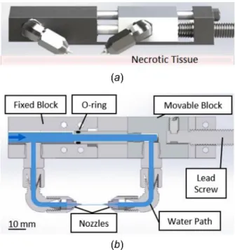
The custom-built handheld device channeled high-pressure water to two nozzles, which were directed to form impinging jets (Fig.1). The position of the nozzles could be adjusted, and tests were performed with the nozzles tips ranging from 2 to 4 mm apart, and the angle of jet intersection varies between 90 deg and 120 deg. Ceramic nozzles (Small Precision Tools MDM-M39C-C) could be inserted and exchanged, permitting the effect of the diameter of the fluid jets to be evaluated. Nozzle diameters tested ranged from 50 lm to 300 lm. A pneumatic piston pump
(Maximator PP72) supplied a continuous flow of water with all the tests performed using pressures ranging from 5 to 30 MPa.
Directing the jets to impinge (Fig.2) was key to the functional-ity of the device. When surrounded by air, the impinging jets atomized to form a fine mist, with droplets retaining a small frac-tion of the pre-atomizafrac-tion kinetic energy. The energy likely goes into heating the water, whose high heat capacity renders this tem-perature change undetectable [5]. When directed into the tissue sample, a single jet was cut to a depth roughly proportional to jet power. When the two impinging jets were directed into the sam-ple, however, they cut the tissue only until they intersected, then abruptly stopped, resulting in a more predictable cut depth. The controlled depth of cut suggests that the jets’ kinetic energy dissi-pated on intersection, preventing further cutting.
Moving the jets longitudinally along the wound would remove a continuous strip of necrotic tissue, triangular in cross section
Fig. 1 The two-nozzle device: (a) rendering of device, front view and (b) cross section showing internal water flow
Fig. 2 The impinging jets causing atomization (a) and cross section of removed tissue (b). Translating the jets (c) would remove a strip of necrotic tissue (d).
1
Accepted and presented at The Design of Medical Devices Conference (DMD2016), April 11–14, 2016 Minneapolis, MN, USA.
DOI:10.1115/1.4033763
Manuscript received March 1, 2016; final manuscript received March 17, 2016; published online August 1, 2016. Editor: William Durfee.
Journal of Medical Devices CopyrightVC2016 by ASME SEPTEMBER 2016, Vol. 10 / 030938-1
(Fig.2). The dimensions of this cross section were dependent on the distance between the nozzles and the jet intersection angle.
Postmortem, ex vivo abdominal porcine tissue was used in all the testing, with the stratum corneum removed by scraping. To mimic the collagen breakdown of the necrotic tissue, 10% acetic acid, an agent known to restructure collagen [6], was applied to the sample surface for 12 hrs prior to testing. The resultant soft tissue was used to mimic the sloughy tissue. Hard eschar was simulated by dehydration of the treated sample, achieved by scorching the sample with a butane torch.
3
Results
The ability of nozzles with orifice diameters ranging from 50 lm to 300 lm to repeatedly cut tissue, and more specifically necrotic tissue, was evaluated. Nozzles with 200 lm diameter or greater proved difficult to control by hand at higher pressures, while the patency of the nozzles with diameters of 50 lm was dif-ficult to maintain. The deepest cuts were produced with nozzles of 75 lm and 100 lm diameters. Because the 75 lm nozzles pro-duced these cuts with a smaller mass flow rate, they were used in subsequent experiments.
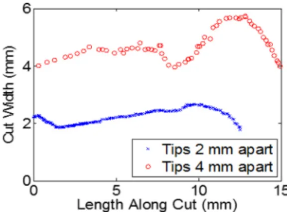
In order to excise sloughy tissue with a jet intersection angle of 120 deg, a pump pressure of 10 MPa was required with the nozzle tips 2 mm apart and 15 MPa with the nozzle tips 4 mm apart. Figure3shows that the width of the necrotic strip was removed, and its variation along the length of the cut was measured for two example cuts. The measurements, made by analyzing photographs of the samples, show that the cut width ranged from 1.8 mm to 2.7 mm for the nozzles 2 mm apart and from 3.8 mm to 5.7 mm for the nozzles 4 mm apart. The imperfect motion of the hand guiding the device may have contributed to this variability.
There was no visible aerosolization when the jets were applied to the sloughy tissue, but some swelling in the sample was visible, indicating that the water was being absorbed. This surface
swelling may have caused the measured cut width in Fig.3above to be wider than the nozzle spacing. Surface deformation caused by the pliability of the tissue furthermore limited control of the handheld device, and precisely controlling the path of the cut was challenging. Further study is needed to quantify the absorbed fluid and assess any damage to the surrounding tissues.
Given the hardness of the eschar, an increased pump pressure of 15 MPa was required during the treatment, with the nozzle tips 2 mm apart and the jets impinging at 120 deg. No swelling was visible in the dehydrated sample, which allowed the user enough control to debride the sample surface without pausing in between removal of each strip, with the resultant sample shown in Fig.4.
4
Interpretation
The handheld device containing the impinging jets was able to remove strips of tissue from both the simulated slough and simulated eschar samples. The pressure required to excise necrotic tissue increased with the spacing between the nozzles and the hardness of the sample. Swelling in the soft tissue indicated fluid absorption, an unwanted side-effect of the device. Yet to be assessed is whether the technique will force bacteria into healthy tissue and how the device will behave when the characteristics of the tissue vary from one jet to the other. Future studies, incorpo-rating samples with integrated healthy and simulated necrotic tis-sues, will investigate these concerns and quantify the fluid absorbed by surrounding healthy tissue.
References
[1] Guo, S., and DiPietro, L., 2010, “Factors Affecting Wound Healing,”J. Dent. Res., 89(3), pp. 219–229.
[2] Falanga, V., Brem, H., and Ennis, W. J., 2008, “Maintenance Debridement in the Treatment of Difficult-to-Heal Chronic Wounds: Recommendations of an Expert Panel,” Ostomy Wound Manage., 8(Suppl.), pp. 2–13.
[3] Moore, Z., 2012, “The Important Role of Debridement in Wound Bed Prepara-tion,”Wounds Int., 3(2), pp. 19–22.
[4] Nusbaum, A. G., Gil, J., Rippy, M. K., Warne, B., Valdes, J., Claro, A., and Davis, S. C., 2012, “Effective Method to Remove Wound Bacteria: Comparison of Various Debridement Modalities in an In Vivo Porcine Model,”J. Surg. Res., 176(2), pp. 701–707.
[5] Giles, D. K., 1988, “Energy Conversion and Distribution in Pressure Atom-ization,”Trans. ASAE, 31(6), pp. 1668–1673.
[6] Davison, P. F., Cannon, D. J., and Andersson, L. P., 1972, “The Effects of Acetic Acid on Collagen Cross-Links,”Connect. Tissue Res., 1(3), pp. 205–216.
Fig. 3 The width of the necrotic tissue removed in a single strip with nozzle tips at 2 and 4 mm apart, 120 deg jet intersection
Fig. 4 The simulated eschar after a single cut—box area (a) and after the competed debridement (b)
030938-2 / Vol. 10, SEPTEMBER 2016 Transactions of the ASME