HAL Id: hal-02344667
https://hal.archives-ouvertes.fr/hal-02344667
Submitted on 4 Nov 2019
HAL is a multi-disciplinary open access
archive for the deposit and dissemination of
sci-entific research documents, whether they are
pub-lished or not. The documents may come from
teaching and research institutions in France or
abroad, or from public or private research centers.
L’archive ouverte pluridisciplinaire HAL, est
destinée au dépôt et à la diffusion de documents
scientifiques de niveau recherche, publiés ou non,
émanant des établissements d’enseignement et de
recherche français ou étrangers, des laboratoires
publics ou privés.
Inhibition, and Formation of Ligand-DNA Adducts
Coralie Caron, Xuan Duong, Régis Guillot, Sophie Bombard, Anton Granzhan
To cite this version:
Coralie Caron, Xuan Duong, Régis Guillot, Sophie Bombard, Anton Granzhan. Interaction of
Func-tionalized Naphthalenophanes with Abasic Sites in DNA: DNA Cleavage, DNA Cleavage Inhibition,
and Formation of Ligand-DNA Adducts. Chemistry - A European Journal, Wiley-VCH Verlag, 2019,
25 (8), pp.1949-1962. �10.1002/chem.201805555�. �hal-02344667�
Sites in DNA: DNA Cleavage, DNA Cleavage Inhibition, and
Formation of Ligand–DNA Adducts
Coralie Caron,
[a,b]Xuan N. T. Duong,
[a,b]Régis Guillot,
[c]Sophie Bombard,
[a,b]and Anton Granzhan*
[a,b]Abstract: Ligands interacting with abasic (AP) sites in DNA may
generate roadblocks in base-excision DNA repair (BER) due to indirect inhibition of DNA repair enzymes (e.g., APE1) and/or formation of toxic byproducts, resulting from ligand-induced strand cleavage or covalent cross-links. Herein, we prepared and syste-matically studied a series of 12 putative AP-site ligands, sharing the common naphthalenophane scaffold but endowed with a variety of substituents. Our results demonstrate that most naphthalenophanes bind to AP-sites in DNA and inhibit the APE1-induced hydrolysis of the latter in vitro. Remarkably, their APE1 inhibitory activity, as characterized by IC50 and KI values, can be directly related to their
affinity and selectivity to AP-sites, assessed from the fluorescence-melting experiments. On the other hand, the molecular design of na-phthalenophanes has crucial influence on their intrinsic AP-site cleavage activity (i.e., ligand-catalyzed β- and β,δ-elimination reactions at the AP site), as illustrated by the compounds either having an exceptionally high AP-site cleavage activity (e.g.,
2,7-BisNP-S, 125-fold more efficacious than spermine) or totally
devoid of this activity (four compounds). Finally, we reveal the unpre-cedented formation of a stable covalent DNA adduct upon reaction of one ligand (2,7-BisNP-NH) with its own product of AP-site cleavage.
Introduction
Apurinic/apyrimidinic (AP, or abasic) sites represent common DNA lesions that naturally occur through deglycosylation of DNA, or through DNA glycosylase-catalyzed removal of damaged (deaminated, alkylated or oxidized) DNA bases.[1–3] In the context of chemotherapy employing DNA-alkylating drugs (e.g., MMS or temozolomide), the major DNA alkylation product, N7
-methylguanine, undergoes spontaneous depurination leading to formation of AP sites,[4] as well as a reaction with histone
proteins leading to generation of DNA–protein crosslinks.[5]
While the latter are thought to be cytotoxic, AP sites are instantly processed by the base excision DNA repair (BER) pathway, which eliminates the drug-induced DNA damage and leads to chemoresistance.[4,6–8] It has been demonstrated that inhibition
of BER increases the sensitivity of cancer cells towards DNA-alkylating drugs in certain cancers, leading to a therapeutically useful synergic effect.[9,10] Accordingly, significant efforts have
been devoted to the development of small-molecule inhibitors of AP endonuclease 1 (APE1, class II AP endonuclease), the key enzyme of the BER pathway that hydrolytically cleaves DNA at AP sites leaving a 3′-OH end (Scheme 1, top).[11–13] An
alter-native promising approach to blocking the BER pathway relies on DNA-binding drugs that selectively bind to AP sites and thereby mask them, or render them unreactive towards from APE1. Thus, methoxyamine (MX, or TRC102) and related alkoxyamines, which form covalent oxime adducts with abasic sites, demonstrate synergy with DNA-alkylating cytotoxic drugs,[14,15] and a combination of MX with temozolomide is
currently being evaluated for glioblastoma treatment in a Phase II clinical trial.[16]
Scheme 1. DNA cleavage at AP sites through APE1-catalysed hydrolysis (top) and β,δ-elimination catalyzed by secondary amines (bottom).
[a] C. Caron, X. N. T. Duong, Dr. S. Bombard, Dr. A. Granzhan CNRS UMR9187, INSERM U1196
Institut Curie, PSL Research University F-91405, Orsay (France)
E-mail: anton.granzhan@curie.fr
[b] C. Caron, X. N. T. Duong, Dr. S. Bombard, Dr. A. Granzhan CNRS UMR9187, INSERM U1196
Université Paris Sud, Université Paris-Saclay F-91405, Orsay (France)
[c] Dr. R. Guillot
CNRS UMR8182, Institut de Chimie Moléculaire et des Matériaux d’Orsay (ICMMO), Université Paris Sud, Université Paris-Saclay 91405 Orsay (France)
Supporting information for this article is given via a link at the end of the document.
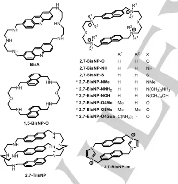
Beyond covalent trapping of abasic sites with reactive drugs such as MX or analogues, several classes of non-covalent ligands were shown to selectively bind to abasic sites in DNA and potentially hinder their recognition and processing by APE1.[17–22] Along these lines, polyazacyclophanes such as
bis-acridine BisA[23,24] and bis-naphthalenes BisNP-O and
2,7-BisNP-NH (Figure 1) bind to abasic sites with high affinity and
selectivity, outperforming other known ligands.[25,26] We
demonstrated that these compounds inhibit the activity of APE1 in vitro with high efficiency, comparable to that of the best catalytic APE1 inhibitors.[25] However, in parallel to inhibiting
APE1 activity, polyazacyclophane ligands also induce an enzyme-independent cleavage of abasic sites via a β-elimination mechanism, due to the presence of secondary amino groups in the ligand structure (Scheme 1, bottom).[23,25] The latter process,
reminiscent to the action of AP lyases (class I endonucleases), was also observed for oligopeptides KWK and KWKK[27–29] as well as several other small-molecule ligands endowed with primary or secondary amino groups, eponymously termed “artificial AP lyases”.[17,30–34] It has been proposed that such
molecules may interfere with the normal BER process because of the accumulation of “dirty ends” (products of β- and β,δ-elimination) that cannot be utilized as substrates by DNA polymerases, and therefore also increase the cytotoxic effect of DNA-alkylating drugs.[35,36]
Figure 1. Structures of previously reported and (*) novel polyazacyclophane ligands. The counter-ions are omitted for clarity.
In order to fully understand the action of ligands targeting abasic sites in biological systems, it would be preferable to decouple the two aforementioned effects, namely the inhibition of the APE1-induced hydrolysis and the ligand-promoted cleavage of AP sites. Towards this end, we designed a novel
series of functionalized macrocycles based on the previously established 2,7-naphthalenophane scaffold (Figure 1) and including, among others, ligands conceived either to be devoid of AP-DNA cleavage activity due to the absence of nucleophilic groups (2,7-BisNP-O8Me, 2,7-BisNP-O4Gua, 2,7-BisNP-Im) or, conversely, to possess an enhanced AP-site cleavage activity (2,7-BisNP-NNH2). Herein, we report the synthesis of novel
ligands, a systematic investigation of their AP-DNA binding affinity and selectivity, in vitro interference with the APE1 processing of abasic sites, as well as the assessment of their intrinsic reactivity towards AP-sites in DNA.
Results and Discussion
Synthesis of novel naphthalenophane ligands
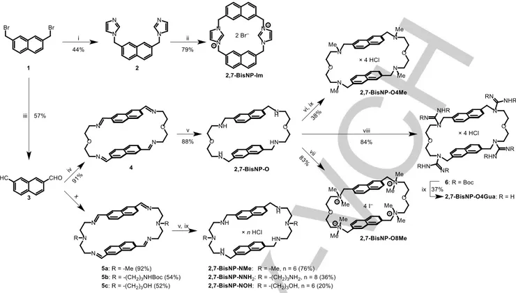
All novel naphthalenophanes were prepared from the common starting material, 2,7-bis(bromomethyl)naphthalene 1 (Scheme 2). The imidazolium-linked cyclophane 2,7-BisNP-Im was obtained, as a bromide salt, in a two-step reaction of 1 with imidazole, performed in an analogy to the reported procedures.[37–40] Kornblum oxidation of 1 gave, in a 52% yield, naphthalene-2,7-dialdehyde 3, a mutual intermediate for the synthesis of all polyamine-type macrocycles obtained through a [2 + 2]-type condensation with the corresponding diamines. Thus, a reaction of 3 with 2,2′-oxybis(ethylamine), followed by the reduction of tetraimine intermediate 4 with NaBH4, gave the
previously described macrocycle 2,7-BisNP-O, which was subsequently converted to the tetramethylated derivative
2,7-BisNP-O4Me via Eschweiler–Clarke reaction (HCHO/HCOOH)
or to the octamethylated analogue 2,7-BisNP-O8Me by exhaustive methylation (MeI, DIPEA). A tetraguanidine derivative 2,7-BisNP-O4Gua was obtained, as a hydrochloride salt, through a treatment of 2,7-BisNP-O with N,N′-di-Boc-S-methylisothiourea in the presence of HgCl2, followed by the
removal of Boc groups (HCl in CHCl3). Finally, three derivatives
of hexaazamacrocycle 2,7-BisNP-NH with varied substituents at two middle nitrogen atoms (2,7-BisNP-NMe, -NNH2 and -NOH)
were prepared by condensation of 3 with the correspondingly substituted derivatives of diethylenetriamine, followed by NaBH4
reduction and conversion to water-soluble hydrochloride salts. The structures of 2,7-BisNP-Im (2 Br−) and
2,7-BisNP-O4Gua × 4 HCl were investigated by single-crystal X-ray
diffrac-tion analysis. In the case of the imidazolium-linked cyclophane, both naphthalene units were found to be strictly coplanar, and the two anti-oriented imidazolium moieties were nearly perpen-dicular to the plane of naphthalene units (Figure 2a). Remarkably, a coplanar geometry of aromatic units is without precedent in the well-studied series of bis-imidazolium cyclo-phanes since, to the best of our knowledge, all previously described anti-conformers adopted a displaced anti-parallel conformation of aromatic units.[38–40] At the same time, 1H NMR
spectra of 2,7-BisNP-Im revealed a sharp singlet for methylene protons at room temperature (δ = 5.77 ppm, in [D6]DMSO),
giving evidence of a rapid interconversion of all possible conformations (presumably, due to the intramolecular rotation of imidazolium rings) on the NMR time scale.
Scheme 2. Synthesis of functionalized naphthalenophane ligands. Reagents and conditions: (i) imidazole, MeOH, reflux, 22 h; (ii) 1, MeCN, 90 °C, 18 h; (iii) NaHCO3, DMSO, 100 °C; (iv) O(CH2CH2NH2)2, MeCN, room temp., 4 days; (v) NaBH4, DCM–MeOH, room temp.; (vi) HCHO, HCOOH, 120 °C, 48 h; (vii) MeI
(excess), DIPEA, DMF, 60 °C, 24 h; (viii) BocN=C(SMe)NHBoc, HgCl2, NEt3, DCM, room temp., 48 h; (ix) HCl (dioxane), CHCl3, room temp., 24 h; (x)
RN(CH2CH2NH2)2, MeCN, room temp., 4 days; then HCl (dioxane), MeOH.
Figure 2. Solid-state structures (ORTEP plots, from single crystal X-ray diffraction analysis): a) 2,7-BisNP-Im (2 Br−), viewed along to the
crystallographic c axis; b) 2,7-BisNP-O4Gua × 4 HCl × 5 H2O, viewed along
the crystallographic b axis. Thermal ellipsoids are drawn at 70 % probability; cyan lines: intramolecular hydrogen bonds. Hydrogen atoms, counter-ions and solvent molecules are omitted for the sake of clarity.
The tetraguanidine macrocycle crystallized with naphthalene units in a displaced anti-parallel conformation (Figure 2b). The conformation of linkers reveals a significant potential of molecular flexibility in aqueous solutions, in spite of the intramolecular hydrogen bonds observed, in the solid state,
between the protonated guanidinium groups and oxygen atoms of the linkers.
AP-DNA affinity of ligands assessed by fluorescence-melting experiments
The interaction of ligands with abasic sites was studied through fluorescence-melting experiments with double-stranded oligo-nucleotides 17-NΦ (Table 1, Φ = THF abasic site), bearing abasic sites opposite to different orphan residues (N = T, C, A or G), a quencher (TAMRA) on the abasic strand, and a fluorophore (6-FAM) on the complementary strand. The use of the stable THF analogue of AP sites in these experiments avoids the ligand-promoted DNA cleavage during the melting ramps, and a relatively low ionic strength of the buffer (20 mM K+,
pH 7.2) allows the observation of even weak stabilization effects, expressed as ligand-induced increase of denaturation temperature (∆Tm). In addition, AP-site selectivity of ligands with
respect to well-matched DNA was estimated through analysis of the drop of ∆Tm values observed in the presence of unlabeled
double-stranded competitor (calf thymus DNA, 200- or 650-fold excess with respect to the AP target). In parallel, thermal denaturation experiments performed with the fully matched duplex (17-TA) provide another measure of the selectivity of ligands with respect to undamaged DNA.
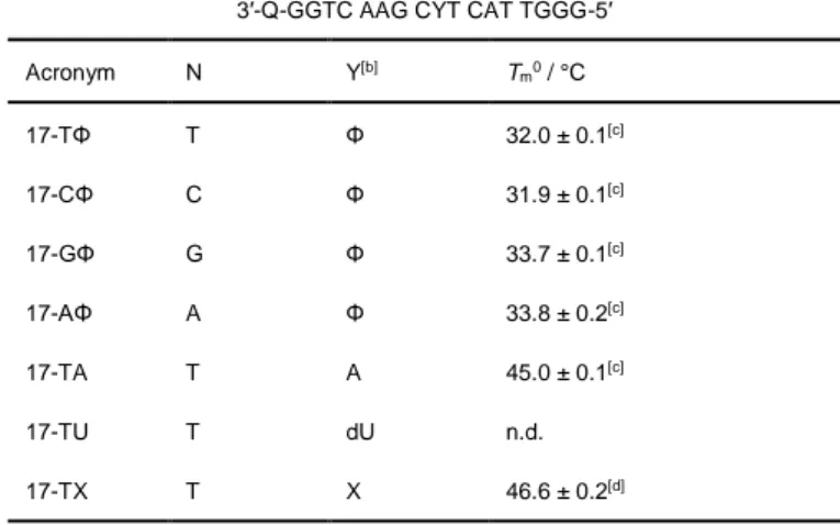
Table 1. Sequences and melting temperatures of 17-mer oligonucleotides used in this work.[a]
5′-F–CCAG TTC GNA GTA ACCC-3′ 3′-Q-GGTC AAG CYT CAT TGGG-5′
Acronym N Y[b] Tm0 / °C 17-TΦ T Φ 32.0 ± 0.1[c] 17-CΦ C Φ 31.9 ± 0.1[c] 17-GΦ G Φ 33.7 ± 0.1[c] 17-AΦ A Φ 33.8 ± 0.2[c] 17-TA T A 45.0 ± 0.1[c] 17-TU T dU n.d. 17-TX T X 46.6 ± 0.2[d]
[a] In the fluorescence-melting assay, F = 6-FAM, Q = TAMRA; in fluorimetric DNA cleavage studies, F = ATTO390, Q = DABCYL. [b] Φ = THF abasic site; X = native abasic site. [c] In 10 mM KAsO2Me2, 10 mM KCl, pH 7.2 (conditions
of the fluorescence-melting assay). [d] In 10 mM KAsO2Me2, 150 mM KCl, pH
7.2 (conditions of the fluorimetric DNA cleavage assay). Data are means ± s.d. from three technical replicates.
The results of fluorescence-melting experiments are presented in Figure 3. All ligands stabilized the abasic duplexes, to an extent depending on the base facing the AP site: 17-TΦ > 17-CΦ > 17-GΦ > 17-AΦ. Remarkably, none of the ligands of the 2,7-BisNP series stabilized the well-matched duplex 17-TA, in contrast to ethidium bromide (used as a control, non-selective DNA binder) and 1,5-BisNP-O, which, in spite of its structural similarity, has a different substitution pattern of naphthalene units and whose non-specific binding to fully matched DNA was previously documented.[41] In addition, the selectivity of most
ligands of the 2,7-BisNP series for abasic oligonucleotides was confirmed by the fact that the presence of double-stranded competitor only slightly decreased the ligand-induced stabiliza-tion effect. Considering the effect of substituents introduced into the naphthalenophane scaffold with respect to parent ligands (2,7-BisNP-NH and 2,7-BisNP-O), the following observations emerge from the results presented in Figure 3:
1) Macrocycles bearing substituents at middle nitrogen atoms of the linkers (2,7-BisNP-NMe, -NNH2, -NOH) demonstrate
the stabilization effect equal to, or even higher than the one induced by “parent” ligands, for all abasic substrates (e.g., ∆TmTΦ ≈ 10 °C), in combination with excellent selectivity
with respect to competitor DNA. This gives evidence that side chains introduced in the middle of polyamine linkers do not hamper the binding of naphthalenophanes to AP sites. In contrast, the sulfur analogue 2,7-BisNP-S was both less efficient (e.g., ∆TmTΦ ≈ 8 °C) and less selective, with respect
to all abasic substrates.
2) The stabilization induced by the ligands bearing substi-tuents at the benzylic amino groups (R1, R2 ≠ H, i.e.,
2,7-BisNP-O4Me, 2,7-BisNP-O8Me, 2,7-BisNP-O4Gua) was
lower, compared to the “parent” ligand 2,7-BisNP-O, in
par-ticular upon binding to 17-TΦ (∆TmTΦ ≤ 8 °C) and 17-GΦ
(∆TmGΦ ≤ 4 °C). The loss of efficiency was most pronounced
in the case of 2,7-BisNP-O4Me (∆TmTΦ = 5.4 °C, ∆TmGΦ =
2.0 °C). These observations support the structural model observed upon binding of 2,7-BisNP-O to a T·T mis-match[42] and suggest that the substituents at the benzylic
amino groups impede the hydrogen bonding of protonated amines to carbonyl groups of orphan G or T residues, redu-cing the overall stabilization of the ligand–AP site complex.
Figure 3. Ligand-induced shifts of melting temperature (∆Tm) of abasic
oligo-nucleotides (17-NΦ) or the well-matched control (17-TA), in the absence (dark bars) or in the presence of ct DNA competitor (medium bars: 20 µM bp, light bars: 65 µM bp). Conditions: c(oligo) = 0.5 µM, c(ligand) = 1 µM in 10 mM KAsO2Me2, 10 mM KCl buffer, pH 7.2. Data are means ± s.d. from three
technical replicates. 2,7-BisNP -NH 2,7-BisNP -O 2,7-BisNP -S 2,7-BisNP -NMe 2,7-BisNP -NNH 2 2,7-BisNP -NOH 2,7-BisNP -O4Me 2,7-BisNP -O8Me 2,7-BisNP -O4Gua 2,7-BisNP -Im 1,5-BisNP -O 2,7-Tri sNP KWKK Ethi dium br omide -2 0 2 4 6 8 17-TA Tm ( °C) 0 2 4 6 8 10 12 17-GΦ Tm ( °C) 0 2 4 6 8 10 17-AΦ Tm ( °C) 0 2 4 6 8 10 12 Tm ( °C) 17-CΦ 0 2 4 6 8 10 12 17-TΦ Tm ( °C)
3) Quite remarkably, the effect of the bicyclophane 2,7-TrisNP and the bis-imidazolium cyclophane 2,7-BisNP-Im is very similar to the one of polyazacyclophane ligands, in spite of their significantly different shapes. In the case of
2,7-TrisNP, this may be rationalized taking into account its
shape similarity to cylindrical metallohelicates which bind to abasic sites with high affinity, even though the structural details of their binding mode have not yet been elucidated.[22] As for 2,7-BisNP-Im, this ligand may bind to
AP sites in a conformation which is significantly different from the one observed in the solid state (cf. Figure 2a), most likely in a folded form allowing the insertion of one of naphthalene units inside the abasic pocket.
Inhibition of enzymatic DNA cleavage
To study the competitive effect of the ligands on the enzymatic processing of AP sites, we used a real-time fluorimetric assay similar to the one employed in our previous work.[25] In this assay,
APE1-induced cleavage of the abasic substrate 17-TΦ (Table 1) is monitored through the fluorescence increase which occurs upon cleavage and dehybridization of the quencher-bearing strand (Figure 4a). However, in preliminary experiments we observed that oligonucleotides labelled with 6-FAM, as well as with several other conventional fluorophores such as Alexa Fluor (AF) 488, AF 546, AF 594, TAMRA, or Texas Red, underwent significant quenching upon addition of most ligands. While this ligand-induced quenching is tolerable in fluorescence-melting experiments (which aim at measuring the temperature of the melting transition and do not account for the exact fluorescence intensity), this phenomenon could bias the output of kinetic assays. Therefore, in order to minimize the extent of ligand-induced quenching, ATTO390 was employed as the fluorophore least prone to this phenomenon, in combination with DABCYL quencher.[43]
The results of the kinetic assay demonstrated that all ligands were able to inhibit APE1 activity in a dose-dependent manner (Figure 4b and Supporting Information, Figure S1), with the corresponding IC50 values ranging from 0.14 µM
(2,7-BisNP-NNH2) to 89 µM (2,7-BisNP-O4Me, Table 2). Taking into
account the fact that indirect inhibition is described by the same mathematical model as competitive inhibition (i.e., Vmax = const,
KMapp = αKM,where α = 1 + [I] / KI), with the only difference that
the inhibition constant (KI) represents the dissociation constant
of the inhibitor–substrate (and not inhibitor–enzyme) complex,[25]
we calculated the corresponding KI values (Table 2) using the
well-known relationship (Eq. 1):[44]
IC50 = KI (1 + S / KM), (1)
where S is concentration of the substrate (0.2 µM) and KM is
Michaelis constant for APE1 (KM = 76 nM, Supporting
Infor-mation, Figure S2).[45] Of note, the K
I values for the macrocycles
2,7-BisNP-NH and 2,7-BisNP-O (≈50 nM) were of the same
order as the ones obtained in our previous work (34 nM) despite a significantly lower concentration of the substrate (25 nM).[25]
Figure 4. Inhibition of APE1 activity by AP-site ligands. a) Principle of the real-time fluorimetric assay for APE1 activity and indirect inhibition of APE1 by ligands binding to AP-sites. F = ATTO390, Q = DABCYL, Φ = THF abasic site. b) Representative dose-response plots for APE1 inhibition by 2,7-BisNP-O (black) and 2,7-BisNP-O8Me (red). Conditions: c(17-TΦ) = 0.2 µM in 50 mM
HEPES, 50 mM NaCl, 1 mM MgCl2, 2 mM DTT buffer, pH 8.0, T = 37 °C,
λex = 395 nm, λem = 465 nm.
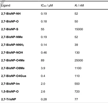
Table 2. Inhibitory activity of macrocyclic ligands with respect to APE1-induced cleavage of 17-TΦ.[a]
Ligand IC50 / µM KI / nM 2,7-BisNP-NH 0.19 52 2,7-BisNP-O 0.18 50 2,7-BisNP-S 55 15000 2,7-BisNP-NMe 0.19 52 2,7-BisNP-NNH2 0.14 39 2,7-BisNP-NOH 0.46 130 2,7-BisNP-O4Me 89 25000 2,7-BisNP-O8Me 3.9 1100 2,7-BisNP-O4Gua 0.4 110 2,7-BisNP-Im 2.0 550 1,5-BisNP-O 2.6 720 2,7-TrisNP 0.28 77
[a] Conditions: see Figure 4 caption. Q F Φ 17-TΦ APE1 a) Φ = + b) KI ligand 0.1 1 10 0.0 0.2 0.4 0.6 0.8 1.0 2,7-BisNP-O IC 50 = 0.18 µM APE1 act ivi ty Ligand concentration (µM) 2,7-BisNP-O8Me IC50 = 3.9 µM 5′ 3′ 5′ 3′ Q F F 3′ Q Φ 5′ 5′
Remarkably, a good correlation could be observed between the 1/KI values (i.e., affinity constants) and the ligand-induced
stabilization of the substrate (∆TmTΦ) observed in the presence of
excess competitor (r 2 = 0.72, Figure 5a): in other words, potent and selective AP-site ligands, whose stabilizing effect was not depressed by the presence of the competitor (2,7-BisNP-NNH2,
2,7-BisNP-NH, 2,7-BisNP-O, 2,7-BisNP-NMe), were most
efficient at inhibiting the APE1-induced cleavage of AP-sites. A correlation made using ∆TmTΦ values obtained in the absence of
the competitor was unsatisfactory (r 2 = 0.26, Figure 5b) due to the fact that the stabilization induced by non-selective binders (e.g., 1,5-BisNP-O) was strongly reduced in the presence of the competitor. Altogether, the comparison made with a series of different ligands, varying in terms of their affinity and selectivity to AP-sites, convincingly support the indirect inhibition model and highlight the efficiency of DNA-targeted inhibition of DNA repair. Moreover, the revealed interplay between the affinity and the selectivity of DNA-targeting inhibitors is of utmost importance for their application in the cellular context, where DNA damage sites are disseminated throughout the large excess of undamaged genome.
Figure 5. Correlation of ligand affinity constants to AP-sites, derived from APE1-inhibition studies interpreted in terms of indirect inhibition model (Ka =
1 / KI) with ligand-induced stabilization of 17-TΦ observed a) in the absence
and b) in the presence of competitor (ct DNA, 65 µM bp).
Intrinsic AP-DNA cleavage activity of ligands
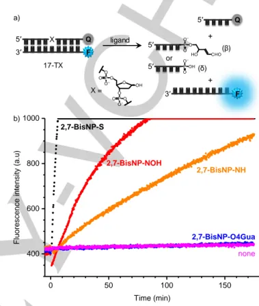
The intrinsic activity of ligands with respect to nucleophile-catalyzed cleavage of AP-sites was also assessed by means of a real-time fluorimetric assay, similar to the one developed by Berthet et al.[46] Our variant of this assay employs the 17-mer
duplex 17-TX (Table 1), double-labelled with ATTO390 and DABCYL and bearing a native abasic site (X), quantitatively produced by UDG treatment of the corresponding uracil-containing precursor 17-TU (Supporting Information, Figure S3). As in the APE1 activity assay, the progress of ligand-induced DNA cleavage is monitored through an increase of fluorescence, taking place upon dehybridization of the cleaved strand (Figure 6a). In order to compensate for the partial ligand-induced fluoro-phore quenching, the extent of this effect was measured in control experiments with a non-cleavable substrate (17-TΦ) and quantified as a ratio of fluorescence intensity in the presence and in the absence of ligands, used at identical concentration of
5 µM (QF, Table 2). This factor was taken into account for calculation of corrected initial reaction rates (vcorr, Table 3), used
as quantitative indicators of AP-DNA cleavage activity of ligands (vcorr = v0 / QF, where v0 are the initial slopes of real-time
fluorescence intensity plots, Figure 6b).
Figure 6. Real-time fluorimetric assay for determination of AP-DNA cleavage activity of ligands. a) Principle of the assay (F = ATTO390, Q = DABCYL, X = native abasic site). b) Representative fluorimetric readout for cleavage of 17-TX with 2,7-BisNP-NH, 2,7-BisNP-S, 2,7-BisNP-NOH and 2,7-BisNP-O4Gua. Conditions: c(17-TX) = 0.2 µM, c(ligand) = 5 µM in 10 mM KAsO2Me2, 150 mM
KCl buffer, pH 7.2, T = 37 °C, λex = 395 nm, λem = 465 nm; the ligand was
added at t = 0. The transient drop of fluorescence intensity after ligand addition (red curve) is due to the ligand-induced quenching effect. The scale of fluorescence intensity (max value = 1000 a.u.) was adjusted in order to allow the best comparison of initial reaction rates induced by different ligands.
In parallel, we assessed the DNA-cleavage activity of ligands through PAGE analysis using the duplex 17-TX, in which the AP strand was 5′-32P-labelled. This complementary assay,
performed in the conditions similar to the ones of the fluorimetric assay (with a fixed incubation time of 1 h), avoids the problems related to the use of fluorescently labeled DNA (ligand-induced quenching) and, in addition, allows to estimate the distribution of various products of strand cleavage (β- and β,δ-elimination products) and secondary reactions. Based on the results of the real-time fluorimetric assay (Table 3) and the PAGE assay (Figure 7), all ligands could be divided into three groups with respect to their AP-site cleavage activity:
0.00 0.01 0.02 0.03 0 2 4 6 8 10 12 r 2 = 0.72 a) Tm TΦ (with com pe titor ) / °C 1 / KI (nM-1) 0.00 0.01 0.02 0.03 0 2 4 6 8 10 12 Tm TΦ (with ou t co mp etito r) / °C 1 / KI (nM-1) b) r 2 = 0.26 17-TX ligand X = b) 0 50 100 150 400 600 800 1000 none 2,7-BisNP-O4Gua 2,7-BisNP-S 2,7-BisNP-NH Flu or esce nce inte nsity (a .u) Time (min) 2,7-BisNP-NOH Q F X a) 5′ 3′ F 3′ Q 5′ 5′ (β) 5′ (δ) or + +
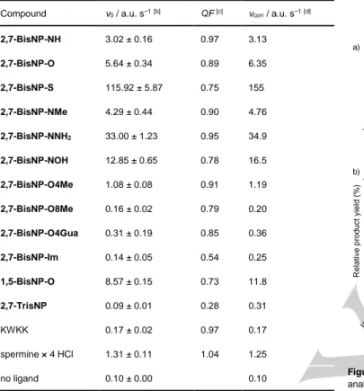
Table 3. DNA AP-site cleavage activity of macrocycles and reference ligands, measured by the real-time fluorimetric assay.[a]
Compound v0 / a.u. s−1[b] QF [c] vcorr / a.u. s−1[d]
2,7-BisNP-NH 3.02 ± 0.16 0.97 3.13 2,7-BisNP-O 5.64 ± 0.34 0.89 6.35 2,7-BisNP-S 115.92 ± 5.87 0.75 155 2,7-BisNP-NMe 4.29 ± 0.44 0.90 4.76 2,7-BisNP-NNH2 33.00 ± 1.23 0.95 34.9 2,7-BisNP-NOH 12.85 ± 0.65 0.78 16.5 2,7-BisNP-O4Me 1.08 ± 0.08 0.91 1.19 2,7-BisNP-O8Me 0.16 ± 0.02 0.79 0.20 2,7-BisNP-O4Gua 0.31 ± 0.19 0.85 0.36 2,7-BisNP-Im 0.14 ± 0.05 0.54 0.25 1,5-BisNP-O 8.57 ± 0.15 0.73 11.8 2,7-TrisNP 0.09 ± 0.01 0.28 0.31 KWKK 0.17 ± 0.02 0.97 0.17 spermine × 4 HCl 1.31 ± 0.11 1.04 1.25 no ligand 0.10 ± 0.00 0.10
[a] Substrate: 17-TX (F = ATTO390. Q = DABCYL), conditions: see Figure 6 legend. [b] Initial slope of the real-time fluorescence intensity plot (mean ± s.d. from three technical replicates). [c] Ligand-induced fluorescence quenching of substrate, QF = Fwith ligand / Fno ligand, measured with the non-cleavable analogue
17-TΦ at c(ligand) = 5 µM. [d] Quenching-adjusted initial rate of AP-DNA cleavage reaction.
1) Very efficient AP-cleaving ligands, namely 2,7-BisNP-S (vcorr = 155 a.u. s−1) and 2,7-BisNP-NNH2 (vcorr = 34.9 a.u.
s−1), for which a total conversion of the substrate was observed after 1 hour, with the products of β- and β,δ-elimination forming in approximately 2 : 1 ratio (according to PAGE analysis). The high efficiency of 2,7-BisNP-NNH2 is,
expectedly, due to the presence of two additional primary amino groups, which lead to an 11-fold higher cleavage rate with respect to the parent ligand 2,7-BisNP-NH (vcorr = 3.11
a.u. s−1). The exceptionally high activity of the sulfur-containing macrocycle is particularly remarkable, taking into account the fact that its affinity for AP sites is significantly lower than the one of 2,7-BisNP-NH and 2,7-BisNP-NNH2,
as demonstrated by both fluorescence-melting experiments (Figure 3) and APE1 inhibition studies (Table 2).
2) Moderately efficient AP-cleaving ligands (2 < vcorr <
20 a.u. s−1): NH, NMe,
2,7-BisNP-NOH, 2,7-BisNP-O and 1,5-BisNP-O, which induced from
~60 to ~90% conversion of the substrate after 1 h (as determined by PAGE), giving predominantly β-elimination products (β : δ = 4.5 to 8). Only one ligand, 2,7-TrisNP,
showed a discrepancy between the two methods: strong cleavage activity was detected by the PAGE assay (con-version = 89% after 1 h), but not in the fluorimetric assay, presumably due to its strong quenching effect (QF = 0.28).
Figure 7. AP-site cleavage activity of ligands assessed by PAGE. a) PAGE analysis of 32
P-labelled 17-TX following incubation with ligands. Lane assignment: 1) untreated control; 2) NaOH treatment (0.5 M); 3) APE1 (0.27 nM); 4) 2,7-BisNP-O; 5) 2,7-BisNP-NH; 6) 2,7-BisNP-S; 7) 1,5-BisNP-O; 8) 2,7-TrisNP; 9) 2,7-BisNP-NMe; 10) 2,7-BisNP-O4Me; 11) 2,7-BisNP-O8Me; 12) 2,7-BisNP-O4Gua; 13) 2,7-BisNP-NNH2; 14) BisNP-NOH; 15) 2,7-BisNP-Im; 16) KWKK; 17) spermine. Conditions: c(17-TX) = 0.2 µM, c(ligand) = 5 µM, incubation for 1 h in 10 mM KAsO2Me2, 150 mM KCl buffer, pH 7.2,
T = 37 °C. Left and right panels are parts of the same gel. b) Quantification of
PAGE data (means ± s.d. from three technical replicates). Band assignment / color codes: * / grey, uncleaved substrate; β / blue, product of β-elimination; δ / cyan, product of β,δ-elimination; † / green, 3′-OH product of APE1 cleavage; ** / pink, putative ligand cross-link.
3) Mostly inactive ligands (vcorr < 2 a.u. s−1, < 10% substrate
conversion in PAGE assay): O4Me,
2,7-BisNP-O8Me, 2,7-BisNP-O4Gua, 2,7-BisNP-Im, spermine and
KWKK. Although spermine and the tetrapeptide KWKK are well-established AP-DNA cleaving agents, in both of our assays their activity was only marginal, likely due to at least 100-fold lower concentration, comparing with previous works.[29,47] This is also in line with the negligible AP-site
binding effect of KWKK observed in FRET-melting experiments (Figure 3).
These results clearly demonstrate the influence of molecular structure of naphthalenophanes on their AP-DNA cleavage activity. Specifically, introduction of side chains at central nitrogen atoms of linkers results in enhanced cleavage activity (NMe, NOH and, particularly,
2,7-BisNP-NNH2) compared to 2,7-BisNP-NH. Conversely, suppression of
nucleophilic centers through introduction of substituents at benzylic amino groups (2,7-BisNP-O8Me, 2,7-BisNP-O4Gua) or
untd control NaOH (0.5 M) APE1 (0. 27 nM) 2,7-B isN P-O 2,7-B isN P-N H 2,7-B isN P-S 1,5-B isN P-O 2,7-Tri sNP 2,7-B isN P-N Me 2,7-B isN P-O4M e 2,7-B isN P-O8M e 2,7-B isN P-O4Gu a 2,7-B isN P-N NH2 2,7-B isN P-N OH 2,7-B isN P-Im KW KK sperm ine 0 20 40 60 80 100 R elativ e produc t y ield (%) * ** δ β a) 1 2 3 4 5 6 7 8 9 10 11 12 13 14 15 16 17 β * δ ** b) † †
removal of these groups (2,7-BisNP-Im) totally abolishes the AP-DNA cleavage activity. In the last series, the derivative
2,7-BisNP-O4Me represents an intermediate case: despite the
presence of four tertiary, putatively nucleophilic, amino groups, the AP-DNA cleavage activity of this ligand is strongly reduced with respect to 2,7-BisNP-NH (vcorr = 1.19 a.u. s−1, conversion =
6%). This behavior is likely due to the combination of two phenomena, namely: (i) the tertiary amino groups do not allow the formation of the pre-incision iminium intermediate of the β-elimination reaction (cf. Scheme 1); (ii) the AP-DNA affinity of
2,7-BisNP-O4Me is reduced with respect to the other ligands of
the series (cf. Figure 3 and Table 2). Notably, a similar decrease of affinity and DNA-cleaving activity upon mono-methylation of secondary amino groups was observed in the series of nucleobase–polyamine–intercalator conjugates.[17]
Formation of covalent adducts with abasic sites
The results of PAGE analysis revealed that, in some cases, the formation of expected products of AP-site cleavage (β- and β,δ-elimination products) was accompanied by appearance of novel bands of intermediate mobility (** in Figure 7). These bands were observed mostly with 2,7-BisNP-NH (20% yield, lane 5), but trace amounts could also be detected with 2,7-BisNP-S (5%) and 2,7-BisNP-O (< 1%). In order to get insight into the nature of these products, we performed several complementary studies. Incubation of 2,7-BisNP-NH with a DNA substrate devoid of abasic site (17-TU) gave no evidence of strand cleavage (Figure 8a, lanes 6–7), therefore excluding the possibility of a strand cleavage at a secondary site (i.e., away from the AP site). Furthermore, the product was stable towards a brief heating at 90 °C (Figure 8a, lane 4), which speaks in favor of a covalent modification of DNA. Next, we performed a trapping reaction through incubation of 17-TX with
2,7-BisNP-NH in the conditions of reductive amination (NaCNBH3, followed
by NaBH4 quenching; Figure 8a, lane 5).[29,33] In this case, we
evidenced the formation of a slower-migrating band (*** in Figure 8a), supposedly corresponding to the reduced pre-incision iminium base intermediate (cf. Scheme 1), as well as another band which migrated closely to the ** band and which could be attributed to the reduced post-incision intermediate (i.e., the product of reductive amination reaction of the polyamine with the α,β-unsaturated aldehyde).[29,48] Taking into account these
results, we may suggest that the ** band in lanes 3–4 (Figure 8a), formed in the absence of reducing agents, corresponds to a stable but unreduced covalent adduct of the ligand with its own product of AP-site cleavage via β-elimination. In addition, its greatly retarded migration with respect to the other cleavage products is consistent with the partial charge neutralization of the oligonucleotide imparted by the remnant of the polycationic ligand (3 to 4 positive charges).
In a separate experiment, we examined the influence of reaction time on the formation of this product. The adduct yield increased with time and reached its maximum (~60%) after 5 h of incubation at 37 °C, when all substrate was consumed (Figure 8b,c). Notably, the increase of the amount of the DNA–ligand adduct was accompanied by a decrease of the yield of the β-elimination product at longer incubation time (from 30% after 1 h
to 18% after 5 h of incubation), giving evidence that the β-elimination product represents an intermediate for the formation of the covalent adduct (and not vice versa).
Figure 8. Formation of a covalent adduct of 2,7-BisNP-NH with abasic sites. a) PAGE analysis showing formation of slow-migrating bands of 2,7-BisNP-NH upon incubation with 17-TX. Lane assignment: 1) 17-TX incubated with 0.5 M NaOH (1 h); 2) untreated 17-TX; 3) 17-TX incubated with 2,7-BisNP-NH (5 µM); 4) same, but heated at 90 °C (5 min) prior to PAGE analysis, 5) 17-TX incubated with 2,7-BisNP-NH (5 µM) and NaCNBH3 (25 mM), followed by
addition of NaBH4 (100 mM) prior to PAGE analysis; 6) untreated 17-TU; 7)
17-TU incubated in the presence of 2,7-BisNP-NH (5 µM). b) Time dependence of the formation of covalent adduct. Lane assignment: 1) 17-TX incubated without additives for 16 h; 2) 17-TX incubated with 0.05 M NaOH (1 h); 3–11) 17-TX incubated with 2,7-BisNP-NH for ¼, ½, ¾, 1, 2, 3, 4, 5 and 16 h, respectively. In all experiments, c(17-TX) = 0.2 µM. Band assignment: *, unmodified substrate; **, covalent ligand–DNA adduct; ***, reduced imine intermediate (lane 5), β, product of β-elimination (or its reduced form in lane 5); δ, product of β,δ-elimination. c) Quantification of PAGE shown in panel b).
Finally, we attempted mass-spectrometric characterization of the product corresponding to the ** band observed in PAGE. A crude reaction mixture of 17-TX with 2,7-BisNP-NH was concentrated, desalted (EtOH precipitation followed by reverse-solid-phase extraction), and analyzed by LC/MS (Figure 9a).
4 5 6 1 2 3 7 δ β ** * *** a) b) 1 2 3 4 5 6 7 8 9 10 11 δ β ** * c) 0 1 2 3 4 5 16 0 20 40 60 80 100 δ β Re la tive amoun t (%) Time (h) * **
Along with an unresolved peak containing cleavage products and the complementary strand, we observed a minor peak with a higher retention time (tR = 4.05 min), consistent with the more
lipophilic character imparted by the presence of the naph-thalenophane remnant. A corresponding mass spectrum (Figure 9b) revealed the presence of two molecular ion peaks (m/z = 1036 and 1555), which could indeed correspond to adduct of the ligand with the β-elimination product of DNA cleavage (M = 3111 Da). Although the structure of this product could not be established unambiguously at this stage, we may suggest that it could be formed through a 1,4-conjugate addition of the neigh-boring amino group of the macrocyclic ligand to the post-incision iminium intermediate (cf. Scheme 1) leading to formation of a cyclic 1,4-diazepine (Figure 9c), which may be subjected to further isomerization. This hypothesis explains the fact that, among all ligands, only 2,7-BisNP-NH (presenting a 1,2-diamino fragment) gives substantial yield of the covalent adduct.
Figure 9. a) TIC chromatogram of the crude reaction mixture of 17-TX (0.2 µM) with 2,7-BisNP-NH (50 µM) (see Experimental Section for details). b) Mass spectrum of the peak with tR = 4.05 min (**). The peak with m/z = 1114.4
could not be assigned. c) Putative structure of the covalent adduct of 2,7-BisNP-NH to the product of β,δ-elimination.
While the reaction of β-elimination products of AP-site cleavage with external alkoxyamines is known and has been exploited for their quantitative detection,[49] the formation of
covalent adducts with 2,7-BisNP-NH represents, to the best of our knowledge, the first example of generation of stable adducts through reaction of cleaved AP-sites with polyamine ligands. In this context, it is interesting to mention the formation of stable interstrand cross-links through reaction of both uncleaved[50,51]
and cleaved[52] AP-sites with purine residues observed in native
(non-reducing) conditions. Notably, in the latter case, the
formation of the cross-link was proposed to occur through a 1,4- conjugate addition of N1 of a proximate adenine residue to the unsaturated aldehyde product of β-elimination cleavage, which is reminiscent to the mechanism proposed above.
Conclusions
In the present work, we systematically explored the 2,7-naphtha-lenophane scaffold as a versatile platform for the development of functionalized ligands interacting, in various ways, with AP-sites in DNA. We demonstrated that careful molecular engineering could be exploited to exacerbate or alleviate some of their biochemical properties, as summarized in Table 4. Specifically, among seven novel compounds based on this scaffold, most ligands conserved the high affinity to abasic sites and excellent selectivity with respect to well-matched DNA. Furthermore, we demonstrated that most compounds were able to inhibit the APE1-induced cleavage of AP-sites in DNA by competing with the enzyme for binding to the substrate, and that the inhibitory activity (as characterized by IC50 and KI values) is
directly related to the affinity and selectivity of ligands to the substrate (i.e., abasic site). Beyond direct implications in the context of APE1-related chemoresistance, these conclusions may be extended to other DNA repair pathways whose DNA substrate may represent a target for small-molecule ligands, such as direct damage reversal[53] and mismatch repair.[54,55]
Table 4. Qualitative summary of AP-DNA-binding properties, APE1 inhibitory activity, and intrinsic reactivity of naphthalenophane macrocycles with respect to AP-sites in DNA.
Ligand AP-DNA binding APE1
inhibi-tion[b] AP-DNA cleavage activity[c] AP cross-linking affinity[a] select.
2,7-BisNP-NH +++ ++ +++ + ++ 2,7-BisNP-O +++ +++ +++ + ± [d] 2,7-BisNP-S ++ + + +++ ± [d] 2,7-BisNP-NMe +++ ++ +++ + − 2,7-BisNP-NNH2 +++ +++ +++ ++ − 2,7-BisNP-NOH +++ ++ +++ + − 2,7-BisNP-O4Me + + + − − 2,7-BisNP-O8Me ++ ++ ++ − − 2,7-BisNP-O4Gua ++ ++ +++ − − 2,7-BisNP-Im + ++ ++ − − 1,5-BisNP-O +++ + ++ + − 2,7-TrisNP ++ +++ +++ + − [a] ∆TmTΦ < 6 (+), 6 to 9 (++), > 9 °C (+++). [b] KI > 2000 (+), 200 to 2000 (++),
< 200 nM (+++). [c] As discussed in manuscript text. [d] Traces.
6.00 Time (min) 5.00 4.00 3.00 2.00 1.00 0.00 4.05 3.47 3.36 0.72 a) % m/z 1000 1100 1200 1300 1400 1500 1600 1700 1800 1900 % 100 0 b) ** unmodified DNA 1555.5 1555.8 1556.3 1554.8 1036.3 1037.1 1114.4 [M−2H]2– [M−3H]3– c) M = 3111.4
We also demonstrated that deliberate incorporation of substituents into the naphthalenophane scaffold may be exploited in order to modulate, or totally suppress, the intrinsic AP-site cleavage activity of ligands (β- and β,δ-elimination reactions at AP-sites). Quite unexpectedly, most efficient AP-site cleavage was observed with the unsubstituted dithiaazamacro-cycle 2,7-BisNP-S, followed by the rationally designed ligand endowed with two auxiliary nucleophilic groups,
2,7-BisNP-NNH2. Even though the reasons of the particularly high activity
of 2,7-BisNP-S are not clear at this point, this observation prompts for a further systematic and mechanistic study of sulfur-containing DNA ligands as AP site-cleaving agents.
Last but not least, we demonstrated the formation of an unexpected covalent adduct upon reaction of at least one ligand,
2,7-BisNP-NH, with its own product of β-elimination cleavage at
AP-sites. In analogy to other intrastrand and small-molecule cross-links of AP-sites, we expect this adduct to be genotoxic, representing a hurdle for DNA polymerases.[50,52,56] It may also
be potentially harnessed for covalent targeting of abasic sites and incorporation of reporter moieties, such as fluorophores or affinity tags for DNA pull-down studies.
Native AP sites are relatively stable in naked DNA, with a half-life of more than 3 weeks at 37 °C. However, AP sites embedded into chromatin undergo an up to 100-fold accelerated cleavage via β-elimination mechanism, catalyzed by lysine-rich N-terminal tails of histone proteins, as well as formation of persistent DNA-protein cross-links.[57–59] In this context, masking AP sites with non-nucleophilic ligands (e.g., 2,7-BisNP-O4Gua and 2,7-BisNP-O8Me) may protect them not only from the APE1-induced hydrolysis, but also from β-elimination cleavage by histones and DNA lyases. Further studies will shed light on these aspects of AP reactivity.
Experimental Section
General remarks: All commercially available chemicals were reagent grade and used without further purification. NMR spectra were measured
with a Bruker Avance 300 (1H: 300 MHz, 13C: 75 MHz) spectrometer at
25 °C; chemical shifts are given in ppm (δ) values. Multiplicities of 13C
NMR signals were determined from DEPT-135 experiments. The melting points were determined in open-end capillaries with a digital melting point instrument (SMP30, Stuart). Elemental microanalysis of all novel compounds was performed by the Service de Microanalyse, CNRS– ICSN, Gif-sur-Yvette, France. Mass spectra (ESI in the positive-ion mode) were recorded with a Waters ZQ instrument. The synthesis of naphthalenophanes BisNP-NH × 6 HCl, BisNP-O × 4 HCl, 2,7-BisNP-S × 4 HCl, 1,5-BisNP-O × 4 HCl[41] and 2,7-TrisNP × 6 HCl[60]
was described elsewhere. Tetrapeptide KWKK (hydrochloride salt) was purchased from Eurogentec. Ethidium bromide and spermine tetrahydro-chloride were purchased from Sigma–Aldrich. Stock solution of ligands were prepared in water at a concentration of 2 mM and stored at 4 °C.
Synthesis
Bis((imidazol-1-yl)methyl)naphthalene (2): To a solution of 2,7-bis(bromomethyl)naphthalene 1 (1.57 g, 5 mmol) in MeOH (100 mL), imidazole (3.4 g, 50 mmol) was added. The resulting mixture was heated under reflux for 22 h. The solvent was evaporated under reduced
pressure and aq. K2CO3 (6%, 100 mL) was added. The yellow precipitate
was collected and flash-chromatographed using gradient elution (SiO2;
eluent: 0 to 10% MeOH in CH2Cl2), to give the product as a yellow
powder (640 mg, 2.20 mmol, 44%), m.p. 139–146 °C. 1H NMR (300 MHz, CDCl3): δ 5.28 (s, 2H), 6.94 (s, 1H), 7.12 (s, 1H), 7.29 (d, 1H), 7.53 (s, 1H), 7.60 (s, 1H), 7.83 (d, 1H); 13C NMR (75 MHz, CDCl 3): δ 50.9 (CH2), 125.5 (CH), 126.2 (CH), 128.8 (CH), 130.1 (CH), 132.6 (Cq), 133.2 (Cq), 134.7 (Cq); MS (ESI+): m/z (%) 289.2 (100) [M + H]+. 1,5(1,3)Diimidazolia-3,7(2,7)dinaphthalenacyclooctaphane dibro-mide (2,7-BisNP-Im, 2 Br−): To a solution of compound 2 (157 mg, 0.54 mmol) in acetonitrile (50 mL), 2,7-bis(bromomethyl)naphthalene 1 (171 mg, 0.54 mmol) was added. The resulting mixture was heated under reflux for 18 h and a white precipitate appeared. After cooling to room temperature, the precipitate was collected, washed with acetonitrile and
ether, and recrystallized from MeOH–H2O to give the product as a white
solid (87.1 mg, 0.14 mmol, 27%). 1H NMR (300 MHz, [D 6]DMSO): δ 5.76 (s, 4H), 7.34 (s, 2H), 7.66 (d, J = 8.4 Hz, 2H), 7.99 (s, 2H), 8.05 (d, J = 8.5 Hz, 2H), 9.53 (s, 1H); 13C NMR (75 MHz, [D 6]DMSO): δ 51.8 (CH2), 123.8 (CH), 124.1 (CH), 126.0 (CH), 128.5 (CH), 132.1 (Cq), 133.0 (Cq), 134.6 (Cq), 137.8 (CH); MS (ESI+): m/z (%): 441.0 (12) [M – H]+, 221.2
(100) [M]2+; anal. calcd. for C
30H26Br2N4 × 1.25 H2O (624.88): C, 57.66; H,
4.6; N, 8.97; found: C, 57.63; H, 4.39; N, 8.75.
Naphthalene-2,7-dicarbaldehyde (3): A solution of
2,7-bis(bromo-methyl)naphthalene 1 (14.0 g, 44.5 mmol) and NaHCO3 (37.9 g, 451
mmol) in anhydrous DMSO (125 mL) was heated at 100 °C during 3.5 h under argon atmosphere. The resulting mixture was cooled to room temperature, and water (200 mL) was added. The aqueous layer was extracted with ethyl acetate (3 × 230 mL). The organic phases were
combined, washed with water, dried with MgSO4, and the solvent was
removed under reduced pressure. The residue was purified by flash
chromatography (SiO2; eluent: using 0 to 20 % AcOEt in cyclohexane), to
give the product as a light-yellow powder (4.63 g, 25.1 mmol, 57%), m.p.
142–143 °C (lit.[41] 145–146 °C). 1H NMR (300 MHz, CDCl
3): δ 8.03 (d, J
= 8.4 Hz, 2H), 8.13 (dd, J = 8.6 Hz, 2H), 8.52 (s, 2H), 10.22 (s, 2H); MS
(ESI+): m/z (%) 185.1 (100) [M + H]+.
General procedure for the synthesis of macrocyclic tetraimine intermediates 4 and 5a–c: A solution of the corresponding diamine (1.0 mmol) in MeCN (60 mL; in the case of a poorly soluble N,N-bis(2-amino-ethyl)aminopropan-1-ol (7), 4 mL of MeOH were used as a co-solvent) was added at room temperature dropwise, within 3 h, to a vigorously stirred solution of 3 (1.0 mmol) in MeCN (60 mL). The reaction mixture was stirred at room temperature for 5–7 days. The formed precipitate was collected, washed with MeCN and dried in vacuo, to give the corresponding macrocyclic tetraimine 5a–c.
6,16-Dioxa-3,9,13,19-tetraaza-1,11(2,7)-dinaphthalenacycloicosa-phane-2,9,12,19-tetraene (4) was obtained in a 91% yield through
reaction of 3 with 1,5-diamino-3-oxapentane. White powder, 1H NMR
(300 MHz, CDCl3): δ 3.78–3.88 (m, 4H), 7.01 (s, 1H), 7.68 (d, J = 8.5 Hz,
1H), 7.98 (d, J= 8.5 Hz, 1H), 8.10 (s, 1H).
6,16-Dimethyl-3,6,9,13,16,19-hexaaza-1,11(2,7)-dinaphthalenacyclo-icosaphane-2,9,12,19-tetraene (5a) was obtained in a 92% yield
through reaction of 3 with 2,2′-diamino-N-methyldiethylamine. White
powder, 1H NMR (300 MHZ, CDCl 3): δ 2.34 (s, 3H), 2.82 (t, 4H), 3.71 (t, 4H), 6.82 (s, 2H), 7.84 (d, J = 8.6 Hz, 2H), 8.09 (s, 2H), 8.16 (d, J = 8.6 Hz, 2H). 6,16-Bis(3-(tert-butoxycarbonyl)aminopropyl)-3,6,9,13,16,19-hexa-aza-1,11(2,7)-dinaphthalenacycloicosaphane-2,9,12, 19-tetraene (5b)
was obtained in a 54% yield through reaction of 3 with tert-butyl
N-{3-[bis(2-aminoethyl)amino]propyl}carbamate.[61] White powder, 1H NMR
(300 MHz, CDCl3): δ 1.35 (s, 18H), 1.55–1.57 (m, 4H), 2.48 (t, 4H), 2.84–
2.87 (m, 8H), 3.04–3.06 (m, 4H), 3.67 (br s, 8H), 4.76 (br s, NH), 6.78 (s, 4H), 7.87 (d, J = 8.6 Hz, 4H), 8.07 (s, 4H), 8.17 (d, J = 8.6 Hz, 4H).
6,16-Bis(3-hydroxypropyl)-3,6,9,13,16,19-hexaaza-1,11(2,7)-dinaph-thalenacycloicosaphane-2,9,12,19-tetraene (5c) was obtained in a 52% yield through reaction of 3 with
N,N-bis(2-aminoethyl)aminopropan-1-ol 7. Light-yellow powder, 1H NMR (300 MHz, CDCl
3): δ 1.75–1.78 (m,
4H), 2.75-2.78 (m, 4H), 2.89-2.91 (m, 8H), 3.73 (br s, 8H), 3.82–3.83 (m, 4H), 6.76 (s, 4H), 7.77 (d, J = 8.3 Hz, 4H), 8.01–8.06 (m, 8H).
General procedure for the synthesis of polyazamacrocyles: To a suspension of the tetraimine intermediate (0.18 mmol) in a mixture of
CH2Cl2 (3.5 mL) and MeOH (1.5 mL), NaBH4 (104 mg, 2.76 mmol) was
added in one portion. The reaction mixture was stirred overnight at room temperature and the solvents were removed under reduced pressure. To the residue, aq. NaOH (1M, 30 mL) was added, and the mixture was
extracted with CH2Cl2 (3 x 20 mL). The combined organic layers were
washed with saturated K2CO3 solution (20 mL), dried with anhyd. K2CO3
and evaporated in vacuo. The residue was flash-chromatographed using
gradient elution with NH4OH (SiO2; eluent: CH2Cl2 / MeOH / aq. NH4OH,
80:20:2 to 80:20:4), to give the macrocyclic polyamine as a free base. Novel macrocycles were fully characterized as hydrochloride salts.
6,16-Dioxa-3,9,13,19-tetraaza-1,11(2,7)-dinaphthalenacycloicosa-phane (2,7-BisNP-O)[41] was obtained from 4 in a 88% yield. White
powder, 1H NMR (300 MHz, CDCl
3): δ 2.84 (t, J = 5.0 Hz, 2H), 3.59 (t,
2H), 3.90 (s, 2H), 7.37 (d, J = 8.6 Hz, 1H), 7.63–7.65 (s + d, 2H). 6,16-Bis(3-hydroxypropyl)-3,6,9,13,16,19-hexaaza-1,11(2,7)-dinaph-thalenacycloicosaphane (2,7-BisNP-NOH) was obtained from 5c in a
28% yield. Yellow oil, 1H NMR (300 MHz, CD
3OD): δ 1.76 (br s, 2H),
2.70–2.76 (m, 6H), 2.92–2.96 (m, 4H), 3.62 (t, 2H), 3.90 (s, 4H), 7.30– 7.38 (m, 2H), 7.43–7.48 (m, 2H), 7.59–7.70 (m, 2H). 2,7-BisNP-NOH × 6 HCl: The free-base macrocycle (31.9 mg, 0.05 mmol) was dissolved in a mixture of MeOH and dioxane (2:1, 1.2 mL). HCl (4 M in 1,4-dioxane, 0.25 mL) was added, and the solution was stirred until the hydrochloride salt precipitated. The volatiles were removed under reduced pressure, to give 2,7-BisNP-NOH × 6 HCl (30.8 mg, 0.04 mmol,
71%) as a white powder. 1H NMR (300 MHz, D 2O): δ 1.63 (quint, 2H), 2.58 (t, 2H), 2.81 (t, J = 5.3 Hz, 4H), 3.16 (t, J = 5.8 Hz, 4H), 3.52 (t, J = 5.8 Hz, 2H), 4.17 (s, 4H), 7.51 (d, J = 8.6 Hz, 2H), 7.89 (s, 2H), 7.99 (d, J = 8.6 Hz, 2H); 13C NMR (75 MHz, D 2O): δ 26.0 (CH2), 41.7 (CH2), 48.5 (CH2), 50.8 (CH2), 51.2 (CH2), 58.8 (CH2), 127.8 (CH), 128.2 (Cq), 129.1 (CH), 130.2 (CH), 132.3 (Cq), 133.3 (Cq); MS (ESI+): m/z (%) 627.5 (40)
[M + H]+; anal. calcd. for C
38H60Cl6N6O2 × 3.8 H2O (914.04): C, 49.93; H,
7.45; N, 9.19; O, 10.15; found : C, 50.06, H, 7.16; N, 8.89; O, 9.88. 6,16-Bis(3-(tert-butoxycarbonyl)aminopropyl)-3,6,9,13,16,19-hexa-aza-1,11(2,7)-dinaphthalenacycloicosaphane (2,7-BisNP-NNHBoc)
was obtained from 5b in a 67% yield. Colorless oil, 1H NMR (300 MHz,
CDCl3): δ 1.41 (s, 9H), 1.66 (br s, 2H), 2.40 (t, 2H), 2.52 (t, J = 5.1 Hz,
4H), 2.69 (t, J = 5.1 Hz, 4H), 3.07–3.08 (m, 2H), 3.86 (s, 4H), 5.85 (br s, NH), 7.36 (d, J = 8.4 Hz, 2H), 7.47 (s, 2H), 7.65 (d, J = 8.4 Hz, 2H). 2,7-BisNP-NNH2 × 8 HCl: The free-base macrocycle (149 mg, 0.18 mmol) was dissolved in a mixture of MeOH and 1,4-dioxane (2:1, 4 mL). HCl (4 M in 1,4-dioxane, 0.8 mL) was added, and the solution was stirred until the hydrochloride salt precipitated. The volatiles were removed under reduced pressure, and the residue was purified by recrystallization from
EtOH–H2O to give 2,7-BisNP-NNH2 × 8 HCl as a white solid (87.7 mg,
0.10 mmol, 53%). 1H NMR (300 MHz, D 2O): δ 1.90–2.01 (m, 2H), 2.90– 3.03 (m, 4H), 3.13 (t, J = 6.4 Hz, 4H), 3.29 (m, 4H), 4.22 (s, 4H), 7.51 (d, J = 8.5 Hz, 2H), 7.96–7.99 (s + d, 4H); 13C NMR (75 MHz, D 2O): δ 22.0 (CH2), 37.0 (CH2), 42.5 (CH2), 48.2 (CH2), 50.1 (CH2), 50.7 (CH2), 127.9 (CH), 128.4 (Cq), 129.2 (CH), 130.2 (CH), 132.4 (Cq), 133.3 (Cq); MS
(ESI+): m/z (%) 625.7 (65) [M + H]+; anal. calcd. for C
38H64Cl8N8 × 4 H2O
(988.65): C, 46.17; H, 7.34; N, 11.33; found : C, 46.47; H, 7.15; N, 10.91.
6,16-Dimethyl-3,6,9,13,16,19-hexaaza-1,11(2,7)-dinaphthalenacyclo-icosaphane (2,7-BisNP-NMe) was obtained from 5a in a 76% yield.
White powder, 1H NMR (300 MHz, CDCl
3): δ 2.20 (s, 3H), 2.48 (t, 4H, J =
6.0 Hz), 2.73 (t, J = 5.7 Hz, 4H), 3.86 (s, 4H), 7.36 (dd, J = 8.4 Hz, 2H), 7.49 (s, 2H), 7.60 (d, J = 8.4 Hz, 1H). 2,7-BisNP-NMe × 6 HCl: The free-base macrocycle (269 mg, 0.5 mmol) was dissolved in a mixture of MeOH and 1,4-dioxane (2:1, 15 mL). HCl (4 M in 1,4-dioxane, 3.13 mL) was added, and a precipitate has formed immediately. The volatiles were removed in vacuo, and the residue was recrystallized from 0.5 M aq. HCl, to give 2,7-BisNP-NMe × 6 HCl (476 mg, 0.58 mmol, quant. yield) as a
colorless crystalline solid. 1H NMR (300 MHz, 0.01 M DCl in D
2O): δ 2.88 (s, 3H), 3.52 (s, 8H), 4.47 (s, 4H), 7.62 (d, J = 8.4 Hz, 2H), 8.06 (d, J = 8.5 Hz, 2H), 8.10 (s, 2H); 13C NMR (75 MHz, 0.01 M DCl in D 2O): δ 41.4 (CH3), 41.8 (CH2), 51.9 (CH2), 52.5 (CH2), 128.5 (CH), 128.9 (Cq), 129.9 (CH), 130.9 (CH), 133.1 (Cq), 134.1 (Cq); MS (ESI+): m/z (%) 539.5 (100)
[M + H]+; anal. calcd. for C
34H52N6Cl6 × 3.5 H2O (820.6): C, 49.76; H
7.25; N, 10.24; found C, 49.60; H, 7.03; N, 10.24.
3,9,13,19-Tetramethyl-6,16-dioxa-3,9,13,19-tetraaza-1,11(2,7)dinaph-thalenacycloicosaphane (BisNP-O4Me): To a solution of 2,7-BisNP-O (200 mg, 0.39 mmol) in HCOOH (0.82 mL), aq. HCHO (37%, 0.82 mL, 7.80 mmol) was added. The resulting mixture was heated at 120 °C for 24 h, evaporated under reduced pressure, and aq. NaOH (1M,
40 mL) was added. The aqueous layer was extracted with CH2Cl2 (3 x
25 mL), and the combined organic layers were dried with anhyd. K2CO3
and evaporated in vacuo. The residue was flash-chromatographed using
gradient elution with NH4OH (SiO2; eluent: CH2Cl2 / MeOH / aq. NH4OH,
80:20:0 to 80:20:2), to give the product as a white solid (140 mg,
0.25 mmol, 63%). 1H NMR (300 MHz, CDCl
3): δ 2.28 (s, 3H), 2.59 (t, J =
5.6 Hz, 2H), 3.52 (t, J = 5.6 Hz, 2H), 3.63 (s, 2H), 7.39 (d, J = 8.4 Hz, 1H), 7.57 (s, 1H), 7.65 (d, J = 8.4 Hz, 1H). 2,7-BisNP-O4Me × 4 HCl: The free-base macrocycle (281 mg, 0.5 mmol) was dissolved in a mixture of MeOH and 1,4-dioxane (2:1, 10 mL). HCl (4 M in 1,4-dioxane, 2 mL) was added and the solution was stirred until the hydrochloride salt precipitated. The volatiles were evaporated under reduced pressure, and the residue was purified by recrystallization from anhydrous isopropanol,
to give the product (214 mg, 0.30 mmol, 60%) as a white solid. 1H NMR
(300 MHz, D2O, 60 °C): δ 3.23 (s, 3H), 3.84 (m, 2H), 4.26 (m, 2H), 4.51
(m, 2H), 7.65 (d, J = 8.0 Hz, 1H), 8.18 (s, 1H), 8.23 (d, J = 8.5 Hz, 1H);
13C NMR (75 MHz, D
2O, 60 °C): δ 41.7 (CH3), 55.4 (CH2), 61.1 (CH2),
65.3 (CH2), 128.0 (Cq), 129.0 (CH), 129.5 (CH), 131.5 (CH), 132.6 (Cq),
133.8 (Cq); MS (ESI+): m/z (%) 569.5 (100) [M + H]+; anal. calcd. for
C36H52N4O2Cl4 × 1.5 H2O (741.64): C, 58.30; H, 7.47; N, 7.55; found : C,
58.38; H, 7.41; N, 7.31.
3,3,9,9,13,13,19,19-Octamethyl-6,16-dioxa-3,9,13,19-tetraazonia-1,11 (2,7)-dinaphthalenacycloicosaphane iodide (2,7-BisNP-O-Me8, 4 I–): In a sealed tube, the macrocycle 2,7-BisNP-O (205 mg, 0.4 mmol), iodomethane (2.00 mL, 4.54 g, 32 mmol) and DIPEA (0.79 mL, 619 mg, 4.8 mmol) in DMF (12 mL) were stirred at 60 °C for 24 h. After cooling to room temperature, the precipitate was collected, washed with DMF, dried
in vacuo, and recrystallized from water to give the product as a white
solid (378 mg, 0.33 mmol, 83%). 1H NMR (300 MHz, [D 6]DMSO:) δ 3.11 (s, 12H), 3.67 (s, 4H), 4.08 (s, 4H), 4.80 (s, 4H), 7.72 (d, J = 8.4 Hz, 2H), 8.09 (d, J = 8.4 Hz, 2H), 8.34 (s, 2H); 13C NMR (75 MHz, [D 6]DMSO): δ 50.2 (CH3), 63.4 (CH2), 64.1 (CH2), 67.4 (CH2), 126.2 (Cq), 128.3 (CH), 131.1 (CH), 131.9 (Cq), 133.6 (CH), 134.1 (Cq); MS (ESI+): m/z (%) 967.6
C40H60N4O2I4 × H2O (1154.58): C, 41.61; H, 5.41; N, 4.85; found: C,
41.77; H, 5.72; N, 4.48.
3,9,13,19-Tetra(N,N′-bis(tert-butoxycarbonyl)carbamimidoyl)-6,16-di-oxa-3,9,13,19-tetraaza-1,11(2,7)dinaphthalenacycloicosaphane (6):
To a solution of 2,7-BisNP-O (512 mg, 1.0 mmol) in CH2Cl2 (50 mL),
1,3-bis(tert-butoxycarbonyl)-2-methyl-2-thiopseudourea (1.45 g, 5.0 mmol),
NEt3 (2.78 mL, 20 mmol) and mercury(II) chloride (1.36 g, 5 mmol) were
added. The resulting mixture was stirred for 2 days at room temperature and then filtered through a bed of Celite. The filtrate was washed with
water (3 × 50 mL), brine (50 mL), dried with MgSO4 and evaporated in
vacuo. The residue was flash-chromatographed using gradient elution
(SiO2; eluent: 0 to 10% MeOH in CH2Cl2), to give the product as a white
powder (1.24 g, 0.84 mmol, 84%). 1H NMR (300 MHz, CDCl
3): δ 1.51 (s,
18H), 3.37 (br s, 4H), 4.78 (s, 2H), 7.37–7.46 (m, 2H), 7.74 (d, J = 8.3 Hz,
1H), 9.42 (br s, NH); MS (ESI+): m/z (%) 741.5 (100) [M + 2H]2+.
3,9,13,19-Tetra(carbamimidoyl)-6,16-dioxa-3,9,13,19-tetraaza-5,22-1,11(2,7)dinaphthalenacycloicosaphane hydrochloride (2,7-BisNP-O4Gua × 4 HCl): To a solution of compound 6 (1.24 g, 0.839 mmol) in chloroform (4 mL), HCl (4 M in 1,4-dioxane, 4.20 mL, 16.8 mmol) was added, the resulting mixture was stirred at room temperature during 24 h. The volatiles were evaporated in vacuo and the product was purified by two recrystallizations, first from EtOH and then from aq. 15% HCl, to give
the product as white crystals (270 mg, 0.31 mmol, 37%). 1H NMR (300
MHz, D2O): δ 3.53–3.54 (m, 4H), 3.61–3.62 (m, 4H), 4.66 (s, 4H), 7.35 (d,
J = 8.5 Hz, 2H), 7.44 (s, 2H), 7.86 (d, J = 8.5 Hz, 2H); 13C NMR (75 MHz,
D2O): δ 50.7 (CH2), 53.7 (CH2), 69.0 (CH2), 124.6 (CH), 125.5 (CH),
129.4 (CH), 132.6 (Cq), 133.4 (Cq), 134.1 (Cq), 157.9 (Cq); MS (ESI+): m/z
(%) 681.5 (100) [M + H]+; anal. calcd. for C
36H52N12O2Cl4 × 2.5 H2O
(869.31): C, 49.60; H, 6.59; N, 19.28; found: C, 49.51; H, 6.31; N, 19.85.
Bis(2-aminoethyl)-3-aminopropan-1-ol (7): To a mixture of
N,N-bis(2-phthalimidoethyl)amine (3.86 g, 10.6 mmol), K2CO3 (8.81 g, 63.8
mmol), KI (88 mg, 0.53 mmol) in MeCN (150 mL), 3-bromo-1-propanol (5.54 mL, 8.86 g, 63.8 mmol) was added via syringe. The mixture was heated under reflux overnight. The solvent was removed under reduced
pressure and H2O (100 mL) was added. The aqueous layer was
extracted with CH2Cl2 (3 × 50 mL). The combined organic layers were
washed with water, dried with MgSO4, filtered and concentrated under
reduced pressure. To the residue, aq. HCl (6 M, 6 mL) was added, and the resulting mixture was heated under reflux overnight and then filtered. The filtrate was stirred in presence of powdered charcoal during 30 min and filtered again; the volatiles were removed under reduced pressure, and MeOH (10 mL) was added. The solution was stirred in presence of
ion exchange resin (Amberlite IRA-402, OH– form, 15 mL), filtered, and
the solvent was removed under reduce pressure, to give the product 7 as
a yellow oil (400 mg, 2.48 mmol, 23%). 1H NMR (300 MHz, CD
3OD): δ
1.73 (m, 2H), 2.53–2.62 (m, 6H), 2.73 (t, J = 6.3 Hz, 4H), 3.62 (t, J = 6.2
Hz, 2H); 13C NMR (75 MHz, CD
3OD): δ 30.6 (CH2), 39.9 (CH2), 52.3
(CH2), 56.9 (CH2), 61.3 (CH2); MS (ESI+): m/z (%) 162.2 (100) [M + H]+.
Single-crystal X-ray crystallography: X-ray diffraction data for 2,7-BisNP-O4Gua × 4 HCl were collected with a Bruker VENTURE / PHOTON 100 CMOS diffractometer with Micro-focus IµS source Mo Kα
radiation. X-ray diffraction data for 2,7-BisNP-Im (2 Br−) were collected
with a Bruker X8 APEX II CCD diffractometer with graphite-monochromated Mo-Kα radiation. Crystals were mounted on a CryoLoop (Hampton Research) with Paratone-N (Hampton Research) as cryoprotectant and then flash-frozen in a nitrogen-gas stream at 100 K. For both compounds, the temperature of the crystal was maintained at the selected value by means of a 700 series Cryostream cooling device to within an accuracy of ±1 K. The data were corrected for Lorentz polarization, and absorption effects. The structures were solved by direct
methods using SHELXS-97[62] and refined against F2 by full-matrix
least-squares techniques using SHELXL-2017[63] with anisotropic displacement
parameters for all non-hydrogen atoms. Hydrogen atoms were located on a difference Fourier map and introduced into the calculations as a riding model with isotropic thermal parameters. All calculations were performed by using the Crystal Structure crystallographic software package
WINGX.[64] The crystal data collection and refinement parameters are
given in Supporting Information (Table S1). CCDC-1834477 & 1834478 contain the supplementary crystallographic data for this paper. These data are provided free of charge by the Cambridge Crystallographic Data Centre.
Nucleic acids: Oligonucleotides (lyophilized, RP-HPLC purity grade) were purchased from Eurogentec, reconstituted in water at a concentration of 100 µM, and stored at −20 °C. The equimolar amounts of complementary strands were mixed in the adequate buffer and the solutions were annealed for 5 min at 80 °C, slowly cooled to ambient
temperature to give the duplex. Calf thymus DNA solution (10 mg mL−1,
Invitrogen) was diluted in a 10 mM KAsO2Me2, 10 mM KCl buffer (pH 7.2)
to a nucleotide concentration of ~5 mM (~50-fold), and the actual concentration was determined by spectrophotometry, using the extinction
coefficient value of 12,824 cm−1 M−1 (base pairs, bp) at 260 nm. Solutions
were stored at +4 °C.
Fluorescence-melting experiments: The experiments were performed with 17-mer oligonucleotides 17-NΦ (5′-(6-FAM)-CCAGTTCGNAGTAAC-CC-3′ / 5′- GGGTTACTΦCGAACTGG-TAMRA-3′, where N = A, T, G or C,
Φ = THF / dSpacer), hybridized in a 10 mM KAsO2Me2, 10 mM KCl buffer
(pH 7.2) at a concentration of 25 µM. The samples (total volume: 25 µL) were prepared by mixing duplex oligonucleotides (final concentration: 0.5 µM) and ligands (final concentration: 1 µM), in the absence or in the presence of ct DNA competitor (final concentration: 60 or 200 µM bp) in a
10 mM KAsO2Me2, 10 mM KCl buffer (pH 7.2). Thermal denaturation
profiles were recorded in 96-well plates with a 7900HT Fast Real-Time PCR apparatus (Applied Biosystems), using fluorescence detection in the FAM channel. After an initial incubation at 25 °C for 5 min, the temperature was increased to 95 °C in 0.5 °C increments every minute. The temperatures of DNA melting transitions were determined from the first-derivative plots of fluorescence intensity versus temperature. Each experimental condition was tested in triplicate.
Fluorimetric APE1 inhibition assay: Recombinant, polyHis-tagged APE1 was purchased from Life Technologies, reconstituted in a buffer
containing 10 mM Tris-HCl, 50 mM NaCl, 1 mM DTT, 0,05 mM Na2EDTA,
200 µg mL-1 BSA, and 50 % glycerol (pH 8.0) at a concentration of
100 nM, aliquoted and stored at −20 °C. The substrate 17-TΦ
(5′-ATTO390-CCAGTTCGTAGTAACCC-3′ /
5′-GGGTTACTΦCGAACTGG-DABCYL-3′) was hybridized in a 250 mM KAsO2Me2, 250 mM KCl (pH
7.2) buffer at a concentration of 20 µM. For kinetic studies, samples of 17-TΦ (final concentration: 0.2 µM, total volume: 1.00 mL) in the absence or in the presence of varied concentrations of ligands were diluted in the
APE1 reaction buffer (50 mM HEPES, 50 mM NaCl, 1 mM MgCl2, 2 mM
DTT, pH 8.0), thermostated at 37 °C in disposable acrylic semi-micro cuvettes (Evergreen Scientific) for at least 5 min, and an aliquot of APE1 was added. The amount of APE1 was adjusted in order to maintain the
same APE1 activity in the absence of inhibitors (v0 ≈ 0.02 s−1). The
real-time fluorescence readings were performed every 12 s with an Agilent Cary Eclipse spectrofluorimeter running the following experimental
para-meters: λex = 395 nm; λem = 465 nm; slit widths = 10 nm; PMT voltage =
700 V, T = 37 °C; run duration = 90 s. In order to account for the partial quenching effect induced by some ligands, all fluorescence intensity plots
were normalized with respect to the initial intensity (F0, before addition of
the enzyme), and initial reaction velocities (v0) were determined by