HAL Id: hal-00428291
https://www.hal.inserm.fr/hal-00428291v2
Submitted on 15 Dec 2009
HAL is a multi-disciplinary open access
archive for the deposit and dissemination of sci-entific research documents, whether they are pub-lished or not. The documents may come from teaching and research institutions in France or abroad, or from public or private research centers.
L’archive ouverte pluridisciplinaire HAL, est destinée au dépôt et à la diffusion de documents scientifiques de niveau recherche, publiés ou non, émanant des établissements d’enseignement et de recherche français ou étrangers, des laboratoires publics ou privés.
Maturation of spontaneous arousals in healthy infants.
Enza Montemitro, Patricia Franco, Sonia Scaillet, Ineko Kato, Jose
Groswasser, Maria Pia Villa, André Kahn, Jean-Pierre Sastre, René Ecochard,
Gérard Thiriez, et al.
To cite this version:
Enza Montemitro, Patricia Franco, Sonia Scaillet, Ineko Kato, Jose Groswasser, et al.. Maturation of spontaneous arousals in healthy infants.: Maturation of spontaneous arousals in infants. SLEEP, American Academy of Sleep Medicine, 2008, 31 (1), pp.47-54. �hal-00428291v2�
Maturation of spontaneous arousals in healthy infants
Enza Montemitro, MD, PhD (1), Patricia Franco, MD, PhD (2), Sonia Scaillet, MD (3), Ineko Kato, MD, PhD (4), Jose Groswasser, MD (3), Maria Pia Villa, MD, PhD (1),
†
Andre Kahn, MD, PhD(3), Jean-Pierre Sastre (2), René Ecochard, MD, PhD (5), Gerard Thiriez, MD (2), Jian-Sheng Lin, MD, PhD (2).
Department of Paediatric, Sleep Disease Centre, University of Rome "La Sapienza"-S Andrea Hospital, Rome, Italy (1)
Pediatric Sleep Unit, Hôpital Debrousse, INSERM U628, Claude Bernard University Lyon 1, Lyon, France (2)
Pediatric Sleep Unit, Free University of Brussels, Brussels, Belgium (3),
Department of Pediatrics, Nagoya City University Medical School, Nagoya, Japan (4)
Biostatistical Department, Claude Bernard University Lyon 1, Lyon, France (5) Short Title: Maturation of spontaneous arousals in infants
†:
Deceased 1st september, 2004
Address for Correspondence: Dr. Patricia Franco
Pediatric Sleep Unit, Hôpital Debrousse 29, rue Soeur Bouvier, 69005 Lyon, France
Tel: (+33).4.72384378, Fax: (+33).4.72385640 E-mail: Patricia.Franco@chu-lyon.fr
HAL author manuscript inserm-00172085, version 1
Key Words: Arousal, Infant, Maturation, Sleep, Sudden Infant death syndrome Absence of financial support - No conflict of interest
ABSTRACT
Objective:
The propensity to arouse from sleep is an integrative part of the sleep structure and can have direct implications in various clinical conditions. This study was conducted to
evaluate the maturation of spontaneous arousals during the first year of life in healthy infants.
Design:
Nineteen infants were studied with night-time polysomnography on 3 occasions: 2-3 months, 5-6 months and 8-9 months. Ten infants with a median age of 3 weeks were
added to the main study to assess the maturation of arousals from birth. The infants were born full-term, were healthy at the time of study, had no history of apnea. Sleep state and cardiorespiratory parameters were scored according to recommended criteria. Arousals
were differentiated into subcortical activations or cortical arousals, according to the presence of autonomic and/or EEG changes. Frequencies of subcortical activations and
cortical arousals were studied at different ages in both REM and NREM sleep.
Results:
During sleep time, the frequency of total arousals, cortical arousals and subcortical
activations decreased with age. The maturation of the arousal events differed according to sleep states and types of arousals. With age, cortical arousals increased in REM sleep (p=.006) and decreased in NREM sleep (p=.01). Subcortical activations decreased with
age in REM (p<.001) and NREM sleep (p<.001).
Conclusions:
During total sleep time, the frequency of cortical arousals and subcortical activations
decreased with maturation. However, the maturation process was different between cortical arousals and subcortical activations. This finding suggests a difference in the
maturational sequence of the different brain centers regulating arousals.
INTRODUCTION
The sudden and unexplained death of sleeping infants aged less than one year, the sudden infant death syndrome (SIDS), is still the principal cause of postneonatal mortality in many industrialized countries. The mechanisms responsible for SIDS are still largely unknown. Failure to arouse from sleep may play a role in SIDS (1). An insufficient propensity to arouse could lower the chance of infants to survive when exposed to noxious conditions during sleep. Arousals reflect a progressive activation of various structures, from
subcortical to cortical areas (2,3). Following CO2 exposure, infants always showed a
specific sequence of stereotyped behaviors before awakenings, from a sigh (i.e., an augmented breath) coupled with a startle, followed by thrashing movements and full arousal (3). Autonomic and brainstem arousals can occur without changes in cortical activity (4). We have previously shown that future SIDS victims had more subcortical activations and fewer cortical arousals than the control infants, suggesting an incomplete arousal process in infants who eventually died of SIDS (5). Few deaths from SIDS occur before 2 months of age. Most deaths from SIDS occur in the first 6 months of life, with a specific peak between 2 and 4 months of age (6). The present study was undertaken to evaluate the maturation of spontaneous arousals during the first year of life in healthy infants.
METHOD
SUBJECTS Main Study: From January 2003 to March 2004, nineteen healthy infants were studied polygraphically during one night at 2-3 months, 5-6 months and 8-9 months. Complementary study: From July 2004 to March 2006, ten newborns were added to the
main study to study the maturation from the first weeks of life. All infants were healthy and were admitted to join a sleep research program on sleeprelated behavior. The infants were eligible for the study if they met the following criteria: they were born full term, from non-smoking parents who used no alcohol or drugs, and they were put to sleep in supine position. This position was their usual sleeping position. They had no family or personal history of apnea or SIDS. At the time of the study, all infants were healthy, not sleep deprived, and receiving no medication. The aim and the methodology of the study were approved by the University Ethical Committee and were explained to the parents, who gave their informed consent.
POLYGRAPHIC STUDIES
The infants were admitted to the sleep laboratory for a 9-hour, night-time monitoring session. Monitoring was performed in a quiet and darkened room at an ambient
temperature ranging from 20 to 23°C. The infants were laid on a hard mattress covered by a single-layer sheet, without any pillow, and allowed to fall asleep in their usual supine
position. Recording began at about 9:00 P.M. The infants were observed continuously by a technician during their recordings. Careful attention was paidto avoid changes in sleep
position during the night. They were fed on demand, and their behavior and any nursing intervention were charted. The following variables were recorded simultaneously: two
scalp EEGs with central and occipital leads, two electrooculograms, digastric
electromyogram (EMG) and electrocardiogram (ECG). Respiratory movements were measured with the use of thoracic and abdominal strain gauges, and airflow by oral and
nasal thermistors. Oxygen saturation was recorded continuously from a pulse oximetry sensor (Nelcor, Hayward, CA). Gross body movements were measured with an actigraph
placed on one arm. The data was collected with a computerized infant sleep recorder (Alice Recording System III; Respironics, Murrysville, PA). During the recording
sessions, care was taken to control the stability of the infant’s environments and to exclude
inadvertent arousals induced by stimuli such as noise, light, touch, nursing intervention, or room drafts.
DATA ANALYSIS
The polygraphic recordings were analysed at 30-second intervals. From birth to 2 months, sleep state was identified according to the criteria of Anders et al (7). After two months of age, sleep state was defined according to the criteria of Guilleminault and Souquet (8). To simplifying the results, sleep stages were presented as NREM (QS) sleep, REM (AS) sleep, indeterminate sleep, or wakefulness. Indeterminate sleep was scored together with REM sleep. Sleep efficiency was defined as the ratio of the total sleep time divided by the total recording time, expressed as percent. Gross body movements were measured from the actigraphs and confirmed visually. Sleep apneas were scored only if they lasted 3 seconds or more (9). Central apneas were defined as the simultaneous recording of flat tracings by both the thoracic and abdominal movements and the thermistors. Obstructive apneas were defined when continuous deflections were shown on the thoracic and abdominal channels while a flat tracing was recorded by the thermistor. Mixed apnea was recorded if a central apnea was directly followed by an obstructive episode, and were scored together with obstructive apneas. The frequencies of apneas were measured by dividing the total number of apneas by the total sleep time in minutes and multiplying by 60. Mean values for
oxygen saturation and heart rate were calculated during the night according to the sleep stages. Drops in heart rate (HR) and in oxygen saturation referred to changes greater than 10% and 3% of basal values, respectively.
CORTICAL AROUSAL AND SUBCORTICAL ACTIVATION Arousals were
subdivided into subcortical activations or cortical arousals, according to the consensus on arousal scoring in healthy infants under 6 months (10). A subcortical activation was scored
if no change in EEG was seen, while at least two of the following changes occurred: a gross body movement detected by movement sensors or seen as an
artefact movement in the somatic channels (ECG, EEG, respiratory parameters) or by
direct observation; changes in heart rate (at least 10% higher than baseline values); changes in breathing pattern (frequency and/or amplitude) in NREM sleep or increase in chin EMG tonus in REM sleep. A cortical arousal was scored using the above criteria, with
the addition of the occurrence of an abrupt change in EEG background frequency of at least 1 Hz, for a minimum of 3 seconds. Total arousals corresponded to the sum of cortical
arousal and subcortical activation. Baseline sleep states that preceded arousal or
subcortical activation were established during 30-sec time periods. At least 10 seconds of
uninterrupted sleep were required between arousals. Cortical arousals and subcortical activations were subdivided into spontaneous and respiratory induced events. Spontaneous
cortical arousals or subcortical activations occurredfor no detected cause. Respiratory arousals correspondedto respiratory related subcortical activations or cortical arousals,
associated with central, mixed or obstructive apnea. Awakening was defined as a cortical arousal (as defined above) lasting 1 minute or more or a cortical arousal (as defined above) directly followed by an epoch meeting the Anders/ Rechtschaffen and Kales criteria for
wakefulness (7, 10). Two independent scorers analysed the sleep recordings to ensure reliability. Scoring discrepancies were discussed and codes thus agreed on were used in the
data analysis.
STATISTICAL ANALYSIS
For the main study, statisticalsignificance was performed with the use of an analysis of
variance for repeated measures for age. Wilcoxon matched-pairs test was used to compare the values obtained during REM and NREM sleep. To compare the results of the newborns
with those of the infants from the main study, theKruskal-Wallis test was used. Mann-Whitney tests were performed to compare the newborn group with each infant group. Statistical significance was defined with a level of p < < .05. Values were expressed as
mean and standard deviation.
RESULTS
The general characteristics of the 19 infants and 10 newborns are shown in table 1. While the frequency of breastfeeding decreased with age, there was no statistical difference between breastfeed infants (complete or mixed breastfeeding) and bottle-feed infants across the different ages. With maturation, there was a significant increase in total sleep time (p< .001) and in % of NREM sleep (p<.001) and a decrease in % of REM sleep (p=.002) (Table 2, Fig.1). Sleep efficiency and % of wake time during sleep did not show significant changes. The same significant trend was found with newborns (table 2, Fig.1). Moreover, newborns had less sleep efficiency (p<.001) and more awake time (%) (p=.002) than the infants.
The frequency of central and obstructive apneas decreased with age (p=.016 and p=.05, respectively) (Table 3). For central apneas, this change was seen in both sleep stages but especially during REM sleep (p<.001). Differences were found for obstructive apneas only during NREM sleep (p=.045). No differences were found in the duration of central and obstructive apneas. The same significant results were found in newborns for the frequency of central and obstructive apneas (p<.001 and p=.011 respectively). The obstructive apneas were longer in newborns compared to infants (p=.029) (Table 3).
Maturation was characterized by significantly lower basal heart rate with age in REM (p<.001) and NREM sleep (p<.001) (Table 3). Newborns had a higher basal heart rate in REM (p<.001) and NREM sleep (p<.001) than infants. Basal oxygen saturation with age
(p<.001). The same significant difference was found when newborns were studied (p=.001). The frequency of heart rate and saturation drops following central apneas
decreased from 2-3 months to 8-9 months of age (p=.02 and p=.03, respectively).
Significant differences after the obstructive apneas were only seen for the saturation drops
(p=.02). The same statistical trend for the frequency of saturation drops after central apnea was found in newborns (p=.034), especially when newborns were compared to 8-9
months-old infants (p=.015).
During total sleep time, the frequency of total arousals (p<.001), cortical arousals (p<.001) and subcortical activations (p<.001) decreased with age (Table 4, Fig 2). With maturation, the frequency of cortical arousals decreased in total sleep (p<.001) and in NREM sleep
(p=.01) but increased in REM sleep (p=.006) (Table 4, Fig 3). The same trend was seen when newborns were taken intoaccount, especially when newborns were compared to 8-9
months. Subcortical activations decreased continuously from 2-3 months to 8-9 months in total sleep (p<.001), NREM (p<.001) and REM sleep (p<.001). The same results were
found when newborns were considered with the infants (Table 4, Fig 4). The ratio of cortical arousal to subcortical activation increased significantly with age in total sleep
(p<.001) and REM sleep (p < .001). No significant difference was seen during NREM sleep. We found the same results when newborns were evaluated with the infants. The frequencies of arousals induced by apneas (p=.011) and the frequency of spontaneous
arousals (p=.009) decreased with age. The same trend was found when newborns were considered but reached statistical significance only with spontaneous arousals (p=.048).
The % of apneas followed by arousals decreased with age ( 26.11+/-20.58 at 2-3 months, 19.36+/-16.07 at 5-6 months and 10.10+/-13.73 at 8-9 months, respectively) (p=.02).
In both REM and NREM sleep, there were more frequent cortical arousals than subcortical activations across the different ages (p<001). At different ages, total arousals and cortical
arousals were more frequent in REM than in NREM sleep (p<.001) (Table 4). There were significant differences in the frequency of subcortical activations between REM and NREM sleep at 5-6 months of age and 8-9 months of age, however not at 2-3 months of
age. For newborns, there were more total arousals (p=.007), cortical arousals (p=.007) and
subcortical activations (p=.009) in REM than in NREM sleep.
DISCUSSION
During sleep time, total arousals, cortical arousals and subcorticalactivations decreased with age. Moreover, the maturation of the arousal events differed
according to sleep stages and types of arousals. While subcortical activations decreased with maturation in the different sleep stages, cortical arousals increased in REM sleep and
decreased in NREM sleep. As all infants were recorded under similar controlled
conditions, the observed differences according to age could not be related to experimental factors that modify arousal thresholds in infants, such as previous sleep deprivation (11),
exposure to sedative drugs (12), type of feeding (13), or high environmental temperatures (14).
As previously reported, we found that the frequency of sleep apneas decreased with age,
especially in REM sleep (9, 15). The heart rate values also decreased with maturation in REM and NREM sleep (16). However, due to the schedule of our study, it was not possible
to see the specific pattern described by Harper consisting of an increase in heart rate from birth to 1 month of life (16). Like Poets et al., we saw a rise in the saturation values during
the first months of life (17).
We must admit several limitations of this study. First, the limited number of infants studied may prevent reaching significance in some analyses. Second, the infants of 3 weeks of age
were different from the infants followed longitudinally. However, the results were similar to the other longitudinal studies from birth to 6 or 12 months of age (18, 19). Third, as our work was focused on the arousal events, to simplify the results, sleep stages analysis was
reported into wakefulness, NREM (QS) sleep and REM (AS) sleep, combining REM and
indeterminate sleep. Fourth, the scoring of cortical arousals and subcortical activations depended on the combination of autonomic and electroencephalographic changes (10). Scoring was based on the evidence that arousal is a continuous process that includes
subcortical structure-induced autonomic changes, and cortical activation (2-3). The visual scoring of cortical arousals corresponded to complete arousals, that included both
autonomic and cortical activations. Arousal reactions that only included autonomic but no cortical changes, were scored as subcortical activation. As scoring was done visually, it
cannot be excluded that spectral or other automatic techniques might lead to a different outcome (20). Fifth, because of the limited number of subjects available for analysis, this
report was limited to the description of arousal characteristics. No multiple analyses were done on various infant characteristics that could lead to an identification of determinant
factors in the arousal processes. Finally, gross body movements were only measured with one actigraph instead of 2 as recommended by the consensus on arousals (10). However, the study on the frequency of arousals in SIDS victims was published with one actigraph as
well. We compared conditions of recordings and analyses between SIDS and healthy infants in the same way.
As already well described before, we found that during the first year of life, there was a
continuous decrease in total sleep time and REM sleep along with a concomitant increase in sleep efficiency and NREM sleep (18, 19, 21-23). During the first six months of life,
spontaneous mobility during sleep, defined by the number of movements and the % of the time spent in movements, decreased (21-23). Although the analysed parameters were
different, we found in the same way that subcortical activations decreased with maturation in REM and NREM sleep. This could reflect the developmental neurophysiological changes that occur within the central nervous system’s structure responsible for sleep
mechanisms. Sleep and waking states are produced by the activity of excitatory and
inhibitory neurons located in several brainstem and forebrain centers. The regions that mature earliest are usually the medulla and pons, followed by the midbrain, thalamus and hypothalamus, and then by the cerebral cortex and striatum (24). The primitive motor
reflexes during the first few months of life originate in and are mediated by subcortical structures since the corticospinal tracts are not yet myelinated. During sleep, the newborn
exhibits a considerable amount of neuromuscular activity that continues until he is about 3 months of age. The response and adaptation to various environmental and internal stimuli
during the neonatal stage are simple and stereotyped. These reflexes responses decreased progressively with age. In response to nasal occlusion, 83% of newborns were able to
establish an oral airway compared to only 54% of 6-week-old infants (25). This effect could be responsible for the decrease in the frequency of arousals induced by apnea in our
study. As there were few arousals induced by apneas, cortical arousals and subcortical activations were considered together. The appearance of spindles during the first 3 months of infancy reflects developmental changes in thalamo-cortical structures
(26) and also in dendrite myelination and growth (27, 28). The disappearance of the
transient reflexes generally parallels anatomical and functional maturation of those parts of
the cerebral cortex that inhibit and control the activity of the corresponding lower centers.
In our study, we observed that the frequency of cortical arousals increased in REM sleep and decreased in NREM sleep. As reported before, we know that the probability of
spontaneous arousals from sleep was significantly higher in REM sleep than in NREM sleep across the different ages (29). The excitatory processes that elicit the brainstem and
cortical responses during sleep are possibly enhanced during REM sleep (30). The level of cortical activity during REM sleep is more closely related to the activity in wakefulness than to the activity during NREM sleep. In NREM sleep, the inhibitory influence that
prevents the spread of arousal activity along the pathways from the brainstem to the cortex
is more prominent than in REM sleep (31). This NREM property could explain the
increase in cortical arousals with age found only in REM sleep. The arousal structures and mechanisms are different in REM and NREM sleep (31). NREM sleep involves the
thalamic reticular nucleus and the cortex. REM sleep includes executive REM-on neurons such as the pedunculopontine nucleus (PPT) and the laterodorsal nucleus (LDT), and
permissive REM-off systems in the pons and in the basal forebrain. During the first weeks of life, different postnatal development occurs in PPT and LDT nuclei in their membrane
properties (32) as well as in the distribution of terminals to the thalamus nuclei (33). There are also significant postnatal changes occurring in the synaptology of corticothalamic
fibers in the thalamus nuclei (34). All these structures are implicated in the control of arousal,
i.e. waking and REM sleep. From childhood to old age, the frequency of spontaneous arousals during sleep increases with age (35). Consistent with this higher arousability, the stimulus intensity required to induce the awakenings decreases (35). Children have higher
arousal threshold than adults; the younger the child, the higher the arousal threshold. Studying electroencephalographic arousals after obstructive apneas in infants and in
children, McNamara et al found fewer cortical arousals in infants than in children (36).
Maturation and structural dysfunction could be implicated within the infants’ arousal system in future SIDS victims. Future SIDS victims, aged 2 months of life, had a similar
frequency of subcortical activations as healthy 3 weeks-old infants, suggesting a delay in maturation (7). However, the frequency of cortical arousals was lower in SIDS victims
than in healthy infants of both 3 weeks and 3 months. Pathological and immunological findings in SIDS victims were in favor of structural (37-39) or functional changes (40-43), especially at the brainstem level. Delayed central nervous system myelination has also
been described in SIDS victims (37) and could be implicated in the reduced propagation of
subcortical to cortical arousals. Compared to control infants, future SIDS victims have been reported to have more and longer obstructive apneas (44-46), higher basal heart rate and reduced heart rate (47) compatible with a decrease in parasympathetic tonus, an
increase in sympathetic activity, or a combination of both conditions (48,49). These alterations in breathing and cardiac controls found in SIDS victims could also result from
the changes within their central nervous system. The simultaneous presence of obstructive breathing, imbalance of autonomic nervous system and decrease in arousability could then
become a potential risk condition for SIDS.
In conclusion, during the first months of life, the frequency of cortical arousals and
subcortical activations decreased in healthy infants. However the maturation of the arousal
events differed according to sleep states and types of arousals. These results could contribute to our understanding of maturation of the brain structures implicated in
sleep-wake behaviours and the mechanisms responsible for the sudden and unexpected death of an apparently healthy infant.
REFERENCES
1. Newman NM, Trindler JA, Phillips KA, Jordan K, Cruickshank J. Arousal deficit:
mechanisms of the sudden infant death syndrome ? Aust Paediatr J 1989;25:196 201.
1 Moruzzi G, Magoun HW. 1995. Brain stem reticular formation and activation of the EEG. J Neuropsychiatry 7:251-267.
2 Lijowska, A.S., Reed, N.W., Chiodini, B.A.M. and Thach, B.T. 1997. Sequential arousal and airway-defensive behavior of infants in asphyxial sleep environments. J Appl Physiol 83:219-228.
3 Rees K, Spence DPS, Earis JE, Calverley PMA. 1995. Arousal responses from apneic events during non-rapid-eye-movement sleep. Am J Respir Crit Care Med 152:1016-1021.
4 Kato I, Franco P, Groswasser J, Scaillet S, Kelmanson I, Togari H, Kahn A. Incomplete arousal processes in infants who were victims of sudden death. Am J Respir Crit Care Med 2003;168:1298-1303.
5 Krous HF, Beckwith JB, Byard RW, et al.Sudden Infant Death Syndrome and Unclassified Sudden Infant Deaths: A Definitional and Diagnostic Approach. Pediatrics
2004;114:234-238.
7. Anders T, Emde E, Parmelee A, eds. A manual of standardized terminology, techniques and criteria for scoring of states of sleep and wakefulness in newborn infants. UCLA Brain information service/BRI: Los Angeles, California; 1971:p.1
11.
6 Guilleminault C, Souquet M. Sleep states and related pathology. In: Korobkin R, Guilleminault C, eds. Advances in Perinatal Neurology. New-York: Spectrum
Publications, 1979:225-47.
7 Kato I, Franco P, Groswasser J, Kelmanson I, Togari H, Kahn A. Prevalence of obstructive and mixed sleep apneas in 1023 infants. Sleep 2000;23,487-492.
10. The International Paediatric North Group on Arousals. The scoring of arousals in healthy term infants (between the ages of 1 and 6 months). J Sleep Res 2005; 14:37
41.
8 Franco P, Seret N, Van Hees JN, Vermeulen F, Scaillet S, Kahn A. Decreased arousals in healthy infants following short-term sleep deprivation. Pediatrics 2004;114:e192-197.
9 Kahn A, Hasaerts D, Blum D. 1985. Phenothiazine-induced sleep apneas in normal infants. Pediatrics 75:844-847.
13. Horne RS, Parslow PM, Ferens D, Watts AM, Adamson TM. Comparison of evoqued arousability in breast and formula fed infants. Arch Dis Child 2004; 89:22
5.
10 Franco P, Scaillet S, Valente F, Chabanski S, Groswasser J, Kahn A. Ambient temperature is associated with changes in infants’ arousability from sleep. Sleep 2001; 24:325-329.
11 Gaultier C. Sleep Apnea in infants. Review article. Sleep Medecine Reviews. 1999;4:303-312.
12 Harper RM, Hoppenbrowers T, Sterman MB, McGinty DJ, Hodgman J.
Polygraphic studies of normal infants during the first six months of life. Heart rate and variability as a function of state. Pediatr Res 1976; 10:945-947.
13 Poets CF, Stebbens VA, Southall DP. Arterial oxygen saturation and breathing movements during the first year of life. J Dev Physiol 1991;15:341-5.
14 Hoppenbrouwers T, Hodgman J, Harper R, Sterman M. Temporal distribution of sleep states, somatic activity, and autonomic activity during the first year of life. Sleep 1982; 5:131-144.
15 Peirano P, Algarin C, Uauy R. Sleep-wake states and their regulatory mechanics throughout early human development .J Pediatr 2003 ;143 :S70-79. 16 Black JE, Guilleminault C, Colrain IM, Carrillo O. Upper airway resistance syndrome. Central electroencephalographic power and changes in breathing effort. Am J Respir Care Med 2000; 162:406-11
17 Louis J, Cannard C, Bastuji H, Challamel MJ. Sleep ontogenesis revisited: A Longitudinal 24-Hour Home Polygraphic Study on 15 Normal Infants During the First Two Years of Life. Sleep 1997;20:323-333.
18 Coons S, Guilleminault C. Development of Sleep-Wake Patterns and Non-rapid Movement Sleep Stages during the First Six months of life in Normal Infants. Pediatrics 1982; 69:793-798.
19 Vecchierini-Blineau MF, Nogues B, Louvet S, Desfontaines O. Maturation de la motricité généralisée, spontanée au cours du sommeil, de la naissance à terme à l’âge de 6 mois Neurophysiol Clin 1994 ;24 :141-154.
20 Yakovlev PI, Lecours AR: The myelogenetic cycles of regional maturation of the brain. In Minlowski A, ed: Regional development of the brain in early life. Philadelphia, 1967, FA Davis.
21 Swift PG, Emery JL. Clinical observations on responses to nasal occlusion in infancy. Arch Dis Child 1973, 48: 947-951.
22 Steriade M, Deschênes M, Domich L, Mulle C. Abolition of spindle oscillation in thalamic neurons disconnected from nucleus reticularis thalami. J Neurophysiol
1985;54:1473-97.
23 Schadé JP, Meeter K. Neuronal and dendritic patterns in the uncinate area of the human hippocampus. Prog Brain Res 1963;3:89-110.
24 Van der Knapp MS, Valk J. MR imaging of the various stages of normal myelination during the first year of life. Neuroradiology 1990;459-70.
25 Horne R, Franco P, Adamson T, Groswasser J, Kahn A. Effects of body position on sleep and arousal characteristics in infants. Early Hum Dev 2002; 69:25-33 (Review). 26 Hess C, Mills K, Murray N, Schriefer T. Excitability of the human motor cortex is enhanced during REM sleep. Neurosci Letters 1987; 82:47-52.
27 Steriade M. Brain electrical activity and sensory processing during waking and sleep states. In: Kryger MH, Roth T, Dement WC, eds. Principles and practice of sleep medicine. Philadelphia, 2005, 93-112.
28 Kobayashi T, Good C, Biedermann J, Barnes C, Skinner RD, Garcia-Rill E. Developmental changes in pedunculopontine nucleus neurons. J Neurophysiol 2004; 91:1470-1481.
29 Hoshino K, Katoh YY, Bai W, Kaiya T, Norita M. Distribution of terminals from pedunculopontine tegmental nucleus and synaptic organization in lateralis medialis-suprageniculate nucleus of cat's thalamus: anterograde tracing, immunohistochemical studies, and quantitative analysis. 2000; 17:893-904.
30 Matsumoto S, Hoshino K, Kobayashi K, Norita M. Postnatal development of the corticothalamic projections from the visual association cortex to the lateralis posterior complex in the cat. Acta Physiol Hung. 2006;93:79-90.
31 Busby KA, Mercier L, Pivik RT. Ontogenetic variations in auditory arousal threshold during sleep. Psychophysiology 1994; 31:182-188.
32 McNamara F, Issa FG, Sullivan CE. Arousal Pattern following central and obstructive breathing abnormalities in infants and children. J Appl Physiol 1996; 81:2651-7.
33 Kinney HC, Brody BA, Finkelstein DM, et al. Delayed central nervous system myelination in the sudden infant death syndrome. J Neuropathol Exp Neurol 1991;50:29-48.
34 Filiano JJ Kinney HC. Arcuate nucleus hypoplasia in the sudden infant death syndrome. J. Neuropath. Exp. Neurol. 1992; 51:394-403.
35 Waters K, Meehan B, Huang J, Gravel R, Michaud J, Côté A. Neuronal apoptosis in sudden infant death syndrome. Pediatr Res 1999; 45:166-172.
36 Carpentier V, Vaudry H, Mallet E, Laquerriere A, Leroux P. Increased density of somatostatin bindings sites in respiratory nuclei of the brainstem in sudden infant death syndrome. Neuroscience 1998;86:159-66.
37 Kinney HC, Filiano JJ, White WF. Medullary serotonergic network deficiency in the Sudden Infant Death Syndrome: Review of a 15-year study of a single dataset. J Neuropathol Exp Neurol 2001;60:228-47.
42. Paterson DS, Trachtenberg FL, Thompson EG, et al. Multiple serotonergic brainstem abnormalities in sudden infant death syndrome. JAMA 2006; 296:2124
32.
38 Kinney HC, Randall LL, Sleeper LA, et al. Serotonergic brainstem abnormalities in Northern Plains Indians with the sudden infant death syndrome. J Neuropathol Exp Neurol. 2003; 62:1178-91.
39 Guilleminault C, Ariagno RL, Forno LS, Baldwin R, Owen M. Obstructive sleep apneas and near miss SIDS: I. Report of an infant with sudden death. Pediatrics 1979; 63:837-843.
40 Kahn A, Groswasser J, Rebuffat E, Sottiaux M, Blum D, Foerster M, Franco P, Bochner A, Alexander M, Bachy A, Richard P, Verghote M, Le Polain D, Wayenberg JL.
Sleep and cardiorespiratory characteristics of infant victims of sudden death: a prospective case-control study. Sleep 1992;15:287-292.
41 Kato I, Groswasser J, Franco P, Scaillet S, Kelmanson I, Togari H, Kahn A. Developmental characteristics of apnea in infants who succumb to sudden infant death syndrome. Am J Respir Crit Care Med 2001;164:1464-1469.
42 Schechtman VL, Harper RM, Kluge KA, Wilson AJ, Hoffman HJ, Southall DP. Cardiac and respiratory patterns in normal infants and victims of the sudden infant death syndrome. Sleep 1988;11:413-424.
43 Kluge AK, Harper RM, Schechtman VL, Wilson AJ, Hoffman HJ, Southall DP. Spectral analysis assessment of respiratory sinus arrhythmia in normal infants and infants who subsequently died of sudden infant death syndrome. Pediatr Res 1988; 24:677-682. 44 Franco P, Szliwowski H, Dramaix M, Kahn A. Polysomnographic study of the autonomic nervous system in potential victims of sudden infant death syndrome. Clin Auton Research 1998b;8:243-249.
TABLE 1: Demographic characteristics of the studied infants.
MAIN STUDY NEWBORN GROUP
GROUP
No. Infants 19 10
Gender (M/F) 9/10 5/5
Gestational age (weeks) 40 (38-41) 40 (37-40)
Birth weight (g) 3318 3835
(2440-4400) (3400-4170)
Age at first sleep study (weeks) 8 (7-13) 3 (0-5)
Weight at the first study (g) 4600 4509 (3270-7200) (3670-5020)
Breastfed/Bottle-fed/mixed 12/4/3 6/2/2
at first sleep study
Age at the second sleep study (weeks) 19 (13-26)
Weight at the second study (g) 6490 (5040-8250)
Breastfed/Bottle-fed/mixed 9/7/3
at the second study
Age at the third sleep study (weeks) 30 (22-42)
Weight at the third study (g) 8000 (6600-10400)
Breastfed/Bottle-fed/mixed 6/8/5
at the third study
The values represent absolute, median, and range values.
TABLE 2: Sleep Characteristics of studied infants NEWBORN
GROUP 0-1 MONTH
MAIN STUDY GROUP 2-3 MONTHS 5-6 MONTHS 8-9 MONTHS
ANOVA KW
Total Recording Time (min) Total Sleep Time (min) Sleep Efficiency (%) NREM sleep (%) REM sleep (%) Awake (%) 547+/-38 418+/-42 78+/-9 19+/-6 59+/-11 22+/-9 537 +/-9 470+/-18 90+/-3 30+/-6 57+/-8 13+/-3 538+/-23 475+/-35 88+/-6 34+/-11 54+/-12 12+/-6 554+/-42 552+/-55 91+/-4 42+/-10 46+/-9 11+/-5 NS <.001 NS <.001 .002 NS NS <.001 <.001 <.001 .001 .002
For the main study, statistical significance was assessed with the use of an analysis of variance for repeated measures for age. Kruskal-Wallis tests (KW) were performed between newborn group and main group study The values represent mean +/- standard deviation.
TABLE 3: Cardio-respiratory Characteristics of studied infants
NEWBORN GROUP 0-1 MONTH
MAIN STUDY GROUP 2-3 MONTHS 5-6
MONTHS 8-9 MONTHS Anova KW
CENTRAL APNEA Frequency (/h sleep) (/h REM sleep) (/hNREM sleep) Duration Mean +/- DS (sec) Range (min-max) (sec) % of heart drops after central apnea % of saturation drops after central apnea 4.8+/-3 5.3+/-2.9 3.2+/-1.5 11.8+/-3.5 (3-18) 33.8+/-26.2 35+/-33.8+/-26.2 1.8+/-0.9 1.9+/-1.1 1. 7+/-1.3 10.1+/-2. 2 (3-16) 47.4+/-22.6 35+/-26.0 1.6+/-0.6 1.9+/-1.0. 1.3+/-1.0 9.5+/-2. 2 (3-16.5) 36.9+/-18.1 29.6+/-23.9 1.1+/-0. 6 0.4+/-0.5 0.6+/-0.6 9.4+/-1.4 (3-12) 29.2+/-13.1 14.5+/-18.2 .016 <.001 .011 NS .003 .0024 <.001 <.001 <.001 NS NS .034
OBSTRUCTIVE APNEA
Frequency (/h sleep) 1.0+/-0.8 0.6+/-0.6 0.6+/-0. 6 0.2+/-0.3 .05 .011
Duration Mean +/- DS (sec) 10.2+/-3.8 6. 4+/-5.1 8+/-4.2 4.9+/-3.9 NS .029
Range (min-max) (sec) (3-16) (3-15.5) (3-15.5) (4-10.5)
HEART RATE
REM (bpm) 129.7+/-10.9 133.1+/-9.8 127.7+/-8.6 120.9+/-4.8 <.001 <.001
NREM (bpm) 129.7+/-10.8 127.3+/-8.8 122.3+/-9.7 115. 6+/-5.6 <.001 <.001
OXYGEN 96.5+/-0.6 96.8+/-1.7 97.4+/-1.1 98.7+/-1.0 <.001 <.001
SATURATION
The values represent mean +/-standard deviation. For the main study, statistical significance was assessed with the use of an analysis of variance for repeated measures for age. Kruskal-Wallis tests (KW) were performed between newborn group and main study group.
TABLE 4: Arousal characteristics of the infants studied
NEWBORN GROUP MAIN STUDY GROUP
0-1 MONTH 2-3 MONTHS 5-6 MONTHS 8-9 MONTHS ANOVA KW Frequency of Total Arousals (/h of sleep) Total sleep 20.5+/-3.5 16.9+/-2.9 17.1+/-1.6 13.1+/-1.8 <.001 <.001 REM sleep NREM sleep 24.0+/-6.6 8.6+/-5. 2 21.7+/-5.1 8.2+/-3.3 26.2+/-5.9 4.9+/-2.8 25.3+/-6.1 4.5+/-2.4 0.04 <.001 NS<.001 P (REM vs NREM) .007 Frequency of Cortical Arousals (h of sleep)/ Total sleep
16.1+/-3.0 <.001 15.1+/-2.8 <.001 16.1+/-1.7 <.001 12.7+/-1.8 <.001 .021 REM sleep 18.7+/-4.9 NREM sleep 7.3+/-4.0 P (REM vs NREM) .007 Frequency of Subcortical arousals /h of sleep) Total sleep
4.4+/-1.6 REM sleep 4.9+/-2.0 NREM sleep 2.2+/-1.3 P(REM vs NREM) .009 Cortical/Subcortical Total sleep 4.4+/-2.5 REM sleep 4.7+/-2.9 NREM sleep 3.3+/-2.0 20.7+/-4.3 4.8+/-3.0 <.001 1.8+/-0.8 1.6+/-0.7 2.3+/-1.4 NS 9.8+/-4.1 14.6+/-5.9 3.1+/-2.6 24.5+/-5.2 5.1+/-3.9 <.001 1.0+/-0.6 1.3+/-0.9 0.6+/-0.5 .003 24.2+/-17.7 27.5+/-17.9 6.5+/-7.1 26.4+/-6.2 2.4+/-1.1 <.001 0.4+/-0.2 0.6+/-0.5 0.2+/-0.2 .002 51.5+/-35.0 59.1+/-32.8 3.2+/-4.3 .006 .01 <.001 <.001 <.001 <.001 <.001 NS .001<.001<.001<.001<.001<.001<.001NS
The values represent mean +/- standard deviation. For the main study, statistical
significance was assessed with the use of an analysis of variance for repeated measures for age. Kruskal-Wallis tests (KW) were performed between newborn group and main study group Figure 1: Maturation of sleep efficiency (%), NREM Sleep (%), REM sleep (%) and
awake time after sleep (%) from 0-1 month to 8-9 months of age.
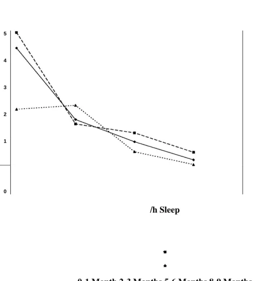
Figure 1 Figure 2: Maturation of total arousals, cortical arousals, subcortical activations during total sleep time from 0-1 month to 8-9 months of age. The frequency of arousals is given as an index (number of arousals/h of sleep).
Figure 1
Figure 2
Figure 3: Maturation of cortical arousals from 0-1 month to 8-9 months of age in total sleep, REM sleep and NREM sleep. The frequency of cortical arousals is given as an index (number of arousals/h of sleep).
Figure 3
Figure 4: Maturation of subcortical activations from 0-1 month to 8-9 months of age in total sleep, REM sleep and NREM sleep. The frequency of subcortical activations is given as an index (number of arousals/h of sleep).
Figure 4 5 4 3 2 1 0 /h Sleep
0-1 Month 2-3 Months 5-6 Months 8-9 Months
Figure 5: Evolution with age of cortical on subcortical ratio in total sleep, REM sleep and NREM sleep. The cortical on subcortical ratio represents the ratio between cortical arousals on subcortical activations.
Figure 5