HAL Id: inserm-01638055
https://www.hal.inserm.fr/inserm-01638055
Submitted on 19 Nov 2017
HAL is a multi-disciplinary open access
archive for the deposit and dissemination of
sci-entific research documents, whether they are
pub-lished or not. The documents may come from
teaching and research institutions in France or
abroad, or from public or private research centers.
L’archive ouverte pluridisciplinaire HAL, est
destinée au dépôt et à la diffusion de documents
scientifiques de niveau recherche, publiés ou non,
émanant des établissements d’enseignement et de
recherche français ou étrangers, des laboratoires
publics ou privés.
Karim Belarbi, Elodie Cuvelier, Alain Destée, Bernard Gressier,
Marie-Christine Chartier-Harlin
To cite this version:
Karim Belarbi, Elodie Cuvelier, Alain Destée, Bernard Gressier, Marie-Christine Chartier-Harlin.
NADPH oxidases in Parkinson’s disease: a systematic review. Molecular Neurodegeneration, BioMed
Central, 2016, 12 (1), pp.84. �10.1186/s13024-017-0225-5�. �inserm-01638055�
R E V I E W
Open Access
NADPH oxidases in Parkinson
’s disease: a
systematic review
Karim Belarbi
1, Elodie Cuvelier
1, Alain Destée
1, Bernard Gressier
1and Marie-Christine Chartier-Harlin
1,2*Abstract
Parkinson’s disease (PD) is a progressive movement neurodegenerative disease associated with a loss of dopaminergic neurons in the substantia nigra of the brain. Oxidative stress, a condition that occurs due to imbalance in oxidant and antioxidant status, is thought to play an important role in dopaminergic neurotoxicity. Nicotinamide adenine dinucleotide phosphate (NADPH) oxidases are multi-subunit enzymatic complexes that generate reactive oxygen species as their primary function. Increased immunoreactivities for the NADPH oxidases catalytic subunits Nox1, Nox2 and Nox4 have been reported in the brain of PD patients. Furthermore, knockout or genetic inactivation of NADPH oxidases exert a neuroprotective effect and reduce detrimental aspects of pathology in experimental models of the disease. However, the connections between NADPH oxidases and the biological processes believed to contribute to neuronal death are not well known. This review provides a comprehensive summary of our current understanding about expression and physiological function of NADPH oxidases in neurons, microglia and astrocytes and their pathophysiological roles in PD. It summarizes the findings supporting the role of both microglial and neuronal NADPH oxidases in cellular disturbances associated with PD such as neuroinflammation, alpha-synuclein accumulation, mitochondrial and synaptic dysfunction or disruption of the autophagy-lysosome system. Furthermore, this review highlights different steps that are essential for NADPH oxidases enzymatic activity and pinpoints major obstacles to overcome for the development of effective NADPH oxidases inhibitors for PD.
Keywords: Alpha-synuclein, Microglia, Mitochondria, Neurodegenerative disorders, Oxidative stress, Synaptic plasticity Background
Parkinson’s disease (PD) is the most prevalent move-ment disorder in elderly adults. It is characterized by the progressive degeneration of dopaminergic neurons in the substantia nigra and by the pathological accumula-tion of alpha-synuclein in protein aggregates named Lewy bodies in surviving neurons [1, 2]. Alpha-synuclein is a protein that is intracellularly localized in presynaptic terminals, where it has been shown to be involved in synaptic vesicle trafficking, synaptic function and plasti-city [3]. However, alpha-synuclein can also be released from neurons and this process can be promoted under stress condition and enhanced by alpha-synuclein mis-folding and aggregation [4, 5]. Sustained inflammation, defined by the presence of chronic microglial activation, is also consistently observed in the brain of patients [6].
These hallmarks are associated with dopamine deficit at the striatum -e.g. the striatal area innervated by the substantia nigra- that is the main factor leading to bra-dykinesia, resting tremor, rigidity and postural instability. It is generally accepted that these motor symptoms appear only after a substantial proportion of substantia nigra dopaminergic neurons are lost [7] suggesting that the disease has been engaged for years. This raises the possibility of adaptive or compensatory mechanisms in the early phase of this disease, involving synaptic plasti-city of the remaining neurons [8, 9].
More than 90% of PD cases are sporadic and attrib-uted to a combination of environment and/or genetic factors. Understanding the function of genes mutated in rare hereditary forms of PD has contributed to a better knowledge of PD pathogenesis. Missense mutations in the gene encoding alpha-synuclein SNCA (synuclein, alpha [non-A4 component of amyloid precursor]) were the first genetic abnormalities to be identified in PD families segregating as an autosomal dominant inherited
* Correspondence:marie-christine.chartier-harlin@inserm.fr
1University Lille, Inserm, CHU Lille, UMR-S 1172 - JPArc - Centre de Recherche
Jean-Pierre AUBERT Neurosciences et Cancer, F-59000 Lille, France
2Inserm UMR S-1172 Team“Early stages of Parkinson’s Disease”, 1 Place de
Verdun, 59006 Lille, France
© The Author(s). 2017 Open Access This article is distributed under the terms of the Creative Commons Attribution 4.0 International License (http://creativecommons.org/licenses/by/4.0/), which permits unrestricted use, distribution, and reproduction in any medium, provided you give appropriate credit to the original author(s) and the source, provide a link to the Creative Commons license, and indicate if changes were made. The Creative Commons Public Domain Dedication waiver (http://creativecommons.org/publicdomain/zero/1.0/) applies to the data made available in this article, unless otherwise stated.
trait [10]. Subsequently SNCA locus triplication and duplication were also shown as a cause of rare familial forms of PD [11–13]. Genetic evidence for a role of alpha-synuclein in sporadic PD emerged from the asso-ciation between polymorphisms regulating SNCA levels and sporadic PD [14, 15], supporting that alpha-synuclein level is instrumental in the most common forms of the disease. Other mechanisms are involved. Mutations in genes encoding proteins of the endosomal/ lysosomal system, vacuolar protein sorting-35 (VPS35), type 5 P-type ATPase ATP13A2, and glucocerebrosidase (GBA1) are also associated with PD [16] and represent a link between autophagy-lysosome function and neurode-generation. Furthermore, overexpression of leucine-rich repeat kinase 2 (LRRK2), another PD-related protein, causes an increase in autophagosome numbers and lyso-somal pH [17]. Other genes with a recessive PD-related inheritance such as DJ-1, PARKIN, PTEN-induced kinase 1 (PINK1) encode proteins playing an important role in the process of autophagy of mitochondria, known as mitophagy [16]. Mutations in one of these genes lead mitochondria to be morphologically aberrant and bioe-nergetically incompetent [18]. Although microglia activation and inflammatory changes are generally con-sidered as a consequence of neuronal destruction, genome-wide analysis evidenced that the HLA-DR region [19] and that genes involved in the ‘regulation of leucocyte/lymphocyte activity’, ‘cytokine-mediated signal-ing’ and more generally in the immune system are asso-ciated with an increased susceptibility to PD [20, 21]. This raises the possibility that a general pro-inflammatory state could be a primary cause of neuronal loss in some cases or at least increases PD risk as a disease modifier genotype. Thus, the identification of these PD-related genes has led to the proposition that the progressive deterioration of dopaminergic neurons may arise from cellular distur-bances produced by misfolding and aggregation of alpha-synuclein, mitochondrial dysfunction, disruption of the autophagy-lysosome system, endoplasmic reticulum stress, dysregulation of calcium homeostasis as well as chronic neuroinflammation [22, 23]. The discovery that environ-mental factors may be associated with PD promoted the creation of toxin-induced animal models designed to eluci-date the mechanisms of neurodegeneration [24, 25]. Some of the most widely used toxins to study PD in animals in-clude 6-hydroxydopamine (6-OHDA), 1-methyl-4-phenyl-1,2,3,6-tetrahydropyridine (MPTP) as well as pesticides such as rotenone (an insecticide) and paraquat (an herbicide). These models can present important features associated with the human disease including alpha-synuclein fibrilla-tion, dopaminergic neuronal cell loss, mitochondrial dys-function as well as oxidative damages (for review, see [26]).
The aforementioned cellular disturbances observed in the genetic and environmental models of PD are all
closely linked to oxidative stress [23]. The term oxidative stress describes a redox imbalance between generations of free radicals or other reactive species and antioxidant defenses, and it may be related to changes in microglia activation, protein clearance, mitochondrial function and the autophagy-lysosome system. Oxidative stress has long been hypothesized to be central in sporadic PD pathogenesis. It was already proposed in 1990 that free radicals generated from oxidation reactions inappropri-ately oxidize macromolecules resulting in cellular dys-function and, ultimately, in cell death [27]. This hypothesis is supported by several data. First, dopamin-ergic neurons of the substantia nigra are particularly sensitive to oxidative stress due to their high neuromela-nin content, the generation and auto-oxidation of dopa-mine generate oxygen species. Second, this area is highly demanding in energy and the discovery in the early 1980s of the mechanism of toxicity of MPTP showed that inhibition of mitochondrial complex I activity causes a degeneration of the nigrostriatal neurons and a parkinsonian syndrome in humans, rodents or primates (as reviewed in [28]). Third, studies of postmortem brain tissues demonstrate increased oxidation of proteins [29], lipids [30] and DNA [31] and decreased levels of the antioxidant glutathione [32, 33] in the substantia nigra of PD patients. Finally, The substantia nigra contains the highest density of microglia in both human [34] and rodents [35] and their activation may therefore encoun-ter an excessively high level of oxidative stress. Uncer-tainty about the molecular mechanisms leading to the oxidative stress in sporadic PD remains. A moderate def-icit in mitochondrial complex I has been repeatedly evi-denced in the substantia nigra of PD patients [36–38]. Furthermore, multiple aforementioned genes in which mutations or polymorphisms increase the risk of PD are linked to mitochondrial function or autophagy. As a consequence, it is considered that accumulation of bioe-nergetically compromised mitochondria could contrib-ute to reactive oxygen generation in PD. However, other sources of reactive oxygen species in the nervous system could also be involved.
In the present review, we consider the role in PD of enzymatic sources that generate reactive oxygen species as their primary function that are the nicotinamide
ad-enine dinucleotide phosphate (NADPH) oxidases.
NADPH oxidases comprise a family of multi-subunit membrane-bound enzymes. Several NADPH oxidases have been evidenced in both neurons and glial cells in the brain where they contribute to a wide range of physiological functions related for example to host-defense or long-term synaptic plasticity. However, an overproduction of reactive oxygen species by NADPH oxidases could be detrimental. Based on the literature, we discuss how both microglial and neuronal NADPH
oxidases could contribute to key cellular disturbances in PD such as microglia activation, alpha-synuclein accu-mulation, mitochondrial and synaptic dysfunction or dis-ruption of the autophagy-lysosome system.
The family of NADPH oxidases
NADPH oxidases are multi-subunit enzymes that primary catalytic function is the generation of reactive oxygen spe-cies. They function as electron transporters, using reduced NADPH as electron donor and molecular oxygen as elec-tron acceptor to generate superoxide and/or hydrogen peroxide. Reactive oxygen species generation by NADPH oxidases was first discovered in polymorphonuclear neu-trophils [39] as the enzyme responsible for the respiratory burst essential to the microbicidal function of these cells [40]. To the phagocytic catalytic subunit Gp91phox, called Nox2 (genomic location Xp21.1-p11.4) in the novel terminology, have been added six additional catalytic sub-units of the same family: Nox1 (Xq22.1) [41, 42], Nox3 (6q25.3) [43], Nox4 (11q14.3) [44, 45], Nox5 (15q23) [46] and dual oxidase 1 and 2 (Duox1 15q21.1 and Duox2 15q21.10) [47, 48]. Duox1 and Duox2 are termed dual
oxidase because they contain both a NADPH oxidase do-main and a peroxidase-like dodo-main. However, according to current knowledge, these two catalytic subunits do not display any peroxidase activity in human [49–51]. They are also named Nox6 and Nox7 according to [52].
All catalytic subunits of the NADPH oxidases family contain a‘NADPH oxidase domain’ that is characterized by at least six membrane-spanning alpha-helical do-mains containing two hemes, a predicted region for flavin adenine dinucleotide (FAD) and a NADPH bind-ing site in the cytosolic C-terminus [53] (Fig. 1). Despite this similar core structure, the NADPH oxidase family members differ in their subunit requirements (Table 1). Some of the NADPH oxidase catalytic subunits require association with other proteins that function as subunits such as p22phox, p40phox, p47phox, and p67phox and necessitate small GTPase Rac1 or Rac2 [54], as listed below. Nox1 requires both p22phox and NoxA1 for acti-vation. The activation of Nox2, that is permanently mem-brane bound and associated with the p22phox subunit [55], needs the cytosolic subunits p40phox, p47phox, p67phox and the small GTPase Rac1 or Rac2 to migrate to the
Fig. 1 Activation of the NADPH oxidase family members. The figure illustrates for each NADPH oxidase the catalytic core region (in blue), the transmembrane maturation and stabilization subunits (in red) as well as the cytosolic subunits and the small GTPases (Rac1 and Rac2). The predicted regions for FAD and NADPH binding sites and the putative peroxidase-like region are also shown, as well as the EF hand motifs (yellow circles) that bind to Ca2+
plasma membrane and assemble [56, 57]. In microglia, the phosphorylation and translocation of p47phox appears as the limiting factor for Nox2 activation [58] and various ki-nases have been implicated in p47phox phosphorylation. These include protein kinase C (PKC) isoforms [59–62], Akt [63, 64] mitogen-activated protein kinases (MAPK) [65], p21-activated kinase (Pak) [66] and extracellular signal-regulated kinase (ERK)1/2 [67]. Regulation of Nox3 appears to depend of the species and in human it needs activator subunits such as p22phox, NoxO1, NoxA1 and Rac for its activity. Nox4 requires p22phox [68] but does not involve cytosolic subunits. It is constitutively active in reconstituted systems [44, 69] and can be regulated in
response to cytokines and growth factors such as insulin-like growth factor-I and transforming growth factor-β [70–72]. Nox5, Duox1 and Duox2 are activated by an ele-vation in intracellular Ca2+ and do not appear to require subunits, either membrane-bound or cytosolic [46, 73].
The patterns of activation of the different NADPH oxi-dase family members are summarized in Fig. 1. It should be noted that nearly all catalytic subunit display different transcripts and isoforms, and thus the properties of these different family members are probably more complex, possibly to enable specific and fine regulation processes (Table 1). Thus, the existence of various catalytic NADPH oxidases with differing activation properties
Table 1 Human NADPH oxidases genes, genomic positions and isoforms
NADPH oxidase catalytic subunit
Official Gene symbol and
Full Name HUGOa Other Proposed Aliases b
Entrez
Gene IDdb Genomiclocationb mRNA (RefSeq Accession Numbers;GRCh38/hg38 human genome)c
Nox1 NOX1 NADPH oxidase 1 MOX1; NOH1; NOH-1; GP91-2 27035 Xq22.1 NM_007052 (isoform 1) NM_013955 (isoform 2) NM_001271815 (isoform 3) Nox2 CYBB cytochrome b-245
beta chain
CGD; NOX2; IMD34; AMCBX2; GP91-1; GP91PHOX; p91-PHOX; GP91-PHOX
1536 Xp21.1-p11.4 NM_000397→ NP_000388
Nox3 NOX3 NADPH oxidase 3 het; GP91-3; nmf250 50508 6q25.3 NM_015718→ NP_056533 Nox4 NOX4 NADPH oxidase 4 KOX; KOX-1; RENOX 50507 11q14.3 NM_016931 (isoform a)
NM_001143836 (isoform b) NM_001143837 (isoform c) NM_001291926 (isoform d) NM_001291927 (isoform e) NM_001291929 (isoform f) NM_001300995 (isoform g) NR_120406
Nox5 NOX5 NADPH oxidase 5 NOX5A, NOX5B 79400 15q23 NM_024505 (isoform 1) NM_001184779 (isoform 2) NM_001184780 (isoform 3) NR_033671
NR_033672
Duox1 DUOX1 dual oxidase 1 LNOX1; THOX1; NOXEF1 53905 15q21.1 NM_017434 (dual oxidase 1 precursor)
NM_175940 (dual oxidase 1 precursor)
Duox2 DUOX2 dual oxidase 2 TDH6; LNOX2; THOX2; NOXEF2; P138-TOX 50506 15q21.1 NM_001190392 dual oxidase 2 precursor NM_177610 dual oxidase 2 precursor NM_213999 dual oxidase 2 precursor NM_024141 dual oxidase 2 precursor a
HUGO:http://www.genenames.org/. Accessed January 5 2017
b
NCBI Entrez Gene:https://www.ncbi.nlm.nih.gov/gene/. Accessed January 5 2017
c
and isoforms, together with various tissue and cell distribution, suggests that NADPH oxidases may have numerous biological functions.
NADPH oxidases in the central nervous system
Since their identification in polymorphonuclear neu-trophils, NADPH oxidases have been evidenced in non-phagocytic cells and in various organs [74], including the brain. A study showed that within total human brain mRNA, Nox2 is predominantly present together with traces of Nox4 and Nox5 transcripts [43]. Another study detected Nox1, Nox3 and Duox1 in the rat brain tissues [75], further confirming that NADPH oxidases are a plausible generators of react-ive oxygen species in the brain.
The distribution of NADPH oxidases in the brain has been studied at the cellular level, in microglia, the resident immune cells of the central nervous system, in neurons and astrocytes (supporting glial cells). In microglia of both humans and rodents, analyses showed that Nox2 is the main NADPH oxidase catalytic subunit present (Fig. 2). Lower levels of Nox1 and Nox4, but not Nox3, have also been documented in microglia at the transcript level [76–81]. In resting microglia and macrophages that can infiltrate the brain under certain pathological conditions, Nox2 localizes to the plasma membrane into cholesterol-enriched membrane microdomains (lipid rafts) together with p22phox [77, 82]. Following macrophage/microglia activation, it is internalized by clathrin-coated pits and redistributed to an intracellular compartment consisting of numerous small (<100 nm) vesicles [83]. Nox1 in microglia appears to localize in intracellular vesicular compartments including lysosomes, and can be recruited to phagosomal membranes [79].
Neurons have been shown to express Nox1 (mRNA; [84]), Nox2 (mRNA and protein; [85, 86]) and Nox4 (mRNA and protein; [87]). Nox4 is predominantly asso-ciated with the internal membranes including the
endo-plasmic reticulum and the endosomes and the
mitochondria membrane [68, 88]. In PD patients, Nox1 and Nox4 have been observed in the nucleus of dopa-minergic neurons [89, 90]. While present in most mam-mals, the Nox5 gene is absent from rodent genomes [53], therefore preventing the study of the endogenous protein in murine experimental models.
Finally, Nox2 appears to be the predominant NADPH oxidase family member expressed in astrocytes where its expression was reported at both the mRNA and protein level. In line, a significant decrease in reactive oxygen spe-cies generation is evidenced in astrocytes from Nox2-deficient mice [91, 92]. The cellular and subcellular expression of NADPH oxidases in the brain is summarized in Fig. 2.
The physiological functions of NADPH oxidase en-zymes are various, including host defense and inflamma-tion, post-translational processing of proteins, cellular signaling and regulation of gene expression (for re-view, see [74, 93]). The majority of publications on the physiological roles of NADPH oxidases in the central ner-vous system have focused on their participation in the host defense and the removal of debris from the brain. NADPH oxidases have also been shown to regulate neur-onal cell fate and function and plasticity, for instance con-tributing to the induction of neuronal apoptosis in response to serum deprivation [85], to N-methyl-D-aspar-tate (NMDA) receptor signaling [94] and to long-term po-tentiation [95]. In the following paragraphs, we provide a thorough review of existing knowledge and information
Fig. 2 Cellular and subcellular expression of NADPH oxidase catalytic subunits in the brain. a Schematic diagram showing the reported cellular localization of NADPH oxidase family members in the brain cells. b Schematic diagram showing the reported subcellular localization of NADPH oxidase family members in a hypothetical cell in the brain
relative to the contribution of specific NADPH oxidase family members in microglia and neurons and highlight the main conclusions regarding PD pathogenesis.
NADPH oxidases and microglia activation in PD
Microglia are the resident mononuclear phagocytes of the central nervous system, belonging to the glial sys-tem of non-neuronal cells that support and protect neuronal functions. Microglia comprise 5–20% of the total glial cell population within the central nervous system parenchyma and most densely populated areas include the substantia nigra [96]. Microglia are not only the first immune sentinels of infection, contrib-uting to both innate and adaptive immune responses locally, but also play intrinsic functions required for normal brain function and neuroprotective effects. Chronic microglia activation is consistently reported in the pathogenesis of PD [97] and can be triggered experimentally by several PD-causing gene products such as alpha-synuclein or DJ-1.
Microglial Nox2 expression is increased in PD and experimental models of PD
In 2003, Przedborski and coworkers reported that post-mortem substantia nigra samples from sporadic PD pa-tients had higher Nox2 (then referred as gp91phox) protein content than samples from control individuals (six PD patients versus three controls; mean duration of disease of 16.8 ± 2.3 years) [98]. Nox2 immunostaining localized with the microglial marker CD68, but not with neuromelanin. An increase in microglial Nox2 was also observed in microglia in the ventral midbrain of mice (brain region containing the substantia nigra pars com-pacta) after repeated intraperitoneal injections of MPTP [98]. In this model, the microglial expression of Nox2 was confirmed by immunostaining co-localization with the marker Macrophage antigen complex-1 (Mac-1) [98] and by ex vivo approaches showing an increase by ap-proximately 3.5-fold in Nox2 expression in microglia acutely isolated, compared to saline-treated mice [99]. The MPTP model also show increased p67phox gene ex-pression [98], translocation of the subunit p67phox from the cytosol to the plasma membrane [100], as well as in-duced p47phox phosphorylation and p47phox–Nox2 complexes in substantia nigra tissues [101], further con-firming the activation of the enzyme. A marked increase in Nox2 protein level in reactive microglia was also doc-umented in experimental models of PD based on com-bined administration of minimally toxic dose of LPS and alpha-synuclein oligomers [102], intraperitoneal injec-tion of paraquat [103] or 6-OHDA [104, 105], on expos-ure to atmospheric ultrafine particles considered as a potential environmental risk factor for PD [106] as well
as with aging and traumatic brain injury known to in-crease the risk of parkinsonism [107–109].
Taken together, these data strongly suggest that micro-glia are the predominant Nox2-expressing cells in PD and in several experimental models of PD and that Nox2 activation could contribute to its pathophysiology.
Nox-2 modulates microglial responses and neurotoxicity
Microglial cells have a wide range of profiles and actions. On one hand, sustained classical microglia acti-vation - consistently detected in the substantia nigra of patients with PD [6, 110–112] - could promote the slow degeneration of dopaminergic neurons. This is demon-strated in rodent models based on lipopolysaccharide administration [113, 114]. On the other hand, microglia act as neuroprotective cells through the elimination of endogenous or exogenous substances and participate to the resolution of the inflammatory response [115]. Moreover microglia have high levels of glutathione and glutathione peroxidase, which act to protect them and possibly neurons from toxic levels of H2O2[116].
Lipopolysaccharide activates microglia primarily through the pattern recognition receptor Toll-like receptor (TLR) 4 signaling pathway [113, 117, 118]. However, lipopolysac-charide stimulation of Nox2 activity in microglia mainly oc-curs through binding of lipopolysaccharide to Mac-1, also named complement receptor (CR) 3 [119, 120]. Mac-1/ CR3 is composed of CD11b (integrin αM) and CD18 (integrinβ2) subunits. In Nox2-deficient mice, lipopolysac-charide fails to induce classical microglia activation as eval-uated by Iba1 immunoreactive cells morphology, intensified F4/80 staining or induction of tumor necrosis factor (TNF)-alpha expression. Furthermore, lipopolysaccharide-induced loss of dopaminergic neurons is attenuated com-pared to Nox2+/+ mice [121, 122]. Also, Nox2 deficient microglia do not migrate towards substance P, a proinflam-matory neuropeptide with high concentrations in the substantia nigra [123], supporting that Nox2 could be im-plicated both in the recruitment and the classical activation of microglia.
Nox2 could play a role in the deleterious effects of microglia in PD. For instance studies using cell culture systems revealed that microglia lacking functional Nox2 fail to produce neurotoxicity in response to MPTP [124, 125], paraquat [126] or rotenone in contrast to Nox2+/+ microglia [127]. This was corroborated by numerous in vivo studies showing that mice lacking Nox2 are less sensitive to dopaminergic degeneration induced by pesti-cides. For example, daily subcutaneous injections of MPTP results in a 32% loss of tyrosine-hydroxylase im-munoreactive neurons in Nox2+/+ mice compared to a 14% loss in Nox2-deficient mice [128]. Similar attenua-tions of neurotoxicity in the Nox2−/− background were observed in mice receiving MPTP injections [98],
paraquat injections [103] and in mice lesioned with 6-OHDA [104, 129]. Consistent with this protection, mice defective in Nox2 show less production of proinflamma-tory cytokines interleukin-1beta, TNF-alpha or inter-feron gamma (6-OHDA model; [104]) and less reactive oxygen species production and protein oxidation (MPTP model; [98]). Recently, Zhang and colleagues investi-gated the mechanisms underlying dopaminergic neuro-degeneration using in vivo and in vitro models based on exposure to minimally toxic dose of LPS and alpha-synuclein oligomers. In their study, synergistic dopamin-ergic neurotoxicity - indicated by reduced dopamine up-take capacity, dopaminergic neuronal numbers in the substantia nigra pars compacta and depleted dopamin-ergic level in striatum – was reduced in Nox2−/− mice compared with Nox2+/+ mice [102]. Furthermore, micro-glial production of superoxides and reactive oxygen spe-cies were robustly reduced in Nox2−/− mice compared to Nox2+/+ mice. Thus, both exogenous and endogenous factors involved in PD appear to propagate classical microglia activation and dopaminergic neurodegeneration through activating microglial Nox2.
A role for Nox2 deficiency to promote neuroprotective role of microglia might emerge from the study of Hernandes and colleagues. In this study, Nox2−/− mice were treated with minocycline or saline and received 6-OHDA injections. Minocycline is a tetracycline derivative that exerts multiple anti-inflammatory effects, including microglial inhibition. Interestingly, the degeneration of dopaminergic neurons after 6-OHDA injections is greater in Nox2−/− mice that were treated with minocycline com-pared to Nox2−/− mice treated with vehicle [104]. Minocy-cline treatment also leads to NF-kappaB activation and increases TNF-alpha release into the substantia nigra of Nox2−/− 6-OHDA lesioned mice [104]. Therefore inhibit-ing Nox2−/− microglia cells likely increases substantia nigra degeneration and parkinsonism, suggesting a protect-ive role for Nox2−/− microglia.
Although more studies are needed, these data demon-strate that Nox2 adds an essential level of regulation to signaling pathways underlying the inflammatory re-sponse. While microglial Nox2 appears to contribute to the chemoattraction, the classical activation and the tox-icity towards dopaminergic neurons of microglial cells, inhibiting Nox2 signaling in microglia could favor their neuroprotective profile and actions in the context of PD.
Alpha-synuclein, DJ-1 and Nox2 in microglia
Among the factors linked to the etiology of PD, alpha-synuclein and DJ-1 have been linked to microglia activa-tion and could modulate NADPH oxidase activity in sev-eral ways as described below. Alpha-synuclein has a direct effect on microglial activation in vitro resulting in an overall increase in proinflammatory molecules and
oxidative stress [130]. The signaling pathways mediating this process are multiple, and may depend of the struc-ture and/or aggregation state of alpha-synuclein. For in-stance, alpha-synuclein monomers and fibrils were shown to induce interleukin-1beta release from mono-cytes via the TLR2 [131] and oligomeric forms of alpha-synuclein also specifically activate TLR2 [132]. However, the TLR4 has also been implicated in alpha-synuclein-induced inflammation [133]. A role for Mac1 has been proposed and for instance the binding of alpha-synuclein to Mac1 has been involved in oxidant release from microglia [134]. Finally, purinergic receptors have been implicated as a direct association between alpha-synuclein and the ionotropic P2X7 purinergic receptor leads to Nox2 activation through the phosphoinositide 3-kinases (PI3K) signaling pathway [135].
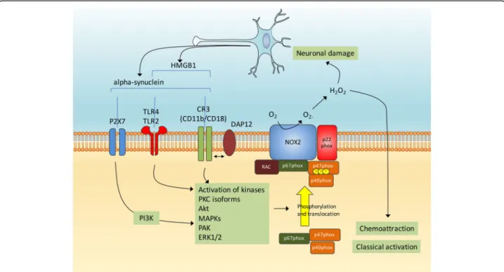
Both wild-type and A53T mutant alpha-synuclein were shown to activate Nox2 in BV2 microglial cells and in pri-mary cultured microglia, with the A53T form producing quickest and sustained effects in terms of oxidative stress and cellular injuries. Interestingly, the process is partly blocked when BV2 cells are pretreated with LY294002, a strong inhibitor of PI3K [135]. Further supporting that alpha-synuclein promotes microglial Nox2 expression in pathological conditions, neuronal alpha-synuclein levels are elevated after spinal cord ischemic/reperfused injury and when cocultured with injured neurons or superna-tants from injured neurons, Nox2 expression, reactive oxygen species generation and TNF-alpha expression are promoted in microglia. In this model, microglia activation is impeded by pretreatment with alpha-synuclein antibody or TLR2 antibodies and Nox2 levels in microglia are re-duced by the pharmacological inhibition of MAPK p38 [136]. The comparison of the ability of various alpha-synuclein peptides to activate microglia allowed the identi-fication of a specific peptide consisting of amino acids A29-V40 of alpha-synuclein that can directly bind to Nox2 resulting in NADPH oxidase complex activation [137]. When administered to wild-type mice, the A29-V40 peptide increases the expression of MHC-II, a cell surface marker of microglia classical activation [138], as well as the amount of malondialdehyde, one of the products dur-ing lipid peroxidation. In contrast, administration of the A29-V40 peptide has no such effects in Nox2−/− mice, suggesting that this alpha-synuclein peptide activates microglia and elicits oxidative stress in vivo in a Nox2-dependent manner [137]. Of note, a recent study indicates that the reactive oxygen species originating from activated Nox2 also serve as a direct signal driving microglial direc-tional migration induced by the binding of alpha-synuclein to CD11b [139]. Thus, it is likely that several signaling pathways link alpha-synuclein with Nox2 and microglia activation (Fig. 3). Altogether, these findings show an important role for microglia Nox2 in mediating
alpha-synuclein elicited microglia chemoattraction, activa-tion and oxidative stress.
DJ-1 is another PD’s related gene product that has been suggested to have several possible functions includ-ing roles as an oxidative stress sensor, a protein chaperone, a protease, an RNA-binding protein, a tran-scription regulator, a regulator of mitochondria function and a regulator of autophagy. Recently, Liu and col-leagues reported that DJ-1 binds to p47phox and that DJ-1 deficiency blunts TLR signaling and impairs NADPH oxidase-dependent reactive oxygen species pro-duction in macrophages. They moreover reported in DJ-1−/− versus control mice increased bacterial burdens, reduced local and systemic inflammation, macrophage paralysis and impaired induction of proinflammatory cytokines under the condition of sepsis. Importantly, in vivo administration of DJ-1 restored macrophages and rescued animals from septic death induced by lipopoly-saccharide [140]. The study by Amatullah and colleagues also demonstrated the binding of DJ-1 with p47phox. In contrast with the aforementioned study, absence of DJ-1 resulted overall in increased reactive oxygen reactive species production and in increased NADPH oxidase ac-tivity, as observed in a mouse model of polymicroglial sepsis (the cecal ligation and puncture model) and in bone marrow-macrophages in response to LPS. The
authors then demonstrated that DJ-1-p47phox interaction could disrupt the NADPH oxidase complex assembly and/or facilitate Nox2 ubiquitination and degradation thereby decreasing reactive oxygen species production [141]. Taken together, these two studies evidence that DJ-1 binds to p47phox and can modulate NADPH-oxidase function. Future studies are needed to precise the implica-tion of this interacimplica-tion regarding oxidative stress and inflammation PD and to determine whether PD-related DJ-1 mutations impact on this interaction.
Altogether, these data show that both
alpha-synuclein and DJ-1 can direct NADPH-oxidases acti-vation in microglia and such signaling could likely interfere with their neurotoxic and neuroprotective ef-fects. The relationship between NADPH-oxidases and other genetic factors associated with PD will require further investigation. Of note, induction of colitis by dextran sulfate sodium leads to an earlier and severer colitismice paralleled by an increased phosphorylation of LRRK2 in p47phox mutated mice [142]. Addressing whether NADPH-oxidase contribute to changes in the im-mune response along with LRRK2 in PD will be of prime interest as both factors are expressed in immune cells [143] and appear to be important player in microglial activation and in alpha-synuclein-mediated microglial activation as well [144].
Fig. 3 Alpha-synuclein and microglial Nox2 activation. The activation of microglia by alpha-synuclein can implicate several cell surface receptors such as P2X7, TLR2/4 and CR3 and subsequent activations of several kinases such as PKC, Akt, MAPKs, PAK and ERK1/2. This in turn could promote the phosphorylation and translocation of p47phox and subsequent Nox2 activation. Released oxygen species appear to promote microglia chemoattraction, activation and oxidative stress. Neuronal damage leads to the release of alpha-synuclein and the TLR-agonist high mobility group box protein 1 (HMGB1)
Neuronal NADPH oxidases and neurodegeneration in PD
As discussed in introduction, defects in several cellu-lar processes may push substantia nigra dopaminergic neurons towards cell death. These include oxidative damage, energy failure associated with mitochondrial dysfunction, altered glutamatergic neurotransmission and alterations in protein degradation efficacy. The finding that NADPH oxidases localize to specific sub-cellular organelles in neurons, such as the nucleus, the mitochondria and pre-synaptic sites [145–148] has raised the possibility that they could contribute to these defects, with implications for the development of neuroprotective therapies.
NADPH oxidases, oxidative damage and alpha-synuclein accumulation in neurons
The presence of oxidative damage to the content of dopa-minergic neurons has been consistently reported in the substantia nigra of PD patients. Suggesting that NADPH oxidases could play a role in these damages, a confocal mi-croscopy imaging study evidenced Nox1 in the nucleus of dopaminergic neurons in the substantia nigra of postmor-tem brains of four PD patients while no signal was detected in those from control individuals [89]. Nox1 expression is also increased in the substantia nigra dopaminergic neu-rons in mice or rats in response to intraperitoneal injections of paraquat [149, 150] or striatal stereotaxic injection of 6-OHDA [89]. In these models, rise in Nox1 expression is as-sociated with an increase in total and proteinase-K resistant alpha-synuclein levels, as well as with lipid peroxidation (paraquat model; [150]) and immunoreactivity for the DNA oxidative stress marker, 8-oxo-dG (6-OHDA model; [89]). Nox1 knockdown or Rac1 inhibition achieved by stereotaxic delivery of adeno-associated virus serotype 2 (AAV2) particles into the rat sub-stantia nigra significantly reduces 6-OHDA-elicited immunostaining with 8-oxo-dG and dopaminergic neuronal loss [89]. The selective knockdown of Nox1 in the substantia nigra also largely attenuates the paraquat-mediated increase of total and proteinase K-resistant alpha-synuclein, oligomer-specific A11 immunoreactivity, oxidative stress and dopaminergic neuronal loss [150]. The Nox1/Rac1 complex was further analyzed in N27 rat dopaminergic cells in which (i) Nox1 tagged with EGFP translocates into the nucleus following 6-OHDA or rote-none treatments and (ii) Nox1 and GTP-bound activated Rac1 were detected by immunoprecipitation of nuclear extracts after a 24 h treatment with 6-OHDA [89, 151]. Therefore Nox1 translocation to the nucleus likely promotes subsequent Nox1/Rac1-derived super-oxide generation responsible for oxidative damages to the neuron and this is associated with increased detri-mental alpha-synuclein levels.
More recently, Nox4 immunoreactivity was noted in the nucleus of dopaminergic neurons in PD patients at Braak stage 6 [90], a stage with widespread alpha-synuclein accumulation [152]. Very interestingly, when looking at the first-affected nigral subregion nigrosome 1, the authors evidenced that the nuclear expression of Nox4 increases stepwise from age-matched controls (n = 7) to asymptomatic (n = 3) to clinically-confirmed PD patients (n = 5). Besides being correlated with nega-tive clinical output, the elevated nuclear expression of Nox4 is also associated with oxidative damage to DNA, caspase-3-mediated cell loss and increased distribution of the angiotensin II type 1 (AT1) receptor in the nu-cleus. Because the activation of the AT1 receptor stimu-lates NADPH oxidase activity [153, 154] and because AT1 receptor increases with age and in response to 6-OHDA treatment [155], the authors propose that angio-tensin II/AT1/Nox4 axis-mediated oxidative stress could contribute to damages in neurons in PD [90].
Cross talk between mitochondria and NADPH oxidases in neurodegeneration
The regulation and maintenance of brain function re-quires high amount of energy, consuming ~20% of total body energy. Neurons depend primarily in oxidative phosphorylation to meet their energy demands, while glucose metabolism is directed towards the pentose phosphate pathway to generate NADPH. Mitochondrial dysfunction in PD is likely to contribute to energy failure and to the excess of reactive oxygen species and subse-quent oxidative damages. Mitochondria and NADPH ox-idases are both major sources of superoxide induction and several lines of evidence suggest that they might be considered along in PD. A first argument is that mito-chondria can control the transcriptional activation of Nox1 as demonstrated in osteocarcinoma cells. In this model the inactivation of mitochondrial genes leads to the down-regulation of Nox1 expression. Conversely, in-creasing mitochondrial superoxide levels by exposing the cells to inhibitors of electron transport chain such as rotenone or antimycin increases the expression of Nox1 [147]. A molecular signaling link between mitochondria and Nox1 was further investigated for its contribution to superoxide production and apoptosis induced by serum withdrawal in human 293 T cells [156]. In this model serum withdrawal promotes the production of reactive oxygen species by stimulating both the mitochondria and Nox1. Mitochondria respond to serum deprivation within a few minutes. The mitochondria-generated re-active oxygen species stimulate PI3K that in turn in-duces the translocation of Rac1 to membrane fractions and the Rac1/Nox1 interaction, leading to sustained ac-cumulation of reactive oxygen species [156]. Serum withdrawal-treated cells eventually loose their viability,
which is prevented by blocking either the mitochondria-dependent induction of reactive oxygen species using rotenone or potassium cyanide or the PI3K/Rac1/Nox1 pathway using dominant negative mutants or small interfering RNAs [156]. Taken together, these data pro-vide great epro-vidence of a signaling link between the mito-chondria and Nox1, which could be crucial for the sustained accumulation of reactive oxygen species and cell death processes in PD.
PINK1, which is linked to autosomal recessive familial PD, is a mitochondria-targeted serine/threonine kinase. It is well established that PINK1 protects neurons from oxida-tive stress [157] and in particular, loss of PINK1 function causes dysregulation of mitochondrial calcium handling, resulting in mitochondrial calcium overload which stimu-lates reactive oxygen species production. Of interest, Gan-dhi and coworkers demonstrated that reduction of NOX2 expression in PINK1 knockdown neuroblastoma cells sig-nificantly attenuates reactive oxygen species production [158], therefore evidencing a new signaling pathway at the crossroad between mitochondrial stress, oxidative stress and NADPH-oxidases.
More recently Nox4 was also directly associated to mito-chondria as evidenced by confocal microscopy imaging showing its colocalization with the mitochondrial dye Mito-traker both in mice and in a murine catecholaminergic cells [88]. As investigated in this study, the expression level of Nox4 increases following angiotensin II exposure and knockdown of Nox4 achieved by adenoviral-encoded small interfering RNA significantly attenuates the angiotensin II-induced increase in mitochondrial-localized superoxide production [88]. Therefore it is likely that Nox4 contributes significantly to superoxide production at the mitochondria in response to angiotensin. Although more studies are needed, one can hypothesize that NADPH oxidases activa-tion at the mitochondria could interact with elements of the electron transport chain within the mitochondria, thus indirectly initiating the production of mitochondrial super-oxide [159, 160].
In conclusion, a cross talk between mitochondria and NADPH oxidases may represent a feed-forward vicious cycle of reactive oxygen species production that could con-tribute to oxidative damages and neurodegeneration in PD. NADPH oxidases-targeted antioxidants might break this vi-cious cycle, reducing NADPH-oxidase and limiting reactive oxygen species production by mitochondria.
NADPH oxidases, synaptic signaling and excitotoxicity
Neurons utilize most of their energy at the synapse and an impairment of the ability of neurons to undergo synaptic plasticity is key in several theories explaining the onset and the progression of PD [161]. Dopaminergic denerv-ation causes a profound network rearrangement, with the appearance of distinct forms of aberrant synaptic
plasticity. Synaptic alterations in PD are also associated with abnormal expression or function of the NMDA re-ceptor, a glutamate receptor and ion channel protein found in nerve cells [162]. Reactive oxygen species are re-quired for NMDA receptor-dependent activation of ERK and long term potentiation [163], both of them being im-plicated in PD [164]. A specific role for NADPH oxidases at the synapse was initially suggested by Tejada-Simon and colleagues who reported that Nox2, p22phox, p40phox, p47phox, p67phox, and Rac are enriched in synaptoneurosome preparations from mouse hippocampal homogenates. Dual immunofluorescent labeling also show that 67phox or Nox2 colocalize with synaptophysin and synaptotagmin, respectively, confirming their localization at pre-synaptic sites [145]. Isolated synaptosomes have been shown to exhibit NADPH-dependent oxygen con-sumption and quantitative production of superoxide radi-cals and these are partially inhibited by the Nox2 inhibitor apocynin [165]. This production appears to be significant as NADPH oxidases rather than mitochondria are identi-fied as the major reactive oxygen species source in isolated synaptosomes from mice, as assayed using spin trapping electron paramagnetic resonance spectroscopy [166]. Taken together, these studies clearly demonstrate the localization of functional NADPH oxidases at the synapse of neurons, making of NADPH oxidases prominent candidates as a source for reactive oxygen species for the control of synap-tic neurotransmission.
In 2005, Kishida and colleagues reported that pharmaco-logical inhibition of the NADPH oxidases using dipheny-lene iodonium or the lack of the p47phox subunit inhibit NMDA receptor-dependent ERK activation in hippocampal slices of mice [94], suggesting a direct role for NADPH oxi-dases in this signaling pathway. In line, pharmacological in-hibition of NADPH-oxidases by diphenylene iodonium or apocynin or knockdown of Nox2 or p47phox in mice block NMDA receptor-dependent early-phase long-term potenti-ation in the hippocampus, leaving the basal synaptic trans-mission intact [95]. Corroborating these results, the rapid increase in superoxide production induced by NMDA re-ceptor activation is blocked by the Nox2 inhibitor apocynin and in neurons lacking the p47phox subunit, both in cellulo and in vivo [167]. Therefore, Nox2 appears as the primary source of NMDA-induced superoxide production in neu-rons, being critical for NMDA receptor-related synaptic plasticity. Although these data were obtained in the hippo-campus, mice lacking Nox2 present deficits not only in memory formation but also in the rotating rod and open field tests, suggesting a role for NADPH oxidases in synap-tic function in several brain areas [95]. Finally, a role for Nox2 has been evidenced in the release of glutamate and dopamine after the administration of the NMDA receptor antagonist ketamine in mice [168]. Future research is needed to explore whether NADPH oxidases deregulations
could control NMDA receptor–dependent glutamate and dopamine release in the context of PD.
While NMDA receptor is critical for synaptic plasti-city, its sustained activation leads to extensive super-oxide production promoting neuronal death [169]. As such, the finding that NMDA receptor stimulation trig-gers NADPH oxidases activation also provides a mech-anistic link between oxidative stress and excitotoxicity. Of interest, PI3K is essential for NMDA receptor-dependent activation of Nox2; for example the PI3K inhibitor wortmannin reduces NMDA-induced Nox2 ac-tivation and cell death in primary neuronal cultures [170]. Perturbations in the PI3K/Akt pathway have been reported in PD patients [171, 172] and, as such, may either increase or decrease Nox2 superoxide production thereby impacting synaptic plasticity, the release of neu-rotransmitters and excitotoxicity in PD. This suggests that deregulation in NADPH oxidases expression, localization or activation could directly contribute to synaptic defects and excitotoxicity in PD.
NADPH oxidases and the autophagy-lysosome system
Autophagy is a dynamic cellular pathway involved in the degradation of misfolded proteins and other cellular con-stituents. Impairment of the autophagic flux has been evi-denced in PD and could promote the accumulation of compromised mitochondria and alpha-synuclein [23]. Several publications reported that exposure to rotenone can impair the autophagic flux, resulting in cytosolic accu-mulation of the autophagosomal membrane form of microtubule-associated protein 1 light chain 3 (LC3) [173], increased p62 levels [174] and aberrant accumulation of alpha-synuclein [175]. This rotenone’s impact on autophagy likely occurs through the PI3K/Akt/mTOR signaling
pathway [173, 176]. To determine whether rotenone-induced autophagy was Nox2 dependent, Pal and col-leagues used the human neuroblastoma SH-SY5Y cell line. They observed that short exposure to rotenone (0.5 μM; 6 h) results in a ~2-fold increase in reactive oxygen species generation, impairs autophagic flux and promotes protein accumulation. Pre-incubation with the Nox2 docking se-quence (Nox2ds)-tat significantly attenuates the levels of LC3 and p62 proteins, indicating that the effect on autoph-agy is mediated by Nox-2 dependent reactive oxygen spe-cies. When SH-SY5Y dopaminergic cells are exposed to higher doses of rotenone (10μM; 24 h), a ~3.5 increase in reactive oxygen species generation is observed compared to untreated cells. Nox2-ds abolish Nox2-generated reactive oxygen species generation (measured using the Nox2-specific redox sensor p47-roGFP) while total intracellular reactive oxygen species (measured using DCF-DA) is par-tially, but not completely, inhibited [177]. This suggests that 10μM rotenone for 24 h stimulates reactive oxygen species generation not only through Nox2, but also possibly from mitochondria. Importantly, preincubation with Nox2-ds par-tially attenuates LC3 and p62 protein levels and protects against rotenone-dependent upregulation in apoptotic sig-naling [177]. These data highlight a novel mechanism by which Nox2-dependent oxidative stress could promote the pathogenesis of PD. Noteworthy, Nox4 was also found to promote autophagy and survival in cancer cells [178] and in cardiomyocytes in response to nutrient deprivation and is-chemia [179], while no studies have been performed yet in the neuronal context. Thus, this emerging evidence indicates the importance of NADPH oxidases in the regulation of au-tophagy. Future studies are warranted to delineate the asso-ciation between NADPH oxidases-dependent impaired autophagy, mitochondria dysfunction and cell death in PD.
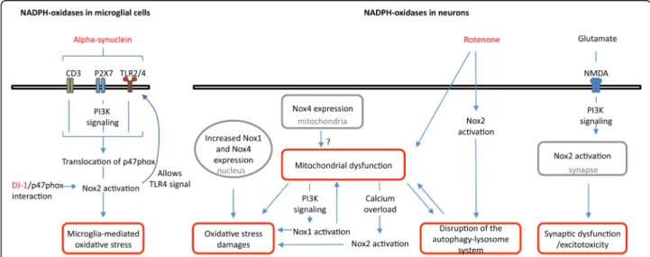
Fig. 4 Proposed role for NADPH oxidases in PD. Schematic view of the link between both microglial and neuronal NADPH oxidases and cellular processes related to PD, e.g. alpha-synuclein signaling, microglia activation, oxidative stress and neuronal damage, mitochondria dysfunction, disruption of the autophagy-lysosome system, synaptic dysfunction and excitotoxicity. Localizations of NADPH oxidases are indicated in grey
Table 2 Therapeutic targeting of NADPH oxidases in selected studies NAD PH oxid ase signali ng step Mod e of action Inhibit ors Exp eriment al mod el Eff ects of the inhib itor on the exper iment al mod el Current the rapeutic roadb locks Re ference Subu nit expre ssion Decre ase spec ific sub unit mR NA and prot ein level s Adeno -associated virus serotype 2 (AAV2) expre ssion cassettes wit h Nox1shR NA Stria tal injec tion of 6-OH DA in the rat Nox1 knoc kdown reduc ed 6-OH DA-induc ed oxidati ve DNA damage an d dopamin ergic neuro nal de generation. Drug delivery [ 89 ] Knockdow n of Nox4 achieved by adeno viral-encode d sm all interfe ring RNA Mou se catechol ami nergic neu ronal cell mod el (CAT H.a) expose d to an gioten sine II Nox4 knoc kdown atte nuated of angiote nsine II-indu ced mi tochon drial O2 ∙-prod uction. Drug delivery [ 88 ] Ligan d-rece ptor binding Block the interac tion be tween alp ha-syn uclein and P2 X7R Brilliant Blue G, a P2 X7R antagonist BV 2 microg lial cells tre ated with wil d type or A53T alp ha-syn uclein Pre treatmen t with Brillian t Blue G red uced the trans locatio n of p47p hox from the cyto plasm to the me mbrane after treatme nt with each form of alp ha-syn uclein. Targets one of many NADPH oxid ase activators [ 135 ] Comp lex assem bly and activ ation AAV2 expressi on cassettes with a T17N dom inant neg ative Rac1 variant Stria tal injec tion of 6-OH DA in the rat Rac1 inhibit ion reduced 6-OH DA-induce d oxid ative D NA dam age and dopaminerg ic neu ron al dege neration. Drug delivery [ 89 ] Pre vent p47ph ox ass ociatio n with NAD PH ox idase com plex Apocy nin Lip opolysacchari de induced PD mod el : sing le injec tion of lipo polysa ccharide at a dose of 5ug/5ul PBS into the SN of rats. Apoc ynin pre vents α -synuclein agg regation, microg lial activ ation , dopaminerg ic neuro degen eration and rel ieves mot or system abnorm ali ty followi ng lipopolysaccharide inje ction. Unsp ecific, dosi ng: [ 189 ] gp91 -ds,a pe ptide inhibit or for NAD PH oxidase ass embl y SH-SY5Y dopamin ergic cells expose d to roteno ne Pre incubation with Nox2-d s partially atte nuated LC3 an d p6 2 prot ein levels and pro tected against ro tenone-de pende nt upr egula tion in apoptotic signaling (Pal et al. , 20 16). Drug delivery, pharmac okin etic an d CNS biod ispon ibility [ 177 ] Block PI3K sign aling neede d for NADPH oxidase activ ation LY2940 02, a potent and specific cell -permeable inhibit or of PI3K [ 135 ] Elect ron transf er Extrac ts electro ns Diphen yleneiod onium Ne uron-glia cultures pretreat ed with lip opolysacchari de, 1-m ethyl-4 -phe nylpy ridiniu m or roteno ne Diphe nylene iodoni um protect ed the dopamin ergic neu rons at subpi comolar conce ntrations . 2 non-specific ity for othe r flavoenzymes an d high cytotoxi city at standard doses (μ M) [ 190 ]
NADPH oxidases inhibitors as potential therapeutic agents in PD
To date, strategies directly targeting oxidative stress in PD have been typically focused on compounds that scavenge re-active oxygen species after they have been produced and on mitochondria-targeting therapeutics. Despite the importance of oxidative stress and mitochondria in PD, these strategies have shown mitigated or no success in clinical trials [180]. Modulating NADPH oxidases activity would represent a logical alternative to modify the course of PD, as it targets enzymatic complexes that are solely dedicated to the pro-duction of reactive oxygen species and that regulate cellular processes that are disrupted in PD (Fig. 4).
Different steps that are essential for NADPH oxidases enzymatic activity can be targeted. These include (i) the expression of NADPH oxidases subunits, (ii) the ligand-receptor binding, (iii) the complex assembly and its activa-tion (including inhibiting signaling pathways such as PI3K signaling) as well as (iv) electron transfer. As illustrated in Table 2, several of these targets have been partially evaluated using either in vitro or in vivo experimental models of PD. These preclinical studies appear overall promising with for example decreases in markers of oxidative stress, in markers of autophagy-lysosomal defects or in neurodegeneration (Table 2). Beside knock-out mouse models and interfering RNAs, several small compounds or antibodies and peptides have been postulated as NADPH oxidases inhibitors. For a critical overview of the NADPH oxidases inhibitors, we refer the reader to the review of Altenhöfer and colleagues [52]. Briefly, the historical Nox2 inhibitors diphenylene iodonium and apocynin are commonly used but are unspecific and their effects cannot be solely attributed to inhibition of Nox2 or even NADPH oxidases [181]. More recently, sev-eral compounds inhibitors have been identified by rational drug discovery approaches and characterized with regards to NADPH oxidases selectivity and potential off-target ef-fects. For example, ML171 is a potent Nox1 inhibitor with IC50 value of 130-150 nM for Nox1 and of 3-5 μM for
Nox2-4. GKT136901 [182, 183] and GKT137831 [184] are two structurally related compounds developed by Genkyo-tex. They potently inhibit Nox1, Nox4 and Nox5 with ~10 lower IC50value than for Nox2 inhibition. They are orally
bio-available and have favorable ADME profiles [183, 184] and have consequently been evaluated across a range of disease models. GKT137831 has progressed through pre-clinical development and into pre-clinical trials and thus ap-pears as a good pharmacological tool for in vivo studies on the role of Nox1, Nox4 and Nox5 NADPH-oxidases enzymes in disease [185]. Peptide-based inhibitors such as Nox2-ds-tat, by their nature, have the potential advantage of being more specific and having fewer off-target effects than small-molecule organic compounds. However these peptides have disadvantages as well, such as low bioavail-ability and metabolic libioavail-ability and - over time - induction
of neutralizing antibodies. Overall, the high degree of structural and catalytic homology between the different catalytic subunits of NADPH oxidases makes finding se-lective inhibitors a challenge. However, as proposed by Diebold and collaborators several protein-protein interac-tions could in principle be targeted with new high-throughput assays. For example, targeting the binding of p67phox to p47phox or the binding of p47 to the mem-brane are suggested specific approaches to block the as-sembly of the active complex of Nox2 [186].
Beside the roadblocks having to do with delivery, phar-macokinetic, biodisponibility and specificity, the use of NADPH oxidases inhibitors will necessitate to determine the percent inhibition that could modify PD develop-ment while preserving physiological activity. NADPH oxidases have essential functions in host defense, regula-tion of cell growth and differentiaregula-tion, regularegula-tion of vas-cular tone and of blood pressure [187], regulation of renal function [188] and, as broached in this review, in normal functioning of neurons. As such, excessive inhib-ition of NADPH oxidases could contribute to increased risk for infections, autoimmune disorders and/or tumor development, as well as cardiovascular diseases for ex-ample. Determining the therapeutic window for efficacy with minimal side effects is therefore needed for the ac-ceptance of NADPH oxidases inhibitors as therapeutic agents in PD.
Conclusions
Data are already available suggesting pathologically rele-vant implications of NADPH oxidases in PD. Due to over-lapping expression of Nox1, Nox2, Nox4 in microglia, and neurons, it remains difficult to dissect the relative involve-ment of the different NADPH oxidases family members in the dopaminergic degeneration. Development of microglia or neuron-selective gene knockout models or functional rescue experiments in a constitutive knockout background may resolve this issue in the future. In parallel, identifica-tion through raidentifica-tional drug discovery approaches of inhibi-tors specific for a given NADPH oxidase is needed to further demonstrate the therapeutic potential of NADPH oxidases in PD. Nox2 potentiates microglia proinflamma-tory phenotype and its overactivation is observed both in patients and in response to several toxins associated with parkinsonism. Because P47phox (i) appears as the rate-limiting factor for Nox2 activation, (ii) directly binds to DJ-1 and (iii) is phosphorylated by several kinases acti-vated downstream of pathways actiacti-vated by alpha-synuclein, it is a potentially important target for the devel-opment of therapeutic agents against PD. The PI3K/Akt/ mTOR pathway that is deregulated in PD and that pro-motes NADPH oxidases activity also appears as a signal-ing pathway to be considered for the development of therapeutic strategies. The neuronal Nox1 and Nox4 that
are upregulated in dopaminergic neurons of patients are also of prime interest although more studies are needed to evaluate the role of other NADPH oxidases in dopamin-ergic degeneration and to potentially identify isoform-specific pathways. Of importance, the evaluation of NADPH oxidases targeting therapeutics should be based not only on the detection of reactive oxygen species and oxidative damages, but also in regards to the energy failure associated with mitochondria dysfunction, the synaptic dysfunction and the disruption of the autophagy-lysosome associated with PD.
Abbreviations
6-OHDA:6-hydroxydopamine; AAV2: Adeno-associated virus serotype 2; ERK: Extracellular signal-regulated kinase; FAD: Flavin adenine dinucleotide; HMGB1: High mobility group box protein 1; LC3: Microtubule-associated protein 1A/1B-light chain 3; MAC-1: Macrophage antigen complex-1; MAPK: Mitogen-activated protein kinases; MPTP: 1-Methyl-4-phenyl-1,2,3,6-tetrahydropyridine; mTOR: Mammalian target of rapamycin;
NADPH: Nicotinamide adenine dinucleotide phosphate; NMDA: N-methyl-D-aspartate; PAK: P21-activated kinase; PD: Parkinson’s disease;
PI3K: Phosphoinositide 3-kinases; PINK1: PTEN-induced kinase 1; Toll-like re-ceptor; TNF: Tumor necrosis factor
Acknowledgements
The authors would like to thank the Neurological Clinic Department of CHRU of Lille and the Department of Pharmacology of the Faculty of Pharmacy of Lille and the JPArc Inserm UMRS 1172 and in particular the other members of the“Early-stages Parkinson’s disease” team.
Funding
This work was supported by funding from the French Ministry of Health, the University of Lille, INSERM, CNRS, the region Haut-de-France and Dementia in Neurological and Mental Diseases (DN2M).
Availability of data and materials Not applicable.
Authors’ contributions
KB did the majority of the appropriate literature search and predominantly contributed to the writing of the article. EC assisted with the literature search and the drafting of the manuscript. AD and BG revised the manuscript providing expertise in PD and Pharmacology. MCCH extensively edited the manuscript providing expertise in molecular neurodegeneration in PD. All authors read and approved the final manuscript.
Ethics approval and consent to participate Not applicable.
Consent for publication Not applicable. Competing interests
The authors declare that they have no competing interests.
Publisher’s Note
Springer Nature remains neutral with regard to jurisdictional claims in published maps and institutional affiliations.
Received: 14 July 2017 Accepted: 25 October 2017
References
1. Dauer W, Przedborski S. Parkinson's disease: mechanisms and models. Neuron. 2003;39:889–909.
2. Spillantini MG, Schmidt ML, Lee VM, Trojanowski JQ, Jakes R, Goedert M. Alpha-synuclein in Lewy bodies. Nature. 1997;388:839–40.
3. Lashuel HA, Overk CR, Oueslati A, Masliah E. The many faces of alpha-synuclein: from structure and toxicity to therapeutic target. Nat Rev Neurosci. 2013;14:38–48.
4. Lee HJ, Patel S, Lee SJ. Intravesicular localization and exocytosis of alpha-synuclein and its aggregates. J Neurosci. 2005;25:6016–24.
5. Jang A, Lee HJ, Suk JE, Jung JW, Kim KP, Lee SJ. Non-classical exocytosis of alpha-synuclein is sensitive to folding states and promoted under stress conditions. J Neurochem. 2010;113:1263–74.
6. Gerhard A, Pavese N, Hotton G, Turkheimer F, Es M, Hammers A, Eggert K, et al. In vivo imaging of microglial activation with [11C](R)-PK11195 PET in idiopathic Parkinson's disease. Neurobiol Dis. 2006;21:404–12.
7. Bezard E, Dovero S, Prunier C, Ravenscroft P, Chalon S, Guilloteau D, Crossman AR, et al. Relationship between the appearance of symptoms and the level of nigrostriatal degeneration in a progressive 1-methyl-4-phenyl-1,2,3,6-tetrahydropyridine-lesioned macaque model of Parkinson's disease. J Neurosci. 2001;21:6853–61.
8. Arkadir D, Bergman H, Fahn S. Redundant dopaminergic activity may enable compensatory axonal sprouting in Parkinson disease. Neurology. 2014;82:1093–8.
9. Lloyd KG. CNS compensation to dopamine neuron loss in Parkinson's disease. Adv Exp Med Biol. 1977;90:255–66.
10. Polymeropoulos MH, Lavedan C, Leroy E, Ide SE, Dehejia A, Dutra A, Pike B, et al. Mutation in the alpha-synuclein gene identified in families with Parkinson's disease. Science. 1997;276:2045–7.
11. Singleton AB, Farrer M, Johnson J, Singleton A, Hague S, Kachergus J, Hulihan M, et al. Alpha-Synuclein locus triplication causes Parkinson's disease. Science. 2003;302:841.
12. Chartier-Harlin MC, Kachergus J, Roumier C, Mouroux V, Douay X, Lincoln S, Levecque C, et al. Alpha-synuclein locus duplication as a cause of familial Parkinson's disease. Lancet. 2004;364:1167–9.
13. Ibanez P, Bonnet AM, Debarges B, Lohmann E, Tison F, Pollak P, Agid Y, et al. Causal relation between alpha-synuclein gene duplication and familial Parkinson's disease. Lancet. 2004;364:1169–71.
14. Maraganore DM, de Andrade M, Elbaz A, Farrer MJ, Ioannidis JP, Kruger R, Rocca WA, et al. Collaborative analysis of alpha-synuclein gene promoter variability and Parkinson disease. JAMA. 2006;296:661–70.
15. Simon-Sanchez J, Schulte C, Bras JM, Sharma M, Gibbs JR, Berg D, Paisan-Ruiz C, et al. Genome-wide association study reveals genetic risk underlying Parkinson's disease. Nat Genet. 2009;41:1308–12.
16. Hernandez DG, Reed X, Singleton AB. Genetics in Parkinson disease: Mendelian versus non-Mendelian inheritance. J Neurochem. 2016;139(Suppl 1):59–74.
17. Gomez-Suaga P, Hilfiker S. LRRK2 as a modulator of lysosomal calcium homeostasis with downstream effects on autophagy. Autophagy. 2012;8: 692–3.
18. Mullin S, Schapira A. The genetics of Parkinson's disease. Br Med Bull. 2015; 114:39–52.
19. Hamza TH, Zabetian CP, Tenesa A, Laederach A, Montimurro J, Yearout D, Kay DM, et al. Common genetic variation in the HLA region is associated with late-onset sporadic Parkinson's disease. Nat Genet. 2010;42:781–5. 20. Holmans P, Moskvina V, Jones L, Sharma M, Vedernikov A, Buchel F, Saad M,
et al. A pathway-based analysis provides additional support for an immune-related genetic susceptibility to Parkinson's disease. Hum Mol Genet. 2013; 22:1039–49.
21. Witoelar A, Jansen IE, Wang Y, Desikan RS, Gibbs JR, Blauwendraat C, Thompson WK, et al. Genome-wide Pleiotropy between Parkinson disease and autoimmune diseases. JAMA Neurol. 2017;74:780-92.
22. Dias V, Junn E, Mouradian MM. The role of oxidative stress in Parkinson's disease. J Parkinsons Dis. 2013;3:461–91.
23. Michel PP, Hirsch EC, Hunot S. Understanding Dopaminergic cell death pathways in Parkinson disease. Neuron. 2016;90:675–91.
24. Elbaz A, Moisan F. The scientific bases to consider Parkinson's disease an occupational disease in agriculture professionals exposed to pesticides in France. J Epidemiol Community Health. 2016;70:319–21.
25. Chin-Chan M, Navarro-Yepes J, Quintanilla-Vega B. Environmental pollutants as risk factors for neurodegenerative disorders: Alzheimer and Parkinson diseases. Front Cell Neurosci. 2015;9:124.
26. Bove J, Perier C. Neurotoxin-based models of Parkinson's disease. Neuroscience. 2012;211:51–76.
27. Olanow CW. Oxidation reactions in Parkinson's disease. Neurology. 1990;40: Suppl 32–7. discussion 37-39.