Lumbar epidural fentanyl: segmental spread and effect on
temporal summation and muscle pain
U. Eichenberger
1*, C. Giani
1, S. Petersen-Felix
1, T. Graven-Nielsen
2, L. Arendt-Nielsen
2and
M. Curatolo
11
Department of Anaesthesiology, Division of Pain Therapy, University of Bern, Inselspital, CH-3010 Bern,
Switzerland.
2Laboratory for Experimental Pain Research, Center for Sensory-Motor Interaction, University
of Aalborg, Aalborg, Denmark
*Corresponding author. E-mail: [email protected]
Background. Despite extensive use, different aspects of the pharmacological action of epidural fentanyl have not been clari®ed. We applied a multi-modal sensory test procedure to investigate the effect of epidural fentanyl on segmental spread, temporal summation (as a measure for short-lasting central hyperexcitability) and muscle pain.
Methods. Thirty patients received either placebo, 50 or 100 mg single dose of fentanyl epidurally (L2±3), in a randomized, double-blind fashion. Heat pain tolerance thresholds at eight dermatomes from S1 to ®fth cranial nerve (assessment of segmental spread), pain threshold to transcutaneous repeated electrical stimulation of the sural nerve (assessment of temporal summation) and pain intensity after injection of hypertonic saline into the tibialis anterior muscle (assessment of muscle pain) were recorded.
Results. Fentanyl 100 mg, but not 50 mg, produced analgesia to heat stimulation only at L2. Surprisingly, no effect at S1 was detected. Both fentanyl doses signi®cantly increased temporal summation threshold and decreased muscle pain intensity.
Conclusions. The ®ndings suggest that a single lumbar epidural dose of fentanyl should be injected at the spinal interspace corresponding to the dermatomal site of pain. Increased effect on L2 compared with S1 suggests that drug effect on spinal nerve roots and binding to opioid receptors on the dorsal root ganglia may be more important than traditionally believed for the segmental effect of epidurally injected fentanyl. Epidural fentanyl increases temporal summation threshold and could therefore contribute to prevention and treatment of central hypersensitivity states. I.M. injection of hypertonic saline is a sensitive technique for detecting the analgesic action of epidural opioids.
Br J Anaesth 2003; 90: 467±73
Keywords: analgesic techniques, epidural; analgesics opioid, fentanyl; pain, threshold Accepted for publication: December 2, 2002
Epidural fentanyl is largely used to provide analgesia for acute pain1 2and to enhance the quality of epidural block for
intraoperative analgesia.3 Despite extensive use, several
aspects of its action remain unknown.
Low doses of epidural fentanyl administered at a lumbar level produce a greater analgesic effect at the foot than at the ®nger,4which suggests that epidural fentanyl has not only a
systemic, but also a segmental effect. Another investigation comparing epidural with i.v. administration came to the same conclusion.5 However, the effect of fentanyl at
different dermatomes has not been quanti®ed. Therefore, it is not known at which dermatomes analgesia is most
profound. This information is important for de®ning the site of injection to achieve a spinal segmental effect.
Repeated nociceptive stimulation produces sensitization of spinal cord neurones.6As a result, innocuous stimuli can
be perceived as painful (central hypersensitivity). Temporal summation occurs when repeated stimuli of constant intensity evoke an increase in perception.7 Short-lasting
neuronal hyperexcitability induced by repeated stimulation probably accounts for this phenomenon.7Facilitated
tem-poral summation has been found in various pain syndromes8±10and may partly explain exaggerated pain in
the presence of minimal nociceptive input. The effect of DOI: 10.1093/bja/aeg100
epidural opioids on temporal summation is still to be investigated.
So far, sensory assessment of epidural opioid analgesia has been performed using cutaneous stimulation. No data are available regarding the effect of epidural opioids on experimentally induced muscle pain. Given the importance of deep pain in clinical conditions, a wider use of deep pain models is desirable.11 For this purpose it is important to
know whether muscle pain models are sensitive for detecting the analgesic effect of epidural opioids.
This study applies a multi-modal test procedure11 to
clarify three aspects of the pharmacological actions of epidural fentanyl: segmental spread, modulation of tem-poral summation and muscle pain.
Methods
Patients
The study was approved by the ethics committee of the University of Bern. Written informed consent was obtained from all patients. The sample size was calculated based on heat tolerance threshold measurements. We arbitrarily chose to detect a minimal temperature difference of 2.5°C. Setting a=0.05 and SD=1.5°C (observed previously21), 10 subjects per group need to be analysed to detect a difference of 2.5°C with a power of b=0.9. To achieve this sample size we had to enrol 34 ASA class I±II patients, undergoing epidural anaesthesia for elective extracorporeal shock wave lithotripsy (ESWL). Exclusion criteria were: age less than 18 or more than 70 yr, a history of alcohol abuse or intake of psychotropic drugs, intake of opioids or non-steroidal anti-in¯ammatory drugs in the past week, intake of other analgesics or sedatives in the last 24 h, coagulation abnormalities, a history of coronary artery disease, preg-nancy, fever, musculoskeletal pain conditions and any other contraindication to epidural block.
The investigation was conducted in a randomized, double-blind, placebo-controlled fashion. Randomization was strati®ed using the minimization method12according to
age (<45 or >45 yr), body weight (<75 or >75 kg) and body height (<170 or >170 cm) and was performed by drawing lots.
Anaesthetic procedure
The patients fasted for at least 6 h and did not receive any premedication on the day of investigation. Electrocardiogram, non-invasive arterial pressure (one measurement every 10 min) and haemoglobin oxygen saturation using pulse oximetry (SpO2) were monitored with a Hellige Servomed monitor (Hellige AG, Freiburg, Germany).
All epidural punctures were performed in the sitting position, with an 18G Tuohy-needle, using the midline
approach at the L2±3 interspace. The L4 spinous process, palpated at the level of the iliac crest, was used as a reference to identify the L2±3 interspace.13 The epidural
space was identi®ed by loss of resistance, injecting no more than 3 ml of saline 0.9%. A multi-pore catheter was inserted 5 cm cephalad in the epidural space. Then the baseline measurements (see below) were performed.
At the end of baseline recordings, patients received in a randomized fashion an epidural injection of either fentanyl 50 or 100 mg, or saline 0.9%. Fentanyl was diluted in saline 0.9%, and the total volume of the three solutions was 15 ml. The solutions were prepared by a person who was not involved with the measurements. The solution to be tested was injected over 10 s via the epidural ®lter and ¯ushed with 1 ml of saline.
At the end of the experiment, 3±5 ml increments of lidocaine 2% with epinephrine 5 mg ml±1were administered
epidurally until a bilateral cranial spread up to T4 as assessed by cold stimulation was reached. Thereafter, the patient was transported to the operating room for ESWL.
Testing procedure
All the tests were performed on the right side. In all threshold assessments, the mean of three measurements was used for data analysis.
Heat pain tolerance thresholds (assessment of segmental effect)
Heat stimulation was performed on the following derma-tomes: S1 (lateral aspect of the foot, 3 cm distal to the lateral malleolus), L4 (5 cm above the middle of the patella, on a line between this point and the anterior superior iliac spine), L2 (on the same line as for L4, 10 cm under the superior iliac spine), T12 (4 cm above the pubic symphysis, 5 cm lateral to the median line), T8 (on a horizontal line passing through the middle between the xyphoid and the umbilicus, 5 cm lateral to the median line), T4 (on a horizontal line passing through the mammilla, 5 cm lateral to the median line), C8 (on the lateral aspect of the hypothenar) and ®fth cranial nerve (2 cm above the middle of the eyebrow). Heat pain tolerance threshold was determined with a computer-ized version of the Thermotest (Somedic AB, Stockholm, Sweden). The hand-held thermode consists of Peletier elements (25350 mm) and was applied in full contact to the skin. For heat pain tolerance thresholds, a starting tempera-ture of 30°C (0.2) and a 2.0°C s±1rate of change (heating
and return to baseline) was used. The patient was instructed to press a button when he could no more tolerate the evoked pain. This temperature was automatically recorded, and the thermode cooled to the baseline temperature. To avoid skin damage a cut-off limit of 52°C was set. If patients did not press the button at 52°C, this value was considered as pain tolerance threshold.
Repeated electrical stimulation (assessment of temporal summation)
After the skin had been degreased with alcohol, bipolar surface Ag±AgCl electrodes were placed just distal to the lateral malleolus for transcutaneous electrical stimulation (sural nerve stimulation, corresponding to root S1). Stimulation was performed with a computer-controlled constant current stimulator (NOXITEST, Aalborg, Denmark). A 25-ms train-of-®ve 1-ms square-wave impulse (perceived as a single stimulus) was used. This stimulus burst was repeated ®ve times with a frequency of 2 Hz (i.e. every 0.5 s).7The current intensity was increased from
1 mA in steps of 0.5 mA, until a subjective pain sensation was evoked. Then the stimulation intensity was reduced and increasing it again using smaller steps the temporal summation threshold was found. Temporal summation pain threshold was de®ned as the current intensity that evoked an increase in perception during the ®ve stimuli, so that the last one to two stimuli were perceived as painful. When pain was evoked at the ®rst of the ®ve impulses, in the absence of increase in perception during the ®ve stimuli, this point was used for data analysis.
Intramuscular injection of hypertonic saline (assessment of muscle pain)
Muscle pain was induced by injection of hypertonic saline. A Harvard 22 infusion Pump (Harvard Apparatus, Edenbridge, Kent, UK) was connected through an extension tube to a stainless disposable needle (27G, 40 mm).14 The
needle was introduced in the tibialis anterior muscle, 14 cm distal from the caudal end of the patella, 2 cm lateral to the anterior edge of the tibia, and 20 mm in depth (correspond-ing to myotomes L4 and L5). Hypertonic NaCl 5% 0.5 ml was administered over 20 s. During 7 min patients rated the pain intensity continuously on a electronic 10-cm visual analogue scale (VAS), where 0 cm indicated `no pain' and 10 cm `the worst imaginable pain'. Data were saved on computer every 5 s. The area below the curve (VASarea)
over the recording period was calculated. The subjects were asked to draw the site and extension of local pain (i.e. pain at the site of i.m. injection) and referred pain (i.e. pain referred to an area at distance from the site of injection) on an anatomical map. The circumference was digitized (ACECAD D9000 + digitizer, Taiwan), and the area calculated (Sigma-Scan, Jandel Scienti®c, Canada).14
Time schedule
After insertion of the epidural catheter, all the test modalities, except i.m. injection of saline (in order not to cause irritation of the muscle before the measurements), were performed for training. Then baseline measurements were recorded for all tests including muscle pain.
End of injection of the epidural solution was considered as time zero. The reported time below represents the start of the test series. The following test series were performed: (1) heat pain tolerance thresholds (dermatomes S1, L2, and
®fth cranial nerve) at 6 and 14 min;
(2) heat pain tolerance thresholds at all tested dermatomes (S1, L2, L4, T12, T8, T4, C8, and ®fth cranial nerve) at 22 min;
(3) electrical stimulation at 34 min;
(4) heat pain tolerance thresholds (dermatomes S1, L2, and ®fth cranial nerve) at 40 min;
(5) i.m. injection of hypertonic saline at 47 min;
(6) heat pain tolerance thresholds (dermatomes S1, L2, and ®fth cranial nerve) at 56 min.
Within each test series, the assessments were made in a randomized order and the exact time difference to time zero was measured.
Statistical analysis
Differences in age, weight, height, amount of lidocaine injected after the experiment among the three treatment groups were analysed by one way analysis of variance (ANOVA, for normally distributed data) or Kruskal±Wallis one wayANOVAon ranks (for not normally distributed data). Differences in gender distribution were analysed by c2test.
To analyse data concerning sensory tests, the differences (assessment after medication) ± (assessment before medi-cation) were used.
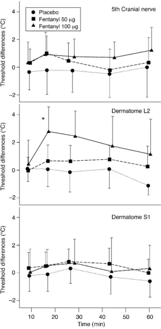
For heat pain tolerance thresholds, data were ®rst analysed graphically. Because the largest differences among groups and among dermatomes were observed at 14 min (Fig. 1), statistical analyses were performed at this time. To ®nd out at which dermatome (S1, L2, or ®fth cranial nerve) and after which treatment (placebo, fentanyl 50 or 100 mg) fentanyl produced signi®cant analgesia, a two way repeated measuresANOVA(with dermatome as repeated factor) was performed. The relatively low number of observations did not allow a comprehensive statistical analysis of all data for all times and dermatomes.
To analyse temporal summation thresholds, VASarea, and
area of local pain after i.m. hypertonic saline injection, one
way ANOVA (for normally distributed data) or
Kruskal±Wallis one wayANOVAon ranks (for not normally distributed data) were used.
In all analyses the Tukey test was used for multiple comparison. A P<0.05 was considered signi®cant. The software used was SigmaStat for Windows, version 2.03 (Jandel Corporation, San Rafael, CA, USA).
Results
Except pruritus in four patients, no side effects were observed. Of the 34 patients enrolled, four were not included in the analyses: unilateral spread of the local anaesthetic after the end of the measurements (n=2), intravascular location of the epidural catheter (n=1), occur-rence of back pain during the investigation that could possibly interfere with the measurements (n=1). Therefore, the analyses were performed on 30 patients, 10 patients in each group.
Patient characteristics and lidocaine dose to achieve a cranial spread up to T4 are shown in Table 1. We found no signi®cant differences among the three groups.
Heat pain tolerance thresholds (assessment of
segmental effect)
Fentanyl 100 mg, but not 50 mg, produced analgesia to heat stimulation only at L2 (P<0.05). Surprisingly, no effect at S1 was detected. The time course of heat pain tolerance thresholds for the dermatomes S1, L2 and ®fth cranial nerve
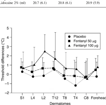
are shown in Figure 1. Figure 2 shows heat pain tolerance thresholds for all dermatomes 22 min after epidural injection of the different solutions.
Repeated electrical stimulation (assessment of
temporal summation)
The results are shown in Table 2. We found statistically signi®cant increases in temporal summation thresholds between the placebo group and both fentanyl 50 and 100 mg groups, respectively. No statistically signi®cant differ-ences between the two fentanyl groups were found.
I.m. injection of hypertonic saline (assessment of
muscle pain)
The results are shown in Table 2. After i.m. injection of hypertonic saline there was a statistically signi®cant decrease in VASarea between placebo and both treatment
groups. No statistically signi®cant differences between the two fentanyl groups were found.
The calculated means (SD) of the differences of the local pain areas drawn by the patients (area after minus area before epidural injection) in arbitrary units were: placebo group 0.75 (1.54), fentanyl 50 mg group ±0.38 (0.70) and fentanyl 100 mg group ±0.07 (0.57). No statistically signi®cant differences among groups were found. Because only one subject in the placebo group, two subjects in the fentanyl 50 mg group and one subject in the fentanyl 100 mg group reported referred pain, we did not calculate these areas.
Discussion
Assessment of segmental effect
Using the heat model, we found a statistically signi®cant effect of epidural fentanyl 100 mg, but not 50 mg, on dermatome L2 (segment of injection). Neither S1 nor ®fth cranial nerve were affected. This ®nding was surprising and challenges the traditional belief that the spinal action of epidural fentanyl is the result of penetration through the dura and diffusion from cerebrospinal ¯uid to dorsal horn neurones. If this was the case, the effect on S1 had to be at least as profound as the effect on L2, since the distance between site of dura penetration at L2±3 and S1 dorsal horn neurones is shorter than the distance to the L2 dorsal horn neurones. A possible explanation for the better effect of epidural fentanyl on L2 than on S1 dermatome could be a direct drug effect on the spinal nerve roots and binding to opioid receptors of dorsal-root ganglia.
Fentanyl was shown to block in vitro rabbit vagus nerve conduction, which suggests a local anaesthetic-type ac-tion.15 16Arendt-Nielsen and colleagues found hypoalgesia
to laser pain 15 min after perineural ulnar injection of 4 mg of morphine in humans.17In the same study lidocaine and
Fig 1 Time course of heat pain tolerance threshold (differences to baseline measurements) over a time period of 60 min after epidural injection of saline, fentanyl 50 and 100 mg. Means andSDare plotted.
*P<0.05 (two way repeated measurements ANOVA). No difference
morphine increased the latency of pain-evoked brain potentials, which may have been the result of the above effect on nerve conduction. An anatomical study18showed a
smaller cross-section area of the thoracic and high lumbar nerve roots compared with the low lumber and sacral roots. This may lead to a better local anaesthetic type action of fentanyl on the smaller root L2 compared with S1.
Animal studies have shown the existence of opioid binding sites in spinal roots.19Therefore, a direct binding of
opioids to receptors in the dorsal-root ganglia may play a role in the antinociceptive effects of opioids.20 The larger
anatomical distance from the epidural site of injection to the dorsal-root ganglion of S1 compared with L2 may be an additional explanation for the better antinociceptive effect of fentanyl on L2 in our study.
Another reason for the limited local action of fentanyl measured with the heat pain model could be the relatively poor rostral spread via the cerebrospinal ¯uid and the trapping of fentanyl in the epidural fat.21
Lack of effect on heat pain threshold measured at S1 and ®fth cranial nerve in our study does not mean that epidural fentanyl has no effect on this area. In fact, we could detect
an effect on L4/L5 and S1 by the muscle pain and the temporal summation model, respectively. Furthermore, our data are not in contrast with the convincing evidence that epidural fentanyl has also or primarily a systemic analgesic effect via vascular absorption from the epidural space.21
Recently, a study by our group showed that heat pain is not the optimal model for detecting the analgesic effect of i.v. alfentanil,22 although heat pain tolerance thresholds are
more sensitive to detect opioids effects than heat pain detection thresholds.23 24Thus, we may have measured the
analgesic action of fentanyl on heat pain only at the site of maximum effect, for example L2. The muscle pain and the temporal summation models may be more sensitive than heat pain to detect opioid induced analgesia. This could also explain the shorter duration of action compared with previous investigations25 26and the lack of effect of the 50
mg dose on heat pain in our study.
Our results differ from those obtained in previous investigations on epidural morphine. Using laser stimula-tion, Arendt-Nielsen and colleagues27found a longer effect
of epidural morphine on S1 than on more cranial dermatomes after injection at L2±3. Angst and colleagues28
found lumbar epidural morphine attenuated heat pain up to trigeminal level. These ®ndings are likely to be explained by the hydrophilicity and spinal cord availability of morphine compared with fentanyl.21These characteristics determine a
higher rostral spread in the cerebrospinal ¯uid and possibly deeper spinal analgesia by morphine than by fentanyl. This may make heat pain models more sensitive for epidural morphine than for epidural fentanyl effects.
Assessment of temporal summation
Neither the temporal summation nor the muscle pain assessments were designed to demonstrate segmental effects of epidural fentanyl. Therefore, the study does not provide information on the extent to which systemic effects contribute to analgesia detected by these sensory modalities. In the present study, repeated electrical stimulation was used to investigate the central integrative mechanism (temporal summation). In previous studies, temporal sum-mation was attenuated, but not completely inhibited, by Table 1 Patient characteristics and lidocaine dose given after the experiment
to achieve T4. Values are mean (SD or range). No statistically signi®cant differences among groups were found in any variable
Saline Fentanyl 50 mg Fentanyl 100 mg
Sex (m/f) 8/2 9/1 7/3
Age (yr) 49.7 (25±68) 49.2 (24±70) 46.7 (33±68) Weight (kg) 72.6 (12.1) 85.9 (14.5) 76.6 (14.5) Height (cm) 170.0 (9.2) 176.0 (6.1) 171.4 (9.1) Lidocaine 2% (ml) 20.7 (6.1) 20.8 (6.1) 20.9 (5.9)
Fig 2 Heat pain tolerance threshold measurements (differences to baseline measurements) in all studied dermatomes 22 min after epidural injection. Means and SD are plotted. The data were not analysed for
statistical signi®cance.
Table 2 Repeated electrical stimulation and i.m. injection of hypertonic saline. Pain thresholds on repeated transcutaneous electrical stimulation (temporal summation) 34 min after epidural injection and continuous VAS rating after i.m. hypertonic saline injection 47 min after epidural injection. Values are differences from baseline measurements, mean (SD). *P<0.05 vs fentanyl 50 and 100 mg
Saline Fentanyl
50 mg Fentanyl100 mg Electrical stimulation (mA) 0.17 (0.35)* 0.95 (0.91) 0.96 (0.59) Hypertonic saline VASarea(cm2) 531 (990)* ±390 (166) ±315 (528)
epidural local anaesthetics29 and epidural clonidine.30
Conversely, intrathecal bupivacaine completely blocked temporal summation.31 In the present study, epidural
fentanyl increased temporal summation threshold, indicat-ing attenuation rather than complete inhibition of temporal summation (Table 2). As mentioned above, we cannot rule out that we measured primarily a systemic effect of fentanyl with this test.
Temporal summation seems to be mediated by the N-methyl-D-aspartate (NMDA) receptor.32 Opioids do not act directly on the NMDA receptor, but may attenuate temporal summation unspeci®cally by reducing the nociceptive input to the dorsal horn neurones. It is conceivable that temporal summation shares common features with central hyperexitability involved in clinical pain.33 Therefore, the temporal summation model may be
more useful than short-lasting transient stimuli for predict-ing the response to analgesics in the clinical environment. Our result may explain why epidural opioids alone may provide only potential prevention of central hypersensitivity states.34
Assessment of muscle pain
This is the ®rst study on regional analgesia that includes an experimental muscle pain model. This is an important development in pain research, given the relevance of deep pain in clinical conditions.
Our data show that i.m. injection of hypertonic saline detects the analgesic effect of epidural fentanyl (Table 2). In a previous study, we found that i.v. remifentanil inhibits pain after i.m. electrical stimulation more profoundly than pain after cutaneous electrical stimulation.35Thus,
includ-ing a muscle pain model in the experimental test of new drugs would probably allow a better evaluation of drug action than procedures including only skin stimulation. It was not possible to show an effect of fentanyl on the area of pain drawn on the anatomical map induced by hypertonic saline although the pain intensity (VASarea) was
signi®-cantly decreased in the fentanyl groups compared with placebo. The diffuse localization of muscle pain may result in high inter-individual variations causing the non-signi®-cant effects on the drawn pain area.
Also, concerning VASarea the muscle pain model was
associated with high inter-individual variability, as shown by the high standard deviations in all groups. This may be the result of the dif®culty of some patients in adjusting continuously the pain intensity on the VAS. Some patients may lose concentration during the procedure and therefore forget to adapt the VAS scale to the real pain intensity. Furthermore, small changes in the needle position during the experiment may lead to stimulation of different muscle locations with possible consequent change in intensity of nociceptive stimulation. Despite its usefulness, this model still needs to be improved.
Acknowledgements
The authors thank Prof. Urs Studer, Head, Department of Urology, University Hospital of Bern, Bern, Switzerland for providing the logistic support.
References
1 Liu SS, Allen HW, Olsson GL. Patient-controlled epidural analgesia with bupivacaine and fentanyl on hospital wards:
prospective experience with 1,030 surgical patients.
Anesthesiology 1998; 88: 688±95
2 Scott DA, Beilby DS, McClymont C. Postoperative analgesia using epidural infusions of fentanyl with bupivacaine. A prospective analysis of 1,014 patients. Anesthesiology 1995; 83: 727±37
3 Curatolo M, Petersen-Felix S, Scaramozzino P, Zbinden AM. Epidural fentanyl, adrenaline and clonidine as adjuvants to local anaesthetics for surgical analgesia: meta-analyses of analgesia and side-effects. Acta Anaesthesiol Scand 1998; 42: 910±20
4 Coda BA, Brown MC, Schaffer R, et al. Pharmacology of epidural fentanyl, alfentanil, and sufentanil in volunteers. Anesthesiology 1994; 81: 1149±61
5 Liu SS, Gerancher JC, Bainton BG, Kopacz DJ, Carpenter RL. The effects of electrical stimulation at different frequencies on perception and pain in human volunteers: epidural versus intravenous administration of fentanyl. Anesth Analg 1996; 82: 98±102
6 Price DD. Characteristics of second pain and ¯exion re¯exes indicative of prolonged central summation. Exp Neurol 1972; 37: 371±87
7 Arendt-Nielsen L, Brennum J, Sindrup S, Bak P.
Electrophysiological and psychophysical quanti®cation of temporal summation in the human nociceptive system. Eur J Appl Physiol Occup Physiol 1994; 68: 266±73
8 Price DD, Long S, Huitt C. Sensory testing of pathophysiological mechanisms of pain in patients with re¯ex sympathetic dystrophy. Pain 1992; 49: 163±73
9 Curatolo M, Petersen-Felix S, Arendt-Nielsen L, Giani C, Zbinden AM, Radanov BP. Central hypersensitivity in chronic pain after whiplash injury. Clin J Pain 2001; 17: 306±15 10 Graven-Nielsen T, Aspegren Kendall S, Henriksson KG, et al.
Ketamine reduces muscle pain, temporal summation, and referred pain in ®bromyalgia patients. Pain 2000; 85: 483±91 11 Curatolo M, Petersen-Felix S, Arendt-Nielsen L. Sensory
assessment of regional analgesia in humans: a review of methods and applications. Anesthesiology 2000; 93: 1517±30 12 Pocock SJ. Clinical Trials. A Practical Approach. Chichester: John
Wiley and Sons, 1983: 80±7
13 Render CA. The reproducibility of the iliac crest as a marker of lumbar spine level. Anaesthesia 1996; 51: 1070±1
14 Graven-Nielsen T, Arendt-Nielsen L, Svensson P, Jensen TS. Quanti®cation of local and referred muscle pain in humans after sequential i.m. injections of hypertonic saline. Pain 1997; 69: 111±7
15 Gissen AJ, Gugino LD, Datta S, Miller J, Covino BG. Effects of fentanyl and sufentanil on peripheral mammalian nerves. Anesth Analg 1987; 66: 1272±6
16 Power I, Brown DT, Wildsmith JA. The effect of fentanyl, meperidine and diamorphine on nerve conduction in vitro. Reg Anesth 1991; 16: 204±8
17 Arendt-Nielsen L, Bjerring P, Dahl JB. A quantitative double-blind evaluation of the antinociceptive effects of perineurally
administered morphine compared with lidocaine. Acta Anaesthesiol Scand 1991; 35: 24±9
18 Hogan Q. Size of human lower thoracic and lumbosacral nerve roots. Anesthesiology 1996; 85: 37±42
19 Fields HL, Emson PC, Leigh BK, Gilbert RF, Iversen LL. Multiple opiate receptor sites on primary afferent ®bres. Nature 1980; 284: 351±3
20 Stein C. The control of pain in peripheral tissue by opioids. N Engl J Med 1995; 332: 1685±90
21 Ummenhofer WC, Arends RH, Shen DD, Bernards CM. Comparative spinal distribution and clearance kinetics of intrathecally administered morphine, fentanyl, alfentanil, and sufentanil. Anesthesiology 2000; 92: 739±53
22 Luginbuhl M, Schnider TW, Petersen-Felix S, Arendt-Nielsen L, Zbinden AM. Comparison of ®ve experimental pain tests to measure analgesic effects of alfentanil. Anesthesiology 2001; 95: 22±9
23 vander Burght M, Rasmussen SE, Arendt-Nielsen L, Bjerring P. Morphine does not affect laser induced warmth and pin prick pain thresholds. Acta Anaesthesiol Scand 1994; 38: 161±4 24 Brennum J, Arendt-Nielsen L, Horn A, Secher NH, Jensen TS.
Quantitative sensory examination during epidural anaesthesia and analgesia in man: effects of morphine. Pain 1993; 52: 75±83 25 Blanco J, Blanco E, Carceller JM, Sarabia A, Solares G. Epidural analgesia for post-caesarean pain relief: a comparison between morphine and fentanyl. Eur J Anaesthesiol 1987; 4: 395±9 26 Grass JA, Sakima NT, Schmidt R, Michitsch R, Zuckerman RL,
Harris AP. A randomized, double-blind, dose-response comparison of epidural fentanyl versus sufentanil analgesia after cesarean section. Anesth Analg 1997; 85: 365±71
27 Arendt-Nielsen L, Oberg B, Bjerring P. Hypoalgesia following epidural morphine: a controlled quantitative experimental study. Acta Anaesthesiol Scand 1991; 35: 430±5
28 Angst MS, Ramaswamy B, Riley ET, Stanski DR. Lumbar epidural morphine in humans and supraspinal analgesia to experimental heat pain. Anesthesiology 2000; 92: 312±24
29 Curatolo M, Petersen-Felix S, Arendt-Nielsen L, Fischer M, Zbinden AM. Temporal summation during extradural anaesthesia. Br J Anaesth 1995; 75: 634±5
30 Curatolo M, Petersen-Felix S, Arendt-Nielsen L, Zbinden AM. Epidural epinephrine and clonidine: segmental analgesia and effects on different pain modalities. Anesthesiology 1997; 87: 785±94
31 Curatolo M, Petersen-Felix S, Arendt-Nielsen L, Zbinden AM. Spinal anaesthesia inhibits central temporal summation. Br J Anaesth 1997; 78: 88±9
32 Arendt-Nielsen L, Petersen-Felix S, Fischer M, Bak P, Bjerring P,
Zbinden AM. The effect of N-methyl-D-aspartate antagonist
(ketamine) on single and repeated nociceptive stimuli: a placebo-controlled experimental human study. Anesth Analg 1995; 81: 63±8
33 Woolf CJ, Salter MW. Neuronal plasticity: increasing the gain in pain. Science 2000; 288: 1765±9
34 Aida S, Yamakura T, Baba H, Taga K, Fukuda S, Shimoji K. Preemptive analgesia by intravenous low-dose ketamine and epidural morphine in gastrectomy: a randomized double-blind study. Anesthesiology 2000; 92: 1624±30
35 Curatolo M, Petersen-Felix S, Gerber A, Arendt-Nielsen L. Remifentanil inhibits muscular more than cutaneous pain in humans. Br J Anaesth 2000; 85: 529±32