Genomics in cardiac metabolism
Jane-Lise Samuel
1,2, Marcus C. Schaub
3, Michael Zaugg
4, Mamas Mamas
5, Warwick B. Dunn
6,
and Bernard Swynghedauw
1,2*
1U689 INSERM, Hoˆpital Lariboisie`re, 41 Bd de la Chapelle, 75475 Paris Cedex 10, France;2Universite´ Denis Diderot, Paris Cedex 7, France;3Institute of Pharmacology and Toxicology, University of Zurich, Zurich, Switzerland;4Institute of Anaesthesiology, University Hospital Zurich, Zurich, Switzerland;5Department of Cardiology, Manchester Royal Infirmary, Manchester, UK; and6The Manchester Centre for Integrative Systems Biology, University of Manchester, Manchester, UK
Received 5 December 2007; revised 3 February 2008; accepted 15 February 2008; online publish-ahead-of-print 7 March 2008 Time for primary review: 15 days
Cell biology is in transition from reductionism to a more integrated science. Large-scale analysis of genome structure, gene expression, and metabolites are new technologies available for studying cardiac metabolism in diseases known to modify cardiac function. These technologies have several limitations and this review aims both to assess and take a critical look at some important results obtained in genomics restricted to molecular genetics, transcriptomics and metabolomics of cardiac metabolism in pathophysiological processes known to alter myocardial function. Therefore, our goal was to delineate new signalling pathways and new areas of research from the vast amount of data already published on genomics as applied to cardiac metabolism in diseases such as coronary heart disease, heart failure, and ischaemic reperfusion.
KEYWORDS Cardiac metabolism; Genome-wide analysis; Metabolomics; Transcriptomics
1. Introduction
This review aims both to assess and critically review the main results obtained in genomics of cardiac metabolism by the end of 2007. Although genomics have been differen-tially defined, we use the most widely accepted one, which was utilized by Gibson and Muse.1 Genomics covers the
overall structure or expression of our genetic inheritance including molecular genetics, transcriptomics, proteomics, and metabolomics.
The present work aims to delineate new signalling path-ways and new areas of research from the vast amount of data already published on genomics as applied to cardiac metabolism in diseases, such as coronary heart disease (CHD) and heart failure (HF). This review was clearly limited by the fact that both the new molecular genetics, based on genome-wide association studies (GWAS) and metabolomics are still in their infancy, at least in the car-diovascular (CV) field. In contrast, the science of transcrip-tomics in the CV arena is more mature and had been developed in more detail.
From the growing flow of new data, a selection was made to illustrate better the potential of such a global approach. (i) Genetics has presently reached a new era based on GWAS. GWAS is, in principle, more liable to identify low-effect genes operative in pathological pathways and disease sus-ceptibility in common diseases.2 (ii) Transcriptomics is the
study of gene expression of either transcripts or proteins. Gene expression is a short-term approach and is based on two main techniques, namely microarrays analysis and pro-teomics.1 Two different aspects of transcriptomics have been developed herein: (a) modifications observed during the time course of a chronic disease of the heart and (b) pre- and post-conditioning and changes induced by short-term metabolic interventions (such as anaesthesia) that confer cardioprotection. (iii) The final chapter is on metabo-lomics that aims to quantify still more rapid modifications in metabolic compounds, but again on a genome-wide scale with the potential of medical applications.
It is worthy to note that with regards to the importance of the research area, we have selected studies, which were the most informative in the field of cardiology.
It has to be underscored that the majority of the studies reported herein, imply a technical approach, which is both costly and frequently requires large groups of investigators, patient cohorts (for GWAS) and multidisciplinary approaches (for metabolomics). Briefly, GWAS is principally based on high-density genotyping arrays that combine the power of association studies with the systematic nature of genome-wide research. Transcriptomics is mainly based on micro-array analysis, i.e. a high-throughput method for screening a collection of microscopic DNA spots attached to a solid surface and to measure the expression levels of large numbers of genes in different samples simultaneously. Pro-teomics commonly utilized two-dimensional polyacrylamide gel electrophoresis to separate proteins, but there are also
*Corresponding author. Tel: þ33 1 5321 6760; fax: þ33 1 53 21 67 39. E-mail address: [email protected]
Published on behalf of the European Society of Cardiology. All rights reserved.&The Author 2008. For permissions please email: [email protected].
many other technical approaches available including protein microarrays. Finally, metabolomics is mainly a multidisci-plinary approach based on the combination of pyrolysis and different spectrometry.
2. Are genome-wide association studies ready
for common diseases?
Medical genetics not only involves the identification of one specific gene associated with a severe risk of having a given disease, but also provides information on associations of gene variants, each providing a moderate risk.3–6GWAS is based on (i) the availability of dense genotyping chips made with single-nucleotide polymorphisms (SNPs) (100 000– 500 000, and recently, one million) covering most of the genome unequally; (ii) the growing resources of the Inter-national HapMap consortium (2007) which documents linkage disequilibrium (LD—a non-random association between alleles in a population due to their tendency to be co-inherited because of reduced recombination between them. Haplotype is the combination of alleles at neighbouring SNPs. Haplotypes blocks are the apparent hap-lotype structures of recombining portions of the genome in which blocks of consecutive co-inherited alleles are separ-ated by short boundary regions), and is a public resource of common SNPs capturing most of the common genome sequence variability. In the human genome, there are 3.2 billion base pairs and approximately 15 million SNPs, indicating that kits using 500 000 SNPs should cover ,0.2% of the genome.4 The second generation human haplotype map now covers over 3.1 million SNPs.7 Nevertheless, the
statistical association among groups of SNPs, i.e. haplotype blocks, suggests that the identification of a few of the SNPs within the blocks can unambiguously identify all associated SNPs without the need to measure them directly. Recent studies have shown that the human genome is organized into a succession of ancestrally conserved distinct haplotype blocks. On the basis of this assumption, it is assumed that a 500 000 SNP scan should cover approximately 90% of the genome.8 This chapter on genetics has been deliberately limited to GWAS and aims both to take a critical look at the main results obtained by the end of 2007 in CV research and to generate a new working hypothesis.
2.1 Main results, new insights into metabolic
genomics
GWAS is, for the moment, dominated by the results of big groups such as The Wellcome Trust consortium (WTCCC)9
and the Framingham Heart Study (FHS).10 The WTCCC focuses on seven common diseases and includes CHD, dia-betes, and hypertension. Most of these results were dupli-cated, especially those concerning CHD in a collaborative study with the German MI Family Study.10,11FHS comprises several working groups each describing specific associations with various traits [biomarkers, body mass index (BMI), and so on]. Several of these results were also duplicated. The WTCCC and FHS utilized approximately 500 000 and 100 000 SNPs, respectively. In addition, there are others groups that usually used smaller SNP density than the WTCCC.
To summarize (Table 1), GWAS has generated, for the time being, three groups of results.
(i) Diseases with phenotypic variance mainly due to genetic factors such as type 1 diabetes. GWAS docu-mented at least a dozen genes strongly associated with type 1 diabetes (HLA class genes, insulin gene, CTLA4 locus, PTPN22, and the IFIH1 region) that mostly belong to the immune system which are the most important targets for research on type 1 diabetes. (ii) On the other hand, diseases such as arterial hyperten-sion and hyperlipidaemias remained poorly associated to simple genetic factors. Despite a considerable hope, genetic influence is weak and there are few risk alleles of large size effects and GWAS has been unable to identify with certainty susceptibility genes of modest effect size and we need, in the least, geno-typic resources of increased density.12 Long-term averages of low-density lipoprotein cholesterol, high-density lipoprotein cholesterol, triglycerides, and blood pressure are highly heritable. Nevertheless, there are no significant associations that could help further research.
(iii) Type 2 diabetes, obesity, and atherosclerosis are in an intermediary position. GWAS has succeeded in detect-ing new loci of interest, which were strongly and repro-ducibly linked to the phenotype. Several new loci and genes of interest have now been identified:
(a) The association of the 9p21 locus with CHD has been found by every GWAS published so far which suggests, at least, that this association is widely dis-tributed. The locus contains two cyclin-dependent kinase inhibitors, which regulate cell cycles. Inter-estingly, the cell cycle pathway, including cyclins, was also over-represented in a large-scale gene expression study, which analysed pathways involved in atherogenesis using a modular approach. For these authors, their data suggest that smooth muscle de-differentiation is a key determinant in atherogenesis, which is new and unexpected.13
(b) FHS has uncovered unexpected genetic associ-ations with various markers of arterial stiffness, including large arteries calcium content and the reflected wave with candidate genes such as LOXL2, an oxidase involved in collagen cross-linking and arterial elasticity.
(c) More than 100 publications have reported associ-ations between genetic variants and BMI, and/or type 2 diabetes, but few have been reproduced so far. The two phenotypes are highly multigenic. Several associated genes such as TCF7L2, KCNJ11, SLC30A8, EXT1 have been linked to pan-creatic development and function.
2.2 Critical evaluation of genome-wide
association studies
GWASs are still in their infancy and many technical and stat-istical problems remain to be solved. The results of promis-ing studies such as FHS are still fragmented and require replication. Nevertheless, GWAS is probably the best tool now available to validate candidate gene associations and to enable unbiased searches for novel variants.
There are approximately seven million variants at a frequency .5%, hence rare alleles may be overlooked.
Good examples may be found in both the WTCCC (for APOE and INS) and FHS (for GCKR).9,10GWAS is based on both SNPs density and haplotype map, and can be improved by increas-ing SNP scan density or improvincreas-ing the haplotype map. The second-generation human haplotype map now covers over 3.1 million SNPs instead of one, and would provide the solution (see http://www.hapmap.org).
The second limitation lies in the fact that GWAS has many caveats when studies were conducted in less severe and more multifactorial diseases, and, in fact, the real problem is probably that GWAS did not catch RNA genes or regulatory segments (see Conclusions) that have not already been identified and could be major determinants in traits generation.
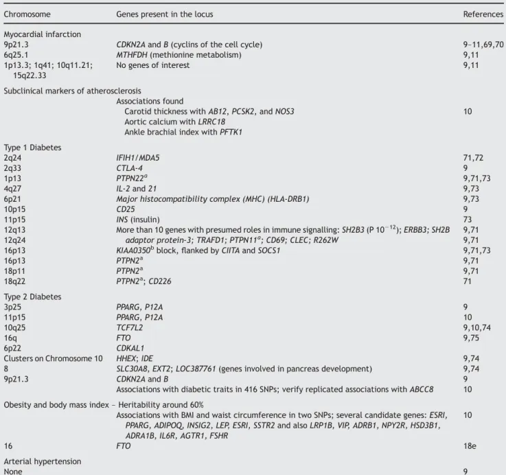
Table 1 Genome-wide analyses in diseases known to modify cardiac function directly or indirectly
Chromosome Genes present in the locus References
Myocardial infarction
9p21.3 CDKN2A and B (cyclins of the cell cycle) 9–11,69,70
6q25.1 MTHFDH (methionine metabolism) 9,11
1p13.3; 1q41; 10q11.21; 15q22.33
No genes of interest 9,11
Subclinical markers of atherosclerosis
Associations found
Carotid thickness with AB12, PCSK2, and NOS3 10 Aortic calcium with LRRC18
Ankle brachial index with PFTK1 Type 1 Diabetes
2q24 IFIH1/MDA5 71,72
2q33 CTLA-4 9
1p13 PTPN22a 9,71,73
4q27 IL-2 and 21 9,73
6p21 Major histocompatibility complex (MHC) (HLA-DRB1) 9,73
10p15 CD25 9
11p15 INS (insulin) 73
12q13 More than 10 genes with presumed roles in immune signalling: SH2B3 (P 10212); ERBB3; SH2B
adaptor protein-3; TRAFD1; PTPN11a; CD69; CLEC; R262W
9,71
12q24 9,71
16p13 KIAA0350bblock, flanked by CIITA and SOCS1 9,71,73
16p13 PTPN2a 9,71 18p11 PTPN2a 9,71 18q22 PTPN2a; CD226 71 Type 2 Diabetes 3p25 PPARG, P12A 9 11p15 PPARG, P12A 10 10q25 TCF7L2 9,10,74 16q FTO 9,75 6p22 CDKAL1
Clusters on Chromosome 10 HHEX; IDE 9,74
8 SLC30A8, EXT2; LOC387761 (genes involved in pancreas development) 9,74
9p21.3 CDKN2A and B 9
Associations with diabetic traits in 416 SNPs; verify replicated associations with ABCC8 10 Obesity and body mass index – Heritability around 60%
Associations with BMI and waist circumference in two SNPs; several candidate genes: ESRI, PPARG, ADIPOQ, INSIG2, LEP, ESRI, SSTR2 and also LRP1B, VIP, ADRB1, NPY2R, HSD3B1, ADRA1B, IL6R, AGTR1, FSHR
10
16 FTO 18e
Arterial hypertension
None 9
Associations with blood pressure or arterial stiffness in seven SNPs; a few candidate genes: MEF2C, SYNE1, LOXL2, TNFSF11
10
Hyperlipidaemia
Associations with LDL-C, HDL-C, and triglycerides in seven SNPs; no new locus identifiable 10
Gene nomenclature can be found on ‘genecards.org’. The papers from FHS are quoted in the work of Cupples et al.10
aThe PTPN family plays a major role in insulin, immune signalling, and autoimmune diseases. PTPN can indirectly dephosphorylate STAT1, a major regulator
of immune signalling.
bKIAA0350 is a widely expressed gene of unknown function. Exon 12 may encode an immune receptor—ITAM—that binds the SH2B3 lymphocyte adaptor
3. Metabolic gene expression in cardiac
hypertrophy and failure
Most of the work, which has been published so far in this area has been mainly descriptive and because they are dealing with the whole genome expression, only a few of them have clearly isolated the metabolic family of genes from the others. However, we now collect these data and attempt to draw some conclusions.
3.1 Transcriptomics
Genomic approaches such as transcriptional profiling by DNA microarrays allow the simultaneous analysis of some 55 000 transcripts in a single assay14–16and provides both qualitative
(switched on/off genes) and quantitative data (transcrip-tional level of single genes), so that subtle differences on gene activation can be detected. Today, transcriptional analysis can be performed on minute tissue samples and, one of the limits due to cellular heterogeneity can now be resolved by laser microdissection, which allowed studies of one cell population. Nevertheless, profiling whole CV tissue samples may generate novel hypotheses, and help to identify unexpected cell components and reveal novel marker genes. As reviewed by Nanni et al.,17a growing number of
transcrip-tome microarray studies have been applied to CV diseases with the aim of recognition of specific disease phenotypes to improve both prognosis and therapeutic assessment.
The comparative study performed by Gao et al.18between
canine tachy-pacing, mouse transgenic, and human HF reveals that, in humans, the disease involves a downregula-tion of genes in a broad range of biological processes. In con-trast to this, in experimental models of HF, downregulation of energy metabolic pathways is observed. Human ischaemic HF and canine HF share a similar over-representation of transcriptional pathways in the upregulated genes. However, in this study, no induction of prominent HF markers, e.g. atrial natriuretic peptides (ANP) and brain natriuretic peptides (BNP) was detected.19In the rat heart
following coronary ligation, Laframboise et al.20 demon-strated that transcripts for signal transduction and inflam-mation gene expression dominated in the infarct zone during late-term recovery. There was recruitment of genes for transcription, metabolism, and detoxification—all classes were depressed in the day 1 infarct zone. In contrast, within one day, the remote zone exhibited an upregulation in many genes particularly in those of the metabolism family or those associated with developmental processes. In contrast, transcripts for contractile proteins matched control values. In the late-term, the metabolic responses of the remote zone was attenuated.
In humans, Kittleson et al.,16 developed a strategy to identify genes differentially expressed between ischaemic (ICM) and non-ischaemic (NICM) cardiomyopathy. When com-pared with controls, 257 genes (over the 22 000 transcripts present on the Affymetrix microarray platform) were differ-entially expressed in NICM and 72 genes in ICM. Only 41 genes were shared between NICM and ICM, and they were mainly involved in cell growth and signal transduction (Table 2). Those specifically expressed in NICM were fre-quently involved in metabolism and included ACE2 and genes involved in fatty acid (FA) and cholesterol metab-olism. The genes specifically upregulated in ICM more
often had catalytic activity, such as SERPINB1, SERPINE1, ATP1B3. Besides these results, using a CardioShip (Cardio-Chip is a custom-made CV-based tag glass slide cDNA micro-array formed by non-redundant 10 848-element human CV-based expressed sequence) in human HF, Barrans et al.21 found more than 100 transcripts upregulated, including stress-response proteins, transcription/translation regulators together with the classical HF genes like ANP, and selected sarcomeric and extracellular matrix (ECM) pro-teins. Conversely, they found a downregulation of cell-signalling channels and mediators, particularly those involved in the Ca2þ pathways of crucial importance. In patients with moderate HF and dilated cardiomyopathy (DCM), the transcriptional profiles demonstrated a switch in the cardiomyocyte energy pathways (higher rate of lipid oxidation), apoptosis, and a downregulation of cell cycle-controlling genes.22In addition, alterations in the
intracellu-lar signalling functions were already present in the early stages of the disease. The genes regulating muscle contrac-tion were deregulated in intermediate stages, whereas apoptosis and the cell cycle regulator gene expression were altered in the late stages.
Different results were obtained by Sanoudou et al.23 The
genes of energy metabolism were predominantly underex-pressed in DCM and hypertrophic cardiomyopathy (HCM), but overexpressed in ICM.23 For instance, activation of the cardiac Peroxisome Proliferator-Activated Receptor-alpha (PPAR-alpha) by Peroxisome Proliferator-Activated Receptor-gamma Coactivator 1-alpha (PGC1-alpha) induces the expression of genes encoding for proteins involved in FA uptake, transport into mitochondria, and beta-oxidation.24
The increased FA utilization leads reciprocally to a decrease in glucose metabolism. On the other hand, the well-known hypoxia-inducible transcription factor 1-alpha (HIF1-alpha) targets approximately 70 genes and among them those that increase oxygen delivery and survival during hypoxia (such as genes involved in the upregulation of glucose metab-olism).25 HIF1 alpha is one of the few transcription factors promoting upregulation of the glucose pathway. The downre-gulation of PPAR-alpha and PGC1-alpha in hypertrophy and in HF could also be considered beneficial, because it indirectly
Table 2 Distribution of genes significantly altered in human cardiac disease
Family genes Up Down
Nucleus 1
Metabolism 4 1
Cytoskeleton 1 1
Cell adhesion, extracellular matrix 1 1 Inflammatory and immune response 2 None
Binding 3 None
Signal transduction 5 None
Cell growth 4 None
Development 2 None
Catalytic activity 1 None
Apoptosis 1 None
Significant changes in gene expression specific to either ischaemic or non-ischaemic cardiomyopathies matching sham cohort verified in differ-ent independdiffer-ent studies (reviewed in Kittleson et al.16). The columns
indicated the activated (up) and inhibited (down) genes comprising each individual gene classification.
favours glucose utilization instead of FA oxidation. Finally, the increased cytoplasmic Ca2þduring diastole in most forms of end-stage HF permeates the mitochondrial matrix, where it stimulates Ca2þ-regulated key enzymes in the tricarboxylic acid cycle as well as ATP synthase, thus accelerating the energy-producing metabolism.26
In vivo measurements of high-energy phosphate com-pounds have shown that the failing heart is an engine out of fuel.24,27 No consent exists, however, whether the FAs or the glucose pathway predominates in end-stage HF. This may, in part, be due to the different pathophysiological aetiologies leading to a final common HF syndrome exhibiting more generalized metabolic dysfunction, rather than altera-tions of specific substrate preference regulating genes.24
Transcriptomic analysis assesses that HF, independent of its aetiology, is characterized by some common final patterns of gene expression, including those coding for a high rate of lipid oxidation with low glycolysis, a dysregulated apoptosis, cell-cycle regulator genes, and ECM remodelling. It was suggested that different gene expression patterns were associated with clinical HF severity14 and some patterns characteristic of clinical syndromes16may open the road for aetiology-specific therapies in NICM-targeting metabolic pathways. In most of these genome-wide analyses, no firm correlation has been established between the altered expression of specific genes with functional parameters.
3.2 Transcriptomics and signalling
Most of the extracellular stimuli (ions, hormones, cell mediators, and mechanical signal) are integrated and transmitted by various intracellular signalling pathways to the cell nucleus, ultimately affecting gene-expression pat-terns.26,28The signalling pattern in response to stimuli may represent an early and sensitive disease-specific fingerprint before the final cellular phenotype has fully developed.
A large body of literature based on ex vivo and in vivo animal models indicates that pathological myocardial remodelling is mainly induced by neurohormonal factors (including angiotensin-II, endothelin-1, and catecholamines) through Gq-coupled (G-protein-coupled receptors, GPCRs) signalling pathways.28,29 Downstream of Gq, the pathway involves phospholipase-C beta, which hydrolyses phospha-tidylinositol bisphosphate (PIP2) into diacylglycerol (DAG) and inositol trisphosphate (IP3). IP3 releases calcium from intracellular stores, which may activate the phosphatase calcineurin-NFAT pathway. In concert, DAG activates protein kinase C (PKC) family members, some of which con-tribute to hypertrophic gene expression.30 In contrast,
exercise-induced hypertrophy appears to be regulated through the PI3K (phosphoinositide-3 kinase)—PKB/Akt— GSK3alpha/beta (glycogen synthase kinases) pathway. Growth (GH) hormone and insulin-like growth factor (IGF) are the major stimuli for physiological hypertrophy.31–33Of note, these signalling pathways are complemented by a panoply of interconnected routes including the mitogen-activated protein kinase (MAPK) cascades and the Janus kinase/signal transducer and activator of transcription JAK/Stat pathway.26,34 Additional signalling components
also contribute to the final phenotype.
Genome-wide transcriptomic analysis of differentially regulated genes in physiological (dubbed ‘adaptive’) and pathological (or ‘maladaptive’) hypertrophy, as well as in
HF of rats was recently reported.35,36 Taken together, the gene activity profiles obtained with Affymetrix Rat Genome U34A microarrays pointed out that (i) gene clusters typically changing in adaptive hypertrophy predominantly comprised genes involved in metabolism and cell growth; (ii) maladaptive hypertrophy was characterized by changes in gene clusters associated with oxidative stress responses, inflammation, and apoptosis; (iii) transition to overt HF was accompanied by an increase in those genes already affected in compensated pathological hypertrophy, by recruitment of additional signalling genes, such as GATA4, RAB7, NRAS, GNA12, STAT3, STAT5B, FYN, CRKO, MYCN, PTEN, AKT1, and IL6ST/gp130.
The most striking and potentially physiologically meaning-ful observation concerns the shift in metabolic gene expression.35 Several genes involved in beta-oxidation of lipids are upregulated in adaptive, but downregulated in maladaptive hypertrophy. On the other hand, a number of genes stimulating glucose metabolism are selectively upre-gulated in adaptive but not in maladaptive hypertrophy. Furthermore, the uncoupling protein UCP2 is downregulated in adaptive and upregulated in maladaptive hypertrophy implying that ATP production through oxidative phosphoryla-tion might be more effective in exercise-trained hearts. Collectively, these findings support the notion that tran-scriptomic changes comprise the basis for functional improvement of cardiac capacity in glucose utilization by adaptation to exercise. It remains to be established whether some or all of the expression changes typical for pathological cardiac hypertrophy can be observed and therapeutically addressed in humans. One major obstacle presents the much slower development in most clinical heart diseases coupled with small alteration in the early phases, which might escape detection.
3.3 Proteomics
There are approximately 10 times more proteins than genes, and expression arrays should preferably be complemented by proteomic evaluation because the transcripts are not always reflected in corresponding protein accumulation, and also because protein may undergo variable co- and post-translational modifications, the information for which is not contained in the original transcript. Therefore, new findings discovered by transcript profiling, may serve as leads but require subsequent functional characterization.23For both
the microarray analysis and proteomics, an unbiased approach requires sophisticated bioinformatics tools and a large panel of controls which will not be detailed in this review (see details in Gibson and Muse,1 Ruiz and Witt,19
da Silva et al.,37and Lucchinetti et al.38).
Proteomic biomarkers differ from traditional biochemical markers, in which multiple interacting protein species are evaluated simultaneously to reflect the response of a cell or an organism to disease. Proteomics might then be utilized to investigate rapid changes in signalling pathways. A resource of web-based two-dimensional electrophoretic maps annotated for healthy and diseased cardiac tissues has recently been highlighted.39Proteomics includes other approaches such as gel-free separation (LC, liquid IEF, CE, FFE) and identification of the purified protein can be per-formed through mass spectrometry (MS or MS/MS).
Lindsey et al.,40 using a multi-dimensional proteomic approach, identified 123 proteins that were differentially expressed during left ventricular hypertrophy in mouse, including LIM proteins, thioredoxin, myoglobin, FA-binding protein 3 (FABP), and myofibrillar proteins. The classification into seven categories is as follows: (1) cell structure and motility; (2) cell signalling and communication; (3) metab-olism; (4) transcription, translation, and trafficking; (5) cell division; (6) cell and organism defense; (7) unknown provides more information. Among the 95% that could be assigned to one of the six known functions, 36% and 21% of the identified proteins belongs to metabolism, cell struc-ture, and motility classes, respectively. Some of these changes were not confirmed using western blotting, as changes in myoglobin. Besides, through a subproteomic analysis, Banfi et al.41demonstrated, among the metabolic changes observed in human HF, a decreased FA oxidation as indicated by the FABP content.
3.4 Critical evaluation of transcriptomics and
proteomics
Both transcriptomics and proteomics are no more in their infancy and require many technical and statistical problems to be solved. As already pointed out above, the results are still fragmented and require further development. One might pay attention to the cell heterogeneity of the heart, and consider the part of non-muscle cell (endothelial cell, fibroblast) into cell signalling changes. Nevertheless, proteo-mics emerges as one of the best tool to bring new insight into applied genomics of cardiac metabolism, mainly through the analysis of post-translational modifications. Yet, proteomics remains to be a field strongly based on technologies.
4. Metabolomics
The study of the collection of small molecular-weight organic and inorganic species present in a biological system is defined as metabolomics.42,43These collection of
metabolites can provide a phenotypic oversight of the organism, either as a snapshot in time or as an integrated picture of biology over a period of time. The human metabo-lome is estimated to contain approximately 1500 endogen-ous metabolites not including many lipids and gut microflora-derived metabolites,44 and is chemically and
physically diverse.45Metabolites are the final downstream product of gene transcription and so reflect more closely the cell activity (or phenotype) at a functional level. Fur-thermore, metabolic control analysis (MCA) describes that small changes in enzyme activity (and the transcripts which encode these) may have minimal influence on meta-bolic fluxes, but large influences on the concentrations of metabolites. Consequently, the metabolome is thought to be a potentially more sensitive marker of cellular processes both in normal physiology and disease.42
The science of metabolomics operates to a workflow or pipeline approach46 and involves multi-disciplinary teams
with the objective to create valid and experimentally robust data and convert this data into biological knowl-edge.47 Analogous to the human genome project, there are numerous approaches to define the human metabo-lome44(http://www.husermet.org; http://www.hmdb.ca).
4.1 Biomarker detection
The application of metabolomics to human disease studies is an emerging science, in which patterns of metabolites in the disease state are compared with those of ‘healthy’ individ-uals to allow identification of potential biomarkers of the disease process.48,49 An advantage of using metabolomics for the identification of biomarkers of a disease state are the inductive approaches applied to studying these complex systems, where many diseases are characterized as multiple disorders or display multiple phenotypes across a population.
In a recent study from our group, the serum of 52 patients with documented HF (left ventricular ejection fraction ,40%) was subjected to metabolomic analyses. Multiple bio-markers were detected, of which 2-oxoglutarate and pseu-douridine were the metabolites showing greatest statistical differences between the case and matched control classes. The combination of both provided a greater sensitivity and specificity for the diagnosis of HF in this patient cohort than the current gold standard biomarker BNP.50Pseudouridine is a modified nucleoside that is found in ribosomal and transfer RNA, is produced post-transcriptionally and is considered to be an excellent measure for RNA degradation, and hence the cell turnover. Tumour cells exhibit an unusually high turnover, and conse-quently has also been proposed as a tumour marker51 where it can have significant prognostic value. In HF its raised level may, in part, reflect the remodelling process in the heart itself or increased catabolic activity in periph-eral tissues.
2-Oxoglutarate is an important intermediate of the Krebs cycle and one of the 12 major precursors for the synthesis of most biochemical substances. In recent years, it has become increasingly clear that alterations in energy metabolism may contribute to the pathophysiol-ogy of HF.52,53 The raised levels of 2-oxoglutarate seen in HF may reflect a decreased flux through the Krebs cycle in HF and overflow of some metabolites into the circulation.
In another study in which blood samples were analysed from subjects undergoing exercise stress testing, a number of biomarkers were identified in those patients whose stress test demonstrated evidence of cardiac ischae-mia54 and noted that six of the metabolites in the Krebs cycle were significantly over-represented in the 23 markers of cardiac ischaemia identified. Indeed, in exper-imental animal models of cardiac ischaemia it has been demonstrated that there is a significant reduction (80%) in efflux of Krebs cycle intermediates from the myocardium with maintenance of intracardiac tissue levels and it is thought that this may be an important pathophysiolocal mechanism to preserve Krebs cycle metabolic intermediates and therefore protect ATP production within the myocardium.
Metabolomic analyses in CV medicine may not only be potentially useful in the identification of potential bio-markers of CV disease in the future, but may provide infor-mation regarding prognosis, response to therapy, and underlying mechanisms of the disease process. It is worthy to note that to reach this goal, the relative specificity for CV diseases of the current metabolomics approaches need to be improved.
5. Genomics and proteomics applied to
myocardial ischaemia and cardioprotection
5.1 Genomics applied to cardioprotection
5.1.1 Cardioprotection obtained by ether-derived halogenated volatile anaestheticsIschaemia, and more recently anaesthesia, are known to confer cardioprotection on either a short- or a long-term basis through different mechanisms. The well documented pre- and post-conditionings (Pre-C and Post-C) are good examples of how genomics can be applied to CV research. The induction of short- and long-term gene reprogramming
was examined by genome-wide gene activity profiling after either ischaemia or anaesthesia obtained with ether-derived halogenated volatile anaesthetics (VAs).37,55 Alterations in gene expression induced by anaesthetic conditioning were compared with the well-known ischaemic conditioning pro-cedures (Figure 1). Subsequently, the insight gained from the experimental approaches could be directly translated into clinical applications. Thereby, the volatile cardiopro-tective sevoflurane in comparison with the widely used intravenous propofol, reduced the expression of genes involved in noxious pathways and shifted the energy metab-olism away from FA oxidation more to glucose utilization.38 Important differences were observed in signalling between ischaemia and VAs-induced conditioning.37,56The two major signalling routes for VAs-Pre-C were the phospholipase-C path-ways and a direct activation of nitric oxide synthase. Both sig-nalling routes converge on activating the mitoKATP channels and blockade of these channels by 5-hydroxydecanoate results in complete inhibition of cardioprotection. Conversely, application of the specific channel opener diazoxide induces cardioprotection just like the VAs.
In contrast, signalling by the ischaemic Pre-C trigger also involves the MAPK pathways in addition to those activated by VAs.37,57 Differences in signalling between the two modes of Pre-C were expected, because the ischaemic trigger exerts a noxious cellular stress for induction of pro-tective responses, while the VAs trigger rather sedates the cells without causing any lesions,58and as a corollary, the two trigger mechanisms affect the gene activity profile in different ways (Figure 1).37,59
The major signalling routes by anaesthetic and ischaemic Post-C comprise the so-called reperfusion injury salvage kinase pathways (RISK).60–63 The RISK pathways comprise several signalling routes all of which converge on inhibiting the mitochondrial permeability transition pore (mPTP) by an as yet unknown mechanism. Therefore, the functionally coupled mitoKATP channel and mPTP represent the major mitochondrial target structures for pre- and post-conditioning, respectively.58,64Finally, functional impairment of either or both structures can initiate apoptosis/necrosis. 5.1.2 Transcriptomic profile during VAs-induced cardioprotection
From visual inspection of the heat map (Figure 1A, first two column groups), one can conclude that both triggers regu-late a number of genes in parallel (same colour in horizontal rows), while a large portion of genes were differentially regulated as indicated by non-corresponding colour rows. On the other hand, a large portion of genes display a similar pattern after test ischaemia alone and after ischae-mic Pre-C followed by test ischaemia (Figure 1A, last two column groups). Finally, a similar correspondence of com-monly regulated genes appears in time-matched perfusion controls when compared with VA Pre-C followed by test ischaemia (Figure 1A, middle two column groups). The different activity profiles between the two conditioning trig-gers and the substantial overlapping of the profiles between (i) VAs Pre-C and untreated controls and (ii) ischaemic Pre-C and unprotected ischaemia are visualized in Figure 1B.37
These findings indicate different gene-activity profiles for the two trigger mechanisms. Although ischaemic Pre-C is able to afford cardioprotection, the subsequent ischaemic cell damage incurred by the test ischaemia brings the
Figure 1 Transcriptome profiles of isolated beating rat hearts after con-ditioning trigger alone and after test ischaemia. (A) Global gene expression matrix (heat map) of 2212 ANOVA-filtered genes (with P-value ¼ 0.01). Hori-zontal rows correspond to genes and vertical columns correspond to samples (numbers of chips on the abscissa of heat map) in the six treatment groups. Colours (dark blue indicates least and red indicates highest degree of expression) represent quantification for gene expression based on normalized and centered robust multichip average values. Coupled two-way clustering for all the six treatment groups (not shown) yielded eight stable gene clus-ters. (B) Principal component analysis of the six treatment groups represent-ing two-dimensional vector projection of individual chip data. APC-TRI, application of 15 min isoflurane (in clinically relevant concentrations) without following test ischaemia (anaesthetic trigger); IPC-TRI, application of three cycles of 5 min ischaemia interspersed by 5 min reperfusion periods without following test ischaemia (ischaemic preconditioning trigger); APC, anaesthetic preconditioning followed by 40 min of global ischaemia; controls (CTL), time-matched perfusion without any treatment; IPC, ischaemic preconditioning followed by 40 min of global ischaemia; ischaemia (ISCH), 40 min global ischaemia without preconditioning. Tissue samples for chip analysis were taken after 3 h of additional perfusion time subsequent to the treatments. Treatment groups are encircled. Note: over-lapping and nearby located treatment groups are encircled in red. Modified from da Silva et al.37
profiles surprisingly close to those of unprotected hearts subjected to severe ischaemia. One may speculate that a number of the cell reactions induced by ischaemic Pre-C are similar or identical to those induced by severe ischaemia in unprotected hearts, thus contributing to the similar tran-scriptome profiles. This constitutes a compelling reason to not apply ischaemic Pre-C therapeutically in the clinical setting for patients with an already compromised vascular system. On the other hand, the completely overlapping pro-files of VAs Pre-C with those of untreated perfused control hearts (Figure 1B) indicates that this procedure does not afflict cell damage in order to induce cardioprotection and may well have clinical potential in perioperative medicine.
5.2 Proteomics and ischaemia reperfusion
Proteomic investigations have been used to reveal altera-tions in mitochondrial signalling mechanisms in different cardiac phenotypes.65 Proteomic analysis of ischaemic/ reperfused rabbit hearts revealed multiple changes associ-ated with stress responses and energy metabolism in mito-chondria. Interestingly, the mitochondrial subproteomic alterations correlated with susceptibility to injury, suggesting that mitochondrial signalling might also serve as biomarker of CV impairments.65Proteomics studies of the mitochondria have provided novel evidences for kinase sig-nalling cascades localized in the mitochondria, some of which are known to involve various isoforms of PKC.66
Finally, post-translational modifications of proteins, protein–protein interactions and the identity, localization and function of signalling complexes can be monitored by proteomic techniques. Agnetti et al.66provided new
con-cepts related to novel cardiac post-translational modifi-cations including (i) PKC-mediated phosphorylation of a myofilament component, troponin I, and of intermediate filaments, desmin; (ii) novel protein modifications that are related to maladaptive cellular processes.
6. Limitations and perspectives
Large-scale quantitative analysis of gene expression, genome structure, and metabolic by-products are now applied to the CV field. Genomics is now producing billions of new data and there is an urgent need for classification, ordering, and finally functional interpretation. At this moment, we are facing two types of problems: (i) how to sort such a flood of new data; (ii) how to explain the fact that GWAS, despite its enormous potential, fails to find more readily disease-related variants of interest.
Traditional approaches have been based upon reduction-ism, whereby small parts of a larger complex system can be investigated by hypothesis-driven experiments. However, in systems of high complexity, a data-driven approach is now applied where an experiment is designed to collect data from which a hypothesis can be deduced.43
Based on the various results coming from the different aspects of genomics, this inductive approach is applied extensively in the post-genomic era. One possibility, among others, to better link these new results to functional interpretation is modular biology. Biological systems are scale-free networks made from genes, proteins, or traits that interact with one another and form functional modules. Networks emerge according to the
‘rich-gets-richer’ mechanism with hubs and nexus as attrac-tive candidate for targeting new pathways.15,67 Such an approach has still been rarely applied.13
Presently, the changing definition of ‘what is a gene’ may contribute to the difficulty in capturing new variants of interest by the GWAS. To account for both the complex pat-terns of dispersed regulation and pervasive transcription uncovered by the ENCODE project and the abundance of non-coding regulatory RNA genes (as miRNA), it was pro-posed to define a gene as ‘a union of genomic sequences encoding a coherent set of potentially overlapping func-tional products’.68Such a definition ‘manifests how integral is the concept of biological function’ and also highlights the limits of the new genetics. In that sense, we could predict that ENCODE, when fully available, will probably modify our approaches to genomic studies like how the two gene-rations of the HapMap programme did.
7. Conclusions
Cell biology is in transition from a reductionistic approach to a more integrated science. Large-scale analysis of genome structure, gene expression, and metabolites are new tech-nologies available for studying cardiac metabolism, includ-ing diseases known to modify cardiac function. These technologies have several limitations, which will hopefully be overcome in the near future. (i) GWAS are a revolution in molecular genetics and have allowed identification of new variants associated with cardiac metabolism in common CV diseases, including variants in the immune system in type 2 diabetes and markers of the cell cycle in atheroma. (ii) The transcriptome is modified in HF with a global change in gene families involved in signal transduc-tion, cell growth, and metabolism with the shift in the meta-bolic genes being predominant irrespective of the disease aetiology. (iii) The VA-induced pre- and post-conditioning is a good example of how genomics can help to decipher the metabolic pathways involved in cardioprotection. (iv) Meta-bolomics is an emerging technique, which has already been able to identify biomarkers of interest, such as pseudo-uridine and 2-oxoglutarate in HF for example.
Acknowledgements
Gene nomenclature can be found on http://www.genecards.org/
Conflict of interest: none declared.
Funding
The work referred to was supported Institut National de la Sante´ et de la Recherche Me´dicale (INSERM, France) and by grants from Swiss National Science Foundation, Swiss University Conference, Swiss Heart Foundation, Novartis Foundation, Fifth Frontiers in Anesthesia Research Award from the International Anesthesia Research Society, BBSRC, and EPSRC in the UK.
References
1. Gibson G, Muse SV. A Primer of Genome Science. 2nd ed. Sunderland, MA: Sinauer Associate Publishers; 2005.
2. Wright A, Hastie N. Genes and Common Diseases. Cambridge, UK: Cambridge University Press; 2007.
3. Swynghedauw B. Susceptibility-conferring polymorphic genotypes in cardiovascular multifactorial syndromes. Eur Heart J 2002;23:271–273. 4. Kruglyak L. Power tools for human genetics. Nat Genet 2005
5. Freimer NB, Sabatti C. Human genetics: variants in common diseases. Nature 2007;445:828–830.
6. Cambien F, Tiret L. Genetics of cardiovascular diseases: from single mutations to the whole genome. Circulation 2007;116:1714–1724. 7. International HapMap Consortium. A haplotype map of the human
genome. Nature 2005;437:1299–1320.
8. Levy S, Sutton G, Ng PC, Feuk L, Halpern AL, Walenz BP et al. The diploid genome sequence of an individual human. PLoS Biol 2007;5:e254. 9. Wellcome Trust Case Control Consortium. Genome-wide association study
of 14,000 cases of seven common diseases and 3000 shared controls. Nature 2007;447:661–678.
10. Cupples LA, Arruda HT, Benjamin EJ, D’Agostino RB Sr, Demissie S, DeStefano AL et al. The Framingham Heart Study 100K SNP genome-wide association study resource: overview of 17 phenotype working group reports. BMC Med Genet 2007;8(Suppl. 1):S1.
11. Samani NJ, Erdmann J, Hall AS, Hengstenberg C, Mangino M, Mayer B et al. Genomewide association analysis of coronary artery disease. N Engl J Med 2007;357:443–453.
12. Hunter DJ, Kraft P. Drinking from the fire hose—statistical issues in genome-wide association studies. N Engl J Med 2007;357:436–439. 13. King JY, Ferrara R, Tabibiazar R, Spin JM, Chen MM, Kuchinsky A et al.
Pathway analysis of coronary atherosclerosis. Physiol Genom 2005;23: 103–118.
14. Steenman M, Lamirault G, Le Meur N, Le Cunff M, Escande D, Leger JJ. Distinct molecular portraits of human failing hearts identified by dedi-cated cDNA microarrays. Eur J Heart Fail 2005;7:157–165.
15. Leger J, Swynghedauw B. From molecular to modular cardiology. How to interpret the millions of data that came out from large scale analysis of gene expression? Arch Mal Coeur Vaiss 2006;99:231–236.
16. Kittleson MM, Minhas KM, Irizarry RA, Ye SQ, Edness G, Breton E et al. Gene expression analysis of ischemic and nonischemic cardiomyopathy: shared and distinct genes in the development of heart failure. Physiol Genom 2005;21:299–307.
17. Nanni L, Romualdi C, Maseri A, Lanfranchi G. Differential gene expression profiling in genetic and multifactorial cardiovascular diseases. J Mol Cell Cardiol 2006;41:934–948.
18. Gao Z, Xu H, Di Silvestre D, Halperin VL, Tunin R, Tian Y et al. Transcrip-tomic profiling of the canine tachycardia-induced heart failure model: global comparison to human and murine heart failure. J Mol Cell Cardiol 2006;40:76–86.
19. Ruiz P, Witt H. Microarray analysis to evaluate different animal models for human heart failure. J Mol Cell Cardiol 2006;40:13–15.
20. LaFramboise WA, Bombach KL, Dhir RJ, Muha N, Cullen RF, Pogozelski AR et al. Molecular dynamics of the compensatory response to myocardial infarct. J Mol Cell Cardiol 2005;38:103–117.
21. Barrans JD, Allen PD, Stamatiou D, Dzau VJ, Liew CC. Global gene expression profiling of end-stage dilated cardiomyopathy using a human cardiovascular-based cDNA microarray. Am J Pathol 2002;160: 2035–2043.
22. Grzeskowiak R, Witt H, Drungowski M, Thermann R, Hennig S, Perrot A et al. Expression profiling of human idiopathic dilated cardiomyopathy. Cardiovasc Res 2003;59:400–411.
23. Sanoudou D, Vafiadaki E, Arvanitis DA, Kranias E, Kontrogianni-Konstantopoulos A. Array lessons from the heart: focus on the genome and transcriptome of cardiomyopathies. Physiol Genom 2005;21:131–143.
24. Kodde IF, van der Stok J, Smolenski RT, de Jong JW. Metabolic and genetic regulation of cardiac energy substrate preference. Comp Biochem Physiol A Mol Integr Physiol 2007;146:26–39.
25. Semenza GL. HIF-1: mediator of physiological and pathophysiological responses to hypoxia. J Appl Physiol 2000;88:1474–1480.
26. Schaub MC, Hefti MA, Zaugg M. Integration of calcium with the signaling network in cardiac myocytes. J Mol Cell Cardiol 2006;41:183–214. 27. Neubauer S. The failing heart—an engine out of fuel. N Engl J Med 2007;
356:1140–1151.
28. Clerk A, Cullingford TE, Fuller SJ, Giraldo A, Markou T, Pikkarainen S et al. Signaling pathways mediating cardiac myocyte gene expression in physiological and stress responses. J Cell Physiol 2007;212:311–322. 29. Dorn GW 2nd. The fuzzy logic of physiological cardiac hypertrophy.
Hypertension 2007;49:962–970.
30. Dorn GW II, Force T. Protein kinase cascades in the regulation of cardiac hypertrophy. J Clin Invest 2005;115:527–537.
31. Markou T, Cullingford TE, Giraldo A, Weiss SC, Alsafi A, Fuller SJ et al. Glycogen synthase kinases 3alpha and 3beta in cardiac myocytes: Regulation and consequences of their inhibition. Cell Signal 2008;20: 206–218.
32. Lupu F, Terwilliger JD, Lee K, Segre GV, Efstratiadis A. Roles of growth hormone and insulin-like growth factor 1 in mouse postnatal growth. Dev Biol 2001;229:141–162.
33. Duerr RL, Huang S, Miraliakbar HR, Clark R, Chien KR, Ross J Jr. Insulin-like growth factor-1 enhances ventricular hypertrophy and function during the onset of experimental cardiac failure. J Clin Invest 1995;95: 619–627.
34. Schaub MC, Hefti MA. The PGE2-Stat3 connection in cardiac hypertrophy. Cardiovasc Res 2007;73:3–5.
35. Strom CC, Aplin M, Ploug T, Christoffersen TE, Langfort J, Viese M et al. Expression profiling reveals differences in metabolic gene expression between exercise-induced cardiac effects and maladaptive cardiac hypertrophy. FEBS J 2005;272:2684–2695.
36. Kong SW, Bodyak N, Yue P, Liu Z, Brown J, Izumo S et al. Genetic expression profiles during physiological and pathological cardiac hyper-trophy and heart failure in rats. Physiol Genom 2005;21:34–42. 37. da Silva R, Lucchinetti E, Pasch T, Schaub MC, Zaugg M. Ischemic but not
pharmacological preconditioning elicits a gene expression profile similar to unprotected myocardium. Physiol Genom 2004;20:117–130. 38. Lucchinetti E, Hofer C, Bestmann L, Hersberger M, Feng J, Zhu M et al.
Gene regulatory control of myocardial energy metabolism predicts post-operative cardiac function in patients undergoing off-pump coronary artery bypass graft surgery: inhalational versus intravenous anesthetics. Anesthesiology 2007;106:444–457.
39. McGregor E, Dunn MJ. Proteomics of the heart: unraveling disease. Circ Res 2006;98:309–321.
40. Lindsey ML, Goshorn DK, Comte-Walters S, Hendrick JW, Hapke E, Zile MR et al. A multidimensional proteomic approach to identify hypertrophy-associated proteins. Proteomics 2006;6:2225–2235. 41. Banfi C, Brioschi M, Wait R, Begum S, Gianazza E, Fratto P et al.
Proteo-mic analysis of membrane Proteo-microdomains derived from both failing and non-failing human hearts. Proteomics 2006;6:1976–1988.
42. van der Greef J, Stroobant P, van der Heijden R. The role of analytical sciences in medical systems biology. Curr Opin Chem Biol 2004;8: 559–565.
43. Kell DB. Metabolomic biomarkers: search, discovery and validation. Expert Rev Mol Diagn 2007;7:329–333.
44. Duarte NC, Becker SA, Jamshidi N, Thiele I, Mo ML, Vo TD et al. Global reconstruction of the human metabolic network based on genomic and bibliomic data. Proc Natl Acad Sci USA 2007;104:1777–1782.
45. Dunn WB, Bailey NJ, Johnson HE. Measuring the metabolome: current analytical technologies. Analyst 2005;130:606–625.
46. Brown M, Dunn WB, Ellis DI, Goodacre R, Handl J, Knowles JD et al. A metabolome pipeline: from concept to data to knowledge. Metabolomics 2005;1:39–51.
47. Broadhurst D, Kell DB. Statistical strategies for avoiding false discoveries in metabolomics and related experiments. Metabolomics 2006;2: 171–196.
48. Kenny LC, Dunn WB, Ellis DI, Myers J, Baker PN; et al. GOPEC Consortium. Novel biomarkers for pre-eclampsia detected using metabolomics and machine learning. Metabolomics 2005;1:227–234.
49. Underwood BR, Broadhurst D, Dunn WB, Ellis DI, Michell AW, Vacher C et al. Huntington disease patients and transgenic mice have similar pro-catabolic serum metabolite profiles. Brain 2006;129:877–886. 50. Dunn WB, Broadhurst DI, Deepak SM, Buch MH, McDowell G, Spasic I et al.
Serum metabolomics reveals many novel metabolic markers of heart failure, including pseudouridine and 2-oxoglutarate. Metabolomics 2007;3:413–426.
51. Amuro Y, Nakaoka H, Shimomura S, Fujikura M, Yamamoto T, Tamura S et al. Serum pseudouridine as a biochemical marker in patients with hepatocellular carcinoma. Clin Chim Acta 1988;178:151–158. 52. Swynghedauw B. Molecular mechanisms of myocardial remodeling.
Physiol Rev 1999;79:215–262.
53. Swynghedauw B, Baillard C. Biology of hypertensive cardiopathy. Curr Opin Cardiol 2000;15:247–253.
54. Sabatine MS, Liu E, Morrow DA, Heller E, McCarroll R, Wiegand R et al. Metabolomic identification of novel biomarkers of myocardial ischemia. Circulation 2005;112:3868–3875.
55. Lucchinetti E, da Silva R, Pasch T, Schaub MC, Zaugg M. Anaesthetic pre-conditioning but not postpre-conditioning prevents early activation of the deleterious cardiac remodelling programme: evidence of opposing
genomic responses in cardioprotection by pre- and postconditioning. Br J Anaesth 2005;95:140–152.
56. Zaugg M, Lucchinetti E, Spahn DR, Pasch T, Schaub MC. Volatile anes-thetics mimic cardiac preconditioning by priming the activation of mito-chondrial K(ATP) channels via multiple signaling pathways. Anesthesiology 2002;97:4–14.
57. Yellon DM, Downey JM. Preconditioning the myocardium: from cellular physiology to clinical cardiology. Physiol Rev 2003;83:1113–1151. 58. Zaugg M, Schaub MC. Signaling and cellular mechanisms in cardiac
pro-tection by ischemic and pharmacological preconditioning. J Muscle Res Cell Motil 2003;24:219–249.
59. Sergeev P, da Silva R, Lucchinetti E, Zaugg K, Pasch T, Schaub MC et al. Trigger-dependent gene expression profiles in cardiac preconditioning: evidence for distinct genetic programs in ischemic and anesthetic pre-conditioning. Anesthesiology 2004;100:474–488.
60. Feng J, Lucchinetti E, Ahuja P, Pasch T, Perriard JC, Zaugg M. Isoflurane postconditioning prevents opening of the mitochondrial permeability transition pore through inhibition of glycogen synthase kinase 3beta. Anesthesiology 2005;103:987–995.
61. Feng J, Fischer G, Lucchinetti E, Zhu M, Bestmann L, Jegger D et al. Infarct-remodeled myocardium is receptive to protection by isoflurane postconditioning: role of protein kinase B/Akt signaling. Anesthesiology 2006;104:1004–1014.
62. Zhu M, Feng J, Lucchinetti E, Fischer G, Xu L, Pedrazzini T et al. Ischemic postconditioning protects remodeled myocardium via the PI3K-PKB/Akt reperfusion injury salvage kinase pathway. Cardiovasc Res 2006;72: 152–162.
63. Hausenloy DJ, Yellon DM. Survival kinases in ischemic preconditioning and postconditioning. Cardiovasc Res 2006;70:240–253.
64. Schaub MC, Lucchinetti E, Zaugg M. Genome reprogramming by triggering cardioprotective mechanisms. Bas Appl Myol 2007;17:151–160. 65. Mayr M, Zhang J, Greene AS, Gutterman D, Perloff J, Ping P.
Proteomics-based development of biomarkers in cardiovascular disease:
mechanistic, clinical, and therapeutic insights. Mol Cell Proteom 2006; 5:1853–1864.
66. Agnetti G, Kane LA, Guarnieri C, Caldarera CM, Van Eyk JE. Proteomic technologies in the study of kinases: novel tools for the investigation of PKC in the heart. Pharmacol Res 2007;55:511–522.
67. Barabasi AL, Oltvai ZN. Network biology: understanding the cell’s func-tional organization. Nat Rev Genet 2004;5:101–113.
68. Gerstein MB, Bruce C, Rozowsky JS, Zheng D, Du J, Korbel JO et al. What is a gene, post-ENCODE? History and updated definition. Genome Res 2007;17:669–681.
69. McPherson R, Pertsemlidis A, Kavaslar N, Stewart A, Roberts R, Cox DR et al. A common allele on chromosome 9 associated with coronary heart disease. Science 2007;316:1488–1491.
70. Helgadottir A, Thorleifsson G, Manolescu A, Gretarsdottir S, Blondal T, Jonasdottir A et al. A common variant on chromosome 9p21 affects the risk of myocardial infarction. Science 2007;316:1491–1493.
71. Todd JA, Walker NM, Cooper JD, Smyth DJ, Downes K, Plagnol V et al. Robust associations of four new chromosome regions from genome-wide analyses of type 1 diabetes. Nat Genet 2007;39:857–864.
72. Smyth DJ, Cooper JD, Bailey R, Field S, Burren O, Smink LJ et al. A genome-wide association study of nonsynonymous SNPs identifies a type 1 diabetes locus in the interferon-induced helicase (IFIH1) region. Nat Genet 2006;38:617–619.
73. Hakonarson H, Grant SF, Bradfield JP, Marchand L, Kim CE, Glessner JT et al. A genome-wide association study identifies KIAA0350 as a type 1 diabetes gene. Nature 2007;448:591–594.
74. Sladek R, Rocheleau G, Rung J, Dina C, Shen L, Serre D et al. A genome-wide association study identifies novel risk loci for type 2 diabetes. Nature 2007;445:881–885.
75. Frayling TM, Timpson NJ, Weedon MN, Zeggini E, Freathy RM, Lindgren CM et al. A common variant in the FTO gene is associated with body mass index and predisposes to childhood and adult obesity. Science 2007;316:889–894.