Cytomegalovirus Retinitis: Decreased Risk of Bilaterality with Increased Use of
Systemic Treatment
Nicole Stalder, Philippe Sudre, Maya Olmari, Milos Opravil, Victor Gabriel, Alessandra Sansonetti, Jan von Overbeck, Carl P. Herbort, Bernard Hirschel, and the Swiss HIV Cohort Study Group*
From the Division of Infectious Diseases and Department of Ophthalmology, University Hospital, Geneva; Division of Infectious Diseases, University Hospital, Zurich; and Division of Infectious Diseases, University Hospital, and Department of Ophthalmology, Ophthalmic Hospital, Lausanne, Switzerland
Cytomegalovirus (CMV) retinitis may be treated systemically or intravitreally. We reviewed retrospectively patients with CMV retinitis, in order to determine whether systemic treatment was associated with less spread of CMV retinitis from one eye to the other. Of 222 cases, 92 patients had bilateral disease at onset of CMV retinitis, leaving 130 for analysis. Bilaterality occurred in 10 patients during 12,687 days of systemic treatment and in 34 during 14,791 days without systemic treatment (odds ratio [OR] = 2.92; confidence interval [CI], 1.44-5.90). Patients who had received systemic treatment for <50% of the follow-up period had a greater risk of bilaterality (OR = 3.7; CI, 2.79-4.54) than did the more intensively treated patients. CD4 cell levels also contributed to increased risk, but multivariate analysis showed that CD4 cell counts and treatment intensity were independent risk factors. CMV retinitis was more likely to become bilateral in patients who received less intravenous therapy. Local treatment can complete but does not replace systemically adminis-tered therapy.
Cytomegalovirus (CMV) retinitis is the most common intra-ocular infection in patients with AIDS, occurring in an esti-mated 20% [1, 2]. Untreated, it destroys the retina and leads to blindness. Two antiviral drugs are currently available for treatment of CMV retinitis: ganciclovir (DHPG, dihydroxy-propoxy-methyl-guanine) and foscarnet (trisodium phosphono-formate). They both inhibit DNA replication of CMV; foscamet also inhibits the reverse transcriptase of HIV.
Each drug can be administered intravenously and is given in an initial high-dose induction course during 2-3 weeks, followed by long-term, low-dose maintenance therapy. Foscarnet [3, 4] and particularly ganciclovir can also be injected intravitreally, or an intravitreal device releasing ganciclovir can be inserted [5, 6]. Oral ganciclovir is being introduced into clinical practice [7, 8]. New drugs and drug combinations are under investigation [9]; of these, cidofovir has recently been licensed and can be used both intravenously and intravitreally [10].
The most common adverse effect of intravenous ganciclovir is bone marrow suppression, mostly as neutropenia and throm-bocytopenia [11, 12]. Zidovudine must usually be withdrawn during intravenous ganciclovir treatment because it also causes neutropenia. Foscarnet is nephrotoxic and may lead to hypocalcemia, hypokalemia, hypophosphatemia, and hypo-magnesemia [12]. Avoidance of these side effects is a
poten-Received 10 June 1996; revised 17 October 1996. * Members are listed at the end of the text.
Reprints or correspondence: B. Hirschel, Division of Infectious Diseases, Geneva University Hospital, CH-1211 Geneva 14, Switzerland.
Clinical Infectious Diseases 1997; 24:620-4
© 1997 by The University of Chicago. All rights reserved. 1058-4838/97/2404-0012$02.00
tial advantage, but while intravitreal therapy is locally effica-cious, it probably does not have systemic effects against CMV infection [5, 6, 13].
Possibly, in patients with unilateral retinitis, the initially uninvolved eye might be protected by systemic therapy but not by intravitreal therapy. To test this hypothesis we reviewed patients with unilateral retinitis to see if the initially uninvolved eye might be protected by systemic but not intravitreal therapy. Methods
Records of the Swiss HIV Cohort Study (SHCS) [14] indi-cated which patients, among the more than 7,000 enrolled, had had CMV retinitis diagnosed. Charts of all such patients with CMV retinitis diagnosed by an ophthalmologist from 1985 to July 1995 at three major university hospitals in Switzerland (Universitatsspital Zurich, Centre Hospitalier Universitaire Vaudois in Lausanne, and HOpital Cantonal Universitaire in Geneva) were reviewed. CMV retinitis was defined by the characteristic ophthalmoscopic picture of necrotizing retinitis with or without hemorrhage.
We excluded all patients whose CMV retinitis was bilateral when first detected. All patients with unilateral CMV retinitis were followed until death or up to July 1995, whichever came first. Patients were seen at least monthly by an ophthalmologist. Date of birth, sex, risk factors for HIV infection, CD4 cell counts, date of diagnosis of CMV retinitis in the first infected eye (and, in cases of bilaterality, the date of diagnosis of that in the second eye), and duration of systemic anti-CMV medica-tions (ganciclovir and/or foscarnet), as well as survival data, were obtained from the SHCS and from chart review.
Patients were treated with intravenous ganciclovir and/or foscarnet and/or intravitreal ganciclovir, or they were not
CID 1997; 24 (April) Systemic Treatment for CMV Retinitis 621 treated. Intravenous treatment consisted of an induction dosage
of 10 mg/(kg • d) of ganciclovir or 180 mg/(kg • d) of foscarnet, followed by a maintenance dosage of 5 mg/(kg • d) of gan-ciclovir, or 90-120 mg/(kg • d) of foscarnet, for 5-6 days out of 7. Intravitreal ganciclovir was injected in a dose of 400 mg twice or three times a week as induction therapy and then once weekly for maintenance therapy.
For patients who developed bilateral disease, follow-up started with the date of diagnosis of CMV retinitis in the first affected eye and ended with the date of diagnosis of such disease in the other eye; for those who did not, follow-up ended on the date of the last examination. For each patient, we determined the number of days on which systemic treatment or no such treatment was given during follow-up. The definition of "no treatment" included anything other than systemic ther-apy with ganciclovir or foscarnet. The intensity of systemic treatment was defined as the percentage of days of systemic treatment.
The probability that the initially healthy eye would become involved with CMV retinitis was analyzed according to the Kaplan-Meier and Cox methods. Possible confounding factors such as the CD4 cell count and the year of treatment were examined by multivariate analysis. Foxpro (Fox Software, Per-rysburg, OH), Epi-Info (Centers for Disease Control and Pre-vention, Atlanta), and SPSS software (SPSS, Chicago) for the Windows operating system (Microsoft, Seattle) were used. Results
We found 222 first episodes of CMV retinitis recorded in the SHCS database. Of these, 92 were bilateral at the time of diagnosis, leaving 130 cases of initially unilateral retinitis in the three centers for analysis (table 1). Treatment strategies differed among the three centers. Whereas physicians in Zurich insisted on intravenous administration, those in Lausanne had an active interest in developing intravitreal therapy. Patients in Zurich received intravenous treatment during 70% of
follow-up, compared with 23% in Geneva and 14% in Lau-sanne. This difference between Zurich and Lausanne is signifi-cant (P < .02 by log-rank test).
Patients with initially unilateral CMV retinitis were followed for a total of 23,560 days. During 11,028 days patients were receiving intravenous ganciclovir or foscarnet; 10 bilateralities occurred during treatment, for an incidence of 0.91 per 1,000 days of follow-up. Similarly, 34 bilateralities occurred during 14,791 days without treatment, for an incidence of 2.71 per 1,000 days of follow-up without treatment. The relative risk of bilaterality without treatment compared with that during treatment was 2.97 (CI, 1.44-5.90).
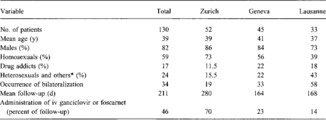
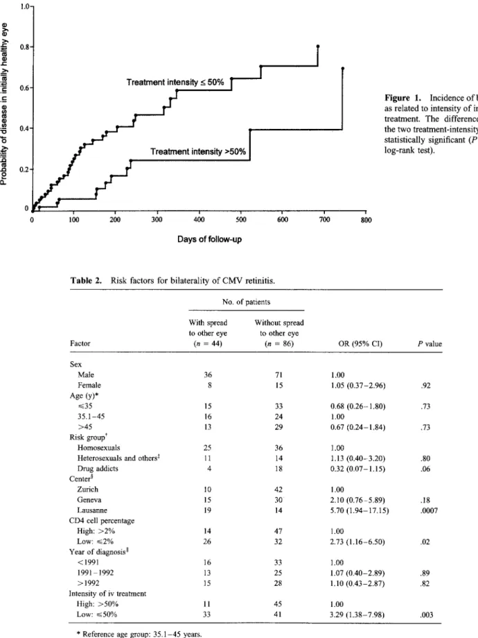
In order to allow for carryover effects, the analysis was repeated, and bilaterality occurring within the first 2 weeks after treatment was started or stopped was assigned to the preceding period. However, the results did not change. In a second analysis, we divided patients into two groups, according to whether they received intravenous treatment for more or less than 50% of the follow-up days. The probability of bilaterality was plotted according to the Kaplan-Meier method and is shown in figure 1. The differences between the two treatment-intensity groups were highly significant (table 2).
As expected, because treatment modalities depended so highly on the center, an analogous analysis for the centers also yielded significant differences. CD4 cell percentages seem to influence the risk of bilaterality (table 2) as well. We therefore performed a multivariate analysis (table 3), in which the treat-ment effect (-_.50%/>50%) was adjusted for CD4 cell percent-ages (<2%/>2%). This adjustment did not notably influence relative risks and confidence intervals, a finding indicating that the two risk factors are independent.
Discussion
The data we have collected shows a strong inverse associa-tion between intravenous treatment and bilaterality of CMV retinitis. In patients who received such treatment for >50% of
Table 1. Data regarding the 130 analyzed patients with initially unilateral CMV retinitis at three centers in Switzerland.
Variable Total Zurich Geneva Lausanne
No. of patients 130 52 45 33
Mean age (y) 39 39 41 37
Males (%) 82 86 84 73
Homosexuals (%) 59 73 56 39
Drug addicts (%) 17 11.5 22 18
Heterosexuals and others* (%) 24 15.5 22 43
Occurrence of bilateralization 34 19 33 58
Mean follow-up (d) 211 280 164 168
Administration of iv ganciclovir or foscarnet
(percent of follow-up) 46 70 23 14
1.0- 0.8- 0.6- Treatment intensity 50%
T
r
ri
0.4-Treatment intensity >50% 0.2-0 100 200 300 400 500 600 700 800 Days of follow-upFigure 1. Incidence of bilaterality as related to intensity of intravenous treatment. The difference between the two treatment-intensity groups is statistically significant (P < .01 by log-rank test).
Table 2. Risk factors for bilaterality of CMV retinitis.
No. of patients Factor With spread to other eye (n = 44) Without spread to other eye (n = 86) OR (95% CI) P value Sex Male 36 71 1.00 Female 8 15 1.05 (0.37-2.96) .92 Age (y)* .---.35 15 33 0.68 (0.26-1.80) .73 35.1-45 16 24 1.00 >45 13 29 0.67 (0.24-1.84) .73 Risk groups Homosexuals 25 36 1.00
Heterosexuals and others1 11 14 1.13 (0.40-3.20) .80
Drug addicts 4 18 0.32 (0.07-1.15) .06 Center Zurich 10 42 1.00 Geneva 15 30 2.10 (0.76-5.89) .18 Lausanne 19 14 5.70 (1.94-17.15) .0007 CD4 cell percentage High: >2% 14 47 1.00 Low: ..--_2% 26 32 2.73 (1.16-6.50) .02 Year of diagnosis II <1991 16 33 1.00 1991-1992 13 25 1.07 (0.40-2.89) .89 >1992 15 28 1.10 (0.43-2.87) .82 Intensity of iv treatment High: >50% 11 45 1.00 Low: --._.50% 33 41 3.29 (1.38-7.98) .003
* Reference age group: 35.1-45 years. t Reference group: homosexuals.
Others includes those with undetermined risk factors (3), multiple risk factors (2), and blood transfusions (1). Reference: Zurich.
CID 1997; 24 (April) Systemic Treatment for CMV Retinitis 623
Table 3. Data regarding a multivariate analysis, in which treatment effect was adjusted for CD4 cell percentages.
Factor OR (CI) before adjustment OR (CI) after adjustment Intensity of treatment High: >50% 1.00 1.00 Low: --50% 3.29 (1.38-7.98) 4.32 (3.40 -5.26) CD4 cell percentage High: >2% 1.00 1.00 Low: 2.73 (1.16-6.5) 2.79 (1.90-3.67) Year of diagnosis <1991 1.00 1.00 1991-1992 2.07 (0.40-2.49) 1.24 (0.16 -2.31) >1992 1.10 (0.43-2.87) 1.67 (0.56-2.75)
the follow-up time, the median interval until bilaterality oc-curred was 175 days; in those who received less intravenous treatment, that interval amounted to only 99 days.
It is tempting to jump from association to causation and conclude that retinitis became bilateral because intravenous
treatment was not intense enough. However, since this was not a prospective, randomized trial, the possibility of con-founding factors needs to be considered. Multivariate analy-sis suggests that CD4 cell percentages were not responsible for the association between treatment and involvement of the initially healthy eye.
Antiretroviral therapy is another potential confounder. Ad-ministration of azidothymidine in Switzerland began in 1988, and that of zalcitabine and didanosine began in 1991; combina-tion therapy became popular in 1993. Patients whose CMV retinitis was diagnosed later received more antiretroviral treat-ment. However, when stratification according to year of diag-nosis was added to the multivariate analysis, this did not change the relative risks associated with intensity of anti-CMV treat-ment. These analyses reinforce the hypothesis of causal rela-tion, although in the absence of randomization other unidenti-fied confounders may exist.
Many of our patients who did not have intravenous therapy received intravitreal injections of ganciclovir. Older studies [13, 15] enrolled few patients with unilateral retinitis [13] or used a combination of intravitreal and intravenous ganciclovir in most patients [15]; for that reason, they may have missed the risk of bilaterality. However, in a larger trial of ganciclovir implants, a high incidence of bilaterality was also observed [6, 16].
Our analysis did not cover side effects and visual outcome; we did not think that retrospective chart review would yield data of sufficient quality. Mortality data showed a trend fa-voring more intensive intravenous therapy, but this may be due to the fact that untreated patients were seen early in the AIDS epidemic, when survival was poor. Therefore, we cannot claim that more intensively treated patients did better overall.
Without results of a randomized, prospective trial, retro-spective data appropriately guide clinical practice. These
data suggest that local therapy by injections or implants may supplement but not replace systemic therapy. Because of the toxicity and expense of intravenous therapy, this is a bitter pill to swallow, sweetened perhaps by the availability of oral ganciclovir.
The Swiss HIV Cohort Study (SHCS) Group
Members of the group are M. Battegay, Ph. Biirgisser, L. Jeannerod, M. Egger, P. Erb (president of the laboratory group), W. Fierz, M. Flepp (president of the clinical group), P. Francioli (president of the SHCS, University Hospital, Lausanne, Switzer-land), P. Grob and U. Grtininger (observers of the Federal Office of Public Health), B. Hirschel (president of the Scientific Board), B. Ledergerber, R. Lathy, R. Malinverni, L. Matter, M. Opravil, F. Paccaud, L. Perrin, W. Pichler, M. Rickenbach (manager of the data coordination center), 0. Rutschmann, P. Vemazza, and J. von Overbeck.
References
1. Gallant JE, Moore RD, Richman DD, Keruly J, Chaisson RE, Zidovud-ine Epidemiology Study Group. Incidence and natural history of cyto-megalovirus disease in patients with advanced human immunodefi-ciency virus disease treated with zidovudine. J Infect Dis 1992; 166: 1223 - 7.
2. Hoover DR, Saah AJ, Bacellar H, et al. Clinical manifestations of AIDS in the era of pneumocystis prophylaxis. Multicenter AIDS Cohort Study. N Engl J Med 1993; 329:1922-6.
3. Berthe P, Baudouin C, Garraffo R, Hofmann P, Taburet AM, Lapalus P. Toxicologic and pharmacokinetic analysis of intravitreal injections of foscarnet, either alone or in combination with ganciclovir. Invest Oph-thalmol Vis Sci 1994; 35:1038-45.
4. Diaz-Llopis M, Espafia E, Mulloz G, et al. High dose intravitreal foscarnet in the treatment of cytomegalovirus retinitis in AIDS. Br J Ophthalmol
1994; 78:120-4.
5. Sanborn GE, Anand R, Torti RE, et al. Sustained-release ganciclovir ther-apy for treatment of cytomegalovirus retinitis: use of an intravitreal device. Arch Ophthalmol 1992; 110:188-95.
6. Martin DF, Parks DJ, Mellow SD, et al. Treatment of cytomegalovirus retinitis with an intraocular sustained-release ganciclovir implant: a ran-domized controlled clinical trial. Arch Ophthalmol 1994; 112:1531-9. 7. Drew WL, Ives D, Lalezari JP, et al. Oral ganciclovir as maintenance treatment for cytomegalovirus retinitis in patients with AIDS. N Engl J Med 1995; 333:615-20.
8. Spector SA, Busch DF, Follansbee S, et al. Pharmacokinetic, safety, and antiviral profiles of oral ganciclovir in persons infected with human immunodeficiency virus: a phase I/II study. J Infect Dis 1995; 171: 1431 -7.
9. Polis MA, Masur H. Promising new treatments for cytomegalovirus retini-tis. JAMA 1995;273:1457-9.
10. Rahhal FM, Arevalo JF, de la Paz EC, Munguia D, Azen SP, Freeman WR. Treatment of cytomegalovirus retinitis with intravitreous cidofovir in patients with AIDS: a preliminary report. Ann Intern Med 1996; 125: 98-103.
11. Studies of ocular complications of AIDS Research Group, in collabo-ration with the AIDS Clinical Trials Group. Morbidity and toxic
effects associated with ganciclovir or foscarnet therapy in a random-ized cytomegalovirus retinitis trial. Arch Intern Med 1995; 155:
65 —74.
12. Maitham A, Faulds D. Ganciclovir: an update of its therapeutic use in cytomegalovirus infection. Drugs 1994; 48:455-84.
13. Cantrill HL, Henry K, Melroe NH, Knobloch WH, Ramsay RC, Balfour
HH Jr. Treatment of cytomegalovirus retinitis with intravitreal gan-ciclovir: long-term results. Ophthalmology 1989; 96:367-74.
14. Ledergerber B, von Overbeck J, Egger M, Liithy R. The Swiss HIV Cohort Study: rationale, organization and selected baseline characteristics. Soz Praventivmed 1994; 39:387 —94.
15. Ussery FM III, Gibson SR, Conklin RH, Piot DF, Stool EW, Conklin AJ. Intravitreal ganciclovir in the treatment of AIDS-associated cytomegalo-virus retinitis. Ophthalmology 1988; 95:640-8.
16. Holland GN, Tufail A. New therapies for cytomegalovirus retinitis [edito-rial]. N Engl J Med 1995; 333:658-9.