Nephrol Dial Transplant (2010) 25: 2225–2231 doi: 10.1093/ndt/gfq008
Advance Access publication 1 February 2010
Glomerular hyperfiltration and increased proximal sodium
reabsorption in subjects with type 2 diabetes or impaired fasting
glucose in a population of the African region
Menno Pruijm
1, Grégoire Wuerzner
1, Marc Maillard
1, Pascal Bovet
2, Claude Renaud
3,
Murielle Bochud
1,2and Michel Burnier
11
Service of Nephrology, University Hospital of Lausanne (CHUV), Switzerland,
2Institute of Social and Preventive Medicine
(IUMSP), University Hospital of Lausanne (CHUV) and University of Lausanne, Switzerland and
3Ministry of Health and Social
Development, Republic of Seychelles
Correspondence and offprint requests to: Michel Burnier; E-mail: [email protected]
Abstract
Background. Glomerular hyperfiltration (GHF) is a
well-recognized early renal alteration in diabetic
pa-tients. As the prevalence of GHF is largely unknown
in populations in the African region with respect to
nor-mal fasting glucose (NFG), impaired fasting glucose
(IFG) and type 2 diabetes [diabetes mellitus (DM)],
we conducted a cross-sectional study in the Seychelles
islands among families including at least one member
with hypertension.
Methods. The glomerular filtration rate (GFR), effective
renal plasma flow (ERPF) and proximal tubular sodium
re-absorption were measured using inulin, p-aminohippurate
(PAH) and endogenous lithium clearance, respectively.
Twenty-four-hour urine was collected on the preceding
day.
Results. Of the 363 participants (mean age 44.7 years),
6.6% had IFG, 9.9% had DM and 63.3% had
hyperten-sion. The prevalence of GHF, defined as a GFR >140
ml/min, was 17.2%, 29.2% and 52.8% in NFG, IFG and
DM, respectively (P trend <0.001). Compared to NFG,
the adjusted odds ratio for GHF was 1.99 [95%
confi-dence interval (CI) 0.73–5.44] for IFG and 5.88 (2.39–
14.45) for DM. Lithium clearance and fractional
excre-tion of lithium were lower in DM and IFG than NFG
(P < 0.001).
Conclusion. In this population of African descent,
sub-jects with impaired fasting glucose or type 2 diabetes
had a high prevalence of GHF and enhanced proximal
so-dium reabsorption. These findings provide further insight
on the elevated incidence of nephropathy reported among
African diabetic individuals.
Keywords: African region; diabetes; glomerular hyperfiltration; inulin clearance; lithium clearance
Introduction
The prevalence of end-stage renal damage (ESRD) is 15–
20% in African-American diabetics, which is four times
higher than in Caucasian diabetics [1,2]. This may be
part-ly explained by socio-economic factors and genetic
differ-ences in renal haemodynamics and sodium handling [3,4].
For example, healthy African-Americans have a 10% lower
renal plasma flow (ERPF) than age-matched Caucasians,
possibly due to a more activated intrarenal
renin–angioten-sin system (RAS) in the former than the latter [5].
African-Americans are also more prone to sodium retention and
salt-sensitive hypertension than Caucasians [3]. Besides,
there is some evidence that glomerular hyperfiltration
(GHF) and a disturbed renal autoregulation are more
com-mon acom-mong hypertensive African-Americans as compared
to age-matched hypertensive Caucasians [6].
GHF and enhanced proximal sodium reabsorption are
also more frequent in type 2 diabetes [diabetes mellitus
(DM)] and in the metabolic syndrome than persons with
normal fasting glucose (NFG) [7–9]. According to the
tu-bulocentric view developed recently by Vallon et al. and
supported by experimental evidence, increased proximal
tubular reabsorption of sodium might be one of the trigger
mechanisms leading to GHF [8]. Together, GHF and
high-er proximal reabsorption of sodium may result in accelhigh-er-
acceler-ated loss of kidney function and hypertension in diabetic
persons [10,11].
One may therefore expect that diabetics of African
de-scent have both a high prevalence of GHF and enhanced
proximal sodium reabsorption, which may make them
more susceptible to kidney function deterioration and
hy-pertension. However, only few studies have examined
re-nal haemodynamics in African subjects, and these studies
included mainly African-Americans [12,13]. To our
knowledge, no study has used gold standard techniques
© The Author 2010. Published by Oxford University Press on behalf of ERA-EDTA. All rights reserved. For Permissions, please e-mail: [email protected]
such as inulin, p-aminohippurate (PAH) and endogenous
lithium clearances to assess the renal function and renal
sodium handling in type 2 DM in the African region so
far. Therefore, the purpose of this analysis was to compare
renal haemodynamic parameters, renal sodium handling
and the prevalence of GHF between NFG, impaired
glu-cose tolerance (IGT) and DM categories in the Seychelles,
a rapidly developing country in the African region, taking
into account possible confounding effects of parameters
such as age, sex, obesity and estimated sodium intake.
Materials and methods
This study was conducted on a sample of families collected prospectively for the primary purpose of a candidate gene study of hypertension [14,15]. The study took place in the Seychelles islands (Indian Ocean), which lie∼1000 km east of Kenya and 1000 km north of Madagascar and Mauritius. The majority of the population is of African descent. We enrolled 494 subjects of East African descent from 76 families en-riched in hypertensive individuals between August 1999 and January 2002. The detailed family selection process has been described previ-ously [4]. The study was approved by the Ethical Committees of the Ministry of Health in the Seychelles and of the University of Lausanne (Switzerland). All participants provided written informed consent. Of the 363 participants with data on inulin clearance, 343 had available valid data on PAH clearance and 329 valid data on endogenous lithium clearance.
Antihypertensive therapy, if any, was stopped 2 weeks before conduct-ing clearance protocols. Clearance studies were performed after an over-night fast, as reported previously [16]. In brief, two intravenous catheters were inserted into antecubital veins, one for the infusion of inulin and PAH, and a second into the contra lateral arm for blood drawing. After an oral water load of 8 ml/kg and a 2-hour equilibration period, two 1-hour inulin and PAH clearances were obtained to measure GFR and ERPF, respectively. The inulin, PAH and endogenous lithium clearances (Cx) were calculated with the formula Cx= (Ux× V) / Px, where Uxand Px
are urinary and plasma concentrations of the x solute, and V is the urine flow rate in millilitre per minute. Renal blood flow (RBF) was calculated as ERPF / [1− (haematocrit / 100)] and renal vascular resistance (RVR) as (mean arterial blood pressure) / RBF. Fractional excretions of endogenous lithium (FELi) and sodium (FENa) were calculated by the standard for-mula FEx= (Ux× Pcreat) / (Px× Ucreat). Fractional sodium reabsorption in
the post-proximal tubule (FDRNa) was estimated as [(FELi− FENa) / FELi] / 100. Filtration fraction (FF) was calculated as GFR divided by ERPF. Creatinine concentration was measured by the picric acid method (Cobas-Mira, Roche, Basel, Switzerland). Mean arterial blood pressure (MAP) was calculated as one-third of systolic blood pressure plus two-thirds of diastolic blood pressure from the mean of six measurements taken with a mercury sphygmomanometer (three on the day preceding
clearances and three on the morning of the clearances). GHF was defined as a GFR >140 ml/min/1.73 m2) [13]. On the day preceding clearance
studies, participants collected their urine for 24 hours to assess sodium intake. Urinary and plasma sodium and potassium concentrations were measured by flame photometry (IL-943, Instrumentation Laboratory, Milan, Italy). Endogenous trace lithium was measured by atomic absorp-tion spectrophotometry [17]. Plasma renin activity (PRA) was measured using the antibody-trapping principle [18,19]. Aldosterone was measured by a direct radioimmunoassay using a very sensitive and specific antise-rum raised in a New Zealand white rabbit [20]. The coefficients of var-iation for within- and among-assay precision were 0.04 to 0.13 for the PRA and aldosterone assays [18,20].
Participants on antidiabetic treatment during the preceding month, or with fasting blood glucose≥7.0 mmol/l (measured on venous whole blood in duplicate using a Glycotronic® C reflectometer, Macherey-Na-gel, Düren, Germany), were considered as having diabetes (DM). Im-paired fasting glucose tolerance (IFG) was defined as fasting glucose ≥5.6 mmol/l and <7 mmol/l and normal fasting glucose (NFG) as fasting glucose <5.6 mmol/l [21]. Body surface area (BSA) was calculated using the Dubois formula [22]. Body mass index (BMI) was calculated as weight (kilogram) divided by squared height (square metre).
All analyses were conducted with Stata 10 (StataCorp, College Station, Texas, USA). We used generalized estimating equations with an ex-changeable correlation structure to account for familial correlation. A Gaussian link was used for continuous phenotypes (e.g. GFR and ERPF) and a binomial link for dichotomous phenotypes (e.g. GHF). To better approximate a normal distribution of the residuals, GFR was log-trans-formed, and FELI and FF were square-root translog-trans-formed, whereas ERPF was not transformed. For fully adjusted models, we used as predictors the age, sex, BMI, 24-hour urinary sodium and potassium excretion, MAP, alcohol consumption, smoking and being taken off antihypertensive treat-ment. We conducted stratified analyses in untreated and treated partici-pants, as well as sensitivity analyses after excluding participants taking diuretics before the treatment was stopped.
Results
Participant characteristics are presented in Table 1. IFG or
DM was associated with age and male sex. Plasma renin
activity and plasma aldosterone levels were similar across
the three blood glucose categories. The mean urinary
cre-atinine excretion was 0.16 mmol/kg/24 hours in women
and 0.20 mmol/kg/24 hours in men, which suggests good
completeness of urine collections. The median urinary
so-dium excretion was similar across diabetes categories, and
also in hypertensive (96 mmol/24 hours) and
non-hypertensive (104 mmol/24 hours) individuals (P = 0.36).
Urinary excretion of potassium was greater in DM than
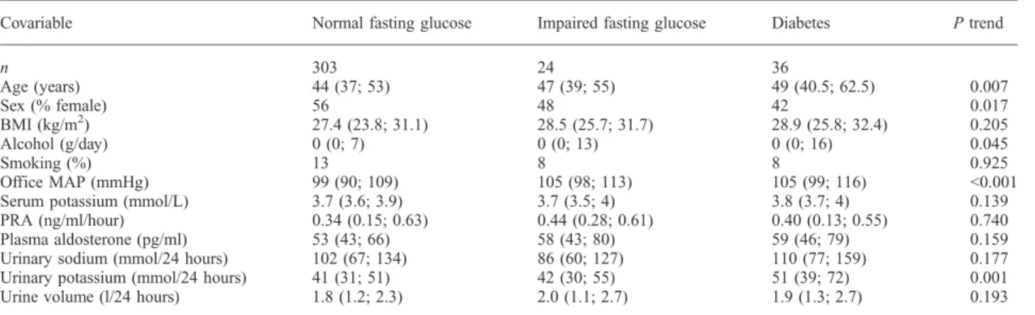
Table 1. Participant characteristics by diabetes status
Covariable Normal fasting glucose Impaired fasting glucose Diabetes P trend
n 303 24 36 Age (years) 44 (37; 53) 47 (39; 55) 49 (40.5; 62.5) 0.007 Sex (% female) 56 48 42 0.017 BMI (kg/m2) 27.4 (23.8; 31.1) 28.5 (25.7; 31.7) 28.9 (25.8; 32.4) 0.205 Alcohol (g/day) 0 (0; 7) 0 (0; 13) 0 (0; 16) 0.045 Smoking (%) 13 8 8 0.925 Office MAP (mmHg) 99 (90; 109) 105 (98; 113) 105 (99; 116) <0.001 Serum potassium (mmol/L) 3.7 (3.6; 3.9) 3.7 (3.5; 4) 3.8 (3.7; 4) 0.139 PRA (ng/ml/hour) 0.34 (0.15; 0.63) 0.44 (0.28; 0.61) 0.40 (0.13; 0.55) 0.740 Plasma aldosterone (pg/ml) 53 (43; 66) 58 (43; 80) 59 (46; 79) 0.159 Urinary sodium (mmol/24 hours) 102 (67; 134) 86 (60; 127) 110 (77; 159) 0.177 Urinary potassium (mmol/24 hours) 41 (31; 51) 42 (30; 55) 51 (39; 72) 0.001 Urine volume (l/24 hours) 1.8 (1.2; 2.3) 2.0 (1.1; 2.7) 1.9 (1.3; 2.7) 0.193 Data are medians (interquartile range), unless otherwise specified. Data are unadjusted. PRA, plasma renin activity.
NFG categories. This was not due to a higher use of
diure-tics (17% in diabediure-tics vs 22% in non-diabediure-tics).
The prevalence of hypertension, defined as having
of-fice blood pressure
≥140/90 mmHg and/or being on
an-tihypertensive treatment, was 63.3%, and the prevalence
tended to be higher in DM and IFG categories than in
NFG participants. Among the 230 hypertensive
partici-pants, 68% were on antihypertensive treatment, which
was stopped in all individuals for a median of 15 days
before clearance studies. The following treatment types
were stopped: angiotensin-converting enzyme (ACE)
inhi-bitors in 30%, calcium channel blockers in 44%, diuretics
(mainly hydrochlorothiazide) in 49%, beta-blockers in
28%. Median (range) diabetes duration was 3 (0–21)
years, and six participants had newly diagnosed type 2
di-abetes mellitus.
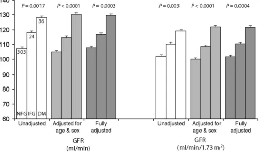
Compared to NFG, GFR was higher in the presence of
DM, and intermediate values were found for IFG,
regard-less of the adjustment procedure used (Figure 1) and
irre-spective of BSA.
The prevalence of hyperfiltration (defined as GFR >140
ml/min) was 17.2%, 29.2% and 52.8% in NFG, IFG and
DM categories, respectively (P trend <0.001). When
cor-rected for body surface area, the corresponding prevalence
of hyperfiltration (defined as GFR >140 ml/min) was
9.9%, 25.0% and 27.8%, respectively (P trend = 0.001).
The odds ratios of having GHF are illustrated in Table 2
after adjustment for different covariate combinations.
Par-ticipants with IFG and DM had an
∼2- to 3-fold increased
risk of GHF when compared to NFG.
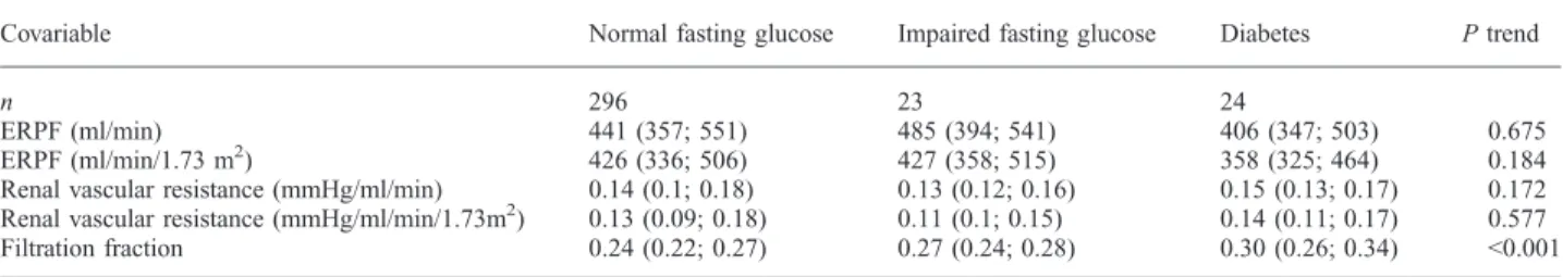
IFG and DM were not associated with ERPF or renal
vascular resistance levels except for a non-significant trend
Fig. 1. Glomerular filtration rate and effective renal plasma flow by diabetes status. Bars are means, and whiskers are standard errors. NFG, normal fasting glucose. IFG, impaired fasting glucose (5.6≤ FG < 7.0 mmol/L). DM, diabetes mellitus. GFR was estimated using inulin clearance. Inulin clearance data were log-transformed to conducted linear regression analyses (generalized estimating equations) and then back-transformed to provide untransformed adjusted data. Fully adjusted models included age, sex, body mass index, alcohol consumption, smoking status, 24-hour urinary sodium and potassium excretion, mean arterial blood pressure and a dummy variable to indicate whether or not the participant was taken off antihypertensive treatment. P-values are from a likelihood ratio test (2 df) comparing a model with and without categorical diabetes status [i.e. normal glucose (reference category), impaired glucose tolerance and diabetes mellitus].
Table 2. Risk of hyperfiltration by diabetes status
Impaired fasting glucose Diabetes Impaired fasting glucose + diabetes P differencea
n 24 36 60
Hyperfiltration (ml/min)
Unadjusted 1.88 (0.77–4.65) 4.73 (2.34–9.56)*** 3.32 (1.84–6.01)*** 0.08 Age- and sex-adjusted 1.98 (0.78–4.98) 6.57 (3.01–14.45)*** 3.95 (2.09–7.48)*** 0.03 Fully adjusted 1.99 (0.73–5.44) 5.88 (2.39–14.45)*** 3.63 (1.79–7.38)*** 0.08 Hyperfiltration (ml/min/1.73 m2)
Unadjusted 2.75 (0.99–7.63) 3.22 (1.39–7.46)** 3.02 (1.49–6.12)** 0.80 Age and sex-adjusted 2.75 (0.97–7.75) 3.58 (1.48–8.67)** 3.20 (1.53–6.70)** 0.67 Fully adjusted 2.63 (0.91–7.57) 2.52 (0.97–6.55) 2.57 (1.18–5.58)* 0.95 Data are odds ratio (OR) (95% CI) for hyperfiltration. Hyperfiltration cutoff: 140 ml/min (high part of the table) or 140 ml/min/1.73 m2(lower part of
the table). Reference category is normal fasting glucose (n = 303).Full models include age, sex, body mass index, urinary sodium and potassium excretion, alcohol consumption, smoking, mean arterial pressure and being taken off antihypertensive treatment. *0.01≥ P < 0.05, **0.001 ≥ P < 0.01, ***P < 0.001.
a
towards lower ERPF in DM (Table 3). Hence, the higher
FF observed in DM resulted essentially from differences in
GFR. Filtration fraction was higher in DM than NFG even
after using the fully adjusted models (data not shown).
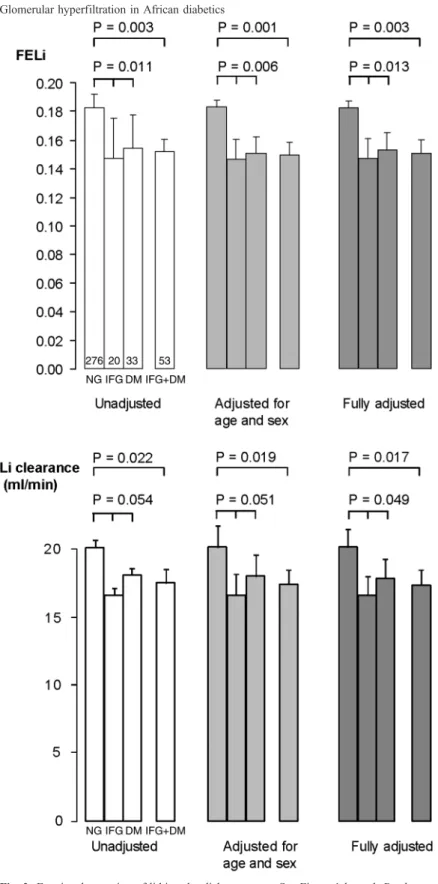
IFG or DM was associated with lower FELi compared to
NFG, indicating increased reabsorption of sodium in
prox-imal segments of the nephron in IFG and DM (Figure 2).
As FELi did not differ between IFG and DM, analyses
were conducted with both groups combined. Similar
re-sults were obtained when using lithium clearance instead
of FELi as the dependent variable. In these latter models,
GFR was included as a covariate in all analyses. The
frac-tional distal reabsorption of sodium (FDRNa) was not
as-sociated with DM or IFG [data not shown, P = not
significant (NS)].
Sensitivity analyses conducted in untreated participants
(n = 186) showed similar results. FELi across diabetes
cat-egories was 18%, 15% and 13% for unadjusted analyses
(P = 0.02), 18%, 16% and 13% for age- and sex-adjusted
analyses (P = 0.02) and 18%, 15% and 13% for fully
adjusted analyses (P = 0.07), respectively. Sensitivity
analyses conducted in participants not taking diuretics
(n = 255) clearly confirmed our results (P < 0.02 for
all analyses).
Discussion
This study in a population in the African region shows that
subjects with DM and IFG have, compared to subjects
with NFG, a higher prevalence of glomerular
hyperfiltra-tion and greater proximal tubular sodium reabsorphyperfiltra-tion, as
measured by FELi, independently of several potentially
confounding variables.
GHF is a well-recognized early renal alteration in DM.
Today, only few studies have reported data on GFR in DM
in individuals in the African region, and data were
ob-tained on small samples (48–162 subjects) [23–25].
More-over, to our knowledge, none of these studies has used
radio-isotopic methods or the
‘gold standard’ inulin
clear-ance to measure GFR, and none has reported values on
re-nal plasma flow. Instead, serum creatinine was used as the
standard for assessing renal function [23–25].
Comparing our data on GHF with studies performed in
Caucasians, Asians and African-Americans is difficult for
several reasons. Firstly, different methods were used to
measure GFR and ERPF between studies (mostly 125
I-la-belled iothalamate or iohexol for GFR and 131 I-laI-la-belled
hippuran for ERPF). Secondly, there is no generally
ac-cepted definition for GHF: cutoff values vary between
125 and 160 ml/min, some but not all being corrected
for body surface area. Furthermore, some authors have
ar-gued against the use of a fixed cutoff value and
recom-mended the use of >1.96 standard deviations (SD) above
the mean GFR as the definition for GHF [26].
Nonethe-less, studies that have used GFR >140 ml/min/1.73 m
2found a prevalence of GHF ranging between 6% and
29% in type 2 diabetic Caucasians [27–29], and between
25% and 36% in type 2 diabetic African-Americans
[13,30], as compared to 27.8% in our study. In our
multi-variate analysis, we used GFR >140 ml/min to define GHF
in order to be able to examine the relationship between
GFR and BMI. Adjusting to BSA would have obscured
this relationship, since BMI itself is highly correlated with
BSA. Studies that used this same cutoff value found a
prevalence of GHF in 16% of Caucasians [7], but no data
are available in African-Americans. In our population, the
prevalence of GHF, when uncorrected for BSA, reached
53% in DM and 29% in IFG. These data suggest that
the prevalence of GHF was rather high in Seychelles
com-pared to other populations, but probably similar to the
prevalence found in African-Americans.
The mechanisms leading to the development of GHF in
IFG and DM are still only partially understood, and several
hypotheses have been proposed. Some animal studies have
suggested that diabetic animals present intraglomerular
hy-pertension because of an inappropriate dilatation of
affer-ent arterioles, leading to raised ERPF and GHF [31].
Studies in humans have also found higher values for
GFR and ERPF in Caucasian patients with newly
diag-nosed type 2 DM as compared to non-diabetic subjects,
which also points to afferent vasodilatation [32]. Why
af-ferent vasodilatation occurs is less clear, but elevated levels
of insulin, insulin-like growth factor-1 (IGF-1), atrial
natri-uretic peptide (ANP) and advanced glycosylation
end-products (AGE) have been postulated [33–35]. In our
study, ERPF was not elevated in IFG and/or DM. On the
contrary, ERPF was relatively low in IFG and DM, which
argues against afferent vasodilatation as a primary
mecha-nism for GHF in subjects of African descent, unless the
former was accompanied by efferent vasoconstriction.
However, in line with previous studies, PRA and
aldoste-rone levels were rather low in all groups [36,37]. More
stringent efferent vasoconstriction in IGT and DM as
com-pared to NFG could thus only be present in case of a more
activated intrarenal renin–angiotensin system or higher
Table 3. Effective renal plasma flow, filtration fraction and renal vascular resistance, by diabetes status
Covariable Normal fasting glucose Impaired fasting glucose Diabetes P trend
n 296 23 24
ERPF (ml/min) 441 (357; 551) 485 (394; 541) 406 (347; 503) 0.675 ERPF (ml/min/1.73 m2) 426 (336; 506) 427 (358; 515) 358 (325; 464) 0.184 Renal vascular resistance (mmHg/ml/min) 0.14 (0.1; 0.18) 0.13 (0.12; 0.16) 0.15 (0.13; 0.17) 0.172 Renal vascular resistance (mmHg/ml/min/1.73m2) 0.13 (0.09; 0.18) 0.11 (0.1; 0.15) 0.14 (0.11; 0.17) 0.577 Filtration fraction 0.24 (0.22; 0.27) 0.27 (0.24; 0.28) 0.30 (0.26; 0.34) <0.001 Data are unadjusted medians (interquartile range). ERPF, effective renal plasma flow, measured using PAH clearance.
sympathetic nervous system activity [5]. Since neither of
the two were measured, this remains hypothetical.
Another explanation might be the role of (intermittent)
high blood glucose levels in DM, which would stimulate
sodium
–glucose co-transport in the proximal tubules, thus
leading to enhanced proximal sodium reabsorption and
di-minished distal sodium delivery. Alternatively, or in
addi-tion, tubular growth has been proposed to increase
Fig. 2. Fractional excretion of lithium by diabetes status. See Figure 1 legend. P-values are either from (i) a likelihood ratio test (2 df) comparing a model with and without categorical diabetes status [i.e. normal glucose (reference category), impaired glucose tolerance and diabetes mellitus] for the three-group comparisons or from (ii) a Wald test (1 df) for the two-group (NFG vs DM + IFG) comparisons.
proximal sodium reabsorption in early DM [8]. Reduced
distal sodium delivery, via the tubuloglomerular feedback
mechanism, leads in turn to dilatation of the afferent
arter-ioles and GHF [8]. This
‘tubulocentric view’ is supported
by animal models and might be mediated by adenosine.
Indeed, in the streptozotocin-induced diabetic mice model,
adenosine knockout mice, which lack a tubuloglomerular
feedback response, also lack GHF [38]. Again, our finding
of a diminished ERPF in DM and IFG compared to NFG
does not fully support such a mechanism in this
popula-tion, although the participants with IFG and/or DM did
have raised proximal sodium reabsorption, as shown by
their diminished lithium clearance.
Enhanced proximal sodium reabsorption has been
re-ported previously in DM type 2 [39], and this might be
one of the mechanisms responsible for diabetes-associated
hypertension [40]. Interestingly, higher renal sodium
reab-sorption is found more often in subjects of African than
Caucasian descent [41,42], although it remains uncertain
in which part of the nephron this reabsorption takes place
[43]. Increased activity of the Na–K–2Cl channel has been
observed in the thick ascending limb in young
normoten-sive African-Americans, while enhanced reabsorption in
the distal [44] or proximal segments of the nephron [45]
have been advocated as well. Our data also support
en-hanced sodium reabsorption in the early tubular segments
of the nephron (proximal tubule and thick ascending limb),
since both the fractional excretion of lithium and lithium
clearance were reduced in DM and IFG as compared to
NFG, but FDRNa was not. The present observation is in
accordance with a recent finding of our group evaluating
another African population, which showed that Black
indi-viduals in South Africa had enhanced proximal sodium
re-absorption compared to Caucasians, regardless of dietary
salt intake [46]. Of note, the estimated 24-hour sodium
intake was rather low in our study at around 5.8 g of salt
(2.3 g of sodium) per day and similar between the three
groups. Indeed, diet in the Seychelles is low in salt and
potassium as it consists mainly of unsalted rice and fish.
The increased proximal sodium reabsorption seen
repeat-edly in subjects of African descent may further explain
why African diabetics are more often hypertensive than
Caucasian diabetics [47].
Our study has some limitations. Firstly, our data rely on
a sample of families enriched in hypertensive persons,
which may limit generalization of our findings. However,
the same tendency of enhanced proximal sodium
reabsorp-tion in diabetics was found in normotensive individuals
and untreated hypertensive individuals. Secondly,
microal-buminuria (MAU) and glycated haemoglobin (HbA1c)
le-vels were not available, and we have limited information
regarding the degree of kidney damage and diabetes
con-trol in our subjects. Finally, the cross-sectional nature of
our data limits causal inferences.
In summary, we found a high prevalence of glomerular
hyperfiltration and increased proximal sodium
reabsorp-tion in individuals in the African region with IFG or DM
as compared to normoglycaemic individuals,
independent-ly of several potentialindependent-ly confounding variables. Our
find-ings are consistent with the view that the high prevalence
of glomerular hyperfiltration and enhanced proximal
sodi-um reabsorption might contribute to the higher incidence
of hypertension and diabetic nephropathy in diabetics of
African descent.
Acknowledgements. This study was supported, in part, by grants from the Swiss National Science Foundation (TANDEM No 31–51115.97), (FN-320000-116364), (SPUM 33CM30-124087), (AMBIZIONE PZ00P3_121655), (PROSPER 3200BO-111362/1 and 3233BO-111361/1) and by the Swiss School of Public Health Plus. We thank the participants to the study as well as the Ministry of Health of the Republic of Seychelles for continued support to epidemiological cardio-vascular research, and Air Seychelles and SkyChef for logistic support in transporting equipment and samples.
Conflict of interest statement. Information in this article was not presented previously.
References
1. Cowie CC, Port FK, Wolfe RA et al. Disparities in incidence of di-abetic end-stage renal disease according to race and type of diabetes. N Engl J Med 1989; 321: 1074–1079
2. Collins AJ, Foley R, Herzog C et al. Excerpts from the United States Renal Data System 2007 Annual Data Report. Am J Kidney Dis 2008; 51: S1–S320
3. Chun TY, Bankir L, Eckert GJ et al. Ethnic differences in renal re-sponses to furosemide. Hypertension 2008; 52: 241–248
4. Bochud M, Elston RC, Maillard M et al. Heritability of renal function in hypertensive families of African descent in the Seychelles (Indian Ocean). Kidney Int 2005; 67: 61–69
5. Price DA, Fisher ND, Osei SY et al. Renal perfusion and function in healthy African Americans. Kidney Int 2001; 59: 1037–1043 6. Kotchen TA, Piering AW, Cowley AW et al. Glomerular
hyperfiltra-tion in hypertensive African Americans. Hypertension 2000; 35: 822–826
7. Vora JP, Dolben J, Dean JD et al. Renal hemodynamics in newly pre-senting non-insulin dependent diabetes mellitus. Kidney Int 1992; 41: 829–835
8. Vallon V, Blantz RC, Thomson S. Glomerular hyperfiltration and the salt paradox in early [corrected] type 1 diabetes mellitus: a tubulo-centric view. J Am Soc Nephrol 2003; 14: 530–537
9. Strazzullo P, Barbato A, Galletti F et al. Abnormalities of renal sodi-um handling in the metabolic syndrome. Results of the Olivetti Heart Study. J Hypertens 2006; 24: 1633–1639
10. Rudberg S, Persson B, Dahlquist G. Increased glomerular filtration rate as a predictor of diabetic nephropathy—an 8-year prospective study. Kidney Int 1992; 41: 822–828
11. Silveiro SP, Friedman R, de Azevedo MJ et al. Five-year prospective study of glomerular filtration rate and albumin excretion rate in nor-mofiltering and hyperfiltering normoalbuminuric NIDDM patients. Diabetes Care 1996; 19: 171–174
12. Parra EJ, Marcini A, Akey J et al. Estimating African American ad-mixture proportions by use of population-specific alleles. Am J Hum Genet 1998; 63: 1839–1851
13. Chaiken RL, Eckert-Norton M, Bard M et al. Hyperfiltration in African-American patients with type 2 diabetes. Cross-sectional and longitudinal data. Diabetes Care 1998; 21: 2129–2134 14. Eap CB, Bochud M, Elston RC et al. CYP3A5 and ABCB1 genes
influence blood pressure and response to treatment, and their effect is modified by salt. Hypertension 2007; 49: 1007–1014
15. Bochud M, Eap CB, Elston RC et al. Association of CYP3A5 geno-types with blood pressure and renal function in African families. J Hypertens 2006; 24: 923–929
16. Burnier M, Rutschmann B, Nussberger J et al. Salt-dependent renal effects of an angiotensin II antagonist in healthy subjects. Hyperten-sion 1993; 22: 339–347
17. Magnin JL, Decosterd LA, Centeno C et al. Determination of trace lithium in biological fluids using graphite furnace atomic absorption spectrophotometry: variability of urine matrices circumvented by
cation exchange solid phase extraction. Pharm Acta Helv 1996; 71: 237–246
18. Nussberger J, Fasanella d'Amore T, Porchet M et al. Repeated admin-istration of the converting enzyme inhibitor cilazapril to normal volunteers. J Cardiovasc Pharmacol 1987; 9: 39–44
19. Poulsen K, Jorgensen J. An easy radioimmunological microassay of renin activity, concentration and substrate in human and animal plas-ma and tissues based on angiotensin I trapping by antibody. J Clin Endocrinol Metab 1974; 39: 816–825
20. Nussberger J, Waeber B, Brunner HR et al. Highly sensitive micro-assay for aldosterone in unextracted plasma: comparison with two other methods. J Lab Clin Med 1984; 104: 789–796
21. Genuth S, Alberti KG, Bennett P et al. Follow-up report on the diag-nosis of diabetes mellitus. Diabetes Care 2003; 26: 3160–3167 22. DuBois M, DuBois EF. A formula to estimate the approximate
sur-face area if height and weight be known. Arch Intern Med 1916; 17: 863–871
23. Agaba EI, Agaba PA, Puepet FA et al. Pattern of glomerular filtration rate abnormalities in type 2 diabetes mellitus in Jos, Nigeria. Niger Postgrad Med J 2004; 11: 262–264
24. Arije A, Akinsola W, Ladipo GO. Renal function in adult Nigerian diabetics. Trop Geogr Med 1988; 40: 334–337
25. Unuigbe EI, Azubike CO, Eregie A. Assessment for markers of ne-phropathy in newly diagnosed type 2 diabetics. West Afr J Med 2005; 24: 134–138
26. Sunder-Plassmann G, Horl WH. A critical appraisal for definition of hyperfiltration. Am J Kidney Dis 2004; 43: 396–397
27. Vedel P, Obel J, Nielsen FS et al. Glomerular hyperfiltration in mi-croalbuminuric NIDDM patients. Diabetologia 1996; 39: 1584–1589 28. Gragnoli G, Signorini AM, Tanganelli I et al. Prevalence of glomerular hyperfiltration and nephromegaly in normo- and mi-croalbuminuric type 2 diabetic patients. Nephron 1993; 65: 206–211
29. Premaratne E, MacIsaac RJ, Tsalamandris C et al. Renal hyperfiltra-tion in type 2 diabetes: effect of age-related decline in glomerular filtration rate. Diabetologia 2005; 48: 2486–2493
30. Palmisano JJ, Lebovitz HE. Renal function in black Americans with type II diabetes. J Diabet Complications 1989; 3: 40–44
31. Bank N. Mechanisms of diabetic hyperfiltration. Kidney Int 1991; 40: 792–807
32. Keller CK, Bergis KH, Fliser D et al. Renal findings in patients with short-term type 2 diabetes. J Am Soc Nephrol 1996; 7: 2627–2635
33. Hirschberg R, Brunori G, Kopple JD et al. Effects of insulin-like growth factor I on renal function in normal men. Kidney Int 1993; 43: 387–397
34. Anderson S, Vora JP. Current concepts of renal hemodynamics in di-abetes. J Diabetes Complications 1995; 9: 304–307
35. Sabbatini M, Sansone G, Uccello F et al. Early glycosylation pro-ducts induce glomerular hyperfiltration in normal rats. Kidney Int 1992; 42: 875–881
36. Helmer OM. The renin–angiotensin system and its relation to hyper-tension. Prog Cardiovasc Dis 1965; 8: 117–128
37. Pratt JH, Jones JJ, Miller JZ et al. Racial differences in aldosterone excretion and plasma aldosterone concentrations in children. N Engl J Med 1989; 321: 1152–1157
38. Vallon V, Schroth J, Satriano J et al. Adenosine A(1) receptors deter-mine glomerular hyperfiltration and the salt paradox in early strepto-zotocin diabetes mellitus. Nephron Physiol 2009; 111: 30–38 39. Mbanya JC, Thomas TH, Taylor R et al. Increased proximal tubular
sodium reabsorption in hypertensive patients with type 2 diabetes. Diabet Med 1989; 6: 614–620
40. Weidmann P, Ferrari P. Central role of sodium in hypertension in di-abetic subjects. Diabetes Care 1991; 14: 220–232
41. Palacios C, Wigertz K, Martin BR et al. Sodium retention in black and white female adolescents in response to salt intake. J Clin Endo-crinol Metab 2004; 89: 1858–1863
42. Barbato A, Cappuccio FP, Folkerd EJ et al. Metabolic syndrome and renal sodium handling in three ethnic groups living in England. Dia-betologia 2004; 47: 40–46
43. Burnier M. Ethnic differences in renal handling of water and solutes in hypertension. Hypertension 2008; 52: 203–204
44. Lifton RP, Gharavi AG, Geller DS. Molecular mechanisms of human hypertension. Cell 2001; 104: 545–556
45. Chiolero A, Maillard M, Nussberger J et al. Proximal sodium reab-sorption: an independent determinant of blood pressure response to salt. Hypertension 2000; 36: 631–637
46. Bochud M, Staessen JA, Maillard M et al. Ethnic differences in prox-imal and distal tubular sodium reabsorption are heritable in black and white populations. J Hypertens 2009; 27: 606–612
47. Tierney WM, McDonald CJ, Luft FC. Renal disease in hypertensive adults: effect of race and type II diabetes mellitus. Am J Kidney Dis 1989; 13: 485–493
Received for publication: 3.9.09; Accepted in revised form: 6.1.10
Nephrol Dial Transplant (2010) 25: 2231–2237 doi: 10.1093/ndt/gfp771
Advance Access publication 18 January 2010
Association of ADIPOQ genetic variants and plasma adiponectin
isoforms with the risk of incident renal events in type 2 diabetes
Riphed Jaziri
1,2, Roberte Aubert
1, Ronan Roussel
1,2,3, Nathalie Emery
1,2, Suliya Maimaitiming
1,2,
Naïma Bellili
1,2, Aurélie Miot
1,2, Pierre-Jean Saulnier
4, Florence Travert
3, Samy Hadjadj
5,6,
Michel Marre
1,2,3, Frédéric Fumeron
1,2and for the DIABHYCAR and SURDIAGENE Study Groups
1
INSERM, U695, Genetic Determinants for Type 2 Diabetes and Its Vascular Complications, Paris, France,
2University Paris
Diderot - Paris 7, Paris, France,
3Assistance Publique-Hôpitaux de Paris, Xavier Bichat Hospital, Department of Endocrinology,
Diabetology, Nutrition and Metabolic Diseases, Paris, France,
4Centre Hospitalier Universitaire Poitiers, and INSERM, CIC 0802,
Poitiers, France,
5Centre Hospitalier Universitaire Poitiers, Endocrinology and Diabetology, Poitiers, France and
6INSERM U927,
University Hospital, Poitiers, France
Correspondence and offprint requests to: Frédéric Fumeron; E-mail: [email protected]
© The Author 2010. Published by Oxford University Press on behalf of ERA-EDTA. All rights reserved. For Permissions, please e-mail: [email protected]