Cite this article as: Trana C, Muller O, Tozzi P, Qanadli SD. Free-floating aortic thrombus originating from the right coronary artery. Eur J Cardiothorac Surg 2015;47:1110–1.
Free-floating aortic thrombus originating from the
right coronary artery
Catalina Trana
a, Olivier Muller
a, Piergiorgio Tozzi
band Salah D. Qanadli
c,*
a Department of Cardiology, University of Lausanne, Lausanne, Switzerland
bDepartment of Cardiovascular Surgery, University of Lausanne, Lausanne, Switzerland
c Cardiothoracic and Vascular Unit, Department of Radiology, University of Lausanne, Lausanne, Switzerland
* Corresponding author. Department of Radiology, CHUV–University of Lausanne, Rue du Bugnon 46, 1011 Lausanne, Switzerland. Tel: +41-79-5562112; fax: +41-21-3144488; e-mail: salah.qanadli@chuv.ch (S.D. Qanadli).
Received 21 March 2014; received in revised form 2 June 2014; accepted 10 June 2014
Keywords:
Aortic thrombus
• Coronary artery thrombus • Free-floating thrombus • Spontaneous thrombus • Cardiac computed tomography
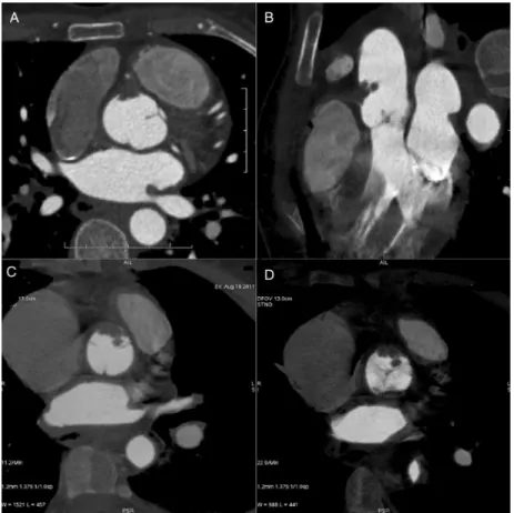
A 48-year old woman presented with atypical chest pain suggestive
of aortic dissection. Computed tomography (Fig.
1
) was performed
and showed a mass protruding from the right coronary artery, the
pathology of which was identi
fied as an organized thrombus (Fig.
2
).
Figure 1:Cardiac computed tomography angiography. (A) Axial–transverse image that shows a low-attenuation material in the right coronary cusp of the sinus of Valsalva. No image suggestive of aortic or coronary dissection was identified. (B) 2D reconstruction in the coronal plane that clearly shows the intracoronary compo-nent of a pseudo mass. (C) Thin slab‘maximum-intensity-projection’ reconstruction in the axial–transverse plane. (D) Thin slab ‘minimum-intensity-projection’ recon-struction showing the extension of the pseudo mass and its relationship to the aortic valve. Similarfindings were found on echocardiography that showed no additional anomalies, particularly no intracardiac thrombus and no patent foramen ovale.
© The Author 2014. Published by Oxford University Press on behalf of the European Association for Cardio-Thoracic Surgery. All rights reserved.
European Journal of Cardio-Thoracic Surgery 47 (2015) 1110–1111
IMAGES IN CARDIO-THORACIC SURGERY
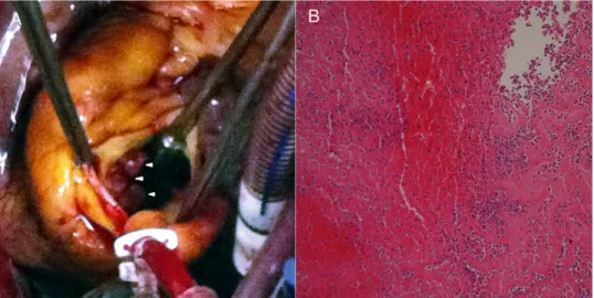
No atherosclerotic changes, prothrombotic factor or embolic sources
were found. We concluded on idiopathic thrombus formation.
Figure 2:(A) After aortotomy, friable material protruding from the right coronary artery into the right coronary cusp of the sinus of Valsalva (arrowheads) was removed with no adherence. (B) The pathology of the removed mass was suggestive of a partially organized thrombus.
IM AG ES IN C A RD IO -THO R A C IC SU RGE R Y