B R I E F R E P O R T
Targeted Nasal Vaccination
Provides Antibody-Independent
Protection Against Staphylococcus
aureus
Karen Misstear,1,aEdel A. McNeela,1,aAlison G. Murphy,2,a Joan A. Geoghegan,3Kate M. O
’Keeffe,2John Fox,4Kin Chan,4 Simon Heuking,5Nicolas Collin,5Timothy J. Foster,3 Rachel M. McLoughlin,2,band Ed C. Lavelle1,6,b
1Adjuvant Research Group and2Host Pathogen Interactions Group, School of
Biochemistry and Immunology, Trinity Biomedical Sciences Institute, Trinity College Dublin, Dublin 2, Ireland;3Microbiology Department, Moyne Institute of Preventive
Medicine, Trinity College Dublin, and4Merrion Pharmaceuticals Ltd, Dublin, Ireland; 5Vaccine Formulation Laboratory, Department of Biochemistry, University of
Lausanne, Epalinges, Switzerland; and6Centre for Research on Adaptive
Nanostructures and Nanodevices, Trinity College Dublin, Dublin 2, Ireland
Despite showing promise in preclinical models, anti– Staphylococcus aureus vaccines have failed in clinical trials. To date, approaches have focused on neutralizing/opsoniz-ing antibodies; however, vaccines exclusively inducneutralizing/opsoniz-ing cellu-lar immunity have not been studied to formally test whether a cellular-only response can protect against infection. We demonstrate that nasal vaccination with targeted nanoparti-cles loaded with Staphylococcus aureus antigen protects against acute systemic S. aureus infection in the absence of any antigen-specific antibodies. These findings can help inform future developments in staphylococcal vaccine devel-opment and studies into the requirements for protective im-munity against S. aureus.
Keywords. vaccine; Staphylococcus aureus; cellular immu-nity; mucosal; adjuvant; nanoparticle.
Infections caused by antibiotic-resistant Staphylococcus aureus are causing a global epidemic and present an urgent and unmet
need for effective vaccines [1]. Although a number of vaccines were effective in preclinical challenge models, these subse-quently failed in clinical trials [2] due at least in part to a lack of insight into what constitutes protective immunity in the human host. Until recently, anti–S. aureus vaccine approaches have focused on the induction of neutralizing/opsonizing antibodies, but there is increasing evidence that cellular immunity may be equally or more important for protective immunity [3]. Indeed, vaccines expanding T-helper type 1 (Th1) and T-helper type 17 (Th17) cells conferred protection in murine models of S. aureus infection [4]. However, to date, systems that exclusive-ly induce cellular immunity in the absence of humoral immu-nity have not been studied to determine if a cellular-only response can protect against systemic staphylococcal infection.
Because mucosal vaccination can potentially stimulate an immune response both at the mucosae and systemically, and has the advantage of being needle-free, our objective was to design a mucosal S. aureus vaccine that selectively promoted cellular immunity. Initiating an immune response at the mucosae is hindered by several factors, particularly poor uptake across epithelial barriers. Targeting mucosal antigen-sampling microfold (M) cells is a promising approach to address ineffi-cient transepithelial vaccine uptake [5]. Ulex europaeus aggluti-nin I (UEA-1), a fucose binding lectin from gorse, binds to [6] and can enhance the transcytosis of nanoparticles across M cells [7], potentially delivering the attached antigen to sube-pithelial dendritic cells. Thus, we investigated the potential of targeting particulate vaccines to M cells with UEA-1 and a UEA-1 peptidomimetic (UEA-1m) [8].
METHODS
Animals
Female BALB/c and C57BL/6 mice were obtained from Harlan Laboratories and Charles River Laboratories and were used at 8–16 weeks of age. Animals were maintained in a specific path-ogen-free environment at the TCD Bioresource facility. All mice were maintained according to European Union regula-tions, and experiments were performed under license from the Irish Department of Health and Children and with ap-proval from the Trinity College Dublin Bioresources Ethics Committee.
Materials
Streptavidin-coated polystyrene nanoparticles (300–390 nm) were supplied by Spherotech Inc and Corpuscular Inc.
Received 12 August 2013; accepted 16 October 2013; electronically published 22 November 2013.
a
K. M., E. A. M., and A. G. M. contributed equally to this work.
b
R. M. M. and E. C. L. contributed equally to this work.
Presented in part: European Vaccine Initiative Rendez-vous, Internationales Wissenschaftsfo-rum der Universitat Heidelberg, Germany, 6 December 2012.
Correspondence: Ed Lavelle, PhD, Adjuvant Research Group, School of Biochemistry and Im-munology, Trinity Biomedical Sciences Institute, Trinity College Dublin, Dublin 2, Ireland (lavellee@tcd.ie).
The Journal of Infectious Diseases 2014;209:1479–84
© The Author 2013. Published by Oxford University Press on behalf of the Infectious Diseases Society of America. All rights reserved. For Permissions, please e-mail: journals.permissions@ oup.com.
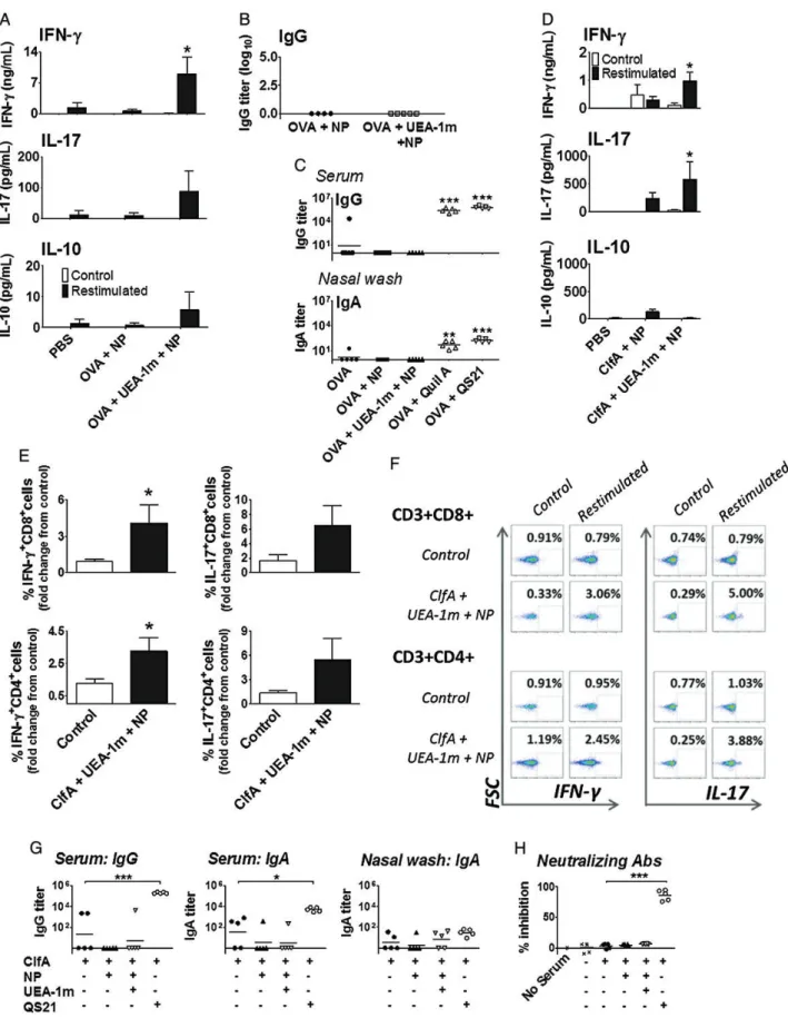
Figure 1. Lectin-targeted mucosal delivery of nanoparticulate antigens induces strong T-cell responses in the absence of a humoral response. Mice were immunized intranasally on days 1, 14, and 28 with phosphate-buffered saline (PBS) only as a control or ovalbumin (OVA; 10 µg) with or without Ulex europaeus agglutinin I peptidomimetic (UEA-1m; 10 µg), attached to nanoparticles (NP). A, Antigen-specific responses by cells isolated from the cervical lymph nodes were determined on day 35, by ex vivo restimulation with OVA (500 µg/mL) for 72 hours, and subsequent enzyme-linked immunosorbent assay (ELISA) to determine cytokine production. Mean ± SEM. *P < .05 vs nontargeted particles; 1-way analysis of variance (ANOVA) and Tukey posttest
Biotinylated UEA-1m (synthesized by Polypeptide), and oval-bumin (OVA) antigen (Sigma) were both provided as lyophilized powders and reconstituted in endotoxin-free phosphate-buffered saline (PBS). The A domain of clumping factor A (ClfA; amino acids 40–559) was expressed as a hexahistidine-tagged recombinant protein and purified by Ni2+chelate chro-matography. Both OVA and ClfA antigens were biotinylated with the EZ-Link Sulfo-NHS-LC biotinylation kit (Thermo Sci-entific). Cytokine and antibody enzyme-linked immunosorbent assay (ELISA) detection kits were obtained from R&D Systems, BD Pharmingen, and BioLegend. Complete RPMI (cRPMI) comprised RPMI 1640 (BioSera), 10% fetal calf serum (BioSera), 1% penicillin-streptomycin (Invitrogen), and 1% L
-glutamine (Invitrogen). Fluorescently conjugated antibodies for flow cytometry were purchased from BD Biosciences (anti–in-terleukin 17A [IL-17A] PerCP-Cy 5 clone TC11-18H10) and eBiosciences (anti–interferon [IFN]–γ PE Cy7 clone XMG 1.2). Quil A saponin was obtained from Brenntag, and the fraction-ated derivative of Quil A (QS21) was provided by the Vaccine Formulation Laboratory in Lausanne, Switzerland.
Vaccine Formulations
Biotinylated antigen (OVA or ClfA) and biotinylated UEA-1m were incubated with streptavidin-coated polystyrene nanoparti-cles for 1 hour at room temperature in sodium phosphate buffer ( pH 5.5). Fifty micrograms of OVA was administered by oral vaccination, but 10 µg OVA and 2 µg ClfA was sufficient for intranasal vaccine formulations (adequate quantity to induce a cellular response to targeted nanoparticles). UEA-1m was incorporated at 10 µg per vaccine formulation (attached to nanoparticles). Protein attachment was determined by bicinchoninic assay (Thermo Scientific) of the supernatant, and the particles were resuspended in PBS ( pH 7.4) before use. The contents of each formulation are detailed in the Figure legends.
Immunization Strategies
Groups of mice (n = 5) were immunized orally on 3 consecutive days (days 0, 1, and 2) and boosted 3 weeks later (days 21, 22, and 23) with PBS only, OVA (50 µg) nanoparticles, or OVA (50 µg) + UEA-1m (50 µg) nanoparticles. Mice were immu-nized intranasally on days 0, 14, and 28 by dropping the vaccine formulation (20 µL maximum volume containing OVA or ClfA antigen in solution or particulate form) onto each nostril and allowing the animal to inhale the vaccine. Group sizes for mucosal vaccinations and challenge were determined based on our previous experience and published reports [9,10]. Blood samples were taken from the tail vein before each immu-nization for analysis of antibody production. Tissues were iso-lated after killing, the cells isoiso-lated by mechanical disruption, and restimulated with antigen in vitro.
Measuring the Cellular and Humoral Response to Antigen
Antigen-specific antibody titers in the serum and nasal washes, and cytokine secretion by restimulated lymphocytes (cultured for 72 hours) were determined by sandwich ELISA as described previously [9]. All samples were analyzed in triplicate. Cytokine production by CD3+CD4+and CD3+CD8+T cells was detected by restimulating the cells for 6 hours in vitro in cRPMI with or without antigen. Brefeldin A (10 µg/mL; Sigma) was added to the culture after 1 hour to prevent cytokine export from the cells. After 6 hours the T cells were labeled with CD3, anti-CD4, and anti-CD8 fluorophore-conjugated antibodies. Fol-lowingfixation and permeabilization with FIX&PERM (ADG Bioresearch GmbH), intracellular IFN-γ and IL-17 were labeled. Data were acquired with a FACSCanto II (BD Biosci-ences) and analyzed using FlowJo software (TreeStar Inc).
Measuring Neutralizing Antibodies in Serum
The presence of neutralizing antibodies was determined by testing the ability of serum from immunized mice to inhibit the
Figure 1 Continued. (n = 5). B, Serum samples harvested from blood taken on day 35 were analyzed for the presence of OVA-specific immunoglobulin G
(IgG). Each data point represents an individual animal. C, Nasal delivery of QS21-adjuvanted OVA promotes humoral immunity. Mice were immunized intra-nasally on days 1, 14, and 28 with PBS or OVA (10 µg) either in solution or conjugated to nanoparticles with UEA-1m (10 µg), or with OVA and Quil A saponin or a purified derivative of Quil A (QS21), both at 10 µg per dose. Blood and nasal cavity washes were taken on day 35. Antigen (OVA)–specific anti-body titers were determined by ELISA. Each data point represents an individual animal (n = 5); the black bars denote the mean. *P < .05, **P < .01, and ***P < .001 vs antigen alone; 1-way ANOVA and Tukey posttest (n = 5). D–H, Nasal immunization with Staphylococcus aureus clumping factor A (ClfA). Female C57BL/6 mice were immunized intranasally on days 1, 14, and 28 with ClfA (2 µg) attached to nanoparticles alone or targeted with UEA-1m (10 µg). Antigen-specific responses by cells isolated from the spleen were determined on day 35, by ex vivo restimulation with ClfA (10 µg/mL), and subse-quent ELISA (D) and intracellular staining (E and F ) to determine cytokine production. Antigen-specific responses by splenic CD3+CD8+and CD3+CD4+T cells were assessed by ex vivo restimulation of the cells with ClfA for 6 hours, and subsequentflow cytometric analysis of interferon (IFN)-γ and interleukin 17 (IL-17)–producing cells. Representative dotplots showing the percentage of IFN-γ or IL-17–producing T cells. Unstimulated cells are on the left of each panel, ClfA-stimulated cells on the right. Pooled data showing the fold change in percentage of cytokine-producing T cells in the spleen (compared to unsti-mulated control). G, Anti-ClfA IgG antibody titers were assessed by ELISA in day 35 serum and nasal wash samples. The presence of neutralizing
antibod-ies was determined by measuring the ability of serum to inhibit ClfA-mediated S. aureus adherence to fibrinogen. Staphylococcus aureus was
preincubated with serum, and adherence tofibrinogen was calculated as a percentage of values measured in control wells lacking serum. Data are repre-sentative of 2–3 independent experiments. Each data point represents an individual animal; the black bars denote the mean. ***P < .001 by 1-way ANOVA and Tukey posttest (n = 5).
adherence of S. aureus to fibrinogen. Microtiter plates were coated withfibrinogen (2 µg/mL) overnight at 4°C and blocked for 2 hours at 37°C with 5% (w/v) bovine serum albumin. Staphylococcus aureus PS80 was grown to stationary phase, washed, and incubated with mouse serum (1:60 dilution) for 30 minutes at room temperature before being added to the wells of afibrinogen-coated plate and incubated for 1.5 hours at 37°C. After washing with PBS, adherent cells werefixed with formal-dehyde (25% v/v) and stained with crystal violet, and the A570
nm was measured. Adherence was expressed as a percentage of bacterial adherence in the absence of serum, and percentage of inhibition was determined by subtracting the percentage adher-ence values from 100.
Staphylococcus aureus Infection
Staphylococcus aureus PS80 has been previously described [10]. Staphylococci were cultivated from frozen stocks on Columbia agar with 2% NaCl. Bacteria were resuspended to an appropri-ate concentration in PBS. Nasally immunized C57BL/6 mice were infected with a sublethal inoculum of S. aureus (5 × 10⁸ colony-forming units [CFU]) by intraperitoneal injection, on day 36, and were sacrificed 3, 24, and 72 hours postinfection (5 mice per treatment group, per timepoint; group size was deter-mined through optimization of the challenge model [10]). At specific timepoints postchallenge, the peritoneal cavity was lavaged with sterile PBS and the kidneys and spleens were har-vested and homogenized in PBS. Serial dilutions of the perito-neal exudates and organ homogenates were prepared and plated onto tryptic soy agar to determine the numbers of S. aureus CFU. Serum was collected from each mouse 1 day prior to each vaccination for analysis of antibody titers. To quantify phago-cyte infiltration, total leukophago-cytes isolated from the peritoneal exudates were enumerated using a hemocytometer and were then stained with monoclonal antibodies against neutrophil (CD11b+Ly6G+F4/80−) and macrophage (CD11b+Ly6G−F4/ 80+) surface markers before analysis by flow cytometry (BD FACSCalibur).
Statistical Analysis
Data are presented as mean (±SEM) from 5 mice per experi-mental group, tested individually in triplicate. P < .05, P < .01, and P < .001 denote statistical significance between groups by 1-way analysis of variance and Tukey posttest, or by 1-tailed unpaired Student t test, as specified in the Figure legends.
RESULTS AND DISCUSSION
Nasal vaccination with UEA-1m–targeted, antigen (OVA)– loaded nanoparticles (300–390 nm diameter) promoted en-hanced antigen-specific T-cell responses compared to antigen conjugated to nontargeted particles (Figure 1A). The most strikingfinding was the Th1- and Th17-biased response elicited
by targeted nanoparticles (Figure 1A), without the induction of any detectable antigen-specific serum immunoglobulin G (IgG) (Figure 1B). Similarly, oral vaccination with UEA-1m–targeted nanoparticles enhanced antigen-specific cellular responses (Supplementary Figure 1), compared to untargeted nanoparticles in the absence of a detectable antibody response. In contrast, antibodies were readily induced both systemically (serum IgG) and locally (nasal wash immunoglobulin A [IgA]) following intranasal administration of OVA in combina-tion with Quil A, a potent saponin adjuvant [11], or QS21 (Figure1C), an adjuvant active fraction of Quil A that displays reduced toxicity [11]. Ourfindings support a report that UEA-1 targeting of a nasal human immunodeficiency virus vaccine elicited enhanced cellular responses [12] but differs in that our system did not induce any local or systemic humoral immunity. Having identified a mucosal adjuvant formulation that selec-tively promoted T-cell responses, we generated a model anti– S. aureus vaccine, based on UEA-1m–targeted nanoparticles coupled with the S. aureus surface antigen ClfA, and investigat-ed its efficacy in protection against systemic S. aureus infection. ClfA is an important S. aureus virulence factor, which pro-motes bacterial adherence tofibrinogen [13]. Whereas immuni-zation of mice with recombinant ClfA A domain formulated with Freund’s complete adjuvant induced protective humoral immunity [13], protection was IL-17A–dependent following
in-jection of the antigen in alum adjuvant [14]. These reports highlight the promise of this ubiquitously expressed staphylo-coccal surface protein as a candidate antigen for the induction of protective cell-mediated immunity. In line with this, a vaccine based on a Candida albicans surface protein with struc-tural similarity to ClfA [15] induced protective anti–S. aureus immunity that was critically dependent on the induction of Th1 and Th17 responses [4].
Initially, we investigated whether UEA-1m–targeted ClfA-loaded nanoparticles could promote antigen-specific T-cell re-sponses in the absence of antibodies. As in the case of OVA, UEA-1m targeting significantly enhanced ClfA-specific IFN-γ and IL-17 production (Figure 1D). Splenic CD3+CD4+ and CD3+CD8+T cells from targeted nanoparticle-vaccinated mice produced IFN-γ and IL-17 upon in vitro restimulation with ClfA, as reflected in an increased percentage of antigen-specific producing T cells and a greater fold increase in cytokine-producing T cells vs controls (Figure1E, F). Anti-ClfA antibodies were undetectable in serum samples from animals vaccinated with ClfA-loaded, UEA-1m–targeted nanoparticles, whereas high titers of antigen-specific serum IgG, IgA (Figure1G), and function-neutralizing antibodies capable of blocking the adher-ence of ClfA-expressing bacteria tofibrinogen were elicited by vaccination with ClfA and QS21 (Figure1H).
Having established the ability of the ClfA-loaded, UEA-1m–targeted nanoparticles to elicit ClfA-specific cellular immune responses in the complete absence of any specific humoral
Figure 2. The induction of cellular immunity by a nasal targeted nanoparticulate Staphylococcus aureus vaccine is sufficient for clearance of a systemic S. aureus infection. A, Immunization strategy. Mice were immunized intranasally on days 1, 14, and 28 with phosphate-buffered saline (PBS) or with clumping factor A (ClfA; 2 µg) and Ulex europaeus agglutinin I peptidomimetic (UEA-1m; 10 µg) attached to nanoparticles (NP). Mice were sacrificed at time 0 (day 35, before infection), and at 3, 24, and 72 hours after intraperitoneal infection with 5 × 10⁸colony-forming units (CFU) S. aureus (strain PS80). Data are represen-tative of 2 independent experiments. B and C, Bacterial counts. The peritoneal exudate, kidneys, and spleen were harvested at each time point, homogenized, and cultured, and CFUs were determined. CFU burden is expressed as the mean ± SEM over time (n = 5, top panel), and in each tissue at 72 hours postinfec-tion. C, Statistical significance between groups is denoted by *P < .05 and **P < .01 (1-tailed unpaired Student t test). D and E, Antibody responses following challenge. Anti-ClfA immunoglobulin G (IgG) antibody titers were determined by enzyme-linked immunosorbent assay on serum samples recovered at each timepoint postinfection. The presence of neutralizing antibodies was determined by measuring the ability of serum to inhibit ClfA-mediated S. aureus adher-ence tofibrinogen. Staphylococcus aureus was preincubated with serum, and adherence to fibrinogen was calculated as a percentage of values measured in control wells lacking serum. Each data point represents an individual animal (n = 5); the black bars denote the mean. ***P < .001 by 1-way analysis of vari-ance and Tukey posttest. F, Phagocytes at the site of infection. The total numbers of peritoneal macrophages and neutrophils in control and vaccinated mice were determined byflow cytometry at 3, 24, and 72 hours following infection. *P < .05 by 1-tailed unpaired Student t test (n = 5).
immunity, we next determined the ability of this vaccine to induce protective immunity against systemic S. aureus infec-tion. Groups of mice were nasally immunized with either ClfA on UEA-1m–targeted nanoparticles or ClfA with QS21 prior to intraperitoneal challenge with a sublethal dose of S. aureus (Figure2A). Bacterial load both locally at the site of infection ( peritoneal cavity) and systemically (kidneys and spleen) was reduced in both vaccinated groups over the course of infection (Figure 2B, Supplementary Figure 2), with a significant
(ap-proximately 2 log) reduction in systemic bacterial dissemina-tion to the kidneys at 72 hours postinfecdissemina-tion, in animals receiving either vaccine compared to unvaccinated control animals (Figure2C,Supplementary Figure 2).
Antigen-specific serum antibody titers induced by the QS21-adjuvanted vaccine remained stable over the course of immuni-zation, and neutralizing antibodies were still present 72 hours postinfection but were completely undetectable in animals re-ceiving the UEA-1m–targeted nanoparticle vaccine (Figure2D and E). In addition to promoting T-cell responses (Figure1D), immunization with ClfA on UEA-1m–targeted nanoparticles also increased local infiltration of neutrophils and macrophages to the peritoneal cavity (Figure 2F ) at 72 hours postinfection compared to unvaccinated animals. In contrast, there was no significant increase in phagocyte infiltration to the peritoneal cavity in animals that received the QS21-adjuvanted vaccine (Supplementary Figure 2C).
Remarkably, these data demonstrate that a targeted nasal vaccine promotes clearance of an acute S. aureus systemic infec-tion, but also that a purely cellular response is sufficient for this protection. This is thefirst formal demonstration that vaccine-induced S. aureus antigen-specific cellular immunity in the absence of detectable antibody responses can protect against staphylococcal infection and expands upon previously pub-lished work indicating the importance of cellular immunity in vaccine-induced protection against S. aureus infection [4, 15]. Our targeted mucosal vaccine approach will prove a valuable tool not only in dissecting protective immunity to S. aureus but also for other bacterial infections where Th1 and Th17 responses have been shown to contribute to protective immunity, including Bordetella pertussis and Streptococcus pneumoniae.
Supplementary Data
Supplementary materialsare available at The Journal of Infectious Diseases
online (http://jid.oxfordjournals.org/). Supplementary materials consist of data provided by the author that are published to benefit the reader. The posted materials are not copyedited. The contents of all supplementary data are the sole responsibility of the authors. Questions or messages regarding errors should be addressed to the author.
Notes
Financial support. This work was supported by a grant from the En-terpise Ireland Innovation Partnerships Scheme (2010). Proposal ID, IP20090022. This publication has emanated from research supported in part by a research grant from Science Foundation Ireland (grant number 12/IA/1421). Part of the work was supported by the EU FP7 project TRANSVAC (FP7-INFRASTRUCTURES-2008-228403). This publication reflects only the author’s views and the European Union is not liable for any use that may be made of the information contained therein.
Potential conflicts of interest. All authors: No reported conflicts. All authors have submitted the ICMJE Form for Disclosure of Potential Conflicts of Interest. Conflicts that the editors consider relevant to the content of the manuscript have been disclosed.
References
1. McKenna M. Vaccine development: man vs MRSA. Nature 2012; 482:23–5.
2. Bagnoli F, Bertholet S, Grandi G. Inferring reasons for the failure of Staphylococcus aureus vaccines in clinical trials. Front Cell Infect Mi-crobiol 2012; 2:16.
3. Spellberg B, Daum R. Development of a vaccine against Staphylococcus aureus. Semin Immunopathol 2012; 34:335–48.
4. Lin L, Ibrahim AS, Xu X, et al. Th1-Th17 cells mediate protective adap-tive immunity against Staphylococcus aureus and Candida albicans in-fection in mice. PLoS Pathog 2009; 5:e1000703.
5. Yamamoto M, Pascual DW, Kiyono H. M cell-targeted mucosal vaccine strategies. Curr Top Microbiol Immunol 2012; 354:39–52. 6. Clark MA, Blair H, Liang L, Brey RN, Brayden D, Hirst BH. Targeting
polymerised liposome vaccine carriers to intestinal M cells. Vaccine 2001; 20:208–17.
7. Foster N, Clark MA, Jepson MA, Hirst BH. Ulex europaeus 1 lectin targets microspheres to mouse Peyer’s patch M-cells in vivo. Vaccine 1998; 16:536–41.
8. Lambkin I, Pinilla C, Hamashin C, et al. Toward targeted oral vaccine delivery systems: selection of lectin mimetics from combinatorial librar-ies. Pharm Res 2003; 20:1258–66.
9. McNeela EA, Burke A, Neill DR, et al. Pneumolysin activates the NLRP3 inflammasome and promotes proinflammatory cytokines inde-pendently of TLR4. PLoS Pathog 2010; 6:e1001191.
10. McLoughlin RM, Lee JC, Kasper DL, Tzianabos AO. IFN-gamma regu-lated chemokine production determines the outcome of Staphylococcus aureus infection. J Immunol 2008; 181:1323–32.
11. Sun HX, Xie Y, Ye YP. Advances in saponin-based adjuvants. Vaccine 2009; 27:1787–96.
12. Manocha M, Pal PC, Chitralekha KT, et al. Enhanced mucosal and sys-temic immune response with intranasal immunization of mice with HIV peptides entrapped in PLG microparticles in combination with Ulex europaeus-I lectin as M cell target. Vaccine 2005; 23:5599–617. 13. Josefsson E, Hartford O, O’Brien L, Patti JM, Foster T. Protection
against experimental Staphylococcus aureus arthritis by vaccination with clumping factor A, a novel virulence determinant. J Infect Dis 2001; 184:1572–80.
14. Narita K, Hu DL, Mori F, Wakabayashi K, Iwakura Y, Nakane A. Role of interleukin-17A in cell-mediated protection against Staphylococcus aureus infection in mice immunized with the fibrinogen-binding domain of clumping factor A. Infect Immun 2010; 78:4234–42. 15. Spellberg B, Ibrahim AS, Yeaman MR, et al. The antifungal vaccine
deri-ved from the recombinant N terminus of Als3p protects mice against the bacterium Staphylococcus aureus. Infect Immun 2008; 76:4574–80.