HAL Id: inserm-00445539
https://www.hal.inserm.fr/inserm-00445539
Submitted on 8 Jan 2010
HAL is a multi-disciplinary open access
archive for the deposit and dissemination of
sci-entific research documents, whether they are
pub-lished or not. The documents may come from
teaching and research institutions in France or
abroad, or from public or private research centers.
L’archive ouverte pluridisciplinaire HAL, est
destinée au dépôt et à la diffusion de documents
scientifiques de niveau recherche, publiés ou non,
émanant des établissements d’enseignement et de
recherche français ou étrangers, des laboratoires
publics ou privés.
Molecular distinctions between Aurora A and B: a single
residue change transforms Aurora A into correctly
localized and functional Aurora B.
Fabienne Hans, Dimitrios Skoufias, Stefan Dimitrov, Robert Margolis
To cite this version:
Fabienne Hans, Dimitrios Skoufias, Stefan Dimitrov, Robert Margolis. Molecular distinctions between
Aurora A and B: a single residue change transforms Aurora A into correctly localized and functional
Aurora B.. Molecular Biology of the Cell, American Society for Cell Biology, 2009, 20 (15),
pp.3491-502. �10.1091/mbc.E09-05-0370�. �inserm-00445539�
Vol. 20, 3491–3502, August 1, 2009
Molecular Distinctions between Aurora A and B: A Single
Residue Change Transforms Aurora A into Correctly
Localized and Functional Aurora B
Fabienne Hans,*
†Dimitrios A. Skoufias,
†‡Stefan Dimitrov,*
and Robert L. Margolis
‡§*Institut National de la Sante´ et de la Recherche Me´dicale, Universite´ Joseph Fourier-Grenoble 1, and Institut
Albert Bonniot U823, Site Sante´-BP 170, 38042 Grenoble, France;
‡Institut de Biologie Structurale Jean Pierre
Ebel (Commissariat a` l’Energie Atomique/Centre National de la Recherche Scientifique/Universite´ Joseph
Fourier), 38027 Grenoble Cedex 1, France; and
§Sidney Kimmel Cancer Center, San Diego, CA 92121
Submitted May 6, 2009; Revised May 19, 2009; Accepted May 22, 2009 Monitoring Editor: Daniel J. Lew
Aurora A and Aurora B, paralogue mitotic kinases, share highly similar primary sequence. Both are important to mitotic progression, but their localizations and functions are distinct. We have combined shRNA suppression with overexpres-sion of Aurora mutants to address the cause of the distinction between Aurora A and Aurora B. Aurora A residue glycine 198 (G198), mutated to asparagine to mimic the aligned asparagine 142 (N142) of Aurora B, causes Aurora A to bind the Aurora B binding partner INCENP but not the Aurora A binding partner TPX2. The mutant Aurora A rescues Aurora B mitotic function. We conclude that binding to INCENP is alone critical to the distinct function of Aurora B. Although G198 of Aurora A is required for TPX2 binding, N142G Aurora B retains INCENP binding and Aurora B function. Thus, although a single residue change transforms Aurora A, the reciprocal mutation of Aurora B does not create Aurora A function. An Aurora A-⌬120 N-terminal truncation construct reinforces Aurora A similarity to Aurora B, because it does not associate with centrosomes but instead associates with kinetochores.
INTRODUCTION
Aurora A (AurA) and Aurora B (AurB) are paralogue mi-totic serine-threonine kinases. Both are important to mimi-totic progression, but both their localization and their functions are distinct (Bischoff and Plowman, 1999; Giet and Prigent, 1999; Giet et al., 2005; Ruchaud et al., 2007).
AurA associates with centrosomes and with centrosome proximal microtubules (Bischoff et al., 1998; Zhou et al., 1998; Kufer et al., 2002), and both binds to and is activated by TPX2, a microtubule-binding protein that localizes AurA to the mitotic spindle (Kufer et al., 2002). AurA is required for mitotic entry (Hirota et al., 2003), centrosome maturation and formation of a bipolar mitotic spindle (Hannak et al., 2001), for mitotic checkpoint function (Anand et al., 2003; Macurek et al., 2008), and for accurate segregation of both centrosomes and chromosomes into daughter cells during mitotic exit (Bischoff and Plowman, 1999; Dutertre et al., 2002; Meraldi et al., 2002).
In contrast, AurB exhibits passenger protein behavior, first associating with the inner centromere, then separating from centromeres to relocate to the spindle midzone during cell
cleavage. It binds to and is activated by INCENP, another passenger protein (Cooke et al., 1987; Adams et al., 2000; Bishop and Schumacher, 2002; Honda et al., 2003; Sessa et al., 2005). AurB is absolutely required for spindle assembly checkpoint function, correct chromosome segregation, and for cell cleavage (Andrews et al., 2003; Carmena and Earn-shaw, 2003; Meraldi et al., 2004; Jeyaprakash et al., 2007; Ciferri et al., 2008; Fuller et al., 2008; Jelluma et al., 2008).
Despite their structural similarities, the two proteins have unique spectra of binding partners (Tien et al., 2004) and of phosphorylation substrates (Meraldi et al., 2004; Ohashi et
al., 2006). We and others have addressed how these distinct
localizations and functions arise in two similar paralogues. Although the central catalytic domains of AurA and AurB share substantial homology, the N termini of the two pro-teins are highly divergent (see Figure 1 for alignment com-parison). Crystal structure analysis of AurA demonstrates that its interaction with the N-terminal region of TPX2 oc-curs in the small lobe of the catalytic domain (Bayliss et al., 2003). AurB binds a strongly conserved region of INCENP, the IN-box, and structural analysis indicates this interaction occurs in a region of the catalytic domain that overlaps with that required for TPX2 binding to AurA (Sessa et al., 2005). The glycine 198 (G198) residue of human AurA is required for association of AurA with TPX2 (Kufer et al., 2002; Bayliss
et al., 2003, 2004; Eyers et al., 2005).
We have searched previously for the sequence require-ments for the distinct targeting and functions of Auroras A and B, by using a procedure of verified suppression of a protein and its rescue by a mutant in the same cell (Scrittori
et al., 2005). Although it would be reasonable to assume that
the divergent sequences relate to the highly divergent
func-This article was published online ahead of print in MBC in Press (http://www.molbiolcell.org/cgi/doi/10.1091/mbc.E09 – 05– 0370) on June 3, 2009.
†These authors have contributed equally to this work.
Address correspondence to: Robert L. Margolis (rmargolis@skcc. org) or Stefan Dimitrov (stefan.dimitrov@ujf-grenoble.fr).
Abbreviations used: AurA, Aurora A; AurB, Aurora B; HA, hem-agglutinin; shRNA, small hairpin RNA.
tions of the two proteins, we established that the N terminus of AurB was apparently dispensable for its mitotic functions. Although the C terminus of AurB was important to its function, substitution of C-terminal AurA sequence could restore AurB activity (Scrittori et al., 2005). A chimeric AurB protein with both AurA N and C termini was as functional as AurB. Despite the substantial divergence of N-terminal sequence between the two proteins, our result strongly sug-gested that key residues that distinguished AurA from AurB function were present uniquely in the catalytic domain. These previous results are summarized in Figure 1A.
Sessa et al. (2005) clarified the role of the C-terminal extension in Aurora function, demonstrating that it folded back to stabilize an open conformation in the catalytic cleft. They noted that the AurB:INCENP complex was remarkably different from the AurA:TPX2 complex, sug-gesting that a switch of binding partners and of function between the two proteins would be difficult to obtain by single site mutation.
In this work, we have determined the domain require-ments that would cause AurA to function as AurB, or AurB to function as AurA. Substitution of G198, the catalytic domain residue critical to AurA interaction with TPX2, with the aligned AurB asparagine 142 (N142) strikingly caused AurA to suppress TPX2 binding and instead bind IN-CENP. This single mutation caused AurA to function like AurB, restoring both correct chromosome segregation and normal mitotic exit in AurB-suppressed cells. The inverse, mutation of AurB N142 to the aligned AurA glycine res-idue neither suppressed INCENP binding nor permitted TPX2 binding. We also assayed N-terminal truncations and chimeras and determined that the N terminus of AurA plays a clear, but not dominant, role in centrosome localization.
Our major conclusion is that, despite the complexity of differences between AurA and AurB interactions with their binding partners (Sessa et al., 2005), AurA will functionally substitute for AurB after a single residue change in the TPX2 binding site, that results in loss of TPX2 binding and gain of INCENP binding. Because the single residue change affects only AurA binding to INCENP, our results permit the fur-ther conclusion that Aurora binding to INCENP is both necessary and sufficient to generate correct Aurora interac-tions with other binding partners and with substrates at the kinetochore. The binding of AurA to INCENP restores both AurB dependent kinetochore function and AurB function in cell cleavage.
MATERIALS AND METHODS
Analysis of Protein Structure
The two three-dimensional (3D) structures used: human AurA/ADP complex (Protein Data Bank [PDB] code 1MQ4) (Nowakowski et al., 2002) and Xenopus AurB/INCENP complex (PDB code 2BFX) (Sessa et al., 2005) were retrieved from the PDB (Berman et al., 2000). Superposition and analysis of the struc-tures were performed using TURBO FRODO (Roussel et al., 1990). The surface accessibility of each amino acid residue in the 3D structures was computed with the DSSP program (Kabsch and Sander, 1983).
Cell Culture and Transfection
All work was done in HeLa cells in culture except for HCT116 cells used for immunoprecipitation (Figure 6). The techniques for suppression of endoge-nous AurB by small hairpin RNA (shRNA) and its functional rescue by Aurora mutants through cotransfection in the same cell, and the antibodies and techniques used for the immunofluorescence microscopy, are all as described in Scrittori et al. (2005). Rabbit polyclonal to INCENP, a generous gift from Dr. Gruneberg (University of Liverpool, Liverpool, United King-dom), was used at 1000-fold dilution. For quantitative purposes, 48 h after transfection, randomly cycling cells were stained for DNA and quantitated by microscopy for binucleate or multinucleate status, to indicate the percentage
of mitotic failure that had occurred in this time period. Usually, four different coverslips and at least 80 cells per coverslip were counted for each of the cDNA Aurora kinase constructs used.
Microscopy
Images were collected with an MRC-600 laser scanning confocal apparatus (Bio-Rad Laboratories, Hercules, CA) coupled to an Optiphot microscope (Nikon, Tokyo, Japan), and with a 510 laser scanning confocal apparatus (Carl Zeiss, Jena, Germany) with a 60⫻ oil immersion objective. Images were also acquired with an inverted IX81 epifluorescence motorized microscope (Olympus, Tokyo, Japan) equipped with a motorized piezo stage (Ludl Electronic Products, Hawthorne, NY) and a Retiga-SRV charge-coupled device camera (QImaging, Surrey, BC, Canada) driven by VOLOCITY software (Improvision, Coventry, United Kingdom) with a binning of 1, using a PlanApo 60⫻ numerical aperture 1.42 objective (Olympus). After deconvolution, images were processed in Photoshop version 7.0 (Adobe Systems, Mountain View, CA) and assembled in CANVAS version 8.0 (Denaba Systems, Miami, FL).
Glutathione Transferase (GST) Pull-Down Assay
GST pull-down assays were performed as described previously (Hagemeier et al., 1994). GST-IN-box (GST fused to the IN-box of the human INCENP protein) (amino acids 826-919) was from Millipore (Billerica, MA).35S-labeled proteins were produced in rabbit reticulocyte lysate from the different pcDNA-hemagglutinin (HA) plasmids encoding wild-type and mutated AurA and AurB regions, by using the TNT transcription/translation kit (Promega, Madison, WI) and [35S]methionine (GE Healthcare, Little Chalfont, Buckinghamshire, United Kingdom). The GST-IN-box was immobilized on glutathione beads and incubated with the different35S-labeled proteins. After pull-down, bound material was subjected to SDS-polyacrylamide gel electro-phoresis (PAGE) and autoradiography. All input lanes were equally loaded, as were all pull-down lanes. The loading of input lanes was uniformly one fifth the loading of pull-down lanes.
Coimmunoprecipitation Assays
HCT166 cells were transfected with the different pcDNA Aurora kinase constructs (encoding HA fusion proteins) by using Lipofectamine 2000 re-agent (Invitrogen, Carlsbad, CA) according to manufacturer’s protocols. Twenty-four hours after transfection, cells were blocked overnight in mitosis with 40 ng/ml nocodazole (Sigma-Aldrich, St. Louis, MO). Mitotic cells were harvested by mitotic shake off, and cells were extracted with radioimmuno-precipitation assay (RIPA) buffer supplemented with 50 mM NaF, 0.5 mM -glycerophosphate, 0.1 phenylmethylsulfonyl fluoride, 10 g/ml aprotinin, and 10g/ml leupeptin. Lysates were centrifuged for 30 min at 13,000 ⫻ g, and 400 g of the supernatant was incubated with 30 l of protein G-Sepharose beads for 2 h at 4°C to eliminate the nonspecific binding. Each cleared extract was then incubated with 30l of protein G-Sepharose coupled to anti-HA (HA.11 monoclonal antibody, Affinity Matrix; Covance Re-search Products, Princeton, NJ) overnight at 4°C, washed three times with RIPA buffer, and then resuspended in 1⫻ SDS-Laemmli sample buffer and boiled. After SDS-PAGE electrophoresis and transfer to nitrocellulose, immunoprecipitated proteins were detected with polyclonal antibodies to Aurora A (Cell Signaling Technology, Danvers, MA) diluted 1000-fold, AurB (ab2254; AbCam, Cambridge, United Kingdom) diluted 1000-fold, and TPX2 (Bethyl Laboratories, Montgomery, TX) diluted 5000-fold. Ex-posure to horseradish peroxidase-conjugated goat anti-rabbit immuno-globulin G secondary antibody (Kirkegaard and Perry Laboratories, Gaith-ersburg, MD) diluted 5000-fold was for 1 h, and blots were washed and subsequently developed with enhanced chemiluminescence (Pierce Chem-ical. Rockford, IL).
Kinase Assays
Recombinant Xenopus laevis full-length histones were produced and purified as described previously (Luger et al., 1999). For kinase assay, 0.5g of recombinant His-Aurora kinase was added to 30l of kinase buffer (50 mM Tris, pH 7.4, 10 mM MgCl2, 1 mM EGTA, and 1 mM dithiothreitol) containing 3g of recombinant substrates. After addition of 20 Ci of [␥-32P]ATP, the reaction was allowed to proceed for 30 min at 30°C. Proteins were separated on SDS polyacrylamide gel, and the gel was dried and processed for autora-diography.
RESULTS
Aurora-A with G198N Substitution Rescues Aurora B Location and Mitotic Function
Our previous work (Scrittori et al., 2005), using shRNA resistant AurB and Aurora A and B chimeras, suggested that specific sequences within the conserved catalytic domain must be critical for distinctive AurA and AurB localization
and function (Figure 1A). Consistent with this, overex-pressed HA-tagged wild-type Aurora A did not localize to centromeres, but to centrosomes, in the absence of Aurora B, and other passenger proteins were distributed throughout the chromosomes rather than on centromeres. These results suggested that Aurora A cannot replace Aurora B in the passenger protein complex (Supplemental Figure 1).
Data from other groups have provided evidence that the patterns of localization of both kinases are strictly depen-dent on unique catalytic domain binding partners (Carmena and Earnshaw, 2003; Fu et al., 2007), indicating that unique catalytic domain sequences are crucial for selective interac-tions with binding partners that determine localization. With this in mind, we first sought to examine the structural differences between the catalytic domains of AurA and AurB kinases with the goal of determining how AurA might be made to bind INCENP. The crystal structures of the catalytic domains of AurA with bound TPX2 (Bayliss et al., 2003), and of AurB with a bound IN-box INCENP fragment (Sessa et al., 2005), have been solved. Using computer model docking to analyze the surface accessibility of amino acid residues of native and complexed forms of AurB, we searched for key residues in AurB that were critical for INCENP binding. Of 29 candidate residues (Sessa et al., 2005), nine were different from AurA. Of these only two Xenopus AurB residues, phe-nylalanine 88 (F88) (equivalent to human AurB F72) and N158 (equivalent to human AurB N142), displayed clear chemical differences from aligned residues in AurA, and the corresponding AurA residues created substantial clashes for INCENP docking.
A crystal structure model (Figure 1B, left) shows human AurB (green) in complex with INCENP (blue). The two key INCENP binding residues on AurB identified by our anal-ysis are indicated with red dots. A close view of Xenopus AurB-N158 interacting with INCENP through multiple hy-drogen bonds is also shown (Figure 1B, right). We estimated that modification of the AurA-G198 residue to the corre-sponding Asparagine would generate a reduction of ⬃5–9 kcal/mol in the interaction energy between Aurora and INCENP, based on the binding energy of 3 kcal/mol for a hydrogen bond in a protein backbone (Shulz and Schirmer, 1979). Interestingly, it was shown previously in vitro that substitution of G198 to Asparagine prevented AurA from binding to TPX2 (Bayliss et al., 2004). Structural analysis of the AurA TPX2 complex argued for a conformation of the activation loop of the kinase that is stabilized in the presence of TPX2 and ready to accept the substrate (Bayliss et al., 2004). Furthermore, a similar mutation in Xenopus AurA resulted in a kinase with lower in vitro intrinsic kinase activity matching that of AurB activity in the absence of any binding partners (Eyers et al., 2005).
For molecular analysis, we used an approach that we established previously (Scrittori et al., 2005) that combines shRNA induced suppression of native AurB with expression of AurB mutants in the same cell. As we reported previously (Scrittori et al., 2005), at 48 h after transfection with AurB shRNA, 46⫾ 1% of all mitotic cells are negative for AurB and this suppression persists to 72 h. As a result, 28⫾ 4% of the shRNA-treated total cell population is bi/multinucleate at 72 h versus 3% of the untreated cells.
Importantly, as demonstrated previously (Scrittori et al., 2005), double transfection with AurB shRNA and wild-type AurB rescue plasmid (HA-AurB*, fully described below) completely rescues AurB function. Thus, 5⫾ 2% of double-transfected cells are multinucleated versus 3⫾ 3% of con-trols at 48 h. These results have validated the approach and
permitted the conclusion that each cell that accepts the shRNA plasmid also incorporates the rescue plasmid, per-mitting genetic rescue analysis on a cell-by-cell basis.
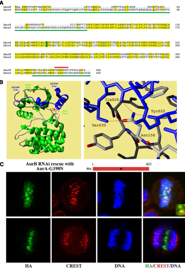
We used the suppression and rescue approach to deter-mine the effect of mutation of AurA residues to their AurB complements. First, we asked whether F72 or N142 were key residues for INCENP binding, centromere targeting, and function for AurB. We found that mutation of W128 of AurA to the aligned phenylalanine (F72) of AurB was without effect on AurA localization or function (data not shown). In contrast, mutation of G198 to the aligned asparagine caused a dramatic change in AurA. In the absence of AurB, ectopi-cally expressed HA-tagged AurA-G198N localized correctly to the centromeres at metaphase and to the spindle midzone in anaphase/telophase (Figure 1C). There were no apparent deficits in mitotic progression that are normally evident in the absence of AurB (Figure 2A). Comparison of AurA-G198N to CREST anti-centromere serum showed that the mutant AurA localized to the inner centromere at meta-phase, the normal position of a passenger protein (Figure 1C, inset). In anaphase, AurA-G198N separated from the centromeres stained with CREST serum, and relocalized at the spindle midzone, mimicking the distribution of native AurB (Figure 1C). Quantitation showed that when AurB suppression was combined with AurA-G198N mutant res-cue, 69 ⫾ 2% of mitotic cells that expressed AurA-G198N contained AurA on centromeres, versus 0% of cells rescued with wild-type AurA.
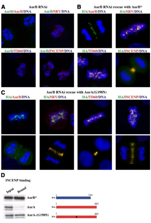
Suppression of AurB by shRNA (Figure 2A) inhibits lo-calization of the passenger proteins to centromeres, causing redistribution of survivin, telophase disc 60 (TD60), and INCENP to the whole chromosome (Figure 2A), whereas rescue of AurB suppression with the shRNA resistant HA-AurB* leads to correct inner centromere targeting of AurB itself and AurB partners such as survivin, TD60, and IN-CENP (Figure 2B; Scrittori et al., 2005). AurB* is HA-tagged AurB, mutated so it cannot be shRNA suppressed nor identified with an antibody to AurB N terminus but is otherwise wild-type in function, as described in Scrittori et
al. (2005). Cells transfected both with shRNA plasmid
tar-geting AurB and with plasmid expressing AurB* can thus be identified as both lacking native AurB and, using HA-anti-body, as expressing HA-AurB*. We have demonstrated pre-viously that HA-AurB* fully rescues AurB localization and function (Scrittori et al., 2005). All rescue experiments with AurB are conducted with shRNA-resistant AurB*. Because AurB* behaves as native AurB (Scrittori et al., 2005), further text does not explicitly mention its use.
Interestingly, rescue of Aurora B suppressed cells by AurA-G198N also rescued correct localization of survivin, TD60, and INCENP, which colocalized with AurA-G198N at the position normally occupied by AurB both on metaphase centromeres and at the spindle midzone during anaphase and telophase (Figure 2C). These results demonstrate that construction of the entire passenger protein complex at the kinetochore critically depends on INCENP binding to Au-rora, and that other than capacity to bind INCENP, there is no specific constraint on Aurora sequence that clearly dis-tinguishes AurB from AurA as a passenger protein.
The localization of AurA-G198N to centromeres and to the spindle midzone, and rescue of passenger protein localiza-tion after AurB suppression, suggested that AurA-G198N could rescue AurB function in chromosome segregation and cell cleavage. If so, AurA-G198N rescue of AurB suppression should retain normal cell morphology after mitosis, reflected in the presence of mononucleate cells. We therefore quanti-tated the effect of rescue on the generation of binucleate and
Figure 1. Rescue of HeLa with G198N AurA after suppression of endogenous AurB. (A) Sequence alignment of human AurB and AurA. Amino acid identity between the two kinases is marked in yellow. Green lines indicate chimeras of AurA sequence swapped onto AurB catalytic domain that retained AurB function (Scrittori et al., 2005). The red line indicates an AurB deletion that suppressed AurB function (Scrittori et al., 2005). The green box indicates the AurB asparagine residue exchange into AurA residue G198 that created AurB function. (B) Structure of Xenopus AurB/INCENP complex (Sessa et al., 2005) (PDB code 2BFX). The AurB structure (left) is green and the INCENP IN-box is blue. The positions of critical AurB residues for interaction with INCENP, Phe88 and Asn158, are shown as red dots. The red arrow indicates the position of the AurB C-terminal. Details of hydrogen bonding between AurB residue Asn158 and three INCENP residues, Tyr
multinucleate cells that result from AurB suppression. The result (see Figure 7D) shows that AurA-G198N rescued AurB suppression with efficiency similar to AurB* rescue, because it restored normal cell cleavage and maintained the mononucleate cell population.
Correct localization of AurB to the inner centromere has been shown to result from interaction with INCENP (Klein
et al., 2006). Because the residue substitution was designed to
enable AurA to interact with INCENP, we conducted an in vitro pull-down assay of the capacity of different35S-labeled
AurA and B constructs to bind the GST-IN-box of INCENP after their in vitro transcription/translation. The results show full-length AurA-G198N binds the GST-IN-box of INCENP, as does the AurB control, whereas, as expected, wild-type AurA kinase does not bind (Figure 2D). We con-clude that the single residue substitution G198N in AurA transforms AurA into an INCENP binding protein that is competent to localize and function as AurB in mitosis.
A previous report showed that the substitution of G198 to asparagine (N) prevented AurA from binding to TPX2 in vitro (Bayliss et al., 2004). Our in vitro results demon-strate that this substitution is also, remarkably, sufficient to create the appropriate protein interface required for
Figure 1 (cont). 825, Ile 828, and Ser 830, are shown in the right panel. AurB is gray and INCENP is blue. Red lines indicate hydro-gen bonds. (C) Confocal microscopy visualization of AurB sup-pressed cells transfected with G198N AurA. G198N AurA (HA stain) localizes to centromeres at metaphase (top images). Centro-meres were identified with CREST antiserum, which recognizes centromere specific antigens. Enlarged inset (bottom right) shows AurA-G198N (green) localized to inner centromeres, between CREST centromere markers (red). In anaphase (bottom images), AurA-G198N (HA stain) localizes to the spindle midzone, duplicat-ing native AurB distribution (Scrittori et al., 2005). Blue is Hoechst stain for DNA.
Figure 2. Effect of expression of AurA-G198N on passenger protein distribution in cells de-pleted of AurB. (A) Alignment of chromosomes in AurB suppressed mitotic cells. In the absence of AurB* rescue, there is no HA antigen signal (left), and other passenger proteins (survivin [SRV] and INCENP) either redistribute from kinetochores to whole chromosomes (purple indicates overlap of passenger protein and DNA signal) or disperse (TD60). (B) Passenger protein distribution in AurB-suppressed cells transfected with AurB*. AurB* is an HA-tagged functional shRNA-resistant mutant of AurB that is not recognized by an N-terminal AurB antibody (Scrittori et al., 2005). HA indi-cates AurB (green). The passenger proteins are labeled in red: SRV. Representative images from different mitotic phases are shown. Equiv-alent results were obtained with all passenger protein markers. (C) Passenger protein distri-bution in AurB-suppressed cells transfected with AurA-G198N. The distribution patterns of AurA-G198N (HA stain), SRV, TD60, and IN-CENP, at centromeres at metaphase (top im-ages), and at the spindle midzone or midbody during late mitosis (bottom images), are shown. Note that all proteins colocalize during the dif-ferent stages of mitosis. (D) GST pull-down analysis of AurA, AurB*, and AurA-G198N mutant binding to the IN-box of INCENP. GST-IN-box fusion was immobilized on glutathione beads and incubated with the indicated 35
S-labeled proteins. GST-IN-box– bound proteins were then detected by autoradiography. A scheme of the different constructs is shown to the right of the autoradiographs (blue, AurB*; red, AurA; G198N mutated form of AurA is indicated by a star).
INCENP binding. Structural analysis of the AurA TPX2 complex argued for a conformation of the activation loop of the kinase that is stabilized in the presence of TPX2 and ready to accept the substrate (Bayliss et al., 2004). The ap-parent binding of INCENP to AurA-G198N in vitro and the observed rescue of AurB suppression by AurA-G198N in our cell-based assays provide evidence that the G198N kinase is active in cells. We confirmed that AurA-G198N is active, because it is competent to phosphorylate histone H3 (see Figure 7).
The result suggests that the binding of INCENP to AurA-G198N not only drives the kinase toward its proper localization but also may confer stability to the activation loop of the kinase in the extended conformation even in the absence of TPX2 by a mechanism similar to the allo-steric mechanism of activation of AurB bound to the INCENP IN-box motif, as described previously (Sessa et
al., 2005).
Aurora B with N142G Substitution Affects Neither Aurora B Location Nor Mitotic Function
Given the unexpected capacity of AurA-G198N to rescue AurB suppression, we tested whether mutation of the criti-cal N142 on AurB to glycine would yield a corresponding loss of AurB function in rescue experiments consequent to a
loss of INCENP binding and, furthermore, if it would bind TPX2 and translocate to the centrosome.
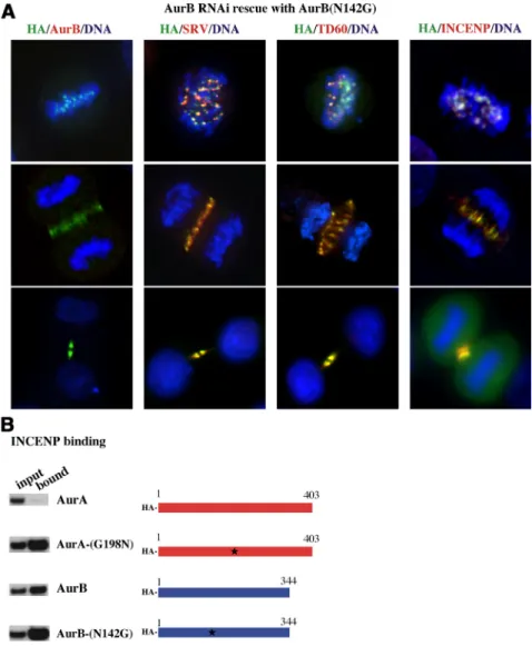
Remarkably, despite the clear requirement for asparagine mutation to transform AurA into an INCENP-binding pro-tein, HA-AurB-N142G rescued suppression of Aurora-B. In-deed, AurB-N142G remained fully competent to associate with centromeres in early mitosis and to migrate to the spindle midzone during anaphase (Figure 3A). Furthermore, other passenger proteins, survivin and TD60, also retained correct cell localization in AurB-N142G rescue cells. In vitro pull-down assays confirmed that AurB-N142G binds to INCENP at levels comparable with AurB or AurA-G198N (Figure 3B). We conclude that although N142 of AurB is important to INCENP binding, other IN-box binding res-idues of AurB must be sufficient to compensate for its absence, retaining INCENP binding capacity. Further-more, we found no evidence that AurB-N142G even par-tially relocates to the centrosome, nor does it bind TPX2 (see Figure 7A). As a consequence, although AurA can be transformed into functional AurB by a single residue substitution, the inverse mutant does not transform AurB into functional AurA. Indeed, quantitative analysis of capacity of AurB-N142G to substitute for AurB showed the mutation did not interfere with its ability to rescue AurB function (see Figure 7D).
Figure 3. Effect of expression of AurB-N142G on passenger protein distribution in cells de-pleted of AurB. (A) Confocal microscopy visu-alization of AurB-suppressed cells transfected with AurB-N142G. Cells, stained for different passenger proteins, are shown in promet-aphase/metaphase, in anaphase and in telo-phase. AurB-N142G (HA stain) localizes to cen-tromeres at metaphase and then transfers to the spindle midzone in anaphase and cells undergo cleavage. Native AurB is absent. Other passen-ger proteins, survivin and TD60, localize nor-mally in different mitotic stages and colocalize with AurB-N142G. (B) GST pull-down analysis of AurA and AurB mutants binding to the IN-box of INCENP. GST-IN-IN-box fusion was immo-bilized on glutathione beads and incubated with the indicated35S-labeled AurA and AurB
proteins. GST-IN-box bound AurA and AurB distinct proteins were detected by autoradiog-raphy.
Role of the Aurora N Terminus in Protein Localization
We had previously demonstrated that the absence of the N-terminal region of AurB did not negatively affect AurB rescue (Scrittori et al., 2005). Indeed, neither N-terminal trun-cation of AurB nor substitution of the N terminus of AurB with N-terminal sequence of AurA affected AurB function. As seen previously (Scrittori et al., 2005), the mitotic distri-bution of a chimera of N-terminal AurA coupled to C-terminal AurB is that of a passenger protein (Figure 4A), although the N-terminal part of AurA alone localizes to the centrosome (Giet and Prigent, 2001).
To reconcile these apparently contradictory data, we con-structed and expressed this N-terminal AurA and C-termi-nal AurB chimera, also containing the AurB INCENP bind-ing site N142G mutation (AurA1-133–AurB78-344 (N142G)).
Expression of this construct showed a protein distribution intermediate between that of AurA and that of AurB. In different cells, either centrosome or centromere staining pre-dominated, or both occurred simultaneously (Figure 4B). In cleaving cells, midbodies were stained. This result permitted two conclusions. First, although the N terminus of AurA is
important for Aurora localization to centrosomes, the kinet-ochore targeting capability of the catalytic domain is domi-nant for chimera localization (Figure 4A). Second, although the N142G substitution in the catalytic domain is of no apparent consequence in full-length AurB, the combination of the N terminus of AurA with the N142G substitution results in centrosomal targeting in addition to kinetochore targeting.
Centrosome localization of the AurA1-133–AurB78-344
(N142G) chimera, however, is not associated with loss of INCENP binding because this chimera binds in vitro to the GST-IN-box (Figure 4D). A possible contributing factor in the redistribution of the AurA1-133–AurB78-344(N142G)
mu-tant could be the lack of the AurB N terminus. To test for this possibility we constructed an AurB-⌬66 N-terminal trunca-tion-N142G mutant and assayed its distribution in rescue of cells where endogenous AurB had been suppressed with shRNA. This mutant showed the same distribution in mi-totic cells as wild-type AurB, and it colocalized with the passenger proteins TD60 and survivin (Figure 4C). Most importantly, the AurB-⌬66 N-terminal truncation-N142G
Figure 4. Mitotic distribution of different AurB chimeric, N142G and truncated constructs. (A) A chimera with N-terminal AurA and C-termi-nal AurB sequence (labeled with HA-tag) dis-tributes as AurB, as described previously (Scrittori et al., 2005). (B) Images showing the distribution of the chimera with N-terminal AurA and C-terminal AurB sequence contain-ing the N142G mutation (labeled with HA-tag) in mitotic cells. (C) Images, showing the distri-bution patterns of transfected AurB-⌬66 N142G in AurB suppressed cells. Cells in different stages of mitosis, with AurB suppressed and rescued by AurB-⌬66 N142G, show the mutant rescues AurB function, and the normal mitotic distribution of survivin and TD60. (D) Binding of different AurA/AurB chimeras and AurB mutants to the IN-box of INCENP. The indi-cated wild-type and mutant Aurora constructs, expressed as35S-proteins, were incubated with
immobilized GST-IN-box and subjected to SDS-PAGE and autoradiography. Data presented in this figure are from different GST pull-downs than for Figure 3, but in each figure positive and negative controls are shown for each indi-vidual experiment. They can thus be cross-com-pared.
mutant behaved like wild-type AurB in rescue experiments, by the criterion of absence of bi/multinucleate cells (see Figure 7D).
GST-IN-box pull-down (Figure 4D) showed that the different AurA/AurB chimeras (AurA1-133–AurB78-344; AurA1-133–
AurB78-344–N142G) and AurB mutants (AurB-⌬66 N-terminal
truncation; AurB-⌬66 N-terminal truncation-N142G) all bound INCENP in vitro, in accord with microscopy results. Although the AurA1-133–AurB78-344–N142G chimera partially
redistrib-uted to centrosomes, it associated well with INCENP (Figure 4D) but not with TPX2 (see Figure 6).
The results with the AurA1-133–AurB78-344–N142G mutant
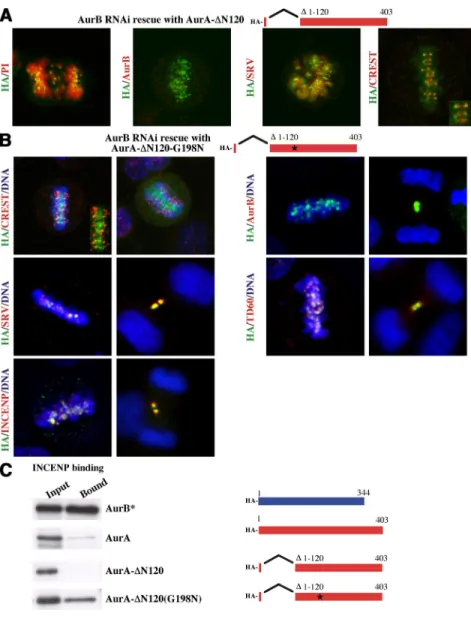
suggested that the N terminus of AurA is important to tethering AurA to the centrosome. To test this possibility, we rescued AurB shRNA suppressed cells with an AurA-⌬120 N-terminal truncation construct. The result (Figure 5A) was striking. AurA-⌬120 shows no association with centrosomes but instead redistributes to centromeres (Figure 5A). We assayed whether AurA-⌬120 would bind INCENP, and we found, remarkably, that redistribution to the centromere occurs in a mutant that does not bind INCENP (Figure 5C). It was further surprising that the AurA-⌬120 mutant did not leave centromeres in anaphase and therefore lacked typical passenger protein mitotic behavior.
It is noteworthy that AurA-⌬120 binds to the kinetochore, rather than the inner centromere site of passenger proteins such as AurB (Figure 5A, CREST, inset). Furthermore, other passenger proteins such as survivin associated with the whole chromosome rather than the centromere with AurA-⌬120 rescue (Figure 5A). This strongly suggested that the catalytic domain of AurA has the “information” to bind to kinetochores and that INCENP is apparently necessary for the relocation from the kinetochore to the inner centromere. To test this, we created an AurA-⌬120-G198N mutant and studied its localization and capacity to rescue AurB suppres-sion. This mutant, in contrast to AurA-⌬120, showed typical passenger protein localization (Figure 5B), and was able to bind INCENP (Figure 5C). Importantly, at metaphase, the AurA-⌬120-G198N mutant apparently localized to the inner centromere, thus lying between the two CREST kinetochore signals in Figure 5B. In addition, all the passenger proteins in cells lacking endogenous AurB and expressing AurA-⌬120-G198N mutant exhibited proper protein passenger lo-calization yielding signals that overlapped with AurA-⌬120-G198N (Figure 5B). We conclude that a single AurA-⌬120-G198N substitution in the catalytic domain of AurA creates the condition for localization to the inner centromere of both Aurora and the other passenger proteins.
Figure 5. AurA-⌬120 and AurA-⌬120-G198N
mitotic distribution and recruitment of IN-CENP, surviving, and TD60. (A) Localization of AurA-⌬120 (HA-tag) in AurB-depleted cells. Inset, AurA-⌬120 localization at the kineto-chore, counterstained with CREST anti-centro-mere serum. (B) AurA-⌬120-G198N shows typ-ical inner centromere localization and protein passenger behavior in cells lacking endogenous AurB. AurA-⌬120-G198N, expressed in AurB-deficient cells, localizes between CREST-posi-tive centromeres (inset) and colocalizes with the passenger proteins survivin, TD60, and IN-CENP, and results in their proper localization in all stages of mitosis. (C) Binding of AurA, AurA-⌬120, AurB*, and AurA-⌬120-G198N to the IN-box of INCENP. The indicated wild-type and mutant Aurora constructs, expressed as
35S-proteins, were incubated with immobilized
GST-IN-box and subjected to SDS-PAGE and autoradiography. The results presented are from the same experiment and the same auto-radiogram as in Figure 2D.
We have assayed for the capacity of different Aurora constructs to bind to TPX2. Just as AurA-⌬120 did not bind INCENP, and yet localized to centromeres (Figure 5), we found that although AurA1-133–AurB78-344–N142G localized
to centrosomes (Figure 4B), it did not bind TPX2 (Figure 6). Indeed, no Aurora N142 or G198 mutant tested detectably bound TPX2 (Figure 6), regardless of localization, under conditions where wild-type AurA binding was evident (Fig-ure 6). In addition, the intracellular mitotic spindle distribu-tion of the TPX2 was not affected by the expression of the different Aurora mutants (data not shown).
Mutant Rescue of Mitotic H3 Phosphorylation and Mitotic Exit after AurB Suppression
If the single residue AurA substitution mutant is indeed transformed into an AurB kinase, then it should substitute for AurB catalytic activity. Because AurB is responsible for phosphorylation of residue S10 on histone H3 during mito-sis (Goto et al., 2002; Soncini et al., 2006), we analyzed the phosphorylation status of S10 upon AurB depletion and rescue.
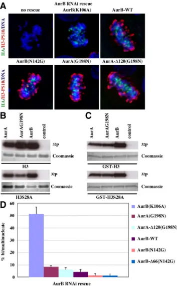
Depletion of AurB with shRNA, abolishes S10 phosphor-ylation on histone H3 as expected (Figure 7A, no rescue). Rescue of AurB depletion with the AurB dead kinase K106A restores proper kinetochore localization of the mutant kinase but not histone H3S10 phosphorylation. In contrast, AurB wild-type and AurB-N142G mutants rescue H3S10 phos-phorylation. In accord with in vitro Aurora kinase assays (Figure 7, B and C), AurA-G198N and AurA-⌬120-G198N mutants rescue AurB suppression and restore histone H3S10 phosphorylation (Figure 7A).
We also determined the ability of the different Aurora kinase constructs to rescue proper mitotic exit in cells where native AurB had been suppressed with shRNA (Figure 7D).
AurB-K106A dead kinase did not rescue the absence of AurB and serves as the control for inability to properly exit mito-sis, as scored by accumulation of bi/multinucleate cells that result from mitotic and cell cleavage errors. The⬃52% mi-totic failure rate obtained with AurB-K106A dead kinase rescue is consistent with the overall 46% of cells in which AurB expression was observed previously to be suppressed by shRNA (Scrittori et al., 2005). In contrast, AurA-G198N and AurA-⌬120-G198N, which bind INCENP (Figure 5C),
Figure 6. Assays of TPX2 binding of Aurora constructs. Extracts, isolated from cells expressing the indicated HA-AurA or HA-AurB mutants, were used for immunoprecipitation using HA anti-body. The presence of AurA, AurB, and TPX2 within the different immunoprecipitates was detected by Western blot analysis, using antibodies against the respective proteins. Results show that no AurA or AurB mutant assayed binds TPX2 under conditions where native AurA does. Loading controls (top lanes) show equivalent amounts of AurA or AurB in all pellet fractions. U, unbound (su-pernatant); B, bound (pellet) fractions.
Figure 7. Assay of histone H3 phosphorylation and functional rescue of AurB suppression by AurB-N142G, AurA-G198N, and AurA-⌬120 G198N constructs. (A) Histone H3 phosphorylation at serine 10 in AurB-depleted cells expressing the indicated Aurora kinase mutants. HA indicates rescue protein expression. Red indi-cates phosphorylated histone H3. (B) In vitro protein kinase assays of the indicated recombinant aurora kinases using as substrates histone H3 and histone H3 mutated at serine 28 to alanine (H3S28A). The autoradiograms demonstrate retained incorporation of32P at the unmutated S10 site. (C) As in B, but using
GST-N-terminal peptide of wild-type H3 (top), or of S28A mutated H3 (bottom), as kinase substrate. (D) Quantitative assay of the rescue of AurB suppression by different mutants. The capacity to rescue AurB function was scored by absence of bi/multinucleate cells, indicative of mitotic failure that results from AurB suppression. Cells were exposed to shRNA to suppress AurB and rescued either with wild-type AurB (AurB-WT) or Aurora mutants as indicated. Four differ-ent coverslips and at least 80 cells per coverslip were counted for each of the cDNA Aurora kinase constructs used.
rescued mitotic exit in a manner statistically indistinguish-able from wild-type AurB (Figure 7D).
DISCUSSION
We have identified key sequences that enable AurA and AurB to localize to specific cellular sites and to respectively function in their unique and distinctive roles. To identify the key residues required to alter AurA to function as AurB, or for AurB to function as AurA, we used an approach we developed previously that enables simultaneous suppres-sion of native AurB and rescue with AurA or AurB mutants in the same cell (Scrittori et al., 2005). Importantly, each cell that received a plasmid for shRNA expression also received a plasmid for mutant rescue.
The validity of this work rests, to some extent, on the expression levels of the rescue constructs relative to the expression of endogenous AurB. For example, a much higher level of relative expression of AurA-G198N than wild-type AurB could overcome a much weaker binding affinity. This is not the case. Western blots of cells transfected with different AurA mutants show they express the mutants at similar, or somewhat lower, levels than the level of en-dogenous protein in mitotic cells (Supplemental Figure 2A), using our transfection conditions. Likewise, AurB mutants are expressed at levels similar to that of wild-type AurB in mitotic cells (Supplemental Figure 2B).
Our most striking result using this approach, was that mod-ification of a single residue, G198, in human AurA to the corresponding aligned N142 residue in human AurB is suffi-cient to apparently transform the mitotic localization of the mutated protein from that of AurA to that of AurB. Further-more, after suppression of native AurB, the other passenger proteins that associate at the kinetochore with AurB are also found to associate with G198N. Apparently, AurA-G198N is able to rescue AurB mitotic function. In contrast, the inverse mutation introduced into AurB-N142G does not alter AurB localization, and rescue experiments after AurB suppres-sion show that it seems to correctly rescue AurB function. In consequence, cells in which these mutants substitute for native AurB faithfully align chromosomes at metaphase and undergo normal cell cleavage, yielding mononucleate daughter cells (Figure 7D).
Despite the substantial differences of localization and function between the two paralogue Aurora kinases, we therefore conclude that AurA retains a latent capacity to mimic AurB and that this capacity can be elicited with modification of a single residue.
Our results suggest that functional specialization among paralogue kinases can arise initially through subtle changes in the catalytic domain. Interestingly, such specialization of paralogue kinases seems to have repeatedly occurred. In
Saccharomyces cerevisiae the three isoforms of protein kinase
A play distinct roles. The Tpk2 subunit activates filamentous growth, whereas the Tpk1 and Tpk3 subunits primarily inhibit filamentous growth. Interestingly, the unique activat-ing function of the Tpk2 subunit is linked to structural differences in the catalytic region of the kinase and not to differences in gene regulation or the unique amino-terminal region of the protein (Pan and Heitman, 1999). Furthermore, two closely related yeast p21-activated kinase paralogues, Ste20 and Cla4, have distinct cell signaling roles. However, analysis of chimeras between the proteins has revealed that substitution of a single amino acid, T818, from Ste20 into Cla4 (D772T), conferred on Cla4 the ability to perform Ste20 specific functions (Keniry and Sprague, 2003). As with the
Aurora kinases, the transformation of function is unidirec-tional.
The Physical Basis of Aurora A Substitution for Aurora B
The principal binding partner of AurB is INCENP, which is both an allosteric activator and a substrate of AurB (Adams
et al., 2000; Kaitna et al., 2000; Bishop and Schumacher, 2002;
Bolton et al., 2002; Kang et al., 2002; Honda et al., 2003). The crystal structure of Xenopus AurB bound to the highly con-served IN-box of INCENP has been solved (Sessa et al., 2005). It shows that 29 residues of AurB make contact with INCENP, of which nine are different from AurA. We found that only two residues of human AurB, F72 and N142, display clear chemical differences from aligned residues in human AurA and that the corresponding AurA residues could, in theory, create substantial clashes for INCENP docking. Mutation of AurA W128 to the aligned AurB tryp-tophan (F72) was without effect. Substitution of asparagine into the aligned G198 residue of AurA caused the mutant protein to lose TPX2 binding (Figure 6), as shown previously (Eyers and Maller, 2004).
Remarkably, we find the G198N mutant also causes AurA to bind INCENP and localize to centromeres (Figure 2, C and D). In contrast, the inverse AurB mutant N142G does not lose INCENP binding or gain TPX2 binding, and it remains at the centromere (Figure 3). The switch in Aurora paralogue function through a single residue change is thus unidirectional.
The reason that AurA can readily switch to AurB local-ization and function, but not the inverse, may lie in the activation of AurB being a two-step process, involving first INCENP-induced allosteric change of AurB to the active conformation; and, second, opening of the catalytic cleft through separation of an ionic link between lysine (K) 97 and glutamate (E) 125 (Sessa et al., 2005). In AurA the equiv-alent ionic pair (K153 and E181) is already separated in the unbound protein (Sessa et al., 2005), reducing activation to a one step process, thus potentially simplifying the binding requirement of AurA-G198N to INCENP.
It is clear from our results that although the G198N mu-tant of AurA is essential to create an INCENP binding site, the aligned N142 of AurB ironically is not critical for IN-CENP binding. Other residues in the binding pocket must be compensating adequately in the N142G mutant.
Role of the Aurora N-Terminal Region
The N-terminal domains of AurA and AurB are highly diver-gent in sequence (Figure 1), suggesting they may play a role in Aurora localization and function. The N terminus of AurA indeed has a function. For example, N-terminal sequence of AurA is required for correct timing of mitotic entry and proper chromosome alignment in Xenopus, although the catalytic do-main alone is sufficient to allow formation of a bipolar spindle (Liu and Ruderman, 2006). Furthermore, GFP-AurA N-termi-nal constructs have demonstrated that the N terminus of AurA plays a role in centrosomal targeting in Xenopus cells (Giet and Prigent, 2001). We have also demonstrated that the N-terminal of AurA has an important function in its localization to the centrosome, but our results now reveal that the AurB N termi-nus has no apparent role in mitosis (Figure 4) (Scrittori et al., 2005) and that the role of the AurA N-terminal region is complex.
We have demonstrated previously that a chimeric protein, consisting of AurA N terminus and AurB catalytic domain, functions indistinguishably from native AurB (Scrittori et al., 2005). Thus, the catalytic domain of AurB has a dominant influence on localization and function. However, our
present results demonstrate that the N-terminal region of AurA seems to have a prominent role in AurA localization. When we rescued AurB shRNA suppressed cells with an AurA-⌬120 N-terminal truncation construct, AurA-⌬120 (containing the catalytic domain only) did not associate with centrosomes but instead associated with kinetochores and did not visibly relocate to the spindle midzone in anaphase. These results lead us to conclude that, even without IN-CENP binding, the AurA catalytic domain has an inherent capacity to associate with the kinetochore that is inhibited by the N-terminal region. These results can be contrasted with those from AurA-G198N-⌬120, which both binds INCENP and locates to the inner centromere. Together, these results show that AurA, relieved of N-terminal constraints, contains the information necessary to associate with the kinetochore, but specific association with inner centromeres, and recruit-ment of other passenger proteins, requires INCENP binding. The AurA-⌬120 construct suggests there seems to be a tug-of-war between the N terminus of AurA and the cata-lytic domain for localization at the centrosome rather than the kinetochore. The AurA1-133–AurB78-344–N142G mutant
reinforces this conclusion. This mutant contains a catalytic domain that retains INCENP binding (Figure 4B), and this construct does not bind TPX2 (Figure 7). Nonetheless, it variably associates with either the centrosome or the kineto-chores and seems to be a physical manifestation of the tug-of-war between the N terminus and the catalytic domain. The gain of centrosome targeting also could be interpreted as a result of a lower affinity for INCENP binding due to the N142G substitution. However, we note that AurB-N142G is competent to behave as an inner centromere passenger protein (Figure 3) that rescues AurB suppression (Figure 7D).
Resolving the Complexity of Aurora Binding Partners and Substrates
Both AurA and AurB have multiple interactions with bind-ing partners, and each has a unique spectrum of phosphor-ylation substrates. Both the binding partners and the sub-strates determine the distinct roles that the two proteins play in cell physiology. In addition to TPX2, Ajuba and phospha-tase inhibitor-2 are allosteric activators of AurA (Hirota et al., 2003; Satinover et al., 2004). AurA is reported to have multiple other binding partners, including TACC3, Bora, HURP, and Hec1/Nuf2 (Barr and Gergely, 2007). AurB associates princi-pally with the chromosome passenger complex (CPC), and INCENP seems to be its principal binding partner that estab-lishes this link. The other CPC proteins seems to associate with AurB through INCENP mediation (Vader et al., 2006), with the possible exception of TD60 (Rosasco-Nitcher et al., 2008).
Perhaps because of their different localizations within the cell, the two Aurora kinases have distinct spectra of substrates. AurA substrates include p53, CENP-A, Lats2, RalA, HURP, BRCA1, Eg5, TACC3, PP1, CDC25B, MBP3, Nuf2, NDEL1, and Plk1 (Meraldi et al., 2004; Ohashi et al., 2006; Fu et al., 2007; Macurek et al., 2008). In addition to histone H3, AurB substrates include survivin, INCENP, Borealin, CENP-A, MCAK, and Hec1/Ndc80 at the centromere (Meraldi et al., 2004; Ohashi et
al., 2006; Fu et al., 2007) and MgcRacGAP, vimentin, and
ZEN4/MKLP1 at the spindle midzone (Ohashi et al., 2006; Fu
et al., 2007). CENP-A uniquely seems to be a substrate first of
AurA and then of AurB during mitotic progression (Kunitoku
et al., 2003). Strikingly, our results show that Aurora
localiza-tion seems to dictate substrate accessibility, at least in the case of histone H3 phosphorylation. As is evident in Figure 7A, AurB suppression yields no apparent H3 phosphorylation, despite the constant presence of AurA, but rescue of AurB
ablation with AurA mutants that localize to chromatin rescues H3 phosphorylation.
Our data permit some simplification of the complexity of substrate selection. Given that a single residue change alters AurA so that it binds to INCENP and rescues AurB mitotic function, we can conclude that INCENP binding seems to be the sole necessity to yield correct AurB localization and func-tion both at the inner centromere and at the spindle midzone. The TPX2 binding site, in contrast, is not sufficient to localize AurA to the centrosome and its proximal microtubules, be-cause N-terminal–deleted AurA localizes to kinetochores. The N terminus of AurA plays a clear role in its localization to the centrosome, whereas the N terminus of AurB, in contrast, plays no clearly identifiable role in the mitotic function of the protein. The fascinating question that our data pose is whether the spectra of other binding partners and of phosphorylation substrates follow changes in localization, or if the two Au-roras partially retain their distinct targets despite different localization. For example, certain roles of AurB in cell phys-iology may not be replaced by rescue with AurA-G198N, and this may compromise long-term survival of such res-cued cells. With the system that we have established, such questions will now be addressable.
ACKNOWLEDGMENTS
We thank Dr. Otto Dideberg (Institut de Biologie Structurale) for aid in 3D modeling of the docking of INCENP to Aurora A to identify steric clashes. We also acknowledge F. Sirot and L. Scrittori for help with mutant constructions in the initial stages of the project. This work was supported by grants from Institut National de la Sante´ et de la Recherche Me´dicale, Region Rhoˆne-Alpes Canceropole CLARA (EpiProNetwork) and la Ligue Nationale Contre le Cancer (Equipe Labelise´: EL2005.LNCC/RM1) [to R.L.M.] and Equipe Labe-lise´ La Ligue ([to S. D.]). S. D. and R.L.M. acknowledge support from the Agence Nationale de la Recherche (NT05-3_42614). R.L.M. also acknowledges support from National Institutes of Health grant R01 GM-068107. While this manuscript was in preparation for submission, an article containing a portion of our results was e-published ahead of print: Fu, J., Bian, M., Liu, J., Jiang, Q., and Zhang, C. (2009) A single amino acid change converts Aurora-A into Aurora-B-like kinase in terms of partner specificity and cellular function. Proc. Natl. Acad. Sci. USA. 2009 Apr 8. [Epub ahead of print].
REFERENCES
Adams, R. R., Wheatley, S. P., Gouldsworthy, A. M., Kandels-Lewis, S. E., Carmena, M., Smythe, C., Gerloff, D. L., and Earnshaw, W. C. (2000). INCENP binds the Aurora-related kinase AIRK2 and is required to target it to chro-mosomes, the central spindle and cleavage furrow. Curr. Biol. 10, 1075–1078. Anand, S., Penrhyn-Lowe, S., and Venkitaraman, A. R. (2003). AURORA-A amplification overrides the mitotic spindle assembly checkpoint, inducing resistance to Taxol. Cancer Cell 3, 51– 62.
Andrews, P. D., Knatko, E., Moore, W. J., and Swedlow, J. R. (2003). Mitotic mechanics: the auroras come into view. Curr. Opin. Cell Biol. 15, 672– 683. Barr, A. R., and Gergely, F. (2007). Aurora-A: the maker and breaker of spindle poles. J. Cell Sci. 120, 2987–2996.
Bayliss, R., Sardon, T., Ebert, J., Lindner, D., Vernos, I., and Conti, E. (2004). Determinants for Aurora-A activation and Aurora-B discrimination by TPX2. Cell Cycle 3, 404 – 407.
Bayliss, R., Sardon, T., Vernos, I., and Conti, E. (2003). Structural basis of Aurora-A activation by TPX2 at the mitotic spindle. Mol. Cell 12, 851– 862. Berman, H. M., Westbrook, J., Feng, Z., Gilliland, G., Bhat, T. N., Weissig, H., Shindyalov, I. N., and Bourne, P. E. (2000). The Protein Data Bank. Nucleic Acids Res. 28, 235–242.
Bischoff, J. R., et al. (1998). A homologue of Drosophila aurora kinase is oncogenic and amplified in human colorectal cancers. EMBO J. 17, 3052–3065. Bischoff, J. R., and Plowman, G. D. (1999). The Aurora/Ipl1p kinase family: regulators of chromosome segregation and cytokinesis. Trends Cell Biol. 9, 454 – 459.
Bishop, J. D., and Schumacher, J. M. (2002). Phosphorylation of the carboxyl terminus of inner centromere protein (INCENP) by the Aurora B kinase stimulates Aurora B kinase activity. J. Biol. Chem. 277, 27577–27580.
Bolton, M. A., Lan, W., Powers, S. E., McCleland, M. L., Kuang, J., and Stukenberg, P. T. (2002). Aurora B kinase exists in a complex with survivin and INCENP and its kinase activity is stimulated by survivin binding and phosphorylation. Mol. Biol. Cell 13, 3064 –3077.
Carmena, M., and Earnshaw, W. C. (2003). The cellular geography of aurora kinases. Nat. Rev. Mol. Cell Biol. 4, 842– 854.
Ciferri, C., et al. (2008). Implications for kinetochore-microtubule attachment from the structure of an engineered Ndc80 complex. Cell 133, 427– 439. Cooke, C. A., Heck, M. M., and Earnshaw, W. C. (1987). The inner centromere protein (INCENP) antigens: movement from inner centromere to midbody during mitosis. J. Cell Biol. 105, 2053–2067.
Dutertre, S., Descamps, S., and Prigent, C. (2002). On the role of aurora-A in centrosome function. Oncogene 21, 6175– 6183.
Eyers, P. A., Churchill, M. E., and Maller, J. L. (2005). The Aurora A and Aurora B protein kinases: a single amino acid difference controls intrinsic activity and activation by TPX2. Cell Cycle 4, 784 –789.
Eyers, P. A., and Maller, J. L. (2004). Regulation of Xenopus Aurora A activa-tion by TPX2. J. Biol. Chem. 279, 9008 –9015.
Fu, J., Bian, M., Jiang, Q., and Zhang, C. (2007). Roles of Aurora kinases in mitosis and tumorigenesis. Mol. Cancer Res. 5, 1–10.
Fuller, B. G., Lampson, M. A., Foley, E. A., Rosasco-Nitcher, S., Le, K. V., Tobelmann, P., Brautigan, D. L., Stukenberg, P. T., and Kapoor, T. M. (2008). Midzone activation of aurora B in anaphase produces an intracellular phos-phorylation gradient. Nature 453, 1132–1136.
Giet, R., Petretti, C., and Prigent, C. (2005). Aurora kinases, aneuploidy and cancer, a coincidence or a real link? Trends Cell Biol. 15, 241–250. Giet, R., and Prigent, C. (1999). Aurora/Ipl1p-related kinases, a new onco-genic family of mitotic serine-threonine kinases. J. Cell Sci. 112, 3591–3601. Giet, R., and Prigent, C. (2001). The non-catalytic domain of the Xenopus laevis auroraA kinase localises the protein to the centrosome. J. Cell Sci. 114, 2095–2104.
Goto, H., Yasui, Y., Nigg, E. A., and Inagaki, M. (2002). Aurora-B phosphor-ylates Histone H3 at serine28 with regard to the mitotic chromosome con-densation. Genes Cells 7, 11–17.
Hagemeier, C., Caswell, R., Hayhurst, G., Sinclair, J., and Kouzarides, T. (1994). Functional interaction between the HCMV IE2 transactivator and the retinoblastoma protein. EMBO J. 13, 2897–2903.
Hannak, E., Kirkham, M., Hyman, A. A., and Oegema, K. (2001). Aurora-A kinase is required for centrosome maturation in Caenorhabditis elegans. J. Cell Biol. 155, 1109 –1116.
Hirota, T., Kunitoku, N., Sasayama, T., Marumoto, T., Zhang, D., Nitta, M., Hatakeyama, K., and Saya, H. (2003). Aurora-A and an interacting activator, the LIM protein Ajuba, are required for mitotic commitment in human cells. Cell 114, 585–598.
Honda, R., Korner, R., and Nigg, E. A. (2003). Exploring the functional interactions between Aurora B, INCENP, and survivin in mitosis. Mol. Biol. Cell 14, 3325–3341.
Jelluma, N., Brenkman, A. B., van den Broek, N. J., Cruijsen, C. W., van Osch, M. H., Lens, S. M., Medema, R. H., and Kops, G. J. (2008). Mps1 phosphory-lates Borealin to control Aurora B activity and chromosome alignment. Cell 132, 233–246.
Jeyaprakash, A. A., Klein, U. R., Lindner, D., Ebert, J., Nigg, E. A., and Conti, E. (2007). Structure of a Survivin-Borealin-INCENP core complex reveals how chromosomal passengers travel together. Cell 131, 271–285.
Kabsch, W., and Sander, C. (1983). Dictionary of protein secondary structure: pattern recognition of hydrogen-bonded and geometrical features. Biopoly-mers 22, 2577–2637.
Kaitna, S., Mendoza, M., Jantsch-Plunger, V., and Glotzer, M. (2000). Incenp and an aurora-like kinase form a complex essential for chromosome segrega-tion and efficient complesegrega-tion of cytokinesis. Curr. Biol. 10, 1172–1181. Kang, D., Chen, J., Wong, J., and Fang, G. (2002). The checkpoint protein Chfr is a ligase that ubiquitinates Plk1 and inhibits Cdc2 at the G2 to M transition. J. Cell Biol. 156, 249 –259.
Keniry, M. E., and Sprague, G. F., Jr. (2003). Identification of p21-activated kinase specificity determinants in budding yeast: a single amino acid substi-tution imparts Ste20 specificity to Cla4. Mol. Cell Biol. 23, 1569 –1580. Klein, U. R., Nigg, E. A., Gruneberg, U., and Zheng, Y. (2006). Centromere targeting of the chromosomal passenger complex requires a ternary
subcom-plex of Borealin, Survivin, and the N-terminal domain of INCENP. Mol. Biol. Cell 17, 2547–2558.
Kufer, T. A., Sillje, H. H., Korner, R., Gruss, O. J., Meraldi, P., and Nigg, E. A. (2002). Human TPX2 is required for targeting Aurora-A kinase to the spindle. J. Cell Biol. 158, 617– 623.
Kunitoku, N., Sasayama, T., Marumoto, T., Zhang, D., Honda, S., Kobayashi, O., Hatakeyama, K., Ushio, Y., Saya, H., and Hirota, T. (2003). CENP-A phosphorylation by Aurora-A in prophase is required for enrichment of Aurora-B at inner centromeres and for kinetochore function. Dev. Cell 5, 853– 864.
Liu, Q., and Ruderman, J. V. (2006). Aurora A, mitotic entry, and spindle bipolarity. Proc. Natl. Acad. Sci. USA 103, 5811–5816.
Luger, K., Rechsteiner, T. J., and Richmond, T. J. (1999). Expression and purification of recombinant histones and nucleosome reconstitution. Methods Mol. Biol. 119, 1–16.
Macurek, L., Lindqvist, A., Lim, D., Lampson, M. A., Klompmaker, R., Freire, R., Clouin, C., Taylor, S. S., Yaffe, M. B., and Medema, R. H. (2008). Polo-like kinase-1 is activated by aurora A to promote checkpoint recovery. Nature 455, 119 –123.
Meraldi, P., Honda, R., and Nigg, E. A. (2002). Aurora-A overexpression reveals tetraploidization as a major route to centrosome amplification in p53⫺/⫺ cells. EMBO J. 21, 483–492.
Meraldi, P., Honda, R., and Nigg, E. A. (2004). Aurora kinases link chromo-some segregation and cell division to cancer susceptibility. Curr. Opin. Genet. Dev. 14, 29 –36.
Nowakowski, J., et al. (2002). Structures of the cancer-related Aurora-A, FAK, and EphA2 protein kinases from nanovolume crystallography. Structure 10, 1659 –1667.
Ohashi, S., Sakashita, G., Ban, R., Nagasawa, M., Matsuzaki, H., Murata, Y., Taniguchi, H., Shima, H., Furukawa, K., and Urano, T. (2006). Phospho-regulation of human protein kinase Aurora-A: analysis using anti-phospho-Thr288 monoclonal antibodies. Oncogene 25, 7691–7702.
Pan, X., and Heitman, J. (1999). Cyclic AMP-dependent protein kinase regu-lates pseudohyphal differentiation in Saccharomyces cerevisiae. Mol. Cell Biol. 19, 4874 – 4887.
Rosasco-Nitcher, S. E., Lan, W., Khorasanizadeh, S., and Stukenberg, P. T. (2008). Centromeric Aurora-B activation requires TD-60, microtubules, and substrate priming phosphorylation. Science 319, 469 – 472.
Roussel, A., Fontecilla-Camps, J. C., and Cambillau, C. (1990). CRYStallize: a crystallographic symmetry display and handling subpackage in TOM/ FRODO. J. Mol. Graph. 8, 86 – 88, 91.
Ruchaud, S., Carmena, M., and Earnshaw, W. C. (2007). Chromosomal pas-sengers: conducting cell division. Nat. Rev. Mol. Cell Biol. 8, 798 – 812. Satinover, D. L., Leach, C. A., Stukenberg, P. T., and Brautigan, D. L. (2004). Activation of Aurora-A kinase by protein phosphatase inhibitor-2, a bifunc-tional signaling protein. Proc. Natl. Acad. Sci. USA 101, 8625– 8630. Scrittori, L., Skoufias, D. A., Hans, F., Gerson, V., Sassone-Corsi, P., Dimitrov, S., and Margolis, R. L. (2005). A small C-terminal sequence of Aurora B is responsible for localization and function. Mol. Biol. Cell 16, 292–305. Sessa, F., Mapelli, M., Ciferri, C., Tarricone, C., Areces, L. B., Schneider, T. R., Stukenberg, P. T., and Musacchio, A. (2005). Mechanism of Aurora B activa-tion by INCENP and inhibiactiva-tion by hesperadin. Mol. Cell 18, 379 –391. Shulz, G. E., and Schirmer, R. H. (1979). Principles of Protein Structure, New York: Springer-Verlag.
Soncini, C., et al. (2006). PHA-680632, a novel Aurora kinase inhibitor with potent antitumoral activity. Clin. Cancer Res. 12, 4080 – 4089.
Tien, A. C., Lin, M. H., Su, L. J., Hong, Y. R., Cheng, T. S., Lee, Y. C., Lin, W. J., Still, I. H., and Huang, C. Y. (2004). Identification of the substrates and interaction proteins of aurora kinases from a protein-protein interaction model. Mol. Cell Proteomics 3, 93–104.
Vader, G., Kauw, J. J., Medema, R. H., and Lens, S. M. (2006). Survivin mediates targeting of the chromosomal passenger complex to the centromere and midbody. EMBO Rep. 7, 85–92.
Zhou, H., Kuang, J., Zhong, L., Kuo, W. L., Gray, J. W., Sahin, A., Brinkley, B. R., and Sen, S. (1998). Tumour amplified kinase STK15/BTAK induces centrosome amplification, aneuploidy and transformation. Nat. Genet. 20, 189 –193.