Early steps in Xenopus neural determination:
Cloning and analysis of opl
by
John Shu-Shin Kuo
A.B. in Biology Harvard College, 1989
SUBMITTED TO THE DEPARTMENT OF BIOLOGY IN PARTIAL FULFILLMENT OF THE REQUIREMENTS FOR THE DEGREE OF
DOCTOR OF PHILOSOPHY IN BIOLOGY AT THE
MASSACHUSETTS INSTITUTE OF TECHNOLOGY
JUNE 1998
© 1998 John S. Kuo. All rights reserved.
The author hereby grants to MIT permission to reproduce and distribute publicly paper and electronic
copies of this thesis document in whole or in part.
Signature of author:
6/
Certified by 7Accepted by: Accepted by: 7 Department of Biology May 27, 1998 Hazel L. Sive Associate Professor of BiologyThesis Advisor
Frank Solomon Professor of Biology Chair, Committee on Graduate Studies
88
Early steps in Xenopus neural determination:
Cloning and analysis of opl
by
John Shu-Shin Kuo
Submitted to the Department of Biology on May 27, 1998 in Partial Fulfillment of the
Requirements for the Degree of Doctor of Philosophy in Biology
Abstract
The early molecular events in vertebrate neural development and the genes that mediate these events during embryogenesis are not well
understood. To identify genes expressed during early neural patterning in
Xenopus, we performed a PCR-based subtractive cloning screen using
micro-dissected gastrula ectoderm. In a non-saturating screen, we isolated over forty genes that are highly expressed in dorsal ectoderm.. One very
interesting neural gene isolated is opl, an early marker of the neurectoderm.
opl resembles the Drosophila pair-rule gene odd-paired (odd-paired-like) and
encodes a zinc finger protein that is a member of the vertebrate zic gene family. When gastrulation starts, opl is expressed throughout the
presumptive neural plate, indicating that neural determination has begun by this stage and correlating with the increased neural competence of the dorsal ectoderm. From early neurula, opl expression is restricted to the dorsal neural tube and neural crest. opl encodes a transcriptional activator with a carboxy terminal regulatory domain whose removal increases opl activity. In animal cap assays, opl strongly increases the response of ectoderm to the neural inducer noggin, suggesting that opl regulates neural competence. opl also alters the spectrum of genes induced by noggin, activating the midbrain neural marker engrailed. Consistent with its later dorsal neural expression, the activated form of opl is able to induce neural crest and dorsal neural tube markers in animal caps and in whole embryos. opl expression in the
ventrolateral ectoderm of whole embryos leads to the formation of large loose aggregates that may indicate early commitment to neural crest lineages. These aggregates do not express an epidermis-specific marker gene,
indicating that opl also suppresses ventral fates. Together, these data suggest that opl may mediate neural competence, and be involved in activation of
midbrain, dorsal neural and neural crest fates.
Thesis advisor: Hazel L. Sive
Dedication
To the ones I love: Linda Chi-Fen Juan
my parents and my brothers.
Acknowledgments
I thank Hazel Sive for her consistent mentoring and insistence on a high scientific standard, and for serving as a role model: as a scientist and a teacher. She provided the scientific and intellectual environment that made
this work possible.
The enthusiasm, curiosity, sense of humor and camaraderie of all of my past and present Sive lab colleagues has made working and studying science
an enjoyable learning experience. Thanks especially to the Opl group: Mukesh Patel, Josh Gamse, Vladimir Apekin, Dave Willison; to the fellow 'senior' graduate students: Laura Gammill, Peggy Kolm; to postdocs: Ben
Sun, Jenya Grinblat, Leila Bradley, Mary Ellen Lane, Charles Sagerstrom, Bertha Kao, Christa Merzdorf, Jacqueline Hoyle; to 'newer' graduate
students Dan Wainstock, Sara Bush, Elizabeth Hick. I also benefited greatly from the members and resources of the Lodish, Jaenisch, Lander and Young labs here at the Whitehead Institute. The resources of the Whitehead
Institute was a key part of making this thesis work possible, especially help from the technical, library and facility staff.
I appreciate the input of present and past thesis committee members: Profs. Richard Hynes, Rudolf Jaenisch, Andrew Chess, Nancy Hopkins and Leonard Zon.
Thanks also to the Harvard-MIT Division of Health Sciences and
Technology (especially Dr. Lee Gehrke, Keiko Oh, and Patricia Cunningham), the Harvard MD-PhD Program (especially Linda Burnley) for their support in my studies at both Harvard Medical School and MIT. Funding from the Medical Scientist Training Program and the Whitehead Cancer Research Training Grant was appreciated.
I am always grateful to my family and to Linda's family for their love, support, encouragement and incredible patience.
Dearest Linda, most of all, I wish to thank you for always knowing what to say to cheer me up, for being a most willing and sympathetic listener, and for offering constant and unwavering encouragement. You are extremely wise in the ways of gently nudging me to realize fresh perspectives, and so patient when I am slow to reach such epiphanies. You strive always for new horizons, and have such zest and enthusiasm for learning and life that I am
moved to stretch myself too. Thank you for going through the many ups and downs with me, and for your sense of humor, unending patience, special love and sensitive affection. My life continues to be immensely, magically
delightful with you as an integral part of it. I look forward to many, many more years of sharing a life of wonder and happiness with you.
Early steps in Xenopus neural determination:
Cloning and analysis of opl
Table of Contents
Abstract ... ... 2
D edication ... . . ... ... 3
Acknowledgm ents ... ... 4
Table of Contents ... ... 5
Chapter 1. Early neural determination in Xenopus ... 10
1.1 Introduction ... 11
1.2A Establishing the DN axis during early Xenopus developm ent ... 12
1.2A Cortical rotation establishes the D/V axis ... 12
1.2B f-catenin/Xtcf3 mediates the dorsal determinant activity ... 13
1.2C Mesoderm induction and determination ... 14
1.2D The dorsal organizer: pattern and function ... 14
1.2E Dorsalization of the early embryo by organizer signals ... 16
1.3 Neural determination in the gastrula ectoderm ... 17
1.3A Enhanced ectodermal competence for neural induction ... ... 17
1.3B Dorsalization of the ectoderm: neural induction... 18
1.3C Initial A/P patterning of the neurectoderm... 19
1.3D Posterior neural inducing molecules ... 21
1.3E Initial neurectodermal patterning along the future D/V neural tube axis ... ... 22
1.3F Later DN patterning of the neural tube ... 23
1.4 What genes are expressed in the gastrula dorsal ectoderm?... 25
1.4A A growing number of neural competence factors ... 25
1.4B Genes that determine and pattern neurectoderm ... 26
1.4C Vertebrate "odd-paired-like" genes ... ... 27
Chapter 2. Identification of early neural-specific genes in Xenopus by subtractive cloning... 36
2.1 Preface ... 37
2.2 Summ ary ... 38
2.3 Introduction ... 39
2.4 R esults ... 4 1 2.4A Subtractive cloning strategy ... . 41
2.4A Monitoring subtractive enrichment ... 41
2.4B Screening the subtracted gastrula dorsal ectoderm library ... 42
2.4B1 Characterizing the subtracted dorsal ectoderm library... ... 43
2.4C Interesting clones... 44 2.4C1 DD 135 ... .... ... 45 2.4C2 DB9 ... ... ... 45 2.343 fkh5... ... ... 45 2.4C4 opl ... . ... ... 46 2.4C5 otx2 ... ... ... 46 2.5 D iscussion ... 47
2.5A Identification of candidate early neural patterning g en es ... 4 7 2.5B Advantages of subtractive cloning ... ... 47
2.5C Improvements on the current subtraction... 48
2.5D Analysis of interesting clones ... 49
2.5D1 Identified clones are early markers of prospective neurectoderm ... 49
2.5D2 Early neural-specific genes may regulate early neural determination ... 49
2.6 Acknowledgments ... ... 51
Chapter 3. Analysis of opl: a gene with multiple roles in early neural developm ent ... ... 70
3.1 Preface ... 71
3.2 Sum m ary ... ... 72
3.3 Introduction ... 73
3.4 R esults ... ... 75
3.4A opl encodes a zinc finger protein with similarity to the Zic fam ily ... ... ... 75
3.4B Temporal and spatial characterization of opl expression ... 75
3.4C opl is a nuclear protein whose carboxy terminal encodes a regulatory domain ... ... . 77
3.4D oplAC activates neural crest and dorsal neural tube m arkers ... 78
3.4E oplAC sensitizes the ectoderm to induction by noggin... 79
3.4F In conjunction with noggin, opl activates posterior and dorsoventral neural markers ... 80
3.4G opl induces cellular aggregates in the absence of DNA synthesis ... .... ... 81
3.4H opl activates expression of neural crest and dorsal neural tube markers and represses epidermal gene expression in whole embryos ... 83
3.5 Discussion ... 85
3.5A opl expression defines an early neurectodermal domain ... 85
3.5B opl can modulate neural competence... 86
3.5C opl can activate engrailed ... 86
3.5D opl as an activator of dorsal neural tube and neural crest fates ... 87
3.5E opl activity is modulated by synergizing factors ... 87
3.5G opl and inhibition of epidermal fates ... 89
3.5H A model for opl function ... 89
3.6 Acknowledgments ... 91
Chapter 4. Future Directions ... 118
4.1 Sum m ary ... 119
4.2 Future directions ... 120
4.2A What regulates opl expression? ..... .. ... .... .. . . 120
4.2A1 What activates early opl expression?... 121
4.2A2 How is opl expression regulated after the gastrula stage? ... .... . . 121
4.2B What are opl's functional roles in neural development? .. .. . . .... .... .... .... ... .... .... .. 122
4.2B1 Ablation of opl function in vivo ... 122
4.2B2 What factors interact with opl to mediate its fu n ction s? ... . .. ... ... ... ... ... ... .. ... ... .. ... 122
4.2C What downstream target genes are regulated by opl? ... ... ... ... ... ... ... ... .. ... ... ... ... ... . . 123
Appendix I. Materials and Methods ... 125
IA Growth, dissection and culture of embryos and explants ... 126
IB.Subtractive cloning of opl ... 126
IC opl constructs ... 127
ID Microinjection... 128
IE In vitro transcription ... ... 129
IF In situ hybridization and sectioning ... 129
IG Isolation of RNA and Northern analysis ... 130
IH Relative quantitative RT-PCR ... 130
IIWestern analysis and immunocytochemistry ... 131
IJ Transient transfection reporter assays ... 131
Appendix II. References ... 132
Appendix III. Translational inhibition by 5' polycytidine tracts in Xenopus embryos and in vitro ... 153
C.1 Preface ... 154
C .2 A bstract ... 155
C.3 Introduction ... 155
C.4 Experimental Design and Discussion... .. 156
C.3A Poly(C) leader sequence depresses translation in Xenopus em bryos ... 156
C.3B A poly(C) leader sequence depresses translation in vitro ... 157
C.5 Acknowledgments ... 160
Figures
Figure 1.1 Early stages of Xenopus development ... 30
Figure 1.2 Establishing the D/V axis during early development ... 32
Figure 1.3 Neural induction and early patterning occurs during gastrulation ... 34
Figure 2.1. Tissues used in subtraction. ... . 52
Figure 2.2. Subtraction scheme ... ... .. 54
Figure 2.3. General removal of tracer by subtraction ... 56
Figure 2.4. Specific enrichment and removal of genes by subtraction ... 58
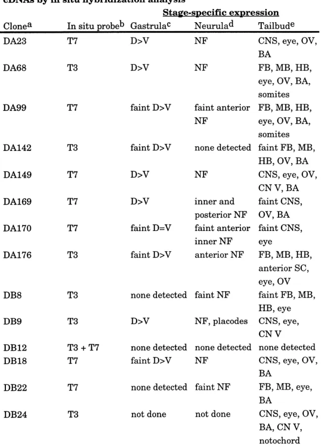
Figure 2.5. Isolated dorsal-specific clones are expressed in the tailbud nervous system ... ... 60
Figure 2.6. Five interesting clones identified in the subtraction screen ... 63
Figure 3.1. opl protein sequence alignments. ... 92
Figure 3.2. Temporal and spatial characteristics of opl expression. ... 94
Figure 3.3. opl constructs ... ... 98
Figure 3.4. opl is a nuclear protein with a regulatory domain in the CO O H -term inal ... ... 100
Figure 3.5. oplAC can activate neural crest and dorsal neural tube marker genes in animal caps. ... 102
Figure 3.6. oplAC sensitizes the ectoderm to induction by noggin. ... 104
Figure 3.7. opl synergizes with noggin to activate more posterior neural markers in animal caps... 106
Figure 3.8. opl induces cellular aggregates without cell division... 108
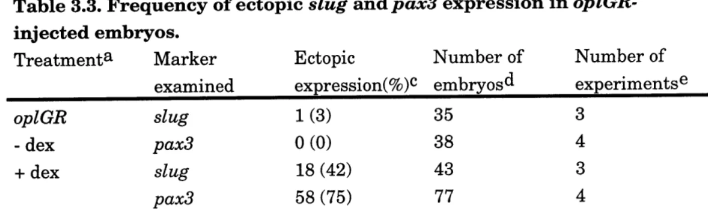
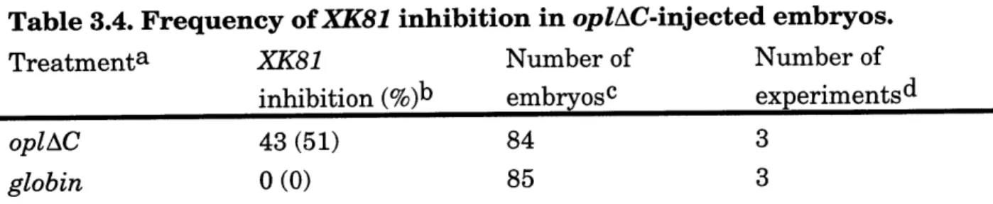
Figure 3.9. opl induces ectopic slug and pax3 and inhibits XK81 expression in em bryos ... 110
Figure 3.10. Model of the roles opl plays in early neural determination... 112
Figure C.1. CAT reporter constructs... 161
Figure C.2. Similar stability in Xenopus embryos of RNAs with various 5' leaders... 163
Figure C.3. Similar sizes of CAT RNAs transcribed by different polym erases ... 165
Tables
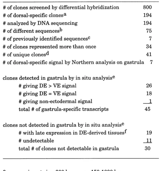
Table 2.1. Summary of screen ... ... 65 Table 2.2. Expression pattern of isolated gastrula dorsal
ectodermal cDNAs by in situ hybridization analysis ... 66
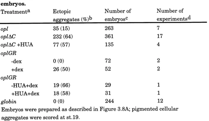
Table 3.1. Frequency of ectopic cellular aggregates in injected
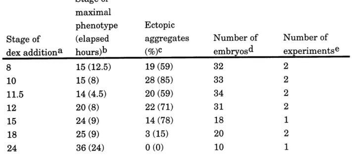
em bry os ... 114 Table 3.2. Induction of cellular aggregates depends on time of
addition and duration of dex treatment in oplGR-injected
embryos ... 115 Table 3.3. Frequency of ectopic slug and pax3 expression in
oplGR-injected em bryos ... 116 Table 3.4. Frequency of XK81 inhibition in oplAC-injected
em bry os... ... 117
Table C.1. Relative CAT activity in Xenopus embryos ... 167 Table C.2. Relative CAT activity in vitro ... 169
1.1 Introduction
To understand the early steps in vertebrate neural development, I studied how the embryonic ectoderm is determined and patterned to start forming a mature Xenopus central nervous system. Neural determination is the process of committing multipotent embryonic cells to the neural lineage. This is followed by neural differentiation, the process where cells complete biochemical and morphological changes to become functional neurons. Neural determination and differentiation are the early and late phases of neurogenesis, the developmental pathway of a cell turning into a neuron. The other key part of elaborating a nervous system is appropriate patterning of the developing neurectoderm. Neural precursor cells must be properly organized along the antero-posterior (A/P) and dorso-ventral (DN) axes of the future nervous system to correctly form a mature brain.
Xenopus is a useful experimental organism to study early neural
development due to the following advantages. External fertilization allows one to obtain many embryos at early stages (Figure 1.1). Development occurs rapidly in these large embryos, which are amenable to experimental
manipulations such as microinjection and dissection. There is also a long history of experimental data accumulated by embryologists that form a basis for the present molecular biological approach to studying development.
Embryologists discovered and defined a dorsal mesoderm tissue, the organizer, as the source of signal(s) that induced and patterned neural tissue (reviewed in Gilbert, et al., 1993). When I started this project, there was great excitement over the identification of candidate neural inducer molecules. Two organizer-derived peptides, noggin and follistatin, were found to directly induce ectoderm to form neural tissue, the major dorsal ectodermal fate (Hemmati-Brivanlou, et al., 1994; Lamb, et al., 1993; Smith, et al., 1992, 1993). Ectodermal cells that do not receive neural induction will adopt the ventral ectodermal fate, epidermis. It was commonly accepted that the blastula ectoderm (animal caps) was a 'naive' tissue that was either
induced at gastrula stage to become neural tissue, or otherwise developed into epidermis. It was also thought that many ectodermal patterning events defining the A/P and D/V organization of the nervous system occurred during gastrulation, many hours before neural differentiation. Since there was a dearth of known markers expressed in the gastrula dorsal ectoderm, such patterning events in the induced neurectoderm were inferred from
specification assays. Explants of gastrula ectoderm were assayed using differentiation markers or histological criteria after incubation in neutral saline. There was a notable gap in the knowledge of genes and mechanisms that act in the gastrula dorsal ectoderm after neural induction, many hours before neural differentiation.
Many interesting questions were raised as promising avenues of inquiry. What genes are activated in the gastrula dorsal ectoderm in response to neural induction? What are the functional roles of such genes? Are they involved in neural determination? Are they involved in establishing A/P or D/V pattern in the induced neurectoderm? Using subtractive cloning, we set out to identify and generate a repertoire of genes expressed in the mid-gastrula dorsal ectoderm. During the course of this work, other mid-gastrula dorsal ectodermal genes were also reported (otx2: (Blitz, et al., 1994; Pannese, et al., 1995); pax3: (Espeseth, et al., 1995); eIF4AII: (Morgan, et al., 1997); XANF: (Mathers, et al., 1995; Zaraisky, et al., 1992, 1995); neurogenin: (Ma, et al., 1996)). Coupled with data from the genes we isolated, these early dorsal ectodermal markers revealed that a complex molecular pattern exists in the gastrula dorsal ectoderm. These genes also mediate many functions in neural determination in the gastrula ectoderm. Since neural tissue is
induced by dorsal mesoderm, and dorsal mesoderm determination results from the fertilization and early cleavage stage events, I will first introduce early development in Xenopus.
1.2A Establishing the D/V axis during early Xenopus development
1.2A Cortical rotation establishes the DIV axis
The sites of action in neural development are on the dorsal side of the embryo. The dorsal ectoderm gives rise to the neural tissue, and neural inducer signals originate from the organizer in the dorsal mesoderm. How is the 'dorsal' side initially established in the zygote?
Xenopus eggs are radially symmetric, composed of the darkly
pigmented animal hemisphere and the yolky vegetal hemisphere (Figure 1.2). The animal hemisphere gives rise to the presumptive ectoderm, the
equatorial regions give rise to mesoderm, and the vegetal hemisphere gives rise to endoderm. Yolk is concentrated in the vegetal hemisphere, and cytoplasm and germinal vesicles are located in the animal hemisphere
hemispheres, and factors localized in the vegetal pole are implicated in mesoderm determination (Rebagliati, et al., 1985). It is also plausible to imagine that similar maternal factors in the animal hemisphere play a role in
ectodermal determination.
The dorso-ventral asymmetry of the embryo begins at fertilization. At fertilization, the sperm enters an egg in the animal hemisphere and
stimulates cortical rotation, which moves dorsal determinants along a
microtubule array underlying the plasma membrane (Figure 1.2) (Elinson, et al., 1988; Rowning, et al., 1997). The dorsal axis-inducing activity moves 300
from the vegetal pole to one side, thereby defining the zygote's dorsal side (roughly 1800 across from the sperm entry point; (Fujisue, et al., 1993), and forming the dorsovegetal Nieuwkoop center. Disruption of the microtubule
array (by treatment with ultraviolet light, or with depolymerizing agents) prevents cortical rotation, and results in the absence of a Nieuwkoop center, which leads to a completely ventralized embryo (Elinson, et al., 1989; Scharf,
et al., 1983). Without cortical rotation, dorsal determinants remain in the vegetal pole and can activate ectopic dorsal-specific gene expression there
(Darras, et al., 1997).
1.2B fcatenin/Xtcf3 mediates the dorsal determinant activity
The wnt genes were first proposed as candidates for the initial dorsalizing activity. But inhibition of upstream components of the wnt signaling pathway did not block dorsal axis formation (Hoppler, et al., 1996; Sokol, 1996), which suggested that a wnt ligand was not involved. What is now accepted is that cortical rotation leads to nuclear localization and activation of the cytoskeletal protein f-catenin, normally a wnt-activated factor, on the dorsal side (Harland, et al., 1997; Larabell, et al., 1997;
Schneider, et al., 1996; Yost, et al., 1996). Activated p-catenin binds with another factor, Xtcf3, to translocate as a complex into dorsal embryonic cell nuclei and activate the dorsal-specific transcriptional program (Brannon, et al., 1997; Molenaar, et al., 1996). The requirement for P-catenin for dorsal determination was shown when antisense-mediated depletion of maternal P-catenin RNAs resulted in ventralized embryos (Heasman, et al., 1994).
In addition to the dorsal vegetal cells, P-catenin is also found in dorsal animal cells at early cleavage stages (Larabell, et al., 1997), and both isolated ectoderm (animal caps) and vegetal explants contain a 0-catenin-dependent
activity for dorsalizing mesoderm (Wylie, et al., 1996). The
0-catenin-dorsalizing activity is less in animal vs. vegetal pole cells (Wylie, et al., 1996), but may be sufficient in combination with unknown animal pole-localized determinants to determine dorsal animal blastomeres. In the 8-cell embryo, dorsal animal blastomeres are less specified to express a ventral epidermal marker (London, et al., 1988). This suggests that ectodermal determination is affected by cortical rotation, which mobilized dorsalizing p-catenin activity into the dorsal ectodermal precursors.
1.2C Mesoderm induction and determination
At the 32-cell stage, mesoderm is induced in the equatorial marginal zone between the animal and vegetal hemispheres (Figure 1.2) (Dale, et al.,
1987b; Jones, et al., 1987). Mesoderm determination is now known to result from both localized cytoplasmic determinants (Lemaire, et al., 1995) and
animal-vegetal signaling by TGF-P family members (Nieuwkoop, 1969). The intracellular activity that contributes to mesoderm determination may be partly ascribed to a localized T-box transcription factor, known as Xombi (Lustig, et al., 1996) or Veg-T (Zhang, et al., 1996), or Antipodean (Stennard, et al., 1996) or Brat (Horb, et al., 1997). Signaling molecules of the TGF-superfamily including activin, processed Vg-1, nodal related factors (Xnr-1, -2) and derriere, a recently isolated novel TGF-P factor (Sun, et al., 1998) are all capable of inducing dorsal mesoderm. Another TGF-3 factor, BMP4, ventralizes mesoderm (rev. in Harland and Gerhart, 1997). FGF is considered a competence or maintenance factor for mesoderm induction because ablation of FGF signaling blocks all mesoderm formation (Amaya, et al., 1993; Cornell, et al., 1994a; LaBonne, et al., 1994).
By blastula stage, mesoderm has formed as an equatorial ring around the embryo, and the organizer is formed from the overlap of the dorsal-most
mesoderm with p-catenin activity. Signals from the dorsal organizer interact with ventral signals to pattern the mesoderm and ectoderm (Figure 1.2).
1.2D The dorsal organizer: pattern and function'
The organizer is a group of cells in the dorsal marginal zone that plays a crucial role in ectodermal and mesodermal patterning. The dorsal
organizer was first discovered by Mangold and Spemann in classical embryological experiments using the amphibian Triturus. Via
transplantation experiments, they defined a region in the dorsal marginal zone that could induce an ectopic embryonic axis in recipient embryos. This organizer tissue ultimately forms mostly dorsal mesodermal structures, but the induced ectopic neural axis is formed from recipient ectoderm (Spemann's original experiments reviewed in Gilbert and Saxen, 1993); in Xenopus: Jacobson, 1984).
The Xenopus organizer consists of a 600 wide arc centered on the dorsal midline marginal zone (Gimlich, et al., 1983; Stewart, et al., 1990; Zoltewicz, et al., 1997). It is formed by the combined activity of the dorsalizing P-catenin signal and mesoderm-inducing factors (rev. in Harland and Gerhart,
1997). The dorsalizing activity, f-catenin, activates molecular markers
expressed in the dorsal organizer. The homeobox gene siamois and the nodal-related factor xnr-3 both contain P-catenin/Xtcf3 sites in their promoters, and are expressed in the regions containing dorsal determinants (anterior
mesoderm and endoderm) (Brannon, et al., 1997; Lemaire, et al., 1995; McKendry, et al., 1997). In turn, siamois activates the homeobox gene
goosecoid and the secreted factors noggin and chordin in mesoderm (Carnac,
et al., 1996; Darras, et al., 1997). Combined with noggin and chordin activity from the mesoderm, expression of siamois in the endoderm activates the secreted factor cerberus in the deep anterior endoderm (Bouwmeester, et al.,
1996; Darras, et al., 1997). Two newly described secreted factors, dickkopf and frzb-1 are also expressed in the deep endoderm (Glinka, et al., 1998; Leyns, et al., 1997; Wang, et al., 1997).
The A/P axis of the organizer is inverted relative to the future A/P axis of the embryo. This is also reflected in the different inductive abilities of the organizer subdomains. The anterior dorsal marginal zone (towards the vegetal pole) is defined as the '"head organizer", and activates only anterior markers when conjugated with uninduced ectoderm. The posterior dorsal marginal zone (towards the animal pole) is the "trunk-tail" organizer induces both anterior and posterior markers in conjugates (Doniach, et al., 1995;
Zoltewicz and Gerhart, 1997). Before gastrulation movements, the restricted expression of transcription factors in the organizer reflect an A/P pattern.
Xbra and Xnot2 are expressed in the posterior dorsal mesoderm (notochord
precursor) (Smith, et al., 1991; von Dassow, et al., 1993; Zoltewicz and Gerhart, 1997), otx2, Xliml, and goosecoid in the anterior dorsal mesoderm (prechordal plate precursor) (Blitz and Cho, 1994; Pannese, et al., 1995;
Taira, et al., 1994; Zoltewicz and Gerhart, 1997), and otx2 and Xliml in anterior endoderm. The expression of secreted factors noggin, chordin, follistatin, frzbl is throughout the organizer, with cerberus located in the
endoderm, and xnr3 in the outer layer of the organizer. Many of these molecules are involved in mediating two critical organizer activites: neural induction in ectoderm and dorsalization of mesoderm.
1.2E Dorsalization of the early embryo by organizer signals
The current view is that mesoderm patterning results from the interaction between dorsalizing, organizer-derived factors (noggin, chordin, xnr3) and ventralizing signals (BMP4, Xwnt8) from ventral mesoderm. In the absence of an organizer or in ventralized embryos, the marginal zone adopts ventral mesodermal fates (Dale and Slack, 1987b; Heasman, et al.,
1994; Stewart and Gerhart, 1990). Dorsalization is the process where the opposing signals from dorsal organizer and ventral mesoderm determine intermediate mesoderm fates (Figure 1.2) (Hansen, et al., 1997; Sasai, et al.,
1994; Smith, et al., 1993). Dorsalization also occurs in the other two embryonic layers. In the endoderm it leads to cerberus and dickkopf
expression and specification of liver and pancreas (Bouwmeester, et al., 1996; Sasai, et al., 1996). Neural induction is the equivalent of dorsalization of
ectoderm by organizer signals (Lamb, et al., 1993; Sasai, et al., 1995). In all cases, dorsalization occurs via repression of ventral signals. The ventralizing signals, BMP4 and xwnt8, disrupt normal mesoderm patterning if ectopically expressed in dorsal mesoderm (Christian, et al., 1993; Dale, et al., 1992; Jones, et al., 1992). Another mechanism by which the marginal zone may be patterned is via different doses or types of inducers at the time of mesoderm induction. When applied in different concentrations, activin induces
differential expression of dorsal and ventral mesodermal markers (Green, et al., 1992).
Furthermore, there are probably determinants within the tissues responding to dorsalization that cooperate in patterning decisions. For example, in the marginal zone, eFGF expression is restricted only to the posterior mesoderm (Isaacs, et al., 1992), and may contribute to the formation of the embryonic A/P axis. Animal caps are induced by noggin to form neural tissue (Lamb, et al., 1993), but ventral marginal zone is induced to express dorsal mesoderm genes instead (Smith and Harland, 1992). The same
molecule, noggin, acting via the same mechanism (described below), results in different readouts when added to different responding tissue. Pre-existing determinants in the responding tissue affect the outcome of dorsalization by organizer-derived signals.
1.3 Neural determination in the gastrula ectoderm
1.3A Enhanced ectodermal competence for neural induction
A general increase in competence of the entire ectoderm to respond to induction is observed during gastrula stages. This was discovered using transplantation experiments, and by exposing isolated ectoderm of various stages to purified factors or mesoderm conjugates (Jones and Woodland,
1987; Kintner, et al., 1991; Knecht, et al., 1997; Lamb, et al., 1993;
Nieuwkoop, 1985; Servetnick, et al., 1991; Sive, et al., 1989). This ectodermal competence is progressively lost by the end of gastrulation, as uninduced ectoderm differentiates into epidermis (Albers, 1987). Recent studies show that there exist at least two pan-ectodermal molecular markers at late blastula: BMP4 (ventral ectodermal marker) and opl (a neural marker) are
expressed throughout the entire late blastula ectoderm (Kuo, et al., 1998; Wilson, et al., 1995). Two intriguing possibilities are that unknown maternal
animal pole determinants activate expression of these ectodermal markers, or that their expression results from the general activation of zygotic genes at the mid-blastula transition.
In addition, the gastrula dorsal ectoderm is biased towards dorsal fates and is more competent for neural induction than ventral ectoderm. By early gastrula, isolated dorsal ectoderm fails to be specified to express epidermal markers that are characteristic of ventral ectodermal fates (Savage, et al., 1989). Sharpe and colleagues showed that dorsal ectoderm conjugated to mesoderm expresses a spinal cord marker (HoxB9), while ventral ectoderm similarly conjugated did not express HoxB9 (Sharpe, et al., 1987) A
reasonable prediction is that this enhanced dorsal competence for neural induction results from differential protein activity or gene expression. Otte
and colleagues found that protein kinase C (PKC) signaling pathways may contribute to enhanced dorsal ectodermal competence. They showed that the PKC-oa isozyme is localized in the dorsal ectoderm, and that its activation or overexpression in ventral ectoderm confers increased competence for neural induction (Otte, 1992a; Otte, et al., 1991). The RNA-helicase translation
initiation factor, eIF4AII, was recently shown to increase ectodermal competence by sensitizing ectoderm to lower levels of the neural inducer noggin (Morgan and Sargent, 1997). eIF4AII and PKC-a genes activate each other in an autocatalytic loop ((Morgan and Sargent, 1997), that could be a molecular mechanism contributing to dorsal ectodermal competence.
Another pathway that may enhance dorsal ectodermal competence is Notch signaling. Overexpression of a constitutively active Xenopus Notch (Xotch) homolog in animal caps enhanced and extended ectodermal competence (Coffman, et al., 1993). Xotch is postulated to act by delaying differentiation and maintaining cells in a competent state to respond to inducing signals.
1.3B Dorsalization of the ectoderm: neural induction'
Analogous to mesodermal patterning, neural induction in the ectoderm occurs during gastrulation by the opposing actions of organizer-derived
signals and ventral signals (Figure 1.2). At the start of gastrulation, bottle cells invaginate and a dorsal blastopore lip is observed on the dorso-vegetal side of the embryo (Nieuwkoop, et al., 1994). As gastrulation proceeds, mesoderm involutes and migrates under the ectoderm to form the
archenteron roof. During gastrulation, signaling occurs via secreted factors between the dorsal mesoderm and the overlying dorsal ectoderm, as well as within the dorsal ectoderm, to induce and pattern the nervous system (Figure
1.3) (reviewed Doniach, 1992). By the end of gastrulation, a neural plate with an elaborate gene expression pattern along the A/P and D/V axes has formed.
Candidate neural inducers have been identified that are expressed in the right place and time (gastrula organizer) and directly induce neural tissue. Noggin, chordin and follistatin are all expressed in the organizer, and their mis-expression in isolated ectoderm led to formation of neural tissue by molecular and histological criteria (Hemmati-Brivanlou, et al., 1994; Lamb,
et al., 1993; Sasai, et al., 1995; Sasai, et al., 1994; Smith and Harland, 1992). Furthermore, these molecules are also active in dorsalizing ventral mesoderm (Figure 1.2) (Sasai, et al., 1995; Sasai, et al., 1994; Smith, et al., 1993),
another key property of the classically defined organizer.
It is now thought that neural induction results from inhibition of an endogenous ventralizing signal that normally determines epidermis in ectoderm (reviewed in Hemmati-Brivanlou, et al., 1997). This mechanism was first suggested by experiments where dissociation of isolated ectoderm
led to autonomous expression of neural markers and loss of epidermal markers (Godsave, et al., 1989; Grunz, et al., 1989). Presumably, this is due to the loss of an endogenous inhibitor(s) that maintains epidermal
specification in ectoderm. The Bone Morphogenetic Protein family member, BMP-4, has been implicated as the signal that maintains ventral epidermis specification (Wilson and Hemmati-Brivanlou, 1995). Graded levels of BMP-4 activity also induce a spectrum of epidermal (high BMP-BMP-4) to anterior neural (low BMP-4) markers (Knecht, et al., 1995; Wilson, et al., 1997). A similar activity gradient was observed by varying the level of a BMP4
signaling effector, Smadl (Wilson, et al., 1997). Overexpression of dominant negative mutant receptors that interfere with BMP-4 signaling induce ectopic neural tissue (Hawley, et al., 1995; Hemmati-Brivanlou, et al., 1994; Sasai, et al., 1995; Wilson and Hemmati-Brivanlou, 1995). Noggin, chordin, follistatin and xnr-3 have all been shown to bind BMP-4 and inhibit its activity
(Hemmati-Brivanlou and Melton, 1997; Piccolo, et al., 1996; Re'em-Kalma, et al., 1995; Sasai, et al., 1995; Zimmerman, et al., 1996). The current
hypothesis is that active BMP-4 signaling maintains the ventral epidermal specification, and neural induction occurs by inhibiting this signal with organizer-derived neural inducers in the dorsal ectoderm (reviewed in Hemmati-Brivanlou and Melton, 1997; Sasai, et al., 1997).
1.3C Initial A/P patterning of the neurectoderm
Many have reported that A/P specification is quite labile in the
gastrula ectoderm (Saha, et al., 1992; Sharpe, et al., 1990; Sive, et al., 1989). The current data support the hypothesis that induced neurectoderm
undergoes an initial, transient anterior specification before committing to its final A/P fate. This transient anterior specification of posterior ectoderm has been observed. Eyal-Giladi observed that forebrain specification in the
induced neurectoderm moved anteriorly as anterior mesoderm involuted more anteriorly, and posterior ectoderm is re-specified to its final posterior fate (Eyal-Giladi, 1954). Sive and colleagues observed the similar progressive determination of induced gastrula ectoderm by assaying markers specific to the anterior ectodermal organ, cement gland (Sive, et al., 1989). This is consistent with the activity of the candidate neural inducers (noggin, chordin, follistatin), which activate primarily general and anterior neural markers (Hemmati-Brivanlou, et al., 1994; Lamb, et al., 1993; Sasai, et al., 1994).
These observations also predict that tissue fated to become posterior neurectoderm is first induced to an anterior state, and another signal(s) is required for induction of the final posterior fate.
Several models have been proposed for establishing the A/P neural axis. The first is a regional inducer model, where the A/P state of the induced neurectoderm is directly induced by the A/P specification of the underlying mesoderm: anterior mesoderm would induce posterior ectoderm to an anterior state via one inducer; then further mesoderm involution during gastrulation would place posterior mesoderm under the posterior ectoderm, and a posterior inducer would then specify posterior neural fates. What is inconsistent with this model is that several studies have shown that posterior mesoderm can induce both anterior and posterior markers by itself (Doniach and Musci, 1995; Hemmati-Brivanlou, et al., 1990; Sharpe and Gurdon,
1990). Another model proposes a neural inducer gradient, with the highest level at the posterior mesoderm that induces posterior neurectoderm, and the lowest level at the anterior mesoderm that induces anterior neural tissue (reviewed in Doniach, 1993). But this is inconsistent with the finding that regardless of the amount of posterior mesoderm present, an anterior neural marker is still induced in isolated ectoderm-mesoderm conjugates (Doniach and Musci, 1995).
The commonly accepted model involves two signals in an 'activation-transformation' model described by (Nieuwkoop, 1952b; Nieuwkoop, 1952c; Nieuwkoop, 1952a), and recently modified by Kolm, et al., 1997). It is based
on Nieuwkoop's experiments, where ectoderm explants were implanted at different A/P positions along the presumptive neural plate in embryos.
Neural tissue was induced in the ectodermal transplants with characteristics related to its implanted A/P position along the neural plate. The proximal
part of the transplant was induced to form neural tissue with the same A/P level as that of the insertion site, and the distal parts of the transplant were induced to form neural tissue of more anterior character. Explants implanted in the most anterior position formed the most anterior neural tissue. This led to the hypothesis that there is first an 'activation' of the anterior neural state in the dorsal ectoderm by neural inducers.. Then a second signal, with an activity gradient highest in the posterior dorsal mesoderm, 'transforms' the induced tissue to more posterior fates. This model correlates with the observations that formation of posterior neurectoderm requires passage
through a labile anterior state. Posterior neurectodermal precursors may be independently specified as posterior or neural in a recently proposed
modification of the Nieuwkoop model (Kolm and Sive, 1997). This takes into account the observations that lateral ectoderm fated to become posterior neural tissue is already induced to express the posterior marker, HoxD1, by mid-gastrula. Subsequently, convergent movement of those cells towards the dorsal midline during gastrulation allows exposure to neural inducers, and completes the induction of posterior neural tissue.
As detailed below, other signals are required for posterior neural induction and result in the final A/P pattern.
1.3D Posterior neural inducing molecules
Secondary treatment of neuralized tissues with several factors, including retinoic acid (RA), fibroblast growth factor (FGF), wnt-3A can induce expression of neural markers more posterior than the forebrain markers induced by organizer signals (Cox, et al., 1995; Kengaku, et al.,
1995; Lamb, et al., 1995; McGrew, et al., 1997; McGrew, et al., 1995; Papalopulu, et al., 1996; Sive, et al., 1991; Sive, et al., 1990; Taira, et al.,
1997). As predicted by Nieuwkoop for posteriorizing ('transformation')
signals, these molecules alone are unable to neuralize isolated ectoderm, and can only act on already induced ('activated') neural tissue.
Much reported data support the role of retinoic acid as an endogenous posteriorizing factor. Retinoic acid eliminates formation of anterior tissue by posteriorizing both mesoderm and ectoderm (Ruiz i Altaba, et al., 1991a; Sive,
et al., 1990). Several different retinoids, receptors and related signal
transducing proteins are present in gastrula embryos (rev. in Kolm and Sive, 1997). Dominant negative retinoic acid receptors block proper patterning of the dorsal ectoderm (Blumberg, et al., 1997; Kolm, et al., 1997).. Retinoic
acid response elements have also been identified in the promoter regions of many genes, such as the anterior patterning gene otx2 (Simeone, et al., 1995) and posterior expressed Hox genes (rev. in (Marshall, et al., 1996). These findings all suggest that retinoic acid has an endogenous role in
determination of posterior neural tissue.
A candidate FGF for posterior neural induction is embryonic FGF (eFGF), which is expressed in a ring around the blastopore, with timing and location consistent with an endogenous posteriorizing role (Isaacs, et al.,
1992). eFGF overexpression from the start of gastrulation results in a posteriorized phenotype (embryos with a reduced head and enlarged
proctodeum), and an increase in expression level and anterior expansion in the territory of posterior neural markers (Isaacs, et al., 1994; Pownall, et al.,
1996). By expressing dominant negative FGF receptors to block FGF
signaling in embryos, it has also been shown that FGF signaling is required for expression of the spinal cord markers Xcad3 and HoxA7, but not needed for patterning anterior neural tissue (Kroll, et al., 1996; Northrop, et al., 1994; Pownall, et al., 1996).
It was reported that overexpression of wnt-3A in noggin-induced
neurectoderm led to expression of posterior neural markers (midbrain marker
en2, hindbrain marker krox20, and spinal cord marker HoxB9) and repression
of anterior ectodermal markers (cement gland marker XAG-1, anterior neural markers XANF-2 and otx2)(McGrew, et al., 1995, 1997). Wnt3a and wnt-8 are two viable candidates for the posteriorizing activity, since they are expressed in the lateral and ventral marginal zones next to the ectoderm fated to become posterior neural tissue, and can repress anterior neural markers (Christian and Moon, 1993; Fredieu, et al., 1997; McGrew, et al.,
1997; Moon, 1993). Consistent with the above hypothesis, expression of dominant negative Xwnt8 caused decreased expression of posterior neural markers and increased expression of anterior neural markers in embryos and in isolated neurectoderm (McGrew, et al., 1997). Intriguingly, these wnts
overlap with eFGF and FGF3 expression domains at gastrula stages,
suggesting that wnt and FGF signaling pathways interact with each other in neurectodermal patterning. Using the relevant dominant negative mutant factors, it was found that FGF signaling is required for wnt3a-mediated downregulation of anterior neural markers, and that FGF posteriorization of neurectoderm also requires active wnt signaling (McGrew, et al., 1997). These interesting studies suggest that retinoic acid, wnt and FGF signaling
pathways all contribute to the 'transformation' of initially induced anterior neurectoderm to elaborate the A/P neural axis.
1.3E Initial neurectodermal patterning along the future DIV neural tube axis
Similar to A/P patterning, initial D/V neural tube patterning also occurs early during gastrula stages (reviewed in Placzek, et al., 1996).
During gastrula and early neurula stages, the presumptive neural plate stays open, and the medio-lateral axis is equivalent to the future DN axis of the neural tube. The lateral margins will form the future neural crest and dorsal neural tube. Organizer-derived factors, such as noggin and chordin, could initiate patterning in the medio-lateral neurectodermal axis (the future DN neural tube axis) (Sasai and DeRobertis, 1997). Such organizer-derived factors are expressed in the chordamesoderm (the notochord precursor) that underlies the neural plate midline (the future floor-plate) (Sasai, et al., 1995; Smith and Harland, 1992). As gastrulation proceeds, organizer signals
diffuse from the underlying mesoderm to the ectoderm, sequestering BMPs to establish neurectodermal territory. BMP4 has been shown to specify
epidermis, the major ventral ectodermal fate (Wilson and
Hemmati-Brivanlou, 1995), and BMPs and the related TGF- family member dorsalin-1, have also been shown to act later in DN neural tube patterning by
antagonizing the floor-plate signal, shh (Liem, et al., 1995).
One testable hypothesis is that a gradient of organizer signal activity exists at a high level at the midline and drops to a low level at the lateral margins of the presumptive neural plate, therefore regionalizing the future
DN neural tube axis. This was shown as a plausible model in a recent study
that compared induction of the general neural marker NCAM, the neural crest marker Xslu and epidermal keratin at different doses of neural inducing activities (noggin, dominant negative BMP-4 receptor and Xlpou2) (Morgan and Sargent, 1997). Upregulation of the epidermal keratin occurred at low doses, activation of the neural crest marker occurred at intermediate doses, and NCAM was activated at high doses of neural inducing activity (Morgan and Sargent, 1997). The changes in marker expression occurred over only a two-fold range of RNA concentrations, suggesting an exquisitely sensitive
patterning response apparatus. Dorsal ectoderm-specific expression of the translation initiation factor eIF4AII is correlated with this effect (Morgan and Sargent, 1997).
1.3F Later DIV patterning of the neural tube
The tissue interactions that contribute to DN patterning in the neural tube are well studied in several vertebrate species. In chick studies, the notochord was shown to be required for inducing floor plate in the overlying neurectoderm (Holtfreter and Hamburger, 1955). Ectopic notochord grafts
induced ectopic floor-plates and ventral cell types (motor neurons) (Yamada, et al., 1993; Yamada, et al., 1991). Floor plate grafts could also mimic
notochord grafts (Placzek, et al., 1991). There are several reports suggesting a requirement for neural and non-neural ectoderm-derived signals in the development of the dorsal neural tube(Dickenson, et al., 1995; Liem, et al., 1995; Mancilla, et al., 1996; Moury, et al., 1990; Selleck, et al., 1995). Neural crest are formed at the borders of neural tissue transplanted to ventral
ectoderm, and neural crest markers can be induced by the ventral epidermal-derived signals (Liem, et al., 1995; Mancilla and Mayor, 1996).
The molecules underlying many of these interactions have been
identified. A great deal of work in many species implicates the ventral floor-plate signal, sonic hedgehog (shh), and dorsal neural tube and epidermal factors (e.g. dorsalin-1, BMP4,) in patterning the D/V neural tube axis during neurula stages (discussed below). Shh is expressed both in the notochord and the floor-plate, and ectopic misexpression of shh induces floor-plate markers in mouse, zebrafish and Xenopus (Echelard, et al., 1993; Riddle, et al., 1993; Roelink, et al., 1994). Both Shh-expressing COS cells and the amino terminal shh autocleavage product can induce ventral neural cell types from lateral and dorsal neural tube explants in vitro (Echelard, et al., 1993; Marti, et al.,
1995; Riddle, et al., 1993; Roelink, et al., 1994). The winged helix
transcription factor hepatocyte nuclear factor 3P (HNF-30) is also required for notochord and floor-plate formation (Ang, et al., 1994; Weinstein, et al.,
1994), and HNF-3P misexpression in dorsal neural tube also leads to
expression of floor-plate markers in mouse and Xenopus (Ruiz i Altaba, et al., 1993; Sasaki, et al., 1994). The current model is that ventral neural tube specification is established by the positive feedback loop of HNF-30 and shh expression, first in the notochord then in the floor-plate, leading to a ventral neural tube shh gradient.
The ventral neural tube shh gradient is antagonized by TGF-P-like factors present in the dorsal neural tube and adjacent non-neural ectoderm (epidermis). BMP4 and 7 and dorsalin-1 have been identified in chick, BMP2 in mouse, and radar (BMP-related molecule) in zebrafish (Basler, et al., 1993; Liem, et al., 1995; Rissi, et al., 1995). These factors originating from the dorsal neural tube and epidermis can induce dorsal neural tube markers (wnt-1, pax3) and cell types (neural crest, roof-plate cells and commisural neurons) (Moury and Jacobson, 1990; Selleck and Bronner-Fraser, 1995).
The interplay between ventral-derived shh and dorsal factors contribute to D/V neural tube patterning.
A large repertoire of neural tube markers have been identified that aid current studies on neural tube patterning. Many are transcription factors that serve as regional markers of the neural tube, and some have been shown to be required for differentiation of specific types of neurons. The following classes of DNA-binding proteins have been found in the neural tube: 1) winged helix (HNF-3p; (Dirksen, et al., 1992; Ruiz i Altaba, et al., 1992b), 2) pax (pax2, 3, 5, 6, 7, 8; reviewed in Gruss, et al., 1992), 3) lim (lim-1, islet-l; (Dawid, et al., 1995; Tsuchida, et al., 1994), 4) msx (msxl, 2; (Davidson, et al., 1991) and 5) nkx (nkx2.2; (Price, 1993; Saha, et al., 1993). Gene targeting studies in mouse showed that pax3 is essential for proper dorsal neural tube formation and closure, and is the gene responsible for the mouse Splotch mutation(Epstein, et al., 1991). Another set of mouse knockout studies show that islet-1 is required for motor neuron development, and for secondary development of adjacent interneurons (Pfaff, et al., 1996). Intensive work is continuing on neural tube patterning by studies on the functional roles of these and other genes.
1.4 What genes are expressed in the gastrula dorsal ectoderm?
During the course of this work, many genes were isolated that are expressed in the gastrula dorsal ectoderm. The following descriptions are of some general classes of genes found by ourselves and others. Identified gastrula dorsal ectodermal genes can be tested for functional involvement in early neural determination: mediating the ectodermal response to neural inducers, patterning the induced neurectoderm, or activating the genetic programs of neuronal types.
1.4A A growing number of neural competence factors
A group of diverse genes have been identified as neural competence factors, regulating the ability of the dorsal ectoderm to respond to neural induction via many different mechanisms. As described before, when
expressed in ectoderm, the translation initiation factor eIF4AII (Morgan and Sargent, 1997) and the signaling kinase isozyme PKC-a (Otte, 1992a)
increase sensitivity to neural inducers, and a constitutively active Xotch receptor extends ectodermal competence (Coffman, et al., 1993). The
Sry-related HMG box chromatin factor, Sox-2, was also reported to sensitize the gastrula ectoderm to FGF signals and cooperatively mediate neuralization (Mizuseki, et al., 1998). Sry-related proteins are thought to regulate gene expression by binding to and causing changes in the topology of promoter regions (Werner, et al., 1995). Chapter 3 details the ability of opl, a zinc finger transcription factor we identified, to sensitize ectoderm to the neural inducer noggin.
1.4B Genes that determine and pattern neurectoderm
During the course of our work, many other genes expressed early in the dorsal ectoderm have also been reported and were tested for possible involvement in neural determination and patterning. Examples include the anterior patterning homebox gene otx2 (Blitz and Cho, 1994; Gammill, et al.,
1997; Pannese, et al., 1995) and distal-less genes (Dirksen, et al., 1994; Papalopulu, et al., 1993), the posterior patterning homebox gene HoxDl (Kolm, et al., 1994; Kolm, et al., 1995a), the neural crest/neural plate border determining factor eIF4AII (Morgan and Sargent, 1997) and the anterior neural fold genes Xanf-1(Zaraisky, et al., 1992) and Xanf-2 (Mathers, et al.,
1995). Some of these genes have also been isolated in other vertebrates, and studies across species yield complementary information on their functional roles in neural patterning.
The vertebrate homologs of two types of genes involved in Drosophila neurogenesis, the proneural and neurogenic genes, are also extensively studied for their activity in neural determination. Proneural genes are
defined in Drosophila as genes involved in determinining the neurectoderm, a region of ventral ectoderm in the fly embryo that gives rise to neurons.
Neurogenic genes are defined in Drosophila as genes that select specific
neurectodermal cells for differentiation into neurons. This arbitrary grouping of neural determination genes in flies is partly based on timing of expression and mechanisms of action, and has been used in the vertebrate neural
determination literature loosely as well.
Identified by homology, multiple basic Helix-Loop-Helix (bHLH) proteins were shown to function as proneural genes in vertebrates. Some examples including vertebrate achaete-scute homologs (Mash-1, Xash-1,
Xash-3) (Ferreiro, et al., 1992; Johnson, et al., 1990; Turner, et al., 1994;
math-3, nex-1, ATH3) (Akazawa, et al., 1995; Bartholoma, et al., 1994; Lee, et
al., 1995; Ma, et al., 1996) and negative proneural regulators (e.g. id, HES) (Benezra, et al., 1990; Sasai, et al., 1992) have been identified. In addition to intriguing expression neural patterns that usually correlate with neural precursors undergoing differentiation, many are also able to convert non-neural cells to the non-neural fate (Lee, et al., 1995; Ma, et al., 1996; Turner and Weintraub, 1994). The proneural bHLH genes, neurogenin and Xash3, are expressed very closely after induction of neurectoderm in early gastrula (Ma, et al., 1996; Turner and Weintraub, 1994) in the regions that will
subsequently express neurogenic genes and give rise to primary
differentiated neurons (Ma, et al., 1996; Turner and Weintraub, 1994).
Neurogenic genes involved in the well-characterized Drosophila lateral inhibition signaling pathway (notch and delta) play a role in selecting cells for neural differentiation in vertebrate species. In Drosophila proneural cell clusters (neurectoderm), delta is the ligand expressed by the cell committed to neural differentiation to inhibit surrounding cells in the cluster via the Notch receptor. This mechanism of selecting cells for neural differentation is preserved in vertebrate species (mouse, chick, xenopus). Xenopus delta expression demarcates differentiating neurons, and ectopic expression inhibits production of neurons (Chitnis, et al., 1995). Expression of a
constitutively active Xotch receptor inhibits expression of neural markers and extends neural competence by delaying differentiation (Coffman, et al., 1993). A related ligand and receptor (jagged, serrate) have analogous activities in mouse and chicks (Lindsell, et al., 1995; Myat, et al., 1996).
1.4C Vertebrate "odd-paired-like" genes
Odd-paired-like (opl) is a zinc finger transcription factor gene we
identified that reaches maximal expression in the gastrula ectoderm and has multiple roles in early neural development (Chapter 3, Kuo et al., 1998). Opl and its zebrafish, mouse and human homologs have zinc finger domains that are highly similar to those encoded by the Drosophila pair-rule gene,
odd-paired (Aruga et al., 1996; Kuo et al., 1998; Grinblat et al., 1998). Drosophila odd-paired (opa) and related vertebrate genes also share the same
exon-intron boundary (Aruga et al., 1996; Grinblat et al., 1998), further suggesting that they evolved from a common ancestral gene.
In Drosophila, opa is involved in early ectodermal segmentation and also required later for proper midgut morphogenesis (Benedyk et al., 1994; Cimbora and Sakonju, 1995). Genetic analysis defined opa as an essential activator for wingless (wg) activation in odd parasegments, and important for the timely activation of both wg and engrailed (en) in all parasegments
(Benedyk et al., 1994). Unlike the segmental expression of other pair-rule genes, opa is ubiquitously expressed in all regions of the blastoderm segment primordium. Therefore, Benedyk and colleagues proposed that opa mediates different functions by cooperating with other, spatially restricted pair-rule factors, in the proper segmentation of Drosophila blastoderm (Benedyk et al., 1994).
Another group identified opa as a gene required in the later development of the visceral mesoderm and proper formation of midgut constrictions (Cimbora and Sakonju, 1995). Their data suggest that opa regulation of the homeobox gene bagpipe (bap) is important for visceral mesoderm determination, and that opa is later regulated by homeotic genes
(antennapedia, abdominal-A, ultrabithorax) and secreted decapentaplegic in
determining the midgut constrictions. Furthermore, proper expression of the endodermal POU-domain gene, pdm-1, depends on opa function in the
mesoderm. These Drosophila studies demonstrate that opa is a gene with many roles in different embryonic processes.
Based on our analysis of Xenopus odd-paired-like (opl), we propose a model (Figure 3.10) for opl's multiple roles in early neural determination. It may mediate different functions by acting alone or by interacting with other spatially restricted factors. Similar to opa, opl and its vertebrate homologs may have roles in multiple distinct developmental processes. The mouse homologs were originally identified as zinc finger genes (zic) in the
cerebellum, and zic-1 was recently shown to be required for proper cerebellar development (Aruga et al., 1994, 1998). It is likely that in vertebrates, as in
Drosophila, these evolutionarily related factors have multiple functions and
interact with distinct regulatory hierarchies of genes during embryogenesis.
With the twin goals of obtaining a repertoire of molecular markers for the induced gastrula ectoderm, and elucidating the molecular mechanism of neural fate determination, we set out to identify genes expressed in gastrula dorsal ectoderm and test them for possible roles in early neural
determination. Chapter 2 of this thesis will describe the successful use of PCR-based subtractive cloning to identify more than 40 genes expressed in the gastrula ectoderm, and the expression profiles of five candidates for neural patterning genes. Chapter 3 presents the expression and analysis of one of the cloned genes, opl, and its multiple roles in early neural
development: regulating neural competence, posteriorizing induced
neurectoderm, and activating dorsal neural tube and neural crest fates (Kuo et al, in press). Chapter 4 will discuss future directions for using these genes to understand neural determination. Appendix I details the Materials and Methods. Appendix II lists the references. Appendix III presents a study on translational inhibition by 5' polycytidine tracts in Xenopus embryos and in vitro.
Figure 1.1 Early stages ofXenopus development
Early stages of Xenopus development, with the stage number and the hours post-fertilization at room temperature below each drawing (d: day). The top row are egg to mid-blastula stages shown in lateral view. The initial gastrula (stage 10) is shown with a vegetal view of the initial dorsal blastopore. Mid-gastrula (stage 11.5) to late neurula (stage 19) are shown in progressively dorsal views. Lateral views of embryos are shown from early tailbud (stage 22) on. All embryos up to stage 30 are drawn to scale. Major morphological features are labeled on the tailbud and hatching stage embryos: CG (cement gland), FB (forebrain), MB (midbrain), HB (hindbrain), E (eye), OV (otic vesicle), TB (tailbud), H (heart). Stage 1 through stage 8 are shown animal pole up, with the view indicated. Stage 10 through stage 11.5 are shown dorsal side up. Stage 12.5 through stage 19 are shown anterior side up. Stage 22 through 25 are shown anterior up and dorsal to the right. Stage 30 is shown anterior to the left and dorsal up. Taken from Nieuwkoop (1994),
stagel stage 2 one-cell two-cell 0 hours 1.5 hours stage 6.5 stage 8.5 48-cell mid-blastula 3.5 hours 5 hours stage 10 initial gastrula 9 hours stage 11.5 stage 12.5 mid-gastrula late gastrula 12.5 hours 14.25 hours
stage 14 early neurula 16.25 hours
stage 16 stage 19 mid-neurula late neurula 18.25 hours 20.75 hours stage 25 tailbud id. 5.5 hours MHB MB stage30 hatching id. 11 hours
Figure 1.2 Establishing the DIV axis during early development
The Xenopus egg begins with radial symmetry and has clear animal pole-vegetal asymmetry. Dorso-ventral asymmetry is introduced by the entry of the sperm in the animal hemisphere (light grey shading) at fertilization. Fertilization causes a 300 cortical rotation, and localizes a dorsal determinant (P-catenin) in the dorsal side of the embryo (hatching). In the vegetal half of the embryo the dorsalized region becomes the Nieuwkoop signaling center (bold hatching); in the dorsal animal hemisphere there is now a 'pre-pattern', observed as enhanced competence to neural inducers. During cleavage stages the Nieuwkoop center induces the formation of the organizer (vertical closed arrow), while a general mesodermalizing signal induces ventral-like
mesoderm throughout the remainder of the marginal zone (vertical open arrows). Mesoderm patterning is further refined during blastula and early gastrula stages when dorsalizing signals (e.g. noggin, chordine, follistatin) from the organizer (horizontal closed arrow) and a ventralizing signal (e.g. BMP4, Xwnt8) from the ventral mesoderm (horizontal open arrow) interact to induce intermediate mesoderm types. In the animal hemisphere, unknown animal pole determinant(s) combine with p-catenin to activate early
ectodermal genes in a rudimentary pattern. By gastrula stage, organizer signals induce neural tissue in the ectoderm by blockade of ventralizing signals. D: dorsal; V: ventral; 0: organizer; DL: dorsolateral mesoderm; VL: ventrolateral mesoderm; V: ventral mesoderm. Based on (Sive, 1993).
Fertilization
Early Cleavage Stages
Sperm ?-,Cortical rotation
Animal Dorsal determinants300
VegetalLate Blastula to Early Gastrula
32-cell Stage to Mid-blastula
induced neurectoderm
ID
Dorsalizing Signal Marginal zone Nieuwkoop CenterI ntral" I
Inducing e rLAnimal
Hemisphere (Ectoderm) *Dorsal
Mesoderm (organizer, presumptive notochord)
-Vegetal
Hemisphere (Endoderm) fDorsolateral Mesoderm (Presumptive muscle and some notochord)
E
Dorsal
RVentrolateral
Mesoderm (presumptive lateral plate and some muscle)
Figure 1.3 Neural induction and early patterning occurs during gastrulation
Diagrams of sagittal sections of Xenopus embryos during gastrulation.
Dorsal is to the right. Before the onset of gastrulation, the embryo is divided into three germ layers; endoderm (yellow) at the vegetal pole, ectoderm (green) at the animal pole, and mesoderm (red, orange) at the marginal zone. At this stage the ectoderm is specified to form epidermis. At initial gastrula (stage 10+), the mesoderm invaginates on the dorsovegetal side of the
embryo. As the dorsal mesoderm (red) passes beneath the ectoderm, there is a series of inductive interactions between the mesoderm and the overlying
and adjacent ectoderm ( 1 ), as well as in the plane of the ectoderm itself
( I). The dorsal ectoderm is initially specified as anterior (purple), then is respecified to more posterior fates (dark blue, light blue). By the end of gastrulation the dorsal ectoderm has been regionalized along the entire anteroposterior axis. Note that as gastrulation proceeds, the population of cells that make up the dorsal lip (organizer) becomes progressively more posterior. D: dorsal, V: ventral, VM: ventral marginal zone, DM: dorsal
marginal zone, 0: organizer; bp: blastopore, bc: blastocoel, arch: archenteron. Diagram is adapted from Slack and Tannahill (1992) and taken from Kolm
Late Blastula
(stage 9)
Initial Gastrula
(stage 10
+
)
Early Gastrula
(stage 10.5)
Mid-gastrula
Chapter 2. Identification of early neural-specific genes in
Xenopus by subtractive cloning
2.1 Preface
The subtractions were performed by M. Patel, and the screening of clones was carried out jointly by J.S. Kuo and M. Patel with substantial help from J. Gamse and David Willison.
2.2 Summary
The molecular basis of early neural determination and patterning in the Xenopus gastrula dorsal ectoderm is largely unknown. Previous efforts to identify genes involved in neural development relied on homology with known genes, therefore potentially missing many novel neural-specific genes. We used a PCR-based subtraction strategy to identify neural-specific genes that are differentially expressed in Xenopus gastrula dorsal ectoderm, by
subtracting an uninduced ectoderm cDNA library from a mid-gastrula dorsal ectoderm cDNA library. Subtractive enrichment was monitored by general removal of radioactive tracer cDNA, removal of ubiquitous translation factor
EF-la and enrichment for known dorsal ectoderm genes. 75 different clones
were identified by differential blot analysis and DNA sequencing from 800 cDNAs randomly chosen from the subtracted dorsal ectoderm library. In situ analysis showed that 44 different clones were specifically expressed in
gastrula ectoderm, with 26 of 44 clones specific to dorsal regions, and 30 other clones (undetectable in gastrula) later expressed in restricted domains in the developing nervous system. The complex molecular pattern in the gastrula dorsal ectoderm revealed by these clones show that gastrula stage neurectoderm is determined and initially regionalized. Some of the isolated clones were novel sequences, while others contain DNA-binding motifs
suggesting that they encode transcriptional regulatory proteins. Five isolated clones (including the known homeobox gene otx2) are candidate genes for involvement in early neural determination and patterning because they are abundantly and differentially expressed in presumptive neurectoderm at gastrula stage, and later persist in various parts of the nervous system.