DIVERSITY AND CHARACTERIZATION OF NOVEL CYTOCHROME P450 2
GENES IN THE MARINE TELEOST FUNDULUS HETEROCLITUS
by
Marjorie Frances Oleksiak S. B. Biology
Massachusetts Institute of Technology, Cambridge, MA 1989 B. A. Mathematics
Wellesley College, Wellesley, MA 1988
SUBMITTED IN PARTIAL FULFILLMENT OF THE REQUIREMENTS FOR THE DEGREE OF DOCTOR OF PHILOSOPHY
at the
MASSACHUSETTS INSTITUTE OF TECHNOLOGY and the
WOODS HOLE OCEANOGRAPHIC INSTITUTION September, 1997 1
© Marjorie F. Oleksiak, 1997 All right reserved.
The author hereby grants to M.I.T. and W.H.O.I. pemission to reproduce and distribute copies of thi cuimen wh le or in part.
Signature of Author
Joint Progr Bio cal Oceanography
Massach tts Institute of Technology/Woods Hole Oceanographic Institution Certified by
John J. Seman, Toeis Supervifor Accepted
by-Mark E. Hahn, Chair
Joint Committe for Biological Oceanography,
Massachusetts Institute of Technology/Woods Hole Oceanograph S~TE INSTITUTE
MAY 19
1998
DIVERSITY AND CHARACTERIZATION OF NOVEL CYTOCHROME P450 2 GENES IN THE MARINE TELEOST FUNDULUS HETEROCLITUS
by
Marjorie Frances Oleksiak
submitted in partial fulfillment of the requirements for the degree of Doctor of Philosophy
ABSTRACT
The multiplicity and roles of cytochrome P450 (CYP) genes in the 2 family are not well known in fish. Characterizations of CYP2 genes were done in the killifish, Fundulus heteroclitus. Multiple cDNAs from three CYP2 subfamilies were isolated from killifish cDNA libraries. Sequence analyses showed that one cloned from liver is related to trout CYP2Kl. The other cDNAs were classified into two new subfamilies, CYP2N and CYP2P. CYP2N1, CYP2P2, and CYP2P3 were cloned from liver, and CYP2N2 was cloned from heart. CYP2P1 was cloned from a killifish genomic DNA library. Northern analyses showed that CYP2P transcripts are expressed strongly in gut and liver. Likewise, CYP2N1 transcripts are expressed strongly in liver and gut and also in heart and brain while CYP2N2 transcripts are expressed strongly in heart and brain and also in liver, gut, eye, kidney, and gill. The CYP2Ns and CYP2Ps are phylogenetically most closely related to the mammalian CYP2Js, and their transcripts also have similar tissue specific sites of expression as the CYP2Js. These phylogenetic and expression site similarities suggest functional similarities as well. To evaluate the functions of the killifish CYP, full length CYP2N1, CYP2N2, and CYP2P3 proteins were expressed in Sf9 insect cells using a baculovirus system, and their metabolism of arachidonic acid was examined. CYP2N1, CYP2N2, and CYP2P3 metabolized arachidonic acid at respective rates of 435, 95, and
135 pmol/nmol CYP/min forming a variety of eicosanoids including epoxyeicosatrienoic acids (14,15-, 11,12-, and 8,9-) and hydroxyeicosatrienoic acids (5-, 9-, 11-, 12-, 16-, 19-, and 20-). Eicosanoids, especially arachidonic acid metabolites, have been shown to modulate epithelial salt and water transport in a wide variety of organisms including mammals, frogs, toads, fishes, molluscs and insects. They also have been shown to act in various aspects of reproductive biology in sea stars, sea urchins, molluscs, insects, fishes, reptiles, and mammals. Arachidonic acid metabolites thus are involved in the regulation of cellular processes that are fundamental to organisms in general, and their synthesis is of primary interest. These results suggest that the CYP2Ns and CYP2Ps may be early vertebrate arachidonic acid catalysts.
ACKNOWLEDGMENTS
This work was dependent on the support and encouragement of many different people. First and foremost, I would like to thank John Stegeman who allowed me freedom to explore different aspects of P450 research. He was always ready with encouragement, good advice, and new ideas. Most importantly, he allowed me to make my own mistakes.
As well, I owe many thanks to the members of John's lab who daily provided help and input. These include Malin Celander, Celine Godard, Renee White, Eli Hestermann, Jennifer Schlezinger, Bruce Woodin, Rachel Cox, Michael Moore, Carolyn Miller, Michael Morss, and Shannon Bard. I especially thank Eli who has been a great office-mate with pertinent insight not only towards research questions, but also towards broad questions of philosophy, politics and life in general. I also thank Jennifer who was my fellow sufferer in North Carolina, Bruce who was always ready to help and who put a lot of effort into trying to purify proteins with me, Michael for his pithy comments on scientific life, Rachel for sequencing/phylogeny discussions, and Malin for her friendship and advice.
I also owe many thanks to the members of Mark Hahn's lab: Bonnie Woodward, Sibel Karchner, Brenda Jensen, Connie Hart, Diana Franks, and Sue Bello. Mark himself was ever helpful with sticky questions of evolution and phylogeny. Thanks to Sue, Fundulus were in constant supply. Sibel not only provided invaluable molecular biolgy advice (as well as that fantastic heart library!), she also was a great support and great friend. So too, was Connie Hart, my fellow graduate student and friend. Without Connie's laughter, friendship, and lessons in diplomacy and connecting senetences, graduate school would have been more formidable.
My thesis committee provided useful insights into this research, and I would like to thank Neal Cornell (MBL), Mark Hahn, Nancy Hopkins (MIT), and Judy McDowell. I also would like to thank Becky Gast who agreed to chair my thesis defense as probably her
first official duty as a new scientist at Woods Hole.
This work would not have been possible without the Fundlus heteroclitus libraries provided by Doug Crawford (University of Missouri) and Sibel Karchner. As well, the arachidonic acid studies were only possible with the help of Darryl Zeldin and Shu Wu (NIEHS). The many members of his lab, especially Jim Boyle and Cindy Moomaw, made my stay in North Carolina more enjoyable. Cosette Sarbjit-Singh and Caroline Lee (Glaxo Pharmaceuticals) provided the vector containing the human reductase that was used in the expression studies. John Curtis (NIEHS) gave me unlimited use of his HPLC apparatus, and Carol Parker (U.S. EPA) injected and analyzed all of my gas chromatography/mass spectroscopy samples. Both Peter Smith and Kasia Hammer devoted time and effort trying to measure calcium flux in fish cells. In addition, I would like to thank Hank Trapedo-Rosenthal for letting me work in his lab at the BBS. Finally, Terry Rioux, Bob Adams, Bill Cruwys, Bob Wichterman, Judy Kleindinst, Lew Karchner, and everyone in the
Education Office helped me in numerous ways.
All the members of Mitch Sogin's lab have helped me with phylogeny, especially Hilary Morrison, Greg Hinkle, and Jeff Silberman. Beck Gast also was always available to answer phylogenetic questions, even those somewhat unique to P450s.
I would not have completed this work without the encouragement and support of my family and friends. Thank you Mom and Dad, Sharon, Stephen, Babka, Sarah Huber and the whole Huber family, Janet Turley, Pratima Rao, Tiong Boon Ong, Karen Angelini and Cara Voutselas, Javier Escartin, Maureen Clayton, Gaspar Taroncher, Ee Lin Lim, Anya Waite, Emilie Hooft, Joanne Briana, my long suffering housemate Ken Gartner, Lew
and Sibel, and Connie and Laird.
This work was funded by the Massachusetts Institute of Technology/ Woods Hole Oceanographic Institution Joint Program, U. S. EPA grant 823890, Seagrant NA46RG04707/P-60, and the MIT fund for research at the Bermuda Biological Station.
TABLE OF CONTENTS Abstract Acknowledgements List of Tables List of Figures Abbreviations
Chapter One: Introduction Introduction
Cytochrome P450 Background CYP Families 1-4
Cytochromes P450 in Fish Specific Objective and Approach
Chapter Two: Diversity of Novel CYP2 Genes in Fundulus heteroclitus Abstract
Introduction
Material and Methods Results
Discussion
Chapter Three: Functional Characterisitics of Novel Cytochromes P450, CYP2N1 and CYP2N2, Highly Expressed in Killifish
Heart and Brain 85
Abstract 88
Introduction 89
Material and Methods 91
Results 94
Discussion 107
Chapter Four: In vitro Expression of a Novel Cytochrome P450, CYP2P3:
an Early Vertebrate Arachidonic Acid Catalyst 117
Abstract 118
Introduction 119
Material and Methods 121
Results 125
Discussion 133
Chapter Five: Summary and Conclusions 143
Cloning and Tissue Distribution 144
Phylogenetic Analyses 146 Functional Studies 146 Future Directions 154 Biological Effects 154 Regulatory Studies 155 Other substrates 157
Appendices 159
Appendix A: Natural Products and P450s 160
Appendix B: Primary Hepatocytes 168
Appendix C: Protein Purification 171
Appendix D: In situ Hybridization 174
LIST OF TABLES
Chapter 1: Table 1:
Chapter 2:
Subfamilies of hepatic cytochrome P450 proteins purified from fish
Table 1: CYP2P1 intron and exon sizes in kilobases (kb)
Table 2a: Percent nucleotide identities between killifish CYP2K2, CYP2N1, CYP2N2, CYP2P1, CYP2P2, and CYP2P3, trout CYP2K 1, and rabbit CYP2J1
Table 2b: Percent amino acid identities between killifish CYP2K2, CYP2N1, CYP2N2, CYP2P1, CYP2P2, and CYP2P3, trout CYP2K 1, and rabbit CYP2J1
Chapter 3:
Table 1: Regio- and stereochemical composition of EETs produced by recombinant CYP2N1
Table 2: Regiochemical composition of HETEs produced by recombinant CYP2N1
Table 3: Regio- and stereochemical composition of EETs produced by recombinant CYP2N2
Table 4: Regiochemical composition of HETEs produced by recombinant CYP2N2
Table 5: Regiochemical composition of DHETs produced by killifish microsomes
102
103
105 Page
LIST OF TABLES, continued
Chapter 3:
Table 6: Regiochemical composition of HETEs produced by killifish microsomes
Table 7: Metabolic rates (pmol/min/nmol CYP) of recombinant
CYP2N 1, CYP2N2, and killifish liver microsomes incubated with different substrates
Table 8a: Comparison of the distribution and stereochemistry of the EETs and DHETs formed by recombinant CYP2N 1, CYP2N2, and killifish liver microsomes
Table 8b: Comparison of the distribution of the HETEs formed by
recombinant CYP2N 1, CYP2N2, and killifish liver microsomes
Chapter 4:
Table 1: Regio- and stereochemical composition of EETs and 19-HETE produced by recombinant CYP2P3
Table 2: Metabolic rates (pmol/min/nmol CYP) of recombinant CYP2P3 incubated with different substrates
Table 3: Comparison of the distribution and stereochemistry of the EETs produced by CYP2P3 to the same regioisomers produced by CYP2J2
Table 4: Comparison of the distribution and stereochemistry of the EETs and HETEs produced by CYP2P3 to the regioisomers produced by CYP2N 1 and CYP2N2
106 108 110 110 130 134 136 139 Page
LIST OF TABLES, continued
Chapter 5:
Table 1: Families and subfamilies of cytochromes P450 sequenced from fish
Page
LIST OF FIGURES
Chapter 1:
Figure 1: Fundulus heteroclitus (killifish)
Chapter 2:
Figure 1: Nucleotide and predicted amino acid sequence for killifish CYP2K2
Figure 2: Nucleotide and predicted amino acid sequence for killifish CYP2N 1
Figure 3: Nucleotide and predicted amino acid sequence for killifish CYP2N2
Figure 4: Nucleotide and predicted amino acid sequence for killifish CYP2P2
Figure 5: Nucleotide and predicted amino acid sequence for killifish CYP2P3
Figure 6: Nucleotide and predicted amino acid sequence for killifish CYP2P1
Figure 7: Deduced amino acid alignment of killifish CYP2K2, CYP2N1 CYP2N2, CYP2P1, CYP2P2, and CYP2P3 with trout CYP2K1 Figure 8: Phylogenetic trees for CYP2 family proteins
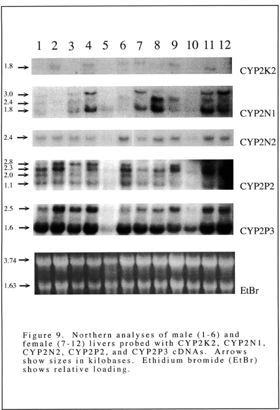
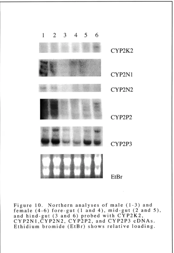
Figure 9: Northern analyses of male and female livers probed with CYP2K2 CYP2N1, CYP2N2, CYP2P2, and CYP2P3 cDNAs Figure 10: Northern analyses of male and female fore-gut, mid-gut and
hind-gut probed with CYP2K2, CYP2N 1, CYP2N2, CYP2P2, and CYP2P3 cDNAs
74 76 78 82 84 85 52 57 59 60 Page
LIST OF FIGURES, continued
Chapter 2:
Figure 11: Northern analyses of male and female extra-hepatic tissues probed with CYP2K2, CYP2N1, and CYP2N2 cDNAs Figure 12: Northern analyses of extra-hepatic tissues of fed/starved/TPA
reated fish probed with CYP2N 1, CYP2N2, CYP2P2, and CYP2P3 cDNAs
Chapter 3:
Figure la: Spectrophotometric analysis of CYP2N1 expressed in Sf9 microsomes
Figure lb: Spectrophotometric analysis of CYP2N2 expressed in SJ9 microsomes
Figure 2: Reverse-phase HPLC chromatogram of the organic soluble metabolites generated during incubation of recombinant CYP2N1 with [1- 14C]arachidonic acid
Figure 3: Reverse-phase HPLC chromatogram of the organic soluble metabolites generated during incubation of recombinant CYP2N2 with [1-1 4C]arachidonic acid
Figure 4: Reverse-phase HPLC chromatogram of the organic soluble metabolites generated during incubation of killifish liver microsomes with [1- 14C]arachidonic acid
61 63 95 95 97 101 104 Page
LIST OF FIGURES, continued
Chapter 4:
Figure 1: Spectrophotometric analysis of CYP2P3 expressed in SJ9 microsomes
Figure 2: Reverse-phase HPLC chromatogram of the organic soluble metabolites generated during incubation of recombinant CYP2P3 with [1-14C]arachidonic acid
Figure 3: Normal-phase HPLC chromatogram of the HETE metabolite generated during incubation of recombinant CYP2P3 with
[1- 14C]arachidonic acid
Figure 4: Endogenous EETs recovered from liver and gut tissues Figure 5: Endogenous DHETs recovered from liver and gut tissues
Chapter 5: Figure 1: Figure 2:
Appendix A:
Arachidonic acid metabolism in vertebrates
Representative metabolites formed from arachidonic acid via cyclooxygenases, lipoxygenases, and cytochromes P450
Figure 1: Total cytochrome P450 levels in Bermuda chub caught in 1988 (88-1-2), 1994 (94-1-5), and 1995 (95-1-15) Figure 2: Immunoblots with Bermuda chub microsomnes prepared
from liver tissues
126 128 129 131 132 149 150 164 165 Page
ABBREVIATIONS AEC: AhR ANOVA: 1xBLOTTO: Barbie BSA: bp: cDNA: cpm: CYP: CYPOR: dCTP: 50xDenhardt's: DEPC: DHET: DMSO: EET: EtBr: EtOH: GC/MS: HETE: HPLC: IgG: ip: 3-amino-9-ethylcarbazole aromatic hydrocarbon receptor analysis of the variance
5% nonfat dried milk, 0.02% sodium azide in H20
barbiturate responsive element bovine serum albumin
base pair
complementary DNA counts per minute cytochrome P450
NADPH cytochrome P450 reductase deoxycytidine triphosphate
1% w/v each Ficoll, polyvinylpyrrolidone, and BSA in H20 diethyl pyrocarbonate dihydroxyeicosatrienoic acid dimethyl sulfoxide epoxyeicosatrienoic acid ethidium bromide ethanol
gas chromatography/mass spectroscopy hydroxyeicosatrienoic acid
high pressure liquid chromatography immunoglobulin G
ABBREVIATIONS, continued
J: joules
kb: kilobase
kDa: kilodalton
MEM: modified Eagle's medium MFO: mixed function oxidase
mRNA: messenger RNA
NADPH: nicotinamide adenine dinucleotide phosphate PBS: phosphate buffered saline
PFA: paraformaldehyde
PFB: pentafluorobenzyl bromide pfu: plaque forming units
PLHC: Poeciliopsis lucida hepatoma carcinoma
P/S: penicillin/streptomycin SDS: sodium dodecyl sulfate SEM standard error of the mean
Sf9 Spodopterafrugiperda
20xSSC 3M NaCI, 0.3 M NaCitrate, pH 7.0
20xSSPE 3M NaC1, 0.2M NaH2PO4, 20 mM EDTA, pH 7.4 TCDD 2,3,7,8-tetrachlorodibenzo-p-dioxin
TMS bis(trimethylsilyl)trifluoracetamide TPA 12-O-tetradecanoyl phorbol-13-acetate
u micro
CHAPTER ONE
INTRODUCTION
Overall Objectives
The long term goal of these studies is to determine the diversity, structure, function, and regulation of cytochromes P450 in early diverging vertebrates and to use this information to evaluate the probable evolution of the cytochromes P450 and their regulatory systems. To address this goal, we have begun to characterize cytochrome P450 2 (CYP2) genes and proteins in the marine teleost Fundulus heteroclitus.
Why P450
Cytochromes P450 are enzymes that transform the structure of organic chemicals via mixed-function oxidase (MFO) or monooxygenase reactions (Ortiz de Montellano, 1986). Cytochromes P450 constitute a gene superfamily (more than 400 sequenced to date), with members in bacteria, plants and animals (Nelson et al., 1996). Multiple forms of cytochrome P450 can be present and induced in a single organism. In vertebrates from fish to mammals the cytochrome P450 enzymes have been found throughout the body except in red blood cells, and they metabolize an extraordinary number and diversity of lipophilic compounds of both exogenous and endogenous origin. Cytochrome P450 activities with exogenously derived substrates might be considered primarily as adaptive functions, involving the protection against toxic chemicals. Cytochromes P450 metabolize many of these diverse compounds to products that are less bioactive and more readily excreted. However, metabolism can activate other compounds to derivatives that are more reactive and more toxic (Guengerich 1985). The broad tissue distribution of cytochromes P450 within a single organism and among many different organisms as well as their ability to oxidize compounds of widely different structure give cytochromes P450 a fundamental role in metabolic processes.
Why CYP2
These studies concentrate on the CYP2 family for several reasons. First, roughly 20% of all CYP sequences are in family 2, and both the number and diversity of CYP2 subfamilies suggest that divergent ecological or physiological pressures may have affected this gene family and its expression. In addition, apparent differences in regulation occur among some CYP2s. Notably, CYP2B-like proteins in fish are not induced by the prototypic mammalian CYP2B inducers (Kleinow et al., 1987; Elskus and Stegeman, 1989; Elskus and Stegeman, 1989; Kleinow et al., 1990). This lack of induction has been speculated to reflect the lack of terrestrial plant material in the diets of the temperate fish most studied (Stegeman, 1993). Finally, CYP2 proteins in mammals metabolize a wide array of both endogenous and exogenous substrates. For example, CYP2A1 in rats is a testosterone 7c-hydroxylase (Sonderfan et al., 1989), and human CYP2A6 is a coumarin hydroxylase (Yun et al., 1991). The CYP2Bs in several species metabolize testosterone and are chlorobiphenyl hydroxylases (Ryan et al., 1982). Human CYP2C8 and CYP2J2 are arachidonic acid epoxygenases (Rifkind et al., 1995; Wu et al., 1996). Many CYP2Cs and CYP2Js also metabolize benzphetamine (Bornheim et al., 1987; Zeldin et al., in press). CYP2D6 in humans activates opiates (Kerry et al., 1994). CYP2E1 in rats metabolizes ketones, ethanol, benzene, and trichloroethylene (Nanji et al., 1994). Given the diversity and number of mammalian CYP2 proteins, one might ask whether CYP2 genes in other vertebrate groups are as diverse and numerous as they are in mammals.
Why Fish
Fish constitute the largest and earliest diverging vertebrate family (-30,000 fish species vs. -4,000 mammalian species). They first evolved 500+ million years ago, roughly 300 million years before mammals (Nelson, 1984). Knowledge of CYP in primitive vertebrates has important implications for understanding the evolution of vertebrate
CYPs in general. Sequence analysis of CYP in different vertebrate groups is an important way to address the evolution of this gene superfamily in vertebrates. Highly conserved CYP between fish and mammals would suggest conserved and possibly endogenous functions. Differences in CYP between fish and mammals could underlie fundamental organismal differences.
Studies to date have shown that fish have multiple CYP2 proteins. In several fish species, immunoblot analyses have shown CYP2B-like and CYP2E-like proteins (Gray et al., 1991; Stegeman and Hahn, 1994). The lauric acid hydroxylases, CYP2K1 and CYP2M1 have been isolated from trout, and again, immunoblot analyses have shown CYP2K-like and CYP2M-like proteins in different fish species (Miranda et al., 1989; Schlenk et al., 1993). As well as having unique CYP proteins, fish probably have either orthologues or paralogues of many mammalian CYP2 proteins.
The myriad proteins in the CYP2 family are postulated to have arisen in order to metabolize dietary plant natural products (Nelson and Strobel, 1987). Unlike mammals, the majority of fish are carnivorous and encounter fewer plant natural products (Horn, 1989). As discussed above, total CYP levels are not induced by the prototypic CYP2 inducer, phenobarbital, in the temperate carnivorous fish most studied, and this lack of induction has been postulated to reflect the lack of terrestrial plants in their diets (Stegeman, 1993). If CYP2 proteins first evolved to metabolize endogenous substrates and later expanded and diversified in order to cope with dietary plant natural products, then fish that have not had to cope with these natural products may have less diverse CYP2 proteins. Thus, fish may be a good vertebrate system with which to examine interactions between CYP and endogenous substrates, and knowledge of fish CYP2 proteins might have important implications for identifying endogenous CYP2 substrates.
This research focuses on fish P450 proteins in the 2 family. Fundamental questions remain concerning their identity and number, their catalytic functions, their
regulation and expression at the genetic as well as the organismal level, their evolution and inter-relatedness, and finally, their endogenous functions, if any. Cytochrome P450 proteins have been studied mainly in mammals. Fish have penetrated virtually every conceivable water habitat and range from the ocean depths (-11,000 m) to high mountain streams (+4,500 m) and from hot springs (430 C) to subfreezing water (-1.80 C) (Lauder and Liem, 1983). Because fish inhabit diverse and extreme environments throughout the world's waters, they can be expected to have novel P450 proteins not found in mammals as well as forms similar to those in mammals.
CYTOCHROME P450 BACKGROUND
General Reaction
Oxidative metabolism involving molecular oxygen is the initial enzymatic process in the biotransformation of a majority of lipophilic organic compounds. The general scheme for monooxygenase reactions is:
RH + NADPH + 02 + H+ --- > ROH + NADP+ + H20
where RH is the substrate and ROH is the hydroxylated product (Stegeman and Hahn, 1994). Cytochrome P450 proteins catalyze oxygenase reactions: they catalyze the incorporation of a single atom of molecular oxygen into its substrate while the other oxygen is reduced to water. They belong to a large and expanding superfamily of heme protein monooxygenases (Nelson et al., 1996). Some P450 proteins have multiple substrates, regulatory mechanisms, and/or biological functions. Altogether, the cytochrome P450 proteins catalyze thousands of different reactions including both oxidative and reductive reactions as well as the release of superoxide anions (Ortiz de Montellano, 1986).
P450 Properties
Most P450 enzymes are discrete gene products of about 57,000 kDa (about 500 amino acid residues) (Soucek and Gut, 1992) and contain one equivalent b-type heme per polypeptide (Black and Coon, 1987). Microsomal P450s contain a highly hydrophobic signal/anchor segment at the amino terminus followed by a short, cationic, halt transfer sequence. In contrast, mitochondrial P450s have a transient targeting sequence at the amino terminus which is proteolytically removed after transport of the precursor to the mitochondrial inner membrane. The mature proteins lack hydrophobic amino termini but otherwise are similar to microsomal P450s. The amino terminus is the principle site of membrane binding, and the majority of the polypeptide is exposed to the cytosolic (inner mitochondria) side of the membrane (Korzekwa and Jones, 1993). All cytochrome P450 proteins possess a noncovalently bound heme (protoporphyrin IX) and a segment of twenty-six amino acids surrounding a cysteine that is highly conserved. This cysteine donates the thiolate fifth ligand to the heme iron (Ortiz de Montellano, 1986).
The amino acid environment surrounding the heme results in a typical cytochrome P450 Soret absorption band at about 450 nm when the iron is reduced by electrons and complexed with carbon monoxide (Ortiz de Montellano, 1986). This property of the cytochrome pigment absorbing at 450 nm gives the enzyme its common name, cytochrome P450. Specific cytochrome P450s are named with the root CYP derived from cytochrome P450 followed by an Arabic numeral denoting the gene family, a letter for the subfamily, and another Arabic numeral designating the gene number (Nelson et al., 1993). P450s within a single gene family generally exhibit more than 40% sequence similarity. P450s within the same subfamilies generally have more than 55% identity within the same species. Cytochromes P450 with the same gene number in different species are considered orthologous.
Evolution of P450 Proteins
Cytochrome P450 proteins are ancient. Estimates based on P450 sequences suggest that P450 genes originated between 1.5 and 3 billion years ago (Stegeman and Hahn, 1994). P450 proteins also are relatively simple. Some P450 catalyzed reactions can be mimicked by heme analogs in an organic solvent: in these model systems, the protein part of the P450 is not essential to the reaction (Mansuy et al., 1989). Thus, the protein part of the enzyme does not seem to have a direct catalytic function. It appears to be largely limited to providing to the heme an environment conducive to the reactions catalyzed and to controlling the access of substrates to the heme. Consequently, even minor alterations of the protein can lead to changes of enzyme specificity: substitutions of a single amino acid residue have been shown to alter substrate recognition patterns (Lindberg and Negishi,
1989).
With the proper modifications, such as increased flexibility or a larger binding site, a P450 protein with very broad specificity probably could have evolved. But instead of only a few P450 proteins with very broad specificities, a vast number of P450 proteins with somewhat limited, but overlapping, specificities evolved, perhaps in order to avoid interference with metabolism of endogenous substrates (Nebert, 1991). Thus, cytochrome P450 proteins capable of metabolizing a diversity of exogenous compounds may have been constrained by the need not to metabolize endogenous compounds which overruled the advantage inherent in making only a few proteins to metabolize many different exogenous compounds (Zimniak and Waxman, 1993). The functional adaptability of P450 proteins may have profound evolutionary consequences. Changes of one or a few amino acids are relatively frequent and could lead to the emergence of cytochrome P450s capable of metabolizing a phenomenal number of compounds such as toxic plant products to which animals may be exposed after colonization of dry land and man-made synthetic compounds which previously were absent from the environment.
In general, membrane bound P450 proteins with a flavoprotein as their immediate redox partner (eukaryotic P450s) are more closely related to each other than to soluble P450 proteins with an iron-sulfur protein as their immediate redox partner (prokaryotic P450s) and vice versa. CYP102, a soluble, prokaryotic enzyme that clusters with the eukaryotic P450s in phlogenetic analyses is the only known exception (Gonzalez and Nebert, 1990). Certain bacterial and eukaryotic cytochromes P450 receive electrons via an iron-sulfur protein which itself receives electrons from a flavin adenine dinucleotide containing enzyme. In eukaryotes, this first major class of P450s is found exclusively in the inner mitochondrial membrane, is involved in highly specific steroid biosynthesis pathways, and does not metabolize xenobiotics. Endogenous P450 functions include sterol metabolism, fatty acid oxidations and oxidations of ethanol and other low molecular weight compounds, and the biosynthesis and metabolism of cholesterol to bile acids and steroid hormones. In contrast to many xenobiotics, endogenous compounds often are hydroxylated by individual P450s with a high degree of positional and stereochemical selectivity (Waxman et al., 1991).
The second major class of P450s is bound to microsomal membranes and electrons are donated to these enzymes via a protein containing a single molecule each of flavin adenine dinucleotide and flavin mononucleotide. A few microsomal P450s are involved in steroidogenesis in specialized tissues. Others are involved in the cholesterol-bile acid biosynthetic pathway (Gonzalez, 1989). However, the majority of microsomal P450s metabolize foreign compounds. In mammals, cytochromes P450 in gene families 1-4 are the most prominent in the metabolism of xenobiotics. Many genes in these families also are induced by xenobiotics. P450s in families 1-4 catalyze many activities with much overlap, and few substrates are exclusive to one protein. As well, orthologous P450s in different
CYP Families 1-4
The majority of information on cytochromes P450 in animals comes from studies in mammalian species. Experiments with rats, mice, rabbits and humans provide most of the fundamental knowledge concerning the enzymes in CYP gene families 1 through 4.
CYP 1: The CYPi family has an affinity for planar substrates and is able to oxygenate molecules in conformationally hindered (bay region) positions, resulting in their activation (Parke et al., 1991). In mammals, the CYPlA subfamily comprises two enzyme proteins, CYPlA1 and CYP1A2. CYP1Al reacts with aromatic hydrocarbon substrates such as benzo[a]pyrene and 3-methylcholanthrene. CYP1A2 reacts with aromatic amines and amides such as B-naphthylamine and 2-acetylaminofluorene (Parke et al., 1991). Regulation of cytochrome P450 activity may occur at the transcriptional, translational, and/or post-translational level. The regulation of CYPlA is the most well characterized of P450 regulation at the molecular level. Briefly, upon treatment with an inducer, an Ah receptor (aromatic hydrocarbon receptor) forms a complex with the hydrophobic ligand inducer and is translocated into the nucleus. The complex reacts with 5' regulatory regions of the CYP1A gene and induces transcription of the CYP1A genes among others (Hankinson, 1995). CYP1B recently has been cloned in mammals and also metabolizes aromatic hydrocarbons (Savas et al., 1994; Sutter et al., 1994). The CYP1B proteins are expressed constitutively at low levels in heart, brain, placenta, lung, skeletal muscle, kidney, spleen, thymus, prostate, gonads, small intestine, colon, and peripheral blood leukocytes (Savas et al., 1994; Sutter et al., 1994).
CYP 2: P450s in gene family 2 are numerous and diverse. Because the mammalian CYP2 family markedly expanded within the past 400-800 million years, about the same time that land plants first evolved, it has been hypothesized that the CYP2 family
diversified to metabolize plant secondary metabolites (Nelson and Strobel, 1987). This large diversity in the CYP2 family may mask orthologous relationships between the CYP2 genes. The following sections briefly summarize some of the information concerning the CYP2 subfamilies.
CYP2A: The rat CYP2A1 and CYP2A2 are differentially regulated and have distinct substrate specificities: CYP2A1 is involved in the 7a-hydroxylation of testosterone in young male and female rats while CYP2A2 is only expressed in adult male rats and has a high level of 15a-hydroxylase activity towards progesterone (Aoyama et al., 1990). CYP2A3 is expressed only in rat lung and is induced by 3-methylcholanthrene via transcriptional activation (Gonzalez, 1989). The sex-specific expression of the rat CYP2As is controlled in part by secretion of pituitary hormone (Waxman et al., 1985; Waxman et al., 1989).
CYP2B: The CYP2B subfamily contains at least ten members. Mammalian CYP2B proteins function in the metabolism of a wide array of drugs, natural products, and pollutants. The proteins are induced by many of their substrates, typified by phenobarbital but including polychlorinated biphenyl congeners, chlorinated pesticides, and many tumor promoters. CYP2B prefers globular molecules for substrates and generally initiates their detoxification. In rat liver, CYP2B 1 and CYP2B2 are the major phenobarbital-inducible P450 enzymes. These enzymes have 97% sequence similarity, but CYP2B2 is generally of lower catalytic activity. CYP2B 1 actively metabolizes a broad spectrum of lipophilic drugs and steroidal substrates including androgens such as androstenedione. CYP2B2 has a similar but distinguishable substrate specificity profile but is several-fold less active than CYP2B1 with many monooxygenase substrates (Gonzalez, 1989). The induction of CYP2B activity by phenobarbital is primarily due to new CYP2B protein synthesis that
results from an increase in steady state levels of CYP2B mRNA resulting from increased transcription of the corresponding CYP2B genes. This transcriptional activation is rapid (30-60 minutes) and can reach a level 20-50 times higher than the basal transcription rate (Adesnik et al., 1981).
CYP2C: The CYP2Cs generally are expressed constitutively and are under developmental and sex-specific regulation (Gonzalez, 1989). CYP2Cs metabolize steroids such as testosterone and progesterone. In humans, CYP2C9 is important in the metabolism of pharmaceuticals. Pharmaceutical substrates include S-warfarin, tolbutamide, phenytoin, tertrahydrocannabinol, ibuprofen, naproxen, tienilic acid, and dicifenac (Smith and Jones, 1992).
CYP2D: The CYP2Ds are among the most important CYP involved in the metabolism of drugs by humans (Smith and Jones, 1992). The CYP2Ds catalyze a suite of reactions including aromatic hydroxylation (propanolol), aliphatic hydroxylation (metoprolol), N-dealkylation (amiflamine), and hydroxyl oxidation (reduced haloperidol). Other CYP2D substrates include oxyprenolol, codeine, debrisoquine, imippramine, and thioridazine, to name a few (Smith and Jones, 1992). Substrates for CYP2D6 have a basic atom about 5-7 A from the site of metabolism which suggests that the enzyme has an acidic residue at approximately this distance from the iron atom (Smith and Jones, 1992).
CYP2E: CYP2E metabolizes small molecules including ethanol, acetone, benzene, and dialkyl nitrosamines and probably is involved in cholic acid biosynthesis (Ryan et al., 1985). CYP2E tends to generate reactive oxygen radicals and is induced by starvation, ethanol, and acetone (Parke et al., 1991). Induction mechanisms include transcriptional activation, mRNA stabilization, and protein stabilization (Soucek and Gut, 1992).
CYP2F: CYP2F was cloned from a human lung cDNA library and has been shown to activate the pneumotoxin naphthylamine (Nhamburo et al., 1989). The endogenous functions of CYP2F proteins are unknown.
CYP2G: Rat CYP2G is a neuroepithelial enzyme and is thought to be involved in olfactory sensing (Nef et al., 1989; Nef et al., 1990). Rabbit CYP2G1 was isolated from a rabbit nasal cDNA library (Ding et al., 1991), is only detected in the olfactory mucosa (Ding and Coon, 1990), and is a principal sex steroid hydroxylase in olfactory microsomes (Ding et al., 1991).
CYP2H: CYP2H1 and CYP2H2 were isolated from chick hepatocytes (Hobbs et al., 1986; Hansen and May, 1989). Reconstituted CYP2H1 and CYP2H2 both demethylate benzphetamine equally but differ in their ability to metabolize p-nitrophenol and acetominophen (Sinclair et al., 1990). In chick hepatocytes, in vivo and in vitro, phenobarbital treatment results in a fifty fold induction of CYP2H1 and CYP2H2 mRNAs. These mRNAs share 92% deduced amino acid similarity with each other and share 51-56% amino acid sequence similarity with several phenobarbital-inducible and constitutive mammalian CYP2C forms (Hobbs et al., 1986; Hansen and May, 1989).
CYP2J: CYP2Js have been identified in humans, rats, mice and rabbits (Wu et al., 1996; Wu et al., 1997; Zhang et al., 1997). Most are highly expressed in gut and liver, and some are expressed in a variety of other tissues including heart. It is not yet known whether the CYP2Js are inducible. Recently, some CYP2J proteins have been shown to metabolize arachidonic acid with a high degree of regio- and stereospecificity (Wu et al.,
CYP2L: The CYP2Ls from lobster are the only full-length, invertebrate CYP2s yet identified (James et al., 1993). The CYP2Ls are highly expressed in the hepatopancreas (digestive organ) and catalyze the monooxygenation of benzphetamine, aminopyrine, benzo[a]pyrene, progesterone, and testosterone. CYP2L1 sequence recently has been published (James et al., 1996), and another CYP2L sequence, CYP2L2, has been identified (Boyle and James, unpublished data).
CYP2K and CYP2M: The trout CYP2K1 and CYP2M1 proteins both are lauric acid hydroxylases (Buhler et al., 1994; Yang et al., 1995) and are discussed further in the section on cytochromes P450 in fish.
CYP2Q: CYP2Q recently was sequenced from frog (Ohi et al., unpublished data). No data on properties other than sequence is available for CYP2Q.
Altogether, the CYP2 family proteins metabolize a wide variety of different compounds. As discussed above, the proliferation and diversification of the CYP2 family has been suggested to have arisen in order to metabolize dietary, plant natural products. Similarly, the large number of substrates including natural products that CYP3 proteins metabolize also suggests involvement of the CYP3 gene family with natural products.
CYP3: The CYP3 family also has a large number of genes in different species, but unlike the CYP2s, the CYP3s all belong to the same subfamily, CYP3A. The CYP3As have an affinity for large, nonplanar substrates such as the macrolide antibiotics and are known to metabolize cyclosporine, ergotamine derivatives, and some steroids. CYP3A proteins are induced by pregnenolone 16-a-carbonitrile. CYP3A is the principal P450 of human liver and is involved in the activation of the dihydrodiol derivatives of polycyclic
aromatic hydrocarbons to the ultimate mutagens and carcinogens (Parke et al., 1991).
CYP4. Enzymes in this family oxidatively metabolize carboxylic acids such as arachidonic acid at the o or o-1 carbon groups and thus may be involved in pathways of fatty-acid and prostaglandin oxidation. This family is induced by the antihypercholesteraemic drugs, clofibrate and ciprofibrate, and the phthalate and sebacate ester plasticizers and is associated with peroxisome proliferation (Parke et al., 1991).
Cytochromes P450 in Fish
Catalytic, immunochemical and regulatory studies indicate that CYP families 1-4 also occur in fish (Stegeman, 1989; Stegeman and Hahn, 1994). Evidence summarized in Table 1 is drawn from recent reviews (Stegeman, 1987; Stegeman, 1993).
Table 1. Subfamilies of Hepatic Cytochrome P450 Proteins Purified from Fish
Subfamily Trivial Name Evidence
lA Scup P450 E P-C, F, I, R, S Trout LM4b P-C, F, I, R, S Cod c P-C, F, I, R, Perch V P-C, F, I, R 2B Scup P450 B I, S 2K Trout LM 2 I, S 2M Trout LMC 1 I, S Trout KM2 I 3A Scup P450 A F, I 3A27 Trout LMC 5 F, I, S Trout P450 con F, I Cod b I
Subfamilies 2E, 2C and 4A have been indicated in one or more species by presence of cross-reactive proteins in microsomes.
Assignment to subfamilies is based on: P-C = physico-chemical properties;
F = functional properties; I = immunological cross reactivity; R = regulatory similarities; S = sequence similarities.
Sequences for CYP1A genes have been identified in a variety of other teleosts. These include plaice (Leaver et al., 1993), oyster toadfish (Morrison et al., 1995), four-eye butterfly fish (Vrolijk et al., 1995), european sea bass (Stien et al., 1995), and wild red sea bream (Mizukami et al., 1994). In additon, a partial CYP3A cDNA, CYP3A30, has been sequenced in killifish (Celander and Stegeman, 1997) and a partial CYP4 cDNA, CYP4T1, has been sequenced in rainbow trout (Falckh et al., 1997).
Teleost CYP 1A
A close immunochemical relationship exists between CYP1A in fish and mammals and among vertebrates generally. This relationship also is based on deduced amino acid sequence similarities. In fish, a single dose of ~-naphthoflavone causes an increase in CYPlA mRNA by 6 hours with a maximum level reached at 30-40 hours. This declines to near control levels at 4-5 days. Functional protein is made by 18 hours with a maximum at three days. Elevated protein levels can persist for 2-3 weeks (in mammals, persistence is generally less than 48 hours) (Kloepper-Sams and Stegeman, 1989). A less rapidly metabolized inducer can cause high mRNA levels for up to two weeks (Hahn and Stegeman, 1994). High dose and/or prolonged exposure causes a decline in activity and in protein even while mRNA levels are maintained (White et al., 1997).
Teleost CYP2B
A number of fish have liver microsomal proteins that cross-react with antibodies to various mammalian CYP2 family proteins. One purified P450 from fish has sequence showing a relationship to the phenobarbital-inducible CYP2B: the amino terminal sequence comparisons show that P450B from scup has 50+ % sequence identity to mammalian CYP2Bs, suggesting identity as a 2B family member.
SCUP P450B M E LS TTLI LEGLILALLLL V
HUMAN 2B6 M E LS VLLFLALLTGLLLLL V
DOG 2B M E LS VLLLLALLTGLLLLM A
MOUSE 2B10 M E PS VLLLLALLVGFLLLL A
RAT 2B1 M E PTI LLLLALLVGFLLLL V
RABBIT 2B4(BO) M E FSLLLLLAFCAGLLLLF R
Anti-P450B recognizes two proteins in scup liver microsomes, one major and one minor. Scup P450B is an immunological counterpart to the PB-inducible 2B 1 forms in mammals based on reciprocal analysis of purified scup P450B and rat P4502B I1 and antibodies to these antigens. Anti-scup B recognizes pure rat 2B 1, 2B2, and 2B3 and only those proteins in rat liver microsomes. Anti-rat 2B 1 recognizes scupB and the same size protein in scup liver microsomes (Stegeman et al., 1990). Specific protein bands in Western blots of all fish, birds, and mammals (except whales) examined are detected identically with antibodies to either rat P450 2B 1 or to scup P450B. In addition, a cDNA probe for rat P450 2B 1/2 detected a hybridizing RNA sequence in scup liver.
Despite the evidence for a teleost 2B-type protein, fish have not been found to respond in any recognizable way to typical mammalian CYP2B inducers such as phenobarbital (Kleinow et al., 1987; Elskus and Stegeman, 1989; Elskus and Stegeman, 1989; Kleinow et al., 1990). Interestingly, some other non-mammalian vertebrates including reptiles and amphibians, also lack 2B-like induction (Stegeman and Hahn, 1994). An examination of the fish CYP that are homologous to the mammalian phenobarbital inducible genes, but are not phenobarbital inducible, may provide insight into the molecular mechanisms that govern the selectivity of phenobarbital induction for individual P450 genes.
Teleost CYP2E
mRNA studies. Fish liver microsomes were shown to dealkylate diethylnitrosamine (Kaplan et al., 1991), an activity associated with CYP2E enzymes in mammals (Gonzalez, 1989). Antibodies to rat CYP2E1 recognized a single protein in the liver of Poeciliopsis sp. Finally, an oligonucleotide probe specific for rat CYP2E1 hybridized with a 3.3 kb mRNA in RNA samples from Poeciliopsis sp. This mRNA also was induced by ethanol
(Kaplan et al., 1991), a prototypic CYP2E inducer.
Teleost CYP2K
Trout LM 2 was classified into the novel CYP2K subfamily based on its sequence (Buhler et al., 1994). CYP2K1 shows marked differences in sex and tissue-specific expression at both the transcriptional and translational level in sexually mature, rainbow trout liver and trunk kidney. CYP2K1 cDNA hybridized with a 2.8 and a 1.9 kb mRNA in mature male trunk kidney and some male livers. In females, the 2.8 kb band generally was not detected. CYP2K1 transcript was expressed more strongly in kidney than in liver and also was expressed more strongly in males than females. These differences showed corresponding differences in protein expression and activities (Buhler et al., 1994). Constitutive CYP2K1 exhibits high estradiol 2-hydroxylase activity and also catalyses the co-1 hydroxylation of lauric acid and the conversion of aflatoxin B1 to the highly electrophilic aflatoxin B 1-8,9-epoxide metabolite (Williams et al., 1984). CYP2K3 also has been identified in trout and shares 96.5% deduced amino acid identity with CYP2K1 (Buhler, unpublished data).
Teleost CYP2M
Trout LMC 1 recently was classified into the novel CYP2M subfamily based on its sequence (Yang et al., 1996). The predicted amino acid sequence of CYP2MI is 57% identical to that of CYP2K1. Northern analyses of trout RNA showed that CYP2M1
hybridized to a single 2.2 kb RNA in liver but not kidney. Like CYP2K1, CYP2M 1 also metabolizes lauric acid; CYP2M 1I shows o)-6 hydroxylase activity towards lauric acid.
Teleost CYP3A
Trout P450con, trout LMC5, scup P450A, and cod P450b are immunochemically related; in addition, each was recognized by polyclonal antibodies to rat CYP3A1 and human CYP3A4 suggesting that these fish proteins all are CYP3A-like and probably members of the CYP3A subfamily (Celander et al., 1996). Furthermore, trout LMC5 and scup P450A are the major hepatic microsomal enzymes that metabolize testosterone at the 63-position; this activity is associated with CYP3A enzymes in mammals (Waxman, 1988). Sequence analysis of trout LMC5 has confirmed this protein as a CYP3A family member (Buhler, unpublished data) and partial sequence for a killifish CYP3A demonstrates this gene in killifish (Celander and Stegeman, 1997).
The fish CYP3A-like proteins are present at high levels in untreated fish (Celander et al., 1989; Gray et al., 1991). In contrast to mammals, little is known about the inducibility of the CYP3As in fish. They seem to be slightly induced by steroids (Celander et al., 1989) and may be influenced by dietary natural products (Vrolijk et al., 1994). Finally, sex-differences may depend on species differences in fish CYP3A-like protein levels and activities (Celander et al., 1996). The mechanism of the regulation of the fish CYP3 genes is still unknown.
Teleost CYP4T
A partial CYP4T1 sequence recently was cloned from trout liver (Falckh et al., 1997). CYP4T1 deduced amino acid sequence is most similar to rat CYP4B2 with 55.4% identity.
In addition to CYP in families 1-4, CYP in other families have been identified in fish. The cholesterol side-chain cleavage cytochrome P450, CYPI1A1, has been cloned from both rainbow trout (Takahashi et al., 1993) and southern stingray (Nunez and Trnat, 1996). The 17 alpha-hydroxylase, CYP17, has been cloned from rainbow trout (Sakai et al., 1992), medaka (Kobayashi et al., 1996), and dogfish (Trant, 1995). Aromatase or CYP19 has been cloned from channel catfish (Trant, 1994), rainbow trout (Tanaka et al., 1992), goldfish (Gelinas et al., 1996), medaka (Tanaka et al., 1995), and tilapia (Chang et al., 1996). Recently, a CYP in a new vertebrate family, CYP26, was isloated from zebrafish (White et al., 1996). CYP26 is a retinoic acid-inducible all trans retinoic acid 4-hydroxylase. Finally, a partial sequence for a CYP52 has been cloned from rainbow trout (Gong, 1994).
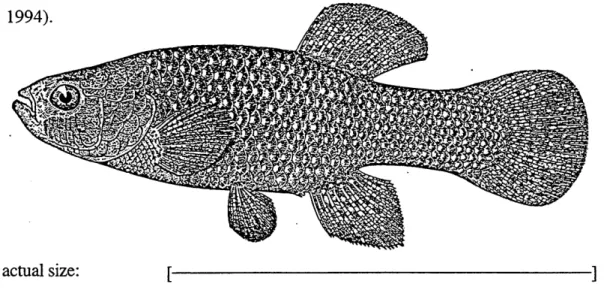
actual size: [
Figure 1. Fundulus heteroclitus (killifish). From (Bigelow and Schroeder, 1953).
Specific Objective and Approach
Although the foregoing indicates that members of gene families 1-4 are expressed in fishes, the studies in fishes have not approached the question of gene diversity in the largest mammalian CYP family, family 2. The specific objective of this research is to explore the diversity, structure, function, and regulation of fish cytochromes P450 in family 2 and to use this information to evaluate the probable evolution of the cytochromes
P450 and their regulatory systems. We approached this objective in the marine teleost, Fundulus heteroclitus or killifish. We used killifish for several reasons.
1. Killifish are an abundant and common fish on the east coast of North America. They are widespread and occur in virtually every salt marsh from Canada to Virginia. We have many different populations in Woods Hole, and they are obtained easily.
2. Killifish reside in salt marshes and range only a few hundred yards from their natal creeks (Lotrich, 1975) making them good species for environmental toxicology studies.
3. Killifish are extremely hardy and adapt well to almost any laboratory condition. They can be cultured easily from eggs and sperm in the laboratory.
4. Killifish move onto marshes with the incoming tide to feed and are an important link in the energy transfer between the shore and open waters (Penczak, 1985). In the winter, killifish hibernate unfeeding in mud (Valiela et al., 1977). They are mainly carnivorous although algal filaments and detritus are common gut contents (D'Avanzo and Valiela, 1990). Because of their significant detrital dietary component and their coastal habitat, they likely are exposed to a wide variety of pollutants from both the water and the sediments.
5. Extensive literature exists on the population genetics of this species in the Eastern United States (Powers et al., 1986; Bernardi et al., 1993).
6. The reproductive physiology of killifish has been studied in detail (Selman and Wallace, 1986; Hsiao and Meier, 1989; Hsiao et al., 1994).
7. The embryology and developmental biology of killifish is known better than that of any other marine or estuarine fish (Armstrong and Child, 1965).
8. Analysis of liver microsomes show that killifish express multiple CYP2-like proteins (Gray et al., 1991).
available. Dr. Douglas Crawford at the University of Chicago provided a genomic DNA library and a liver cDNA library. Dr. Sibel Karchner at Woods Hole Oceanographic Institution provided a liver cDNA library and a heart cDNA library.
The approach taken for this work was to design degenerate oligonucleotide probes to conserved sequences in multiple CYP2 genes. These probes were used to screen the killifish libraries. Initially, partial sequences in three CYP2 subfamilies, CYP2K, CYP2N, and CYP2P were isolated. These partial sequences were used to further screen the libraries and obtain full length sequences. Chapter 2 details these initial experiments and describes the phylogenetic relationships between the killifish CYP2 genes and other CYP2 genes. The next question addressed concerned the functions of these genes. To approach this question, three of these genes, CYP2N1, CYP2N2, and CYP2P3, were co-expressed with human NADPH cytochrome P450 reductase (oxidoreductase) in SJ9 insect cells using a baculovirus expression system. Functional P450 and reductase proteins were co-expressed in the insect cells and used in catalytic studies. The results of these experiments are presented in Chapters 3 and 4. Finally, Chapter 5 briefly summarizes the results of these studies and discusses future research directions.
CHAPTER TWO
ABSTRACT
The cytochrome P450 2 (CYP2) genes occupy 15 subfamilies in mammals (Nelson et al., 1996). The diversity of non-mammalian vertebrate genes in the CYP2 family is not known but could shed light on the functional significance of CYP2 diversity. Genes in the CYP2 family were sought in the killifish (Fundulus heteroclitus). Multiple cDNAs related to known CYP2 genes were isolated from a killifish liver cDNA library. Sequence analyses showed that one is related to CYP2K1 previously identified in trout (Buhler et al., 1994). The other cDNAs were classified into two new subfamilies, CYP2N and CYP2P, with one gene in CYP2N (CYP2N1) and two genes in CYP2P (CYP2P2 and CYP2P3). Analyses of killifish RNA demonstrated that CYP2K2, CYP2P2, CYP2P3, and CYP2N 1 transcripts are expressed in both liver and gut tissues. CYP2N1 transcripts also are expressed in heart and brain. Subsequently, a killifish heart cDNA library was screened with CYP2N1, and another CYP2N gene was isolated. Cardiac CYP2N2 shares 75% amino acid identity with liver CYP2N 1 and is expressed most highly in heart and brain. CYP2P and CYP2N gene expression was examined in fish that were held without food and in fish that were treated with the diacylglycerol mimic, 12-O-tetradecanoyl phorbol-13-acetate (TPA) (Kraft and Anderson, 1983). RNA analyses of these treated fish showed an apparent down-regulation of expression of the CYP2P and CYP2N transcripts in the extra-hepatic tissues of the TPA treated fish. In addition, a CYP2P gene was isolated from a killifish genomic DNA library. Sequence analysis indicates that the intron-exon pattern for this gene, CYP2P1, is similar to that of other CYP2 genes (Suwa et al., 1985). Altogether, these six killifish CYP2 genes in three different subfamilies suggest a diversity and multiplicity of CYP2 genes in early diverging vertebrates.
INTRODUCTION
The cytochrome P450 (CYP) superfamily contains both a great number and a great diversity of proteins. These proteins are found in most if not all phyla, and they have key functions in toxicology, endocrinology, pharmacology, and carcinogenesis. Many CYP proteins such as aromatase (CYP19) have similar functions in different species (Evans et al., 1987) while others such as P450cam have specialized functions in particular species (Sligar and Murray, 1986). Knowledge of the similarities and differences between diverse CYP proteins is fundamental to understanding the CYP superfamily as a whole.
The CYP2 family is the largest family in the cytochrome P450 superfamily. Currently, it has fifteen subfamilies, and about 20% of all CYP sequences are in family 2 (Nelson et al., 1996). The CYP2 proteins in mammals metabolize a wide array of both endogenous and exogenous substrates. For example, CYP2A1 in rats is a testosterone 70o-hydroxylase (Sonderfan et al., 1989). The CYP2Bs in several species are chlorobiphenyl hydroxylases (Ryan et al., 1982). Human CYP2C8 and CYP2J2 are arachidonic acid epoxygenases (Rifkind et al., 1995; Wu et al., 1996). CYP2D6, also in humans, activates opiates (Kerry et al., 1994). CYP2E1 in rats metabolizes ethanol, ketones, benzene, and trichloroethylene (Nanji et al., 1994). Both the number and the diversity of the CYP2 subfamilies in mammals suggest that divergent ecological or physiological pressures may have influenced this gene family and its expression. Determining whether other vertebrate groups have a similar diversity of CYP2 genes may establish when in phylogeny this gene radiation occured and may indicate those ecological or physiological features contributing to this diversity.
Fish constitute the largest and earliest diverging vertebrate group. They first evolved 500+ million years ago, roughly 300 million years before mammals (Nelson, 1984). Knowledge of CYP in early diverging vertebrates has important implications for
understanding the evolution of vertebrate CYP in general. If CYP are highly conserved between fish and mammals, this would suggest conserved and possibly endogenous functions. On the other hand, wide differences in CYP between fish and mammals may underlie fundamental organismal differences. Finally, sequence analysis of CYP in different vertebrates addresses the evolution of this gene superfamily in vertebrates.
The myriad CYP in the CYP2 family are postulated to have arisen in order to metabolize dietary plant natural products (Nelson and Strobel, 1987). Unlike mammals, the majority of fish are carnivorous and encounter fewer dietary plant natural products (Horn, 1989). In fact, in the temperate carnivorous fish most studied, total CYP levels are not induced by the prototypic CYP2 inducer, phenobarbital (Kleinow et al., 1987; Elskus and Stegeman, 1989; Elskus and Stegeman, 1989; Kleinow et al., 1990), and this lack of induction has been postulated to reflect the lack of terrestrial plants in their diets (Stegeman, 1993). If CYP2 proteins first evolved to metabolize endogenous substrates and later expanded and diversified in order to cope with dietary plant natural products, then fish that have not had to cope with dietary plant natural products may have less diversity of CYP2 proteins. Thus, fish may be a good vertebrate system with which to examine interactions between CYP and endogenous substrates, and knowledge of fish CYP2 proteins might have important implications for identifying endogenous CYP2 substrates.
Several lines of evidence indicate that fish have multiple CYP2 proteins. In numerous fish species, immunoblot analyses have shown CYP2B-like and CYP2E-like proteins (Stegeman, 1989; Stegeman and Hahn, 1994; Kaplan et al., 1991). As well, the lauric acid hydroxylases, CYP2K1 and CYP2M1 have been isolated from trout, and again, immunoblot analyses have shown CYP2KI-like and CYP2M1-like proteins in other fish species (Buhler et al., 1994; Yang et al., 1995). Whether fish have either orthologues or paralogues of many mammalian CYP2 proteins and whether unique CYP proteins occur in fish is not known.
Our present objectives are to examine the diversity of CYP2 genes in the marine fish Fundulus heteroclitus. Multiple CYP2 proteins have been studied in mammals, but less is known about the diversity, number and functions of non-mammalian CYP2 proteins. Since the profusion of CYP2 subfamilies obscures the functional relationships between CYP2 proteins, establishing the diversity of non-mammalian CYP2 proteins and their relationships to known CYP2 proteins may clarify these functions.
MATERIALS AND METHODS
Materials: [a3 2P]dCTP was purchased from Amersham, and [y3 2P]ATP was
purchased from DuPont NEN. Restriction enzymes, T4 DNA ligase, T4 polynucleotide kinase, and the Nick Translation System were purchased from Promega. Infra-red dye labelled sequencing primers were purchased from Licor. Long Ranger acrylamide was purchased from FMC Biochemicals. Cycle sequencing kits were purchased from Epicentre Technologies. General contractor cloning kits were purchased from 5 Prime to 3 Prime. Bluescript vector was purchased from Stratagene. Primers were purchased from Operon. RNA Stat-60 was purchased from Tel-Test "B," Inc. All other chemicals and reagents were purchased from Sigma unless otherwise noted.
Screening of the libraries: One killifish liver cDNA library was constructed in lambda gtl0 from a single female fish liver. It had an amplified titer of 1.5x10⁸ pfulml and average insert size of 1.0 kb. A second killifish liver cDNA library was constructed in Lambda Zap from a pool of female fish. It had an amplified titer of 1.6x10¹⁰ pfu/ml and average insert size of 1.8 kb. The killifish heart cDNA library was constructed in Lambda Zap from a pool of male and female hearts. It had an amplified titer of 1.5x10¹⁰ pfu/ml and average insert size of 1.5 kb. The killifish genomic library was constructed in Lambda Fix II from a male fish (Stratagene). It had an amplified titer of 5x10⁹ pfu/ml and insert
sizes from 9-23 kb.
Approximately 5X10⁵ phage from the lambda gtlO liver cDNA library were plated with XL1-Blue E. coli as host and screened as described (Sambrook et al., 1989). These were screened with two degenerate probes end-labelled with [y3 2P]ATP. The
oligonucleotide probes were designed based on conserved regions found in multiple CYP2 proteins from multiple species (Hobbs et al., 1986; Graves et al., 1990; Gotoh, 1992). The CYP2 proteins were rat 2A1, 2B1, 2B2, 2G1, 2D1, and 2E1, mouse 2A5, human 2B 1 and 2F1, rabbit 2B4, 2C3, and 2C4, dog 2C21, chicken 2H1 and 2H2, and P. putida
101Al. The primers were based on conserved regions corresponding to alpha helices in CYP101Al as determined by x-ray crystallography (Gotoh, 1992). Thus, they likely code for structural parts of the protein which probably are more conserved than substrate binding regions. The 5' end CYP2 probe had a PstI and a XhoI restriction enzyme recognition sequence at its 5' most end. Its sequence was
5'-CTGCAGCTCGAGGAGCGVATYCAGGASGARGC-3'. The 3' end CYP2 probe had a BamHI and a XbaI restriction enzyme recognition sequence at its 5' end. Its sequence was 5'-GGATCCTCTAGATSACBGCVTCNGTGTAKGGCA-3'. Where appropriate, codon usage tables for teleosts were used to eliminate degeneracies (Fitzgerald et al., 1993).
Hybridizations were done in 6XSSPE, 0.05X Blotto, and 20% formamide at 420 C overnight. Approximately 90 positive clones were identified. 35 of these were subcloned into pBluescript SK(+) and cycle sequenced by the dideoxy chain termination method (Sanger et al., 1977) using DNA polymerase (Epicentre) and infra-red labelled primers (Licor). Approximately 6X10⁵ phage from each of the Lambda Zap liver and heart cDNA
libraries were screened with the CYP2K2, CYP2P2, and CYP2N 1 fragments isolated from the lambda gtlO0 library. These fragments were random primer-labelled with [a3 2P]dCTP
using Rediprime (Amersham). Hybridizations were done in 6XSSPE, 0.05X Blotto and 50% (liver) or 20% (heart) formamide at 420 C overnight. Positive clones were isolated
from each library, rescued into pBluescript SK(+) (Stratagene), and sequenced as above. Approximately 5X10⁵ phage from the lambda Fix II library were plated with XL1-Blue E. coli as host. Probes and hybridization conditions were identical to those used for the lambda gtl0 library. The 14 kb CYP2P1 clone was restriction enzyme digested, subcloned in pBluescript SK(+), and sequenced as above.
Sequence Analyses: The CYP2A, CYP2B, CYP2C, CYP2D, CYP2E, CYP2F, CYP2G, CYP2H, CYP2J, CYP2K, CYP2L, CYP2M, and CYP2Q genes were retrieved from the GenBank database. Accession numbers were: rat CYP2Al (M33312), rabbit CYP2B1 (M20857), rat CYP2C23 (U04733), mouse CYP2D10 (M27168), hamster CYP2E1 (D17449), mouse CYP2F2 (M77497), rat CYP2G1 (M33296), chicken CYP2H2 (M25469), rabbit CYP2J1 (D90405), trout CYP2K1 (L11528), lobster CYP2L1 (U44826), trout CYP2M1 (U16657), and frog CYP2Ql (D50560). The killifish sequences were assembled and translated using MacVector/AssemblyLign sequence analysis software (Oxford Molecular Group). The killifish nucleotide and deduced amino acid sequences were aligned with the CYP2 published sequences using CLUSTAL (Higgins and Sharp, 1988); refinements to the alignment were made manually. Phylogenetic trees based on amino acid alignments and nucleotide alignments (all three positions) were constructed using the minimum evolution criterion (Neighbor-Joining algorithm (Saitou and Nei, 1987)) and maximum parsimony (PAUP 3.1 (Swofford, 1990)). Phylogenetic trees based on nucleotide alignments also were constructed using distance-matrix (Olsen, 1988). Bootstrap analyses (Felsenstein, 1985) were performed to assess relative confidence in the topologies obtained.
RNA Analyses: Total RNA was prepared from killifish tissues using RNA Stat-60 according to manufacturer's directions except DNA sometimes was precipitated by centrifugation of the first homogenate for 5 minutes at 12000 g. 10 ug of total RNA was
denatured and electrophoresed on 1.0% agarose gels containing 0.41 M formaldehyde and transferred to nylon membranes by downward alkaline capillary as described (Chomczynski, 1992) except 4 bridge sheets were used rather than 2 . The blots were hybridized with the CYP2K, CYP2N, and CYP2P subclones nick translation labelled with [o(3 2P]dCTP. Hybridizations were at 420 C in 50% formamide, 6XSSPE, 1% (w/v) SDS, and 100 ug/ml of heat-denatured calf-thymus DNA. Even loading and complete transfer of RNA were monitored by detection of ethidium bromide stain in the gels and on the filters. Correlation between the stain on the filters and the amount of RNA was ascertained by probing the filters with labelled ribosomal RNA (data not shown).
Experimental Treatments Including TPA and Starvation: Sixty female fish that had been held in clean water for greater than six months were acclimated from 200 C to 140 C for 1 week. Six fish were sacrificed immediately and liver, gut, kidney, heart, and brain tissues were frozen immediately in liquid nitrogen for RNA analyses. Gut was divided into three equal parts: the fore-gut, the mid-gut, and the hind-gut. Twenty-four of the fish were fed excess TetraMin (Tetra Sales, Blacksburg, VA) throughout the experiment. The rest were held without feeding. After ten days, six fish from each group were sacrificed and liver, gut, kidney, heart, and brain tissues were frozen immediately in liquid nitrogen for RNA analyses. After two weeks, six fish from both groups were dosed intraperitoneally (ip) with 0.02 ug TPA/g fish in 80% normal saline/19.8% acetone/0.2% ethanol (solvent), six fish from both groups were dosed ip with solvent, six fish from both groups were untreated, and six of the starved fish were fed excess TetraMin. Fish were sacrificed 72 hours later and liver, gut, kidney, heart, and brain tissues were frozen immediately in liquid nitrogen for RNA analyses. All tissues except liver and fore-gut were pooled. RNA from fore-gut was degraded and was not analyzed.


![Figure 2. Reverse-phase HPLC chromatogram of the organic soluble metabolites generated during incubation of recombinant CYP2N1 with [1- 14 C]arachidonic acid.](https://thumb-eu.123doks.com/thumbv2/123doknet/14752111.580706/97.918.146.678.144.872/figure-reverse-chromatogram-metabolites-generated-incubation-recombinant-arachidonic.webp)
