HAL Id: hal-02386325
https://hal.archives-ouvertes.fr/hal-02386325
Submitted on 3 Jun 2021
HAL is a multi-disciplinary open access
archive for the deposit and dissemination of
sci-entific research documents, whether they are
pub-lished or not. The documents may come from
teaching and research institutions in France or
abroad, or from public or private research centers.
L’archive ouverte pluridisciplinaire HAL, est
destinée au dépôt et à la diffusion de documents
scientifiques de niveau recherche, publiés ou non,
émanant des établissements d’enseignement et de
recherche français ou étrangers, des laboratoires
publics ou privés.
Distributed under a Creative Commons Attribution| 4.0 International License
impaired intestinal epithelial barrier function and
inflammatory disease
B. Williams, N. Tebbutt, M. Buchert, T. Putoczki, K. Doggett, S. Bao, C.
Johnstone, F. Masson, Frédéric Hollande, A. Burgess, et al.
To cite this version:
B. Williams, N. Tebbutt, M. Buchert, T. Putoczki, K. Doggett, et al.. Glycoprotein A33
defi-ciency: a new mouse model of impaired intestinal epithelial barrier function and inflammatory
dis-ease. Disease Models & Mechanisms, Cambridge Company of Biologists, 2015, 8 (8), pp.805-815.
�10.1242/dmm.019935�. �hal-02386325�
ABSTRACT
The cells of the intestinal epithelium provide a selectively permeable barrier between the external environment and internal tissues. The integrity of this barrier is maintained by tight junctions, specialised cell-cell contacts that permit the absorption of water and nutrients while excluding microbes, toxins and dietary antigens. Impairment of intestinal barrier function contributes to multiple gastrointestinal disorders, including food hypersensitivity, inflammatory bowel disease (IBD) and colitis-associated cancer (CAC). Glycoprotein A33 (GPA33) is an intestinal epithelium-specific cell surface marker and member of the CTX group of transmembrane proteins. Roles in cell-cell adhesion have been demonstrated for multiple CTX family members, suggesting a similar function for GPA33 within the gastrointestinal tract. To test a potential requirement for GPA33 in intestinal barrier function, we generated Gpa33−/− mice and subjected them to experimental regimens designed to produce food hypersensitivity, colitis and CAC. Gpa33−/−mice exhibited impaired intestinal barrier function. This was shown by elevated steady-state immunosurveillance in the colonic mucosa and leakiness to oral TRITC-labelled dextran after short-term exposure to dextran sodium sulphate (DSS) to injure the intestinal epithelium. Gpa33−/− mice also exhibited rapid onset and reduced resolution of DSS-induced colitis, and a striking increase in the number of colitis-associated tumours produced by treatment with the colon-specific mutagen azoxymethane (AOM) followed by two cycles of DSS. In contrast, Gpa33−/− mice treated with AOM alone showed no increase in sporadic tumour formation, indicating that their increased tumour susceptibility is dependent on inflammatory stimuli. Finally, Gpa33−/− mice displayed hypersensitivity to food allergens, a common co-morbidity in humans with IBD. We propose that Gpa33−/− mice provide a valuable model to study the mechanisms linking intestinal
permeability and multiple inflammatory pathologies. Moreover, this model could facilitate preclinical studies aimed at identifying drugs that restore barrier function.
KEY WORDS: GPA33, Intestinal permeability, Colitis, Colorectal cancer, Oral tolerance
INTRODUCTION
Impaired intestinal epithelial barrier function is an emerging factor in the aetiology and pathology of a range of gastrointestinal disorders (Turner, 2009), including food hypersensitivity (Ventura et al., 2006), inflammatory bowel disease (IBD) (Heller et al., 2005; Zeissig et al., 2007) and colitis-associated cancer (CAC) (Kim and Kim, 2014). It is becoming clear that gastrointestinal homeostasis depends on the capacity of the epithelial lining to exclude luminal bacterial and food antigens from the lamina propria (LP) and thereby prevent local and systemic inflammation. Accordingly, restoration of barrier function is an important therapeutic ambition for the treatment and prevention of inflammation-mediated pathologies (Fries et al., 2013).
The intestinal barrier is maintained by a number of interacting components: production of mucus and anti-microbial peptides, epithelial integrity, cell-cell adhesion and innate immune responses– defects in any of which can increase intestinal permeability (Pastorelli et al., 2013). Cell-cell adhesion within the intestinal epithelium is regulated by tight junctions (TJs), the most apical of the junction complexes, which block the passage of luminal contents via a paracellular route between cells. TJs are composed of a number of proteins, including occludins, claudins and members of the cortical thymocyte marker for Xenopus (CTX) group of type I transmembrane proteins of the immunoglobulin (Ig) superfamily (Chrétien et al., 1998). The CTX group includes members of the junction adhesion molecule (JAM) (Laukoetter et al., 2007; Vetrano and Danese, 2009; Vetrano et al., 2008), the Coxsackie and adenovirus receptor (CAR) (Morton et al., 2013; Raschperger et al., 2006) and the endothelial cell-selective adhesion molecule (ESAM) (Nasdala et al., 2002) families. Notably, the loss of JAM-A has been implicated in changes to intestinal permeability with subsequent inflammation, cytokine production and colitis in mice (Laukoetter et al., 2007) and IBD in humans (Vetrano and Danese, 2009; Vetrano et al., 2008).
A relatively understudied founding member of the CTX family (Chrétien et al., 1998) is glycoprotein A33 (GPA33) (Heath et al., 1997). In human and mouse, the expression pattern of GPA33 is exquisitely restricted to epithelial cells lining the entire length of the intestine. Within this compartment, GPA33 is present on the basolateral membranes of the epithelial cells of both absorptive and secretory lineages, with notable enrichment of GPA33 protein evident at cell-cell junctions in whole-mount preparations of colonic epithelium (Johnstone et al., 2000). Its robust expression from the
Received 2 February 2015; Accepted 5 May 2015
1
The Walter and Eliza Hall Institute of Medical Research, Parkville,
Victoria 3052, Australia.2Department of Medical Biology, University of Melbourne, Parkville, Victoria 3052, Australia.3Ludwig Institute for Cancer Research, Melbourne-Parkville Branch, Parkville, Victoria 3050, Australia.4Ludwig Institute for Cancer Research, Melbourne-Austin Branch, Heidelberg, Victoria 3084, Australia.
5
Discipline of Pathology, School of Medical Science and Bosch Institute, University of Sydney, Camperdown, NSW 2006, Australia.6Trescowthick Research Laboratories, Peter MacCallum Cancer Centre, East Melbourne, Victoria 3002, Australia.7Department of Pathology, University of Melbourne, Parkville, Victoria, Australia.
*Present address: Olivia Newton-John Cancer Research Institute, Heidelberg, Victoria 3084, Australia.‡Present address: School of Cancer Medicine, La Trobe University, Melbourne, Victoria 3086, Australia.
§
Author for correspondence ( [email protected])
This is an Open Access article distributed under the terms of the Creative Commons Attribution License (http://creativecommons.org/licenses/by/3.0), which permits unrestricted use, distribution and reproduction in any medium provided that the original work is properly attributed.
Disease
Models
&
base of the crypts to the tips of the villi and along the entire rostrocaudal axis of the intestinal tract established GPA33 as a definitive marker of the intestinal epithelium (Heath et al., 1997; Johnstone et al., 2000). Clinically, GPA33 expression has been observed in over 95% of colorectal cancers (CRCs) and associated metastases (Garinchesa et al., 1996), an observation that has underpinned an ongoing and concerted effort to use radionuclide-bound murine (Welt et al., 1994, 1990) and humanised (huA33 mAb) monoclonal antibodies to target GPA33-expressing tumours (Ciprotti et al., 2014; Infante et al., 2013; Scott et al., 2005; Welt et al., 2003, 1996). A recent clinical study established the efficacy of the huA33 mAb in penetrating CRC metastases (Ciprotti et al., 2014), promoting continued interest in GPA33 as a therapeutic target in antibody-based treatment of late-stage CRC.
Despite the highly tissue-specific expression pattern and associated clinical interest, relatively little is known about the
function of GPA33. Insights into its transcriptional regulation (Johnstone et al., 2002; Mao et al., 2003), and implied roles in cell-cell adhesion (Ackerman et al., 2008) and immune signalling (Mallegol et al., 2005, 2007; Van Niel et al., 2003), constitute the most prominent findings to date. In a prior report, conducted with Gpa33−/−mice on a mixed background (C57BL/6×129Sv/J), we noticed a mild elevation in spontaneous colonic ulceration and a more severe response to the colonic injury induced by trinitrobenzenesulfonic acid (TNBS), compared to wild-type (WT) mice (Pereira-Fantini et al., 2010).
In this study, we analysed the role of GPA33 in intestinal barrier function using Gpa33−/−mice on a pure C57BL/6 background. Our findings indicate a role for GPA33 in the maintenance of intestinal barrier function and the prevention of associated pathologies such as IBD.
RESULTS
Gpa33−/−mice do not exhibit spontaneous gastrointestinal
pathology
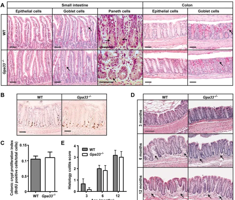
A null allele of Gpa33 was generated by replacing most of the Gpa33 coding sequence with a promoter-lessβ-geo cassette as described in supplementary material Fig. S1A. Expression of GPA33 was clearly detectable in the intestinal epithelium of WT mice and undetectable in Gpa33−/−mice, as shown by RT-qPCR (supplementary material Fig. S1B) and immunohistochemistry (supplementary material Fig. S1C), confirming the allele as a bona fide null. Gpa33−/− mice were born at the expected frequency, are fertile and have a normal lifespan. Intestinal epithelial cell lineage differentiation (Fig. 1A) and proliferation (Fig. 1B,C) were found to be normal in Gpa33−/−mice and, apart from an extremely mild, progressive colitis with age, which is indistinguishable from that seen in WT, Gpa33−/− mice did not exhibit steady-state colitis (Fig. 1D,E).
Gpa33−/−mice have impaired intestinal barrier function
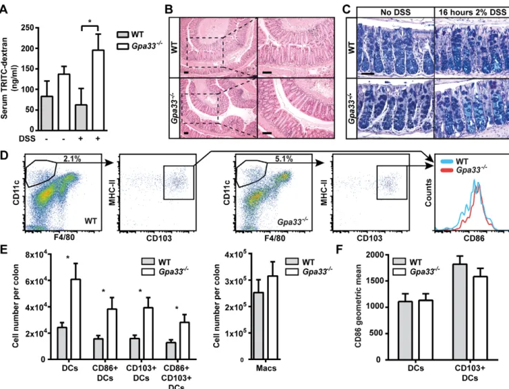
To study the requirement for GPA33 in the maintenance of intestinal barrier function, we delivered TRITC-labelled dextran (4 kDa) to the gastrointestinal tract by gavage and measured its levels in the circulation 4 h later. WT and Gpa33−/− mice did not exhibit significant, detectable intestinal permeability to dextran in the steady-state. To test barrier function under challenge conditions, the experiment was repeated with a cohort of mice that received 2% dextran sodium sulphate (DSS) in the drinking water for 16 h prior to TRITC-dextran gavage (Brandl et al., 2009). This short-term DSS treatment did not cause morphological epithelial damage in the colon of either genotype (Fig. 2B,C). WT mice were unaffected by 2% DSS treatment, in accord with a published report in which effects on intestinal permeability were first observed in WT mice only after 3 days of 5% DSS treatment (Kitajima et al., 1999). In contrast, after DSS treatment, Gpa33−/−mice demonstrated a more than threefold increase in the level of serum TRITC-dextran (Fig. 2A) over WT mice exposed to DSS, indicating a relative loss of barrier function in Gpa33−/−mice compared to WT.
In order to further interrogate steady-state barrier function in Gpa33−/−mice, dendritic cells (DCs) within the colonic LP were analysed by flow cytometry. A range of DC populations were elevated in the colonic LP (Fig. 2E) but not the spleen (supplementary material Fig. S2B) of Gpa33−/− mice, most notably activated DCs and activated CD103+ gut-resident DCs
(Fig. 2D,E). Although more total DCs were present in Gpa33−/− mice, the relative activation state of DC populations in the colonic LP were similar in Gpa33−/− and WT mice (Fig. 2D,F). This elevated immunosurveillance phenotype is not dependent on Gpa33 TRANSLATIONAL IMPACT
Clinical issue
Intestinal permeability is recognised as an aetiological and pathological factor in a number of intestinal disorders. These include food hypersensitivity, inflammatory bowel disease (IBD) and colitis-associated cancer (CAC), which commonly occur as co-morbidities. The two main forms of IBD, Crohn’s disease (CD) and ulcerative colitis (UC), are chronic conditions that increase the lifetime risk of colorectal cancer development 4- to 10-fold over the unaffected population and lower the age of onset by about 20 years. At present, anti-inflammatory agents make up the main treatments for IBD. However, restoration of barrier function would be an attractive therapeutic option for the treatment of IBD and prevention of CAC development. To achieve this goal, model systems with impaired intestinal barrier function and susceptibility to food hypersensitivity, in which IBD and CAC can be experimentally induced, are required. Such models offer the potential to study the mechanistic link between intestinal permeability and inflammatory disorders and to test novel therapeutic agents for the restoration of barrier function in multiple disease states.
Results
In this study, the authors characterised a knockout mouse with no expression of the intestinal epithelium-specific glycoprotein A33 (GPA33). Gpa33–/–mice exhibit elevated intestinal immunosurveillance in the normal state, indicating a mild increase in intestinal permeability. This is markedly increased by environmental challenge with dextran sodium sulphate (DSS), an intestinal luminal irritant. Using this mouse model, the authors investigated the effect of impaired intestinal barrier function on a range of inducible intestinal pathologies. Gpa33–/–mice exhibited hypersensitivity to food antigens and a markedly increased severity of colitis upon exposure to DSS. Coupled with administration of azoxymethane, a colon-specific mutagen, Gpa33–/–DSS-treated mice developed severe CAC. These observations indicate a fundamental role for impaired barrier function in the susceptibility of Gpa33–/–mice to a range of related inflammatory intestinal pathologies. Moreover, the analysis of publicly available gene expression data revealed that GPA33 RNA expression is reduced in the inflamed bowel of individuals with either CD or UC, suggesting that GPA33 deficiency might contribute to intestinal permeability in human disease.
Implications and future directions
This study demonstrates a non-redundant role for GPA33 in the maintenance of intestinal barrier function. Future work will determine whether this function is achieved through interactions of GPA33 with components of junctional complexes that promote cell-cell adhesion in the intestine. The evidence that loss of GPA33 contributes to increased severity of multiple intestinal disorders associated with intestinal permeability advocates Gpa33–/–mice as an advantageous model in which to test novel therapeutics designed to restore barrier function.
Disease
Models
&
expression in haematopoietic cells: Gpa33 expression was undetectable in 39 blood cell populations encompassing both myeloid and lymphoid lineages (supplementary material Fig. S3). Collectively, these data suggest that Gpa33−/− mice exhibit an intestinal epithelial cell-intrinsic mild and sub-pathological intestinal permeability (IP) defect that sensitises them to intestinal challenge.
Gpa33−/−mice are susceptible to colitis induction
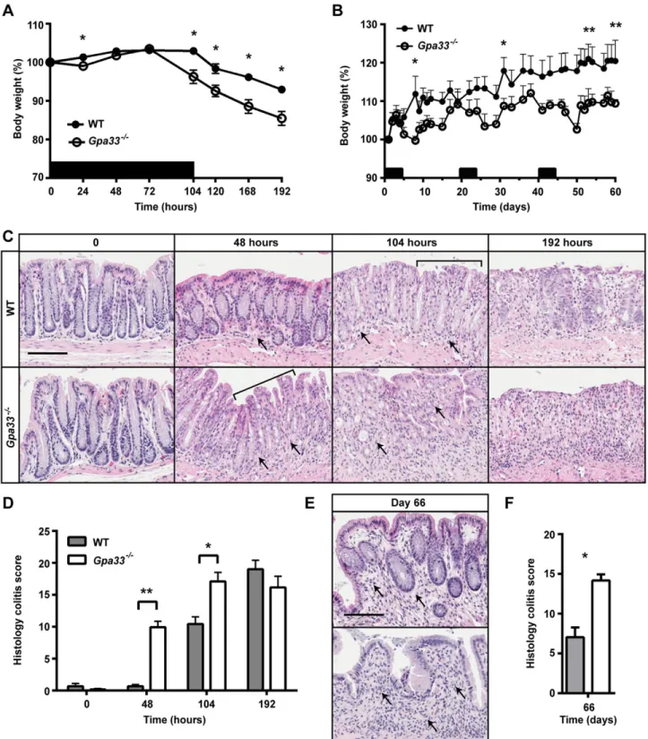
To investigate the effect of GPA33 deficiency on susceptibility to colitis induction, we treated Gpa33−/−mice with 2% DSS. Two different DSS treatment regimens were employed to induce acute or chronic colitis. In the acute colitis model, mice were exposed to 2% DSS in the drinking water ad libitum for 104 h and then switched to
normal drinking water for a further 88 h (192 h in total). Gpa33−/− mice exhibited a more rapid onset of colitis compared to WT mice, shown by increased weight loss (Fig. 3A), and histological damage (Fig. 3C,D). Maximal colitis in Gpa33−/− mice was observed at 104 h, whereas the colitis score significantly increased between 104 h and 192 h (P<0.01) in WT mice (Fig. 3C,D), demonstrating accelerated onset and progression of DSS-induced colitis in Gpa33-deficient animals. At the 192 h time point, both genotypes exhibited similar colitis severity, suggesting that Gpa33 deficiency mostly affects colitis onset (Fig. 3C,D). In the chronic colitis model, mice were exposed to three cycles of 2% DSS in the drinking water for 5 days followed by normal drinking water for 16 days. Gpa33−/− mice also exhibited increased susceptibility to chronic colitis as shown by more exaggerated weight loss following each cycle of Fig. 1.Gpa33−/−mice do not exhibit steady-state gastrointestinal pathology. (A) Representative histological images of small intestine and colon from 10-week-old Gpa33−/−and WT mice on a pure C57Bl/6 background. H&E stains all epithelial cells, Periodic acid-Schiff stains the mucins in goblet cells pink (arrows), and phloxine and tartrazine highlights the granules in Paneth cells (arrows). (B) No difference in proliferation is observed in colonic crypts of Gpa33−/− and WT mice. Representative BrdU immunohistochemistry in the colon of 10-week-old Gpa33−/−and WT mice collected 2 h after intraperitoneal BrdU injection (100 mg/kg). (C) Quantitation of BrdU-positive cells expressed as a fraction of total cells per crypt. A minimum of ten crypts were evaluated per mouse. (D) Both Gpa33−/−and WT mice exhibit mild progressive colitis with age. Representative images of H&E-stained distal colon sections from mice aged 3, 6 and 12 months. Arrows indicate sub-mucosal thickening and immune cell infiltration, characteristic of colitis. (E) Quantitation of colitis using the scheme shown in supplementary material Table S1. Mean±s.e.m., n=3. Scale bars: 50 µm.
Disease
Models
&
DSS (Fig. 3B) and impaired crypt regeneration and resolution of colonic inflammation compared to WT (Fig. 3E,F). Together, these observations demonstrate rapid onset of acute colitis and delayed resolution of chronic colitis in Gpa33−/−mice.
Gpa33−/−mice are susceptible to colitis-associated cancer
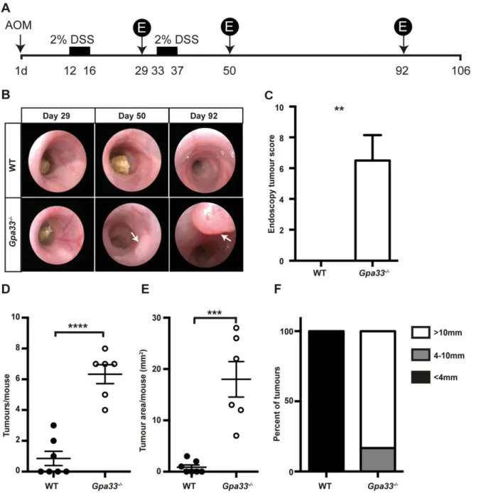
(CAC)
Chronic colitis is a susceptibility factor for CRC (Eaden et al., 2001; Kim and Chang, 2014). To test whether the rapid induction and impaired resolution of colitis in Gpa33−/−mice leads to increased development of CAC, we employed the azoxymethane (AOM)/DSS model (Neufert et al., 2007; Wirtz et al., 2007). Mice were injected with the colon-specific mutagen AOM and subjected to two cycles of 2% DSS in drinking water to induce chronic colitis (Fig. 4A). Colon tumour burden was markedly elevated in Gpa33−/− mice
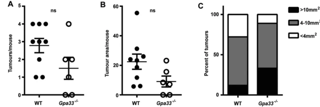
compared to WT mice, as shown by endoscopy of the most distal part of the colon in live animals (Fig. 4B,C) and by quantitating the number and area of the tumours at the end of the experiment (Fig. 4D-F). Both WT and Gpa33−/−mice exhibited activation of the WNT pathway, shown by elevated β-catenin protein (supplementary material Fig. S4A) and upregulated expression of the WNT target genes, CD44 and Lgr5, in tumours compared to normal epithelium (supplementary material Fig. S4B), consistent with CTNNB (encodingβ-catenin) being a known AOM target.
Gpa33−/−mice are not predisposed to sporadic CRC
To investigate the requirement for inflammation in the susceptibility of Gpa33−/−mice to CAC, a model of sporadic CRC was employed. Mice were injected weekly with AOM alone (10 mg/kg) for 6 weeks and aged until 18 weeks to allow outgrowth of induced lesions. In Fig. 2.Gpa33−/−mice are sensitised to impaired intestinal barrier function. (A) Mice were provided with 2% DSS in the drinking water ad libitum for 16 h following which TRITC-dextran was administered by gavage. Four hours later, the TRITC signal in the serum was quantitated as a measure of intestinal permeability. DSS-treated Gpa33−/−but not WT mice exhibit an increase in serum TRITC-dextran, which is indicative of impaired intestinal barrier function. (B) Representative images of H&E-stained distal and mid colon sections from mice treated with 2% DSS in the drinking water for 16 h, illustrating morphological integrity of the epithelial layer after DSS treatment. The right-hand column provides higher magnifications of the boxed areas. (C) Representative histological images of mid colon from 10-week-old Gpa33−/−and WT mice stained with alcian blue and Periodic acid-Schiff for neutral (magenta) and acidic (blue) mucins, respectively, in the presence and absence of DSS treatment. Scale bars: (B) 100 µm and (C) 50 µm. (D) Single-cell suspensions of colon LP were prepared and the numbers of DCs and macrophages were determined by FACS. The gating strategy for activated CD103+DCs derived from viable, single, haematopoietic cells (Epcam−CD45+) is shown. (E) Absolute cell numbers for populations within the whole colon LP were calculated using relative cell populations obtained by FACS and viable single-cell counts. Gpa33−/−mice exhibit an increase in activated DCs within the colonic LP relative to WT controls, indicating elevated
immunosurveillance. Populations represent: DCs (Epcam−CD45+CD11c+F4/80−MHC-II+), CD86+activated DCs, gut resident CD103+DCs and macrophages (Epcam−CD45+CD11c+F4/80+). (F) Geometric mean for CD86 in DCs and CD130+DC populations. Mean±s.e.m., (A) n=7-8, (E,F) n=6, *P<0.05.
Disease
Models
&
Fig. 3.Gpa33−/−mice are more susceptible to DSS-induced colitis. (A,B) Black bars indicate cycles of 2% DSS treatment provided ad libitum in drinking water. (A) Percentage body weight changes during acute colitis reveal that Gpa33−/−mice lose significantly more weight than WT mice in response to treatment with 2% DSS (asterisks) for 104 h. (B) Percentage body weight changes during chronic colitis reveal that Gpa33−/−mice lose significantly more weight than WT mice after each cycle of DSS treatment, indicated by asterisks on days 8, 31, 50 and 61. Gpa33−/−mice also exhibited significantly reduced percentage body weight on days 10, 12, 47, 51, 52 and 54 (P<0.05). (C) Representative images of H&E-stained distal colon sections from mice that were exposed to acute DSS treatment for 0, 48 and 104 h and after an additional 88 h of normal water (192 h time-point). Crypt degradation is evident in Gpa33−/−mice at 48 h but not in WT mice until 104 h (brackets). Arrows indicate immune cell infiltration and thickening of the LP, which is first conspicuous at 48 h in Gpa33−/−mice but not until 104 h in WT controls. (D) Histology colitis scores at 0, 48, 104 and 192 h show accelerated colitis onset in Gpa33−/−mice. Also, the histology colitis score is significantly higher at 192 h than at 104 h in WT mice (P<0.01). (E) Representative images of H&E-stained distal colon sections from mice (day 66) that underwent chronic DSS treatment. Arrows indicate immune cell infiltration and thickening of the LP, which is most pronounced in the Gpa33−/−mice. (F) Histology colitis score of mice at day 66 following chronic colitis regimen. Mean±s.e.m. for A,B,D,F; n=5 (A), n=5-8 (B), n=3 (D), n=4 (F); *P<0.05, **P<0.01. Scale bars: 50 µm.
Disease
Models
&
this model, the absence of DSS removes the major inflammatory driver of tumorigenesis present in the AOM/DSS CAC model. Gpa33−/− mice did not exhibit an increase in sporadic CRC incidence (Fig. 5A) or tumour size (Fig. 5B,C) compared to WT controls. This finding indicates that inflammation is required for the increased susceptibility of Gpa33−/−mice to colonic tumorigenesis.
Gpa33−/−mice exhibit increased sensitivity to food antigen
A clinical consequence of increased IP is the loss of tolerance to food antigens, with increasing severity of hypersensitivity correlated with diminishing barrier function (Ventura et al.,
2006). To examine delayed-type hypersensitivity (DTH) to oral ovalbumin (OVA) in Gpa33−/− and WT controls, mice were gavaged with OVA to induce tolerance, injected intraperitoneally 7 days later with OVA and Freund’s complete adjuvant to boost any primed immune response, and injected with OVA in the ear to elicit local inflammation and changes in ear thickness (Wang et al., 2007). Ear thickness was measured with a calliper at 0, 24 and 48 h after intra-ear injection of OVA. In the positive control cohorts, which were gavaged with PBS alone, or OVA in combination with cholera toxin (OVA+CT) to break tolerance to OVA, topical injection of OVA caused an increase in ear thickness 24 and 48 h later. The Fig. 4.Gpa33−/−mice are more susceptible to colitis-associated cancer (CAC). (A) Schematic representation of the regimen used to induce CAC, whereby a single AOM injection (10 mg/kg body weight) is followed by two cycles of 2% DSS provided ad libitum in the drinking water. Tumour development was monitored by endoscopy at the indicated times (days) post-AOM treatment (denoted by‘E’ in the schematic). (B) Representative endoscopy images indicating the presence of tumours (arrows) in the distal portion (final 2.5 cm) of the colon in Gpa33−/−mice at day 50 and day 92 post-AOM injection. (C) Endoscopy tumour score at day 92 post-AOM administration indicates that Gpa33−/−mice exhibit increased incidence of CAC. (D,E) At collection (106 days post-AOM injection), tumour number (D) and total tumour area (E) per mouse are elevated in Gpa33−/−mice compared to WT mice. (F) The mean area of tumours in WT mice is less than 4 mm2, whereas in Gpa33−/−mice a large proportion (>80%) of tumours are greater than 10 mm2in area. Mean±s.e.m.; n=6-7 (C-F);
**P<0.005, ***P<0.0005, ****P<0.0001.
Disease
Models
&
development of DTH and broken oral tolerance in these mice occurred irrespective of genotype, as expected (Fig. 6). For WT mice that had been gavaged with OVA alone, the mean ear thickness was significantly less than that of the PBS and OVA+CT controls, indicating that oral tolerance had been established. In contrast, the mean ear thickness in Gpa33−/−mice that had been gavaged with OVA alone was not significantly different from that of Gpa33−/− mice that had been gavaged with PBS, or OVA+CT, indicating systemic hypersensitivity to OVA and broken oral tolerance (Fig. 6).
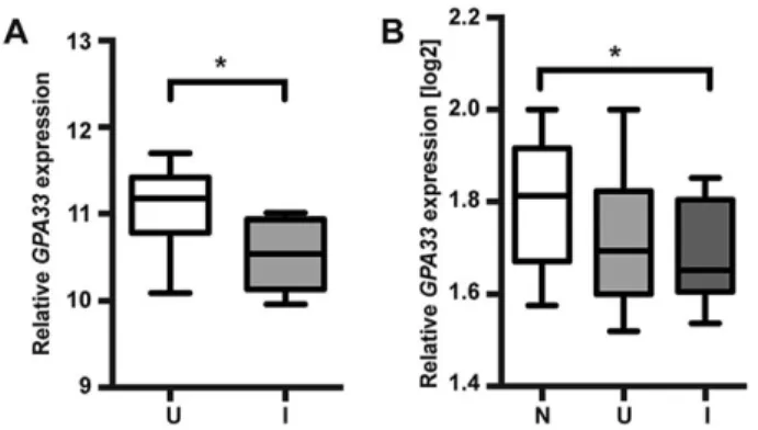
GPA33 expression is reduced in the inflamed bowel of individuals with IBD
In order to investigate the potential role of GPA33 deficiency in human IBD, GPA33 expression was analysed in two published human IBD microarray datasets (Noble et al., 2008; Olsen et al., 2009). In individuals with either Crohn’s disease (CD) or ulcerative colitis (UC), mean GPA33 expression was reduced in inflamed bowel compared to uninflamed and normal control tissue, respectively (Fig. 7). To address the possibility that the reduction in the mean level of GPA33 expression is related to the epithelial cell content of the inflamed samples, we also analysed the expression of EPCAM, an epithelial marker that is also highly expressed in intestinal epithelium. We found that EPCAM was not differentially expressed between uninflamed and inflamed tissue in both the CD
and UC datasets (supplementary material Fig. S5A,B). We also analysed the expression of FAP (encoding fibroblast activation protein, expressed in activated stromal fibroblasts of epithelial cancers and healing wounds) and found that its expression was unchanged between uninflamed and inflamed tissues from individuals with CD and elevated in the inflamed tissues of individuals with UC (supplementary material Fig. S5C,D). Analysis of the expression of an additional epithelial marker (CDH1, encoding E-cadherin) and another stromal marker (VIM, encoding vimentin) reiterated the EPCAM and FAP results, respectively (not shown). Collectively, these data suggest that the observed reduction in mean GPA33 expression in the inflamed tissue of individuals with CD or UC is not simply due to reduced epithelial cell content of the samples. These data suggest that GPA33 deficiency correlates with intestinal inflammation in IBD and that it might contribute to IP in this context.
DISCUSSION
Gpa33−/−mice on a pure C57BL/6 background are healthy and do not develop spontaneous colitis or any other gastrointestinal pathology up to 1 year of age. Strikingly, however, Gpa33−/− mice are more susceptible than WT to DSS-induced loss of intestinal barrier function and severe colitis and, when DSS treatment is coupled with prior administration of the mutagen Fig. 5.Gpa33−/−mice are not prone to sporadic CRC. Mice were injected with AOM (10 mg/kg body weight) six times at weekly intervals to induce sporadic CRC. (A) Tumour number and (B) tumour area per mouse colon at collection was similar in Gpa33−/−and WT mice. (C) Size distribution of sporadic CRC tumours across all Gpa33−/−and WT mice at the end of the experiment. Mean±s.e.m.; n=6-9.
Fig. 6.Gpa33−/−mice lack tolerance to oral antigen. Gpa33−/−mice exhibit lack of oral tolerance to OVA administered by gavage, with resultant systemic delayed-type hypersensitivity (DTH) response manifested as increased ear thickness at the topical OVA injection site. Administration by gavage of PBS, or OVA in combination with cholera toxin (OVA+CT), make up the positive controls for lack of oral tolerance. Whereas WT mice gavaged with OVA alone exhibit oral tolerance compared to WT mice treated with PBS and OVA+CT (mean±s.d., n=6-7, **P<0.01), Gpa33−/−mice gavaged with OVA exhibit increased ear thickness comparable to positive controls, indicative of lack of oral tolerance and systemic DTH. Compared to baseline levels of ear thickness at the time of local OVA injection (0 h), the percentage increases in ear thickness of Gpa33−/−mice at 24 h and 48 h are 33% (P<0.0001) and 41% (P<0.0001), respectively.
Disease
Models
&
AOM, they develop significantly more and larger colon tumours. Further evidence that GPA33 plays a role in barrier function is the observation that Gpa33−/−mice fail to establish tolerance to the oral antigen OVA and develop DTH.
To investigate the contribution of GPA33 to intestinal barrier integrity, we assessed immunosurveillance in the colon and paracellular penetration of 4 kDa dextran in WT and Gpa33−/− mice. Although leakiness to dextran was not significantly elevated in Gpa33−/−mice under steady-state conditions, Gpa33−/− mice exhibited a twofold increase in activated CD103+DCs, the major
class of antigen-presenting cells required for induction both of tolerance to benign antigen and inflammation to clear pathogens (Scott et al., 2011). This observation suggests that Gpa33−/−mice exhibit a mild increase in luminal antigen flux into the colonic LP. To analyse the pathological consequences of increased IP and inflammation on Gpa33−/−mice, we exploited several DSS models of colitis (Wirtz et al., 2007; Wirtz and Neurath, 2007). Short-term DSS treatment (2% DSS for 16 h) reduces mucus protection of the most luminal epithelial cells, and allows penetration of bacteria closer to the epithelium (Johansson et al., 2010). Although this did not affect epithelial cell integrity or induce paracellular permeability in WT mice, which is consistent with previous findings (Kitajima et al., 1999), Gpa33−/−mice exhibited dextran permeability upon mucus layer degradation, indicating an underlying defect in cell-cell adhesion. Similar findings have been reported for mice individually deficient for CAR and Occludin, indicating that a number of junctional proteins might play non-redundant roles in the steady-state maintenance of TJs and barrier function (Pazirandeh et al., 2011; Saitou et al., 2000).
Using a harsher regime of DSS treatment (2% DSS in drinking water for 104 h), Gpa33−/−mice exhibited a rapid onset of acute colitis, indicative of luminal antigen penetration into the LP, most likely caused by TJ impairment and enhanced paracellular permeability. Similar observations have been reported for DSS-treated Jam-A-knockout mice, demonstrating a direct link between TJ dysfunction and severe colitis (Laukoetter et al., 2007; Vetrano et al., 2008). When exposed to repeated DSS cycles to induce chronic colitis akin to the flares of inflammation in IBD, Gpa33−/−
mice were less able to resolve multiple bursts of epithelial damage and inflammation than WT controls. Considered in conjunction with the reduced GPA33 expression observed in the inflamed bowel of humans with either CD or UC, this suggests a potential role for GPA33 impairment in the progression of IBD.
Individuals with IBD have an increased risk of developing CRC (Eaden et al., 2001; Itzkowitz and Yio, 2004; Kim and Kim, 2014). This fundamental role for inflammation in CRC is modelled using AOM/DSS treatment of mice to induce CAC (Neufert et al., 2007). Strikingly, Gpa33−/−mice develop increased numbers of and larger tumours than WT control mice, suggesting that loss of GPA33 leads to both an increase in the incidence of tumour formation and the rate of tumour growth. These observations are consistent with increased inflammation and cytokine production stimulating outgrowth of AOM-mutated tumour-initiating cells in Gpa33−/− mice (Wirtz et al., 2007). Meanwhile, induction of sporadic CRC in Gpa33−/− mice by injection with AOM alone did not result in elevated tumorigenesis, confirming the essential requirement for inflammation and the underlying loss of permeability as the fundamental defect.
The pathological consequences of IP are not restricted to IBD and localised inflammation in the intestinal tract, but can also lead to loss of oral tolerance and systemic inflammatory effects. A leaky epithelium can allow luminal contents including food antigens to bypass normal antigen processing and initiate an inflammatory response in the LP with consequent systemic sensitisation to that food antigen (Wirtz and Neurath, 2007). Indeed, food hypersensitivity can precede IBD onset and is a recognised co-morbidity (Kraus et al., 2004). Using an established OVA model of oral DTH (Wang et al., 2007) we observed an increased response in Gpa33−/− mice, providing further evidence for IP in GPA33-deficient contexts.
In addition to the results presented here, and the demonstration that other CTX family members, such as JAM-A, play a role in cell-cell adhesion, in vitro experiments have also suggested an epithelial cell-intrinsic requirement for GPA33 in barrier formation and junctional complex integrity (Ackerman et al., 2008). Further work will establish the precise mechanism by which GPA33 acts to maintain barrier function and protect against disease, although the cumulative evidence to date points to a role in cell-cell adhesion.
The highly restricted expression of GPA33 to the intestinal epithelium and CRC is a hallmark that has sustained the interest in GPA33 as a target for antibody-based cancer therapy. Interestingly, however, Sakamoto et al. (2000) also detected weak expression of GPA33 in murine salivary glands. This observation, coupled with two studies regarding Sjögren’s syndrome (SS; Ewert et al., 2010; Nguyen et al., 2008), suggests a possible extra-intestinal role for GPA33 in barrier function. SS is an autoimmune disorder in which the salivary and lacrimal glands are destroyed by infiltrating lymphocytes, leading to symptomatic dry mouth and eyes in affected individuals. Although the aetiology of this disorder is thought to be polygenic, a recent study implicated TJ disruption and resultant pro-inflammatory cytokine exposure within the salivary glands as a key pathogenic factor (Ewert et al., 2010). Interestingly, investigation of SS using inbred mouse models identified Gpa33 as one of a handful of genes deleted in two SS susceptibility loci (Nguyen et al., 2008). This tantalising observation suggests that GPA33 might play a role in barrier function outside the intestine.
A number of genetically modified mouse models with deranged junctional complexes and barrier dysfunction have been reported, for which a subset develop spontaneous or inducible inflammatory Fig. 7.GPA33 expression is reduced in the inflamed bowel of individuals
with IBD. Relative GPA33 expression in normal (N), uninflamed (U) and inflamed (I) bowel of individuals with IBD represented as whisker and box plots; *P<0.05. (A) GPA33 expression is elevated in inflamed compared to uninflamed bowel from individuals with CD (uninflamed n=18, inflamed n=8; dataset GDS3119) (Olsen et al., 2009). (B) GPA33 expression is elevated in inflamed bowel from individuals with UC compared to normal bowel (normal n=22, uninflamed n=15, inflamed n=19; dataset GDS3268) (Noble et al., 2008). Relative gene expression values were normalised using (A) Human Genome U133 Plus 2.0 Array Normalisation Controls (Affymetrix) and (B) Stratagene Universal Human Reference (Stratagene).
Disease
Models
&
and Eliza Hall Institute of Medical Research and the University of Sydney (Australia). All mice were maintained on a pure C57BL/6 genetic background except those used to assess oral tolerance. Cohorts for the oral tolerance experiments were maintained on a mixed 129/SvJ, C57BL/6 background with littermate controls used at all times. Mice were bred and housed in the same room in a specific pathogen-free facility at the Ludwig Institute of Cancer Research to minimise variation in gut microbiota. All mice were 8 to 12 weeks old at the commencement of treatment, or when analysed, unless otherwise indicated.
Generation ofGpa33−/−mice
We generated embryonic stem (ES) cells (129Sv/J background) containing a null Gpa33 allele by replacing 20 kb of endogenous Gpa33 sequence from the 5′ end of exon 2 to the 5′ end of exon 7 with a promoter-less targeting vector. The targeting vector contained a translational stop codon (TGA) at codon 35 of exon 2 to terminate translation of an encoded truncated (13 amino acid) GPA33 protein. This was followed by an internal ribosome entry site (IRES) to permit translation of a β-geo cassette encoding a β-galactosidase–neomycin fusion protein (supplementary material Fig. S1A) to allow selection of targeted neomycin-resistant ES cells and visualisation of Gpa33 promoter activity in vivo via blue staining following X-gal metabolism. A congenic Gpa33−/−C57BL/6 strain was generated by backcrossing the Gpa33−/−129Sv/J×C57BL/6 founder mice to C57BL/6 mice for a minimum of ten generations.
RNA analysis
RNA was extracted from purified epithelial cell fractions harvested from freshly dissected small and large intestines (Whitehead et al., 1987) and dissected tumour tissue using TRIzol (Life Technologies, Carlsbad, CA, USA). RNA integrity was analysed on a 2100 Bioanalyzer (Agilent, Santa Clara, CA, USA) and cDNA generated using the High Fidelity cDNA synthesis kit (Applied Biosystems, Waltham, MA, USA). Quantitative RT-PCR was performed using SYBR green (Quantace, London, UK) on an ABI 7300 real-time PCR machine using primers designed to amplify Gpa33, Lgr5, CD44 and Hprt1 (sequences available on request). Expression data was normalised to Hprt1 expression.
In vivo assessment of intestinal permeability
Mice were exposed to either normal drinking water or DSS (2% w/v, MW 35-50 kDa; MP Biomedicals, Santa Ana, CA, USA) in drinking water ad libitum for 16 h prior to oral gavage with 200 µl of TRITC-dextran (40 mg/ml TRITC-dextran 4 kDa in PBS; Sigma-Aldrich, St Louis, MO, USA). Circulating TRITC signal was quantitated by fluorometry on serum collected 4 h post-gavage, as described previously (Brandl et al., 2009).
Fluorescence-activated cell sorting (FACS) analysis
Analyses of haematopoietic populations in the colonic LP and spleen were performed by FACS on single-cell suspensions. Colon LP was prepared by incubating washed, finely chopped colon in 20 ml dissociation solution comprising FCS (2.5%; Life Technologies, Carlsbad, CA, USA), Hanks’ Balanced Salt Solution (HBSS) without Ca++ and Mg++ (Life Technologies, Carlsbad, CA, USA), EDTA (5 mM; BDH Chemicals, Radnor, PA, USA) and DTT (1 mM; Promega, Madison, WI, USA) for
of Medical Research, Biotechnology Centre, Melbourne, Victoria, Australia), CD103-PE (557495; BD Pharmingen), CD86-PE-Cy7 (A15412; Life Technologies, Carlsbad, CA, USA) and lymphoid cell populations: Epcam-FITC (11-5791; eBioscience), CD45-BV421 (563890; BD Pharmingen), CD11b-PE (553311; BD Pharmingen), TCRβ-PerCP (557019; BD Pharmingen), B220-biotin (553086; BD Pharmingen), Streptavadin-BV650 (405231; BioLegend, San Diego, CA, USA), CD4-APC (GK1.5; Walter and Eliza Hall Institute of Medical Research, Biotechnology Centre) and CD8-PE-Cy7 (552877; BD Pharmingen). Acquisition was performed on a Fortessa X20 (BD Biosciences, San Jose, CA, USA) with viability assessed using propidium iodide (Sigma-Aldrich, St Louis, MO, USA) and data analysed using FlowJo software (FlowJo, Ashland, OR, USA). Absolute cell numbers for haematopoietic populations in colonic LP or spleen were calculated using whole organ, single, viable cell counts and the derived relative populations obtained by FACS.
Induction of acute and chronic colitis
One cycle of colitis was induced by ad libitum exposure to DSS (2%; Sigma-Aldrich, St Louis, MO, USA) in drinking water for 104 h, followed by normal drinking water. In the acute colitis experiment, mice were euthanised after 0, 48 and 104 h of DSS treatment and after a further 88 h of normal water (192 h time-point). To induce chronic colitis, mice were treated with three cycles of 2% DSS interspersed with normal water for 16 days. Mice in both models were weighed daily to observe weight loss consequent to colitis. For histological analyses, the colons were opened lengthwise from anus to caecum, rolled and fixed in 10% neutral buffered formalin (Pathtech, Melbourne, Victoria, Australia).
Colitis-associated and sporadic colon cancer
CAC was induced through a single intraperitoneal injection of AOM (10 mg/kg; Sigma-Aldrich, St Louis, MO, USA) in PBS, followed 12 days later by two cycles of DSS treatment. Both cycles comprised ad libitum exposure to 2% DSS for 5 days in drinking water, interspersed with 16 days of normal drinking water. To mimic sporadic CRC, mice received weekly intraperitoneal injections of AOM (10 mg/kg) for 6 weeks. Tumour burden in the colon was monitored using a Coloview high-resolution mouse video endoscopic system (Karl Storz GmbH & Co, Tuttlingen, Germany) as described previously (Becker et al., 2006; Ernst et al., 2015). Tumours were counted and measured in two dimensions with callipers to calculate the area. Prior to fixation, samples of tumour and adjacent normal epithelium were collected for molecular analysis.
Histological techniques
Observation of intestinal lineages was performed by staining histological sections (4 µm) with haematoxylin and eosin (H&E; all epithelial cells), phloxine and tartrazine (Paneth cells) and Alcian blue Periodic acid-Schiff stain (goblet cells). To detect cells in the S phase of the cell cycle, mice received an intraperitoneal injection of bromodeoxyuridine (BrdU; 100 mg/kg; Sigma-Aldrich, St Louis, MO, USA) 2 h prior to collection of tissues. Visualisation of apoptosis was performed using the Apop-Tag Peroxidase In Situ Apoptosis Detection Kit (Merck Millipore, Billerica, MA, USA) according to the manufacturer’s instructions. Antibodies used to assess cell proliferation (BrdU; BD555624; Life Technologies, Carlsbad,
Disease
Models
&
CA, USA), Wnt signalling activation (β-catenin; BD610154; BD Biosciences, San Jose, CA, USA) and GPA33 expression (rabbit polyclonal antibody raised against amino acids 302-319 of mouse GPA33) (Johnstone et al., 2000) were incubated with histological sections and processed as follows. Antigen retrieval was performed by heating to 100°C in 10 mM pH 6 citrate buffer (BDH, Radnor, PA, USA), endogenous peroxidase activity was inhibited with 3% H2O2(v/v; BioLab, Lawrenceville, GA, USA) and non-specific antibody
binding was blocked using 2% bovine serum albumin (w/v; Sigma-Aldrich, St Louis, MO, USA). Primary antibody binding was followed by incubation with a species-appropriate HRP-conjugated secondary antibody (Dako, Carpinteria, CA, USA), and the signal detected with Liquid Diaminobenzidine substrate Chromogen System (Dako, Carpinteria, CA, USA) prior to counterstaining with haematoxylin. Histological assessment of colitis was performed in a blinded manner using a system comprising crypt loss and inflammation in the LP and submucosa (supplementary material Table S1) (Wirtz et al., 2007).
Oral tolerance to ovalbumin
On day 0 mice were anaesthetised by intraperitoneal injection with Avertin (0.5 mg/g; Sigma-Aldrich, St Louis, MO, USA) and gavaged with 200 µl sodium bicarbonate (0.75%; BDH Chemicals, Radnor, PA, USA) 15 min prior to immunisation. WT and Gpa33−/−mice were then gavaged with 200 µl PBS, or 200 µl ovalbumin (OVA) (100 mg/ml, grade III, Sigma-Aldrich, St Louis, MO, USA) in PBS, or 200 µl OVA in combination with cholera toxin B (50 µg/ml; Calbiochem, Billerica, MA, USA) in PBS. On day 7 all mice were injected subcutaneously with a mixture containing 50 µl OVA (1 mg/ml, grade V, Sigma-Aldrich, St Louis, MO, USA) in PBS and 50 µl Freund’s Complete Adjuvant (Sigma-Aldrich, St Louis, MO, USA). On day 28, 10 µl of OVA (1 mg/ml, grade V) in PBS was injected subcutaneously into the ear of all mice. Ear thickness was immediately measured with a calliper by a blinded observer (t=0 h) and then 24 and 48 h later.
Statistical data analysis
Data are expressed as mean±s.e.m. unless indicated otherwise. P-values were calculated using a two-tailed, unpaired t-test with *P<0.05 considered significant and **P<0.01.
Acknowledgements
The authors thank Andrew Naughton, Melanie Asquith, Anthony Gomes, Krystal Dredge and Cara Mansfield (mouse husbandry), Cary Tsui (histology), Adele Preaudet and Ryan Farid (mouse endoscopy), and John Mariadason (discussion and critical review of the manuscript).
Competing interests
The authors declare no competing or financial interests.
Author contributions
B.B.W., N.C.T., M.B., S.B., F.H., M.E. and J.K.H. conceived and designed the experiments. B.B.W., N.C.T., M.B., T.L.P., S.B., C.N.J. and F.M. performed the experiments. B.B.W., N.C.T., M.B., T.L.P., K.D., S.B., F.M., A.W.B., A.M.S., M.E. and J.K.H. analysed the data. B.B.W. and J.K.H. wrote the paper.
Funding
This research was supported by Program grant 487922 (A.W.B., A.M.S., M.E. and J.K.H.), Senior Research Fellowships 6031222 and 1022870 (M.E. and J.K.H., respectively) and a Medical Postgraduate Award (N.C.T.) from the National Health and Medical Research Council (NHMRC) of Australia. This work was also funded by Ludwig Cancer Research and operational infrastructure grants from the Australian Federal Government (IRISS) and the Victorian State Government (OIS).
Supplementary material
Supplementary material available online at
http://dmm.biologists.org/lookup/suppl/doi:10.1242/dmm.019935/-/DC1
References
Ackerman, M. E., Chalouni, C., Schmidt, M. M., Raman, V. V., Ritter, G., Old, L. J., Mellman, I. and Wittrup, K. D. (2008). A33 antigen displays persistent surface expression. Cancer Immunol. Immunother. 57, 1017-1027.
Becker, C., Dornhoff, H., Neufert, C., Fantini, M. C., Wirtz, S., Huebner, S., Nikolaev, A., Lehr, H.-A., Murphy, A. J., Valenzuela, D. M. et al. (2006). Cutting
edge: IL-23 cross-regulates IL-12 production in T cell-dependent experimental colitis. J. Immunol. 177, 2760-2764.
Brandl, K., Rutschmann, S., Li, X., Du, X., Xiao, N., Schnabl, B., Brenner, D. A. and Beutler, B. (2009). Enhanced sensitivity to DSS colitis caused by a hypomorphic Mbtps1 mutation disrupting the ATF6-driven unfolded protein response. Proc. Natl. Acad. Sci. USA 106, 3300-3305.
Chrétien, I., Marcuz, A., Courtet, M., Katevuo, K., Vainio, O., Heath, J. K., White, S. J. and Du Pasquier, L. (1998). CTX, a Xenopus thymocyte receptor, defines a molecular family conserved throughout vertebrates. Eur. J. Immunol. 28, 4094-4104.
Ciprotti, M., Chong, G., Gan, H. K., Chan, A., Murone, C., MacGregor, D., Lee, F.-T., Johns, T. G., Heath, J. K., Ernst, M. et al. (2014). Quantitative intratumoural microdistribution and kinetics of (131)I-huA33 antibody in patients with colorectal carcinoma. EJNMMI Res. 4, 22.
Eaden, J. A., Abrams, K. R. and Mayberry, J. F. (2001). The risk of colorectal cancer in ulcerative colitis: a meta-analysis. Gut 48, 526-535.
Ernst, M., Preaudet, A. and Putoczki, T. (2015). Non-invasive assessment of the efficacy of new therapeutics for intestinal pathologies using serial endoscopic imaging of live mice. J. Vis. Exp., e52383.
Ewert, P., Aguilera, S., Alliende, C., Kwon, Y.-J., Albornoz, A., Molina, C., Urzúa, U., Quest, A. F. G., Olea, N., Pérez, P. et al. (2010). Disruption of tight junction structure in salivary glands from Sjö gren’s syndrome patients is linked to proinflammatory cytokine exposure. Arthritis Rheum. 62, 1280-1289.
Fries, W., Belvedere, A. and Vetrano, S. (2013). Sealing the broken barrier in IBD: intestinal permeability, epithelial cells and junctions. Curr. Drug Targets 14, 1460-1470.
Garinchesa, P., Sakamoto, J., Welt, S., Real, F., Rettig, W. and Old, L. (1996). Organ-specific expression of the colon cancer antigen A33, a cell surface target for antibody-based therapy. Int. J. Oncol. 9, 465-471.
Heath, J. K., White, S. J., Johnstone, C. N., Catimel, B., Simpson, R. J., Moritz, R. L., Tu, G.-F., Ji, H., Whitehead, R. H., Groenen, L. C. et al. (1997). The human A33 antigen is a transmembrane glycoprotein and a novel member of the immunoglobulin superfamily. Proc. Natl. Acad. Sci. USA 94, 469-474. Heller, F., Florian, P., Bojarski, C., Richter, J., Christ, M., Hillenbrand, B.,
Mankertz, J., Gitter, A. H., Bü rgel, N., Fromm, M. et al. (2005). Interleukin-13 is the key effector Th2 cytokine in ulcerative colitis that affects epithelial tight junctions, apoptosis, and cell restitution. Gastroenterology 129, 550-564. Hermiston, M. L. and Gordon, J. I. (1995). Inflammatory bowel disease and
adenomas in mice expressing a dominant negative N-cadherin. Science 270, 1203-1207.
Infante, J. R., Bendell, J. C., Goff, L. W., Jones, S. F., Chan, E., Sudo, T., Burris, H. A. and Berlin, J. D. (2013). Safety, pharmacokinetics and pharmacodynamics of the anti-A33 fully-human monoclonal antibody, KRN330, in patients with advanced colorectal cancer. Eur. J. Cancer 49, 1169-1175.
Itzkowitz, S. H. and Yio, X. (2004). Inflammation and cancer IV. Colorectal cancer in inflammatory bowel disease: the role of inflammation. Am. J. Physiol. Gastrointest. Liver Physiol. 287, G7-G17.
Johansson, M. E. V., Gustafsson, J. K., Sjö berg, K. E., Petersson, J., Holm, L., Sjö vall, H. and Hansson, G. C. (2010). Bacteria penetrate the inner mucus layer before inflammation in the dextran sulfate colitis model. PLoS ONE 5, e12238. Johnstone, C. N., Tebbutt, N. C., Abud, H. E., White, S. J., Stenvers, K. L., Hall,
N. E., Cody, S. H., Whitehead, R. H., Catimel, B., Nice, E. C. et al. (2000). Characterization of mouse A33 antigen, a definitive marker for basolateral surfaces of intestinal epithelial cells. Am. J. Physiol. Gastrointest. Liver Physiol. 279, G500-G510.
Johnstone, C. N., White, S. J., Tebbutt, N. C., Clay, F. J., Ernst, M., Biggs, W. H., Viars, C. S., Czekay, S., Arden, K. C. and Heath, J. K. (2002). Analysis of the regulation of the A33 antigen gene reveals intestine-specific mechanisms of gene expression. J. Biol. Chem. 277, 34531-34539.
Kim, E. R. and Chang, D. K. (2014). Colorectal cancer in inflammatory bowel disease: the risk, pathogenesis, prevention and diagnosis. World J. Gastroenterol. 20, 9872-9881.
Kim, E. R. and Kim, Y.-H. (2014). Clinical application of genetics in management of colorectal cancer. Intest. Res. 12, 184-193.
Kitajima, S., Takuma, S. and Morimoto, M. (1999). Changes in colonic mucosal permeability in mouse colitis induced with dextran sulfate sodium. Exp. Anim. 48, 137-143.
Kraus, T. A., Toy, L., Chan, L., Childs, J. and Mayer, L. (2004). Failure to induce oral tolerance to a soluble protein in patients with inflammatory bowel disease. Gastroenterology 126, 1771-1778.
Laukoetter, M. G., Nava, P., Lee, W. Y., Severson, E. A., Capaldo, C. T., Babbin, B. A., Williams, I. R., Koval, M., Peatman, E., Campbell, J. A. et al. (2007). JAM-A regulates permeability and inflammation in the intestine in vivo. J. Exp. Med. 204, 3067-3076.
Mallegol, J., van Niel, G. and Heyman, M. (2005). Phenotypic and functional characterization of intestinal epithelial exosomes. Blood Cells Mol. Dis. 35, 11-16. Mallegol, J., Van Niel, G., Lebreton, C., Lepelletier, Y., Candalh, C., Dugave, C., Heath, J. K., Raposo, G., Cerf-Bensussan, N. and Heyman, M. (2007). T84-intestinal epithelial exosomes bear MHC class II/peptide complexes potentiating antigen presentation by dendritic cells. Gastroenterology 132, 1866-1876.
Disease
Models
&
exocrinopathy. Arthritis Res. Ther. 10, R137.
Noble, C. L., Abbas, A. R., Cornelius, J., Lees, C. W., Ho, G.-T., Toy, K., Modrusan, Z., Pal, N., Zhong, F., Chalasani, S. et al. (2008). Regional variation in gene expression in the healthy colon is dysregulated in ulcerative colitis. Gut 57, 1398-1405.
Olsen, J., Gerds, T. A., Seidelin, J. B., Csillag, C., Bjerrum, J. T., Troelsen, J. T. and Nielsen, O. H. (2009). Diagnosis of ulcerative colitis before onset of inflammation by multivariate modeling of genome-wide gene expression data. Inflamm. Bowel Dis. 15, 1032-1038.
Pastorelli, L., De Salvo, C., Mercado, J. R., Vecchi, M. and Pizarro, T. T. (2013). Central role of the gut epithelial barrier in the pathogenesis of chronic intestinal inflammation: lessons learned from animal models and human genetics. Front. Immunol. 4, 280.
Pazirandeh, A., Sultana, T., Mirza, M., Rozell, B., Hultenby, K., Wallis, K., Vennströ m, B., Davis, B., Arner, A., Heuchel, R. et al. (2011). Multiple phenotypes in adult mice following inactivation of the Coxsackievirus and Adenovirus Receptor (Car) gene. PLoS ONE 6, e20203.
Pereira-Fantini, P. M., Judd, L. M., Kalantzis, A., Peterson, A., Ernst, M., Heath, J. K. and Giraud, A. S. (2010). A33 antigen-deficient mice have defective colonic mucosal repair. Inflamm. Bowel Dis. 16, 604-612.
Raschperger, E., Thyberg, J., Pettersson, S., Philipson, L., Fuxe, J. and Pettersson, R. F. (2006). The coxsackie- and adenovirus receptor (CAR) is an in vivo marker for epithelial tight junctions, with a potential role in regulating permeability and tissue homeostasis. Exp. Cell Res. 312, 1566-1580. Rudolph, U., Finegold, M. J., Rich, S. S., Harriman, G. R., Srinivasan, Y., Brabet,
P., Bradley, A. and Birnbaumer, L. (1995). Gi2 alpha protein deficiency: a model for inflammatory bowel disease. J. Clin. Immunol. 15, S101-S105.
Saitou, M., Furuse, M., Sasaki, H., Schulzke, J.-D., Fromm, M., Takano, H., Noda, T. and Tsukita, S. (2000). Complex phenotype of mice lacking occludin, a component of tight junction strands. Mol. Biol. Cell 11, 4131-4142.
Sakamoto, J., Kojima, H., Kato, J., Hamashima, H. and Suzuki, H. (2000). Organ-specific expression of the intestinal epithelium-related antigen A33, a cell surface target for antibody-based imaging and treatment in gastrointestinal cancer. Cancer Chemother. Pharmacol. 46 Suppl., S27-S32.
Scott, A. M., Lee, F.-T., Jones, R., Hopkins, W., MacGregor, D., Cebon, J. S., Hannah, A., Chong, G., U, P., Papenfuss, A. et al. (2005). A phase I trial of humanized monoclonal antibody A33 in patients with colorectal carcinoma:
permeability in patients with adverse reactions to food. Dig. Liver Dis. 38, 732-736. Vetrano, S. and Danese, S. (2009). The role of JAM-A in inflammatory bowel
disease: unrevealing the ties that bind. Ann. N. Y. Acad. Sci. 1165, 308-313. Vetrano, S., Rescigno, M., Cera, M. R., Correale, C., Rumio, C., Doni, A., Fantini,
M., Sturm, A., Borroni, E., Repici, A. et al. (2008). Unique role of junctional adhesion molecule-a in maintaining mucosal homeostasis in inflammatory bowel disease. Gastroenterology 135, 173-184.
Wang, Y., Mei, Y., Bao, S. and Xu, L. (2007). Vasoactive intestinal polypeptide enhances oral tolerance by regulating both cellular and humoral immune responses. Clin. Exp. Immunol. 148, 178-187.
Welt, S., Divgi, C. R., Real, F. X., Yeh, S. D., Garin-Chesa, P., Finstad, C. L., Sakamoto, J., Cohen, A., Sigurdson, E. R., Kemeny, N. et al. (1990). Quantitative analysis of antibody localization in human metastatic colon cancer: a phase I study of monoclonal antibody A33. J. Clin. Oncol. 8, 1894-1906. Welt, S., Divgi, C. R., Kemeny, N., Finn, R. D., Scott, A. M., Graham, M., Germain,
J. S., Richards, E. C., Larson, S. M., Oettgen, H. F. et al. (1994). Phase I/II study of iodine 131-labeled monoclonal antibody A33 in patients with advanced colon cancer. J. Clin. Oncol. 12, 1561-1571.
Welt, S., Scott, A. M., Divgi, C. R., Kemeny, N. E., Finn, R. D., Daghighian, F., Germain, J. S., Richards, E. C., Larson, S. M. and Old, L. J. (1996). Phase I/II study of iodine 125-labeled monoclonal antibody A33 in patients with advanced colon cancer. J. Clin. Oncol. 14, 1787-1797.
Welt, S., Ritter, G., Williams, C., Jr, Cohen, L. S., Jungbluth, A., Richards, E. A., Old, L. J. and Kemeny, N. E. (2003). Preliminary report of a phase I study of combination chemotherapy and humanized A33 antibody immunotherapy in patients with advanced colorectal cancer. Clin. Cancer Res. 9, 1347-1353. Whitehead, R. H., Brown, A. and Bhathal, P. S. (1987). A method for the isolation
and culture of human colonic crypts in collagen gels. In Vitro Cell Dev. Biol. 23, 436-442.
Wirtz, S. and Neurath, M. F. (2007). Mouse models of inflammatory bowel disease. Adv. Drug Deliv. Rev.59, 1073-1083.
Wirtz, S., Neufert, C., Weigmann, B. and Neurath, M. F. (2007). Chemically induced mouse models of intestinal inflammation. Nat. Protoc. 2, 541-546. Zeissig, S., Burgel, N., Gunzel, D., Richter, J., Mankertz, J., Wahnschaffe, U.,
Kroesen, A. J., Zeitz, M., Fromm, M. and Schulzke, J.-D. (2007). Changes in expression and distribution of claudin 2, 5 and 8 lead to discontinuous tight junctions and barrier dysfunction in active Crohn’s disease. Gut 56, 61-72.