HAL Id: inserm-00829466
https://www.hal.inserm.fr/inserm-00829466
Submitted on 3 Jun 2013
HAL is a multi-disciplinary open access
archive for the deposit and dissemination of
sci-entific research documents, whether they are
pub-lished or not. The documents may come from
teaching and research institutions in France or
abroad, or from public or private research centers.
L’archive ouverte pluridisciplinaire HAL, est
destinée au dépôt et à la diffusion de documents
scientifiques de niveau recherche, publiés ou non,
émanant des établissements d’enseignement et de
recherche français ou étrangers, des laboratoires
publics ou privés.
Similar early characteristics but variable neurological
outcome of patients with a de novo mutation of KCNQ2.
Mathieu Milh, Nadia Boutry-Kryza, Julie Sutera-Sardo, Cyril Mignot,
Stéphane Auvin, Caroline Lacoste, Nathalie Villeneuve, Agathe Roubertie,
Bénédicte Heron, Maryline Carneiro, et al.
To cite this version:
Mathieu Milh, Nadia Boutry-Kryza, Julie Sutera-Sardo, Cyril Mignot, Stéphane Auvin, et al.. Similar
early characteristics but variable neurological outcome of patients with a de novo mutation of KCNQ2..
Orphanet Journal of Rare Diseases, BioMed Central, 2013, 8 (1), pp.80. �10.1186/1750-1172-8-80�.
�inserm-00829466�
R E S E A R C H
Open Access
Similar early characteristics but variable
neurological outcome of patients with a de novo
mutation of KCNQ2
Mathieu Milh
1,2*, Nadia Boutry-Kryza
3, Julie Sutera-Sardo
1,2, Cyril Mignot
4,5,6, Stéphane Auvin
7, Caroline Lacoste
8,
Nathalie Villeneuve
2, Agathe Roubertie
9,10, Bénédicte Heron
6, Maryline Carneiro
9, Anna Kaminska
11,
Cécilia Altuzarra
12, Gaëlle Blanchard
13, Dorothée Ville
13, Marie Anne Barthez
14, Delphine Heron
4,5, Domitille Gras
7,
Alexandra Afenjar
5,6, Nathalie Dorison
6, Dianne Doummar
6, Thierry Billette de Villemeur
5,6, Isabelle An
15,
Aurélia Jacquette
4, Perrine Charles
4, Julie Perrier
16, Bertrand Isidor
17, Laurent Vercueil
18, Brigitte Chabrol
1,2,
Catherine Badens
1,8,19, Gaétan Lesca
3and Laurent Villard
1,19Abstract
Background: Early onset epileptic encephalopathies (EOEEs) are dramatic heterogeneous conditions in which
aetiology, seizures and/or interictal EEG have a negative impact on neurological development. Several genes have
been associated with EOEE and a molecular diagnosis workup is challenging since similar phenotypes are
associated with mutations in different genes and since mutations in one given gene can be associated with very
different phenotypes. Recently, de novo mutations in KCNQ2, have been found in about 10% of EOEE patients. Our
objective was to confirm that KCNQ2 was an important gene to include in the diagnosis workup of EOEEs and to
fully describe the clinical and EEG features of mutated patients.
Methods: We have screened KCNQ2 in a cohort of 71 patients with an EOEE, without any brain structural
abnormality. To be included in the cohort, patient’s epilepsy should begin before three months of age and be
associated with abnormal interictal EEG and neurological impairment. Brain MRI should not show any structural
abnormality that could account for the epilepsy.
Results: Out of those 71 patients, 16 had a de novo mutation in KCNQ2 (23%). Interestingly, in the majority of the
cases, the initial epileptic features of these patients were comparable to those previously described in the case of
benign familial neonatal epilepsy (BFNE) also caused by KCNQ2 mutations. However, in contrast to BFNE, the interictal
background EEG was altered and displayed multifocal spikes or a suppression-burst pattern. The ongoing epilepsy and
development were highly variable but overall severe: 15/16 had obvious cognitive impairment, half of the patients
became seizure-free, 5/16 could walk before the age of 3 and only 2/16 patient acquired the ability to speak.
Conclusion: This study confirms that KCNQ2 is frequently mutated de novo in neonatal onset epileptic
encephalopathy. We show here that despite a relatively stereotyped beginning of the condition, the neurological and
epileptic evolution is variable.
Keywords: Epilepsy, Genetics, KCNQ2, Encephalopathy
* Correspondence:mathieu.milh@ap-hm.fr
1INSERM, UMR_S 910 Faculté de médecine, Boulevard jean MOULIN F13005,
Marseille, France
2APHM, Service de neurologie pédiatrique, CHU Timone, Marseille, France
Full list of author information is available at the end of the article
© 2013 Milh et al.; licensee BioMed Central Ltd. This is an Open Access article distributed under the terms of the Creative Commons Attribution License (http://creativecommons.org/licenses/by/2.0), which permits unrestricted use, distribution, and reproduction in any medium, provided the original work is properly cited.
Milh et al. Orphanet Journal of Rare Diseases 2013, 8:80 http://www.ojrd.com/content/8/1/80
Background
KCNQ2
encodes a channel subunit carrying the neuronal
Im current whose inherited mutations were first described
in autosomal dominant benign familial neonatal epilepsy
(BFNE, OMIM#121200) [1-3]. Patients affected by a BFNE
displayed stormy phase of motor seizures during the
neo-natal period, lasting 2 to 6 weeks in average. Interictal
EEG was normal or slightly modified [4]. Subsequently,
seizure frequency quickly decreased and the vast majority
of patients became seizure free before the age of three
months [5]. Motor and cognitive outcome were usually
normal. Recently, de novo mutations of KCNQ2 have
been described in early onset epileptic encephalopathies
(EOEEs; OMIM#613720) [6-8]. EOEEs are a group of
devastating epilepsies beginning before three months of
age, with frequent seizures and abnormal interictal EEG
leading to a rapid deterioration of motor, cognitive and
sensori-neuronal functions. Patients carrying de novo
KCNQ2
mutations displayed abnormal interictal EEG
that could reveal multifocal spikes or a
suppression-burst pattern, and all had poor neurological outcome
[7,8]. This dramatic form of KCNQ2-related epilepsy, with
very poor neurological outcome, was unexpected. In order
to assess the importance of KCNQ2 screening for the
molecular diagnosis of early onset epilepsies, and mostly
to describe the outcome of the sporadically mutated
pa-tients, we have analyzed a cohort of 71 patients with an
early onset, severe epilepsy, without any familial history
of epilepsy.
Methods
This study was approved by CPP Sud Méditerannée
(Comité de protection des personnes). Seventy one
pa-tients were included in a cohort of subjects who
displayed an early onset epileptic encephalopathy. All the
patients or their parents gave their informed consent to
join the cohort. Inclusion in the cohort was decided
according to the following criteria; (1) epilepsy onset
within the first 3 months of age; (2) abnormal interictal
EEG (3) brain MRI without obvious cortical malformation
or hypoxic lesion; (4) normal metabolic screening
(exclu-sion of nonketotic hyperglycinemia, hyperammonemia,
urea cycle defect, organic aciduria, hyperlactacidemia,
pyridoxine-dependent and pyridoxal-dependent seizures);
(5) No mutation of STXBP1, a major gene involved in
early onset epileptic encephalopathy with or without
suppression-burst [9]; (6) No mutation of ARX [10] in
male patients (n=35); (7) patients must be regularly followed
till now. All the girls that displayed early onset epileptic
spasms and/or tonic seizures without any
suppression-bursts were tested for CDKL5 (n=36). The epilepsy began
during the neonatal period for 47/71 patients, the EEG
showed a suppression-burst or discontinuous traces in
33 of them (Groupe A), and multifocal spikes in the
remaining 14 (Groupe B). Epilepsy began between 1 and
3 months for the 24 patients of groupe C. The 18 coding
exons (including alternative exons) of KCNQ2 were
se-quenced. Primer sequences are available upon request.
The identified mutations were numbered according to
the KCNQ2 reference sequence NM_172107.2.
Results and discussion
We found heterozygous mutations in KCNQ2 in 16/71
patients (Table 1). All of them have occurred de novo.
Typically, the first seizure was observed before the 5
thday of life (n=14/16), taking the form of clonic and/or
tonic seizures resembling those observed in BFNE (12/16).
These seizures were very frequent, rapidly leading to
obvious neurological impairment before the end of the
first week (10/16). Eight patients carrying a KCNQ2
mutation were initially diagnosed with an Ohtahara
syn-drome, with a typical suppression-burst pattern on the
EEG (Table 1, Figure 1). The first EEG did not show any
suppression-burst pattern, but discontinuous traces in
the remaining patients (Table 1, Figure 1). In three cases,
EEGs evolved into a hypsarythmic pattern, but the
ma-jority quickly developed into a continuous pattern with
multifocal asynchronous spikes and/or slowing of the
traces (13/16). The outcome of epilepsy was highly
vari-able: 9/16 patients became seizure free during the
follow-up, 6 of them before the end of the first year of life, while
7/16 patients were still epileptic, three of them had only
myoclonic jerks, two of them had recurrent generalized
tonic clonic seizures and two had focal seizures (Table 1).
Fifteen patients had obvious developmental delay: 4/15
could walk but 3/4 had no language and 1/3 had autistic
features; 11/15 were profoundly impaired with poor or
absent head control and eye contact (8/15) or global/
axial hypotonia with poor or absent hand use (3/15). One
patient had a good evolution with normal neurological
evaluation at age 6. The initial brain MRI was normal or
showed very slight and transitory brain signal
abnormal-ities in 12/16 patients, while in 3 patients, abnormal signal
intensity were found, as previously described [7] (Table 2).
Only one patient had extra-neurological features:
congeni-tal left hip luxation and cleft palate (patient 2). Fifteen
patients had a mutated KCNQ2 in the group A (45%,
n=33), one patient had a mutation of KCNQ2 in the group
B (7%, n=14) and none of the patient had a KCNQ2
muta-tion in the group C. Thus, KCNQ2 was mutated in half
of patients with a neonatal onset epileptic
encephal-opathy and an EEG showing either discontinuous or
suppression-burst pattern. Here, we confirm that, besides
well described entity BFNE, KCNQ2 mutations can also
be associated with severe epileptic and cognitive
pheno-types defining early onset epileptic encephalopathies [7].
Hence, it should be considered in the diagnosis workup
of neonatal onset epilepsies especially those beginning
Milh et al. Orphanet Journal of Rare Diseases 2013, 8:80 Page 2 of 8
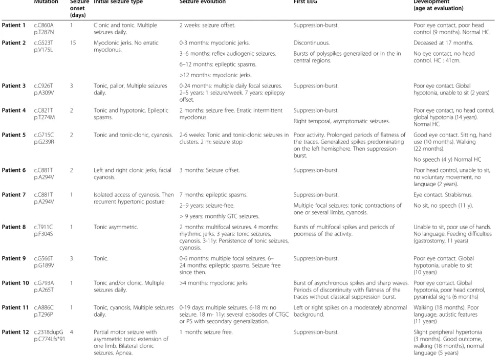
Table 1 KCNQ2 mutations and main features of the patients
Mutation Seizure onset (days)
Initial seizure type Seizure evolution First EEG Development
(age at evaluation) Patient 1 c.C860A
p.T287N
1 Clonic and tonic. Multiple seizures daily.
2 weeks: seizure offset. Suppression-burst. Poor eye contact, poor head control (9 months). Normal HC. Patient 2 c.G523T
p.V175L
15 Myoclonic jerks. No erratic myoclonus.
0-3 months: myoclonic jerks. Discontinuous. Deceased at 17 months. Bursts of polyspikes generalized or in the in
central regions.
No eye contact, no head control. HC : 41cm. 3–6 months: reflex audiogenic seizures.
6–12 months: epileptic spasms. >12 months: myoclonic jerks. Patient 3 c.C926T
p.A309V
3 Tonic, pallor, Multiple seizures daily.
0-24 months: multiple daily focal seizures. 2–5 years: 1 seizure/week. 7 years: epilepsy offset.
Suppression-burst. Poor eye contact. Global hypotonia, unable to sit (2 years) Patient 4 c.C821T
p.T274M
2 Tonic and hypotonic. Epileptic spasms.
2 months: seizure free. Erratic intermittent myoclonus.
Suppression-burst. Poor eye contact, no head control, global hypotonia (14 years). Normal HC.
Right temporal, asymptomatic seizures. Patient 5 c.G715C
p.G239R
2 Tonic and tonic-clonic, cyanosis. 2-6 weeks: Tonic and tonic-clonic seizures in clusters. 2 m: seizure stop
Poor activity. Prolonged periods of flatness of the traces. Generalized spikes predominating on the left hemisphere. Then suppression-burst.
Good eye contact. Sitting, hand use (10 months). Walking (22 months).
No speech (4 y) Normal HC Patient 6 c.C881T
p.A294V
2 Left and right clonic jerks, facial cyanosis.
3 months: Seizure offset. Suppression-burst. Poor head control, unable to sit, no voluntary movement, no language (2 years). Patient 7 c.C881T
p.A294V
1 Isolated access of cyanosis. Then recurrent hypertonic posture.
7 months: epileptic spasms. Suppression-burst. Eye contact. Strabismus. No sit, no speech (11 y). Multiple focal seizures: tonic contractions of
one or several limbs, cyanosis. 2–9 years: seizure-free.
> 9 years: monthly GTC seizures. Patient 8 c.T911C
p.F304S
1 Tonic asymmetric. 2 months: multifocal seizures. 4 months: rhythmic jerks. 3 years: tonic seizures, cyanosis. 3-11y: Persistence of tonic seizures, cyanosis.
Bursts of multifocal spikes and periods of poorness of the activity.
Unable to sit, poor use of hands. No language. Feeding difficulties (gastrostomy, 11 years) Patient 9 c.G566T
p.G189V
3 Tonic. 0-6 months: multiple focal seizures. 6– 24 months: epileptic spasms. Seizure free since then.
Suppression-burst. Poor eye contact. Global hypotonia, unable to sit (10 years)
Patient 10 c.G793A p.A265T
1 Tonic and/or clonic, Multiple seizures daily.
>4 months: myoclonic jerks Burst of asynchronous spikes and sharp waves. Periods of discontinuity with flatness of the traces without classical suppression burst.
Poor eye contact. Global hypotonia, poor head control, pyramidal signs (6 months) Patient 11 c.A886C
p.T296P
1 Tonic, cyanosis, Multiple seizures daily.
0-19 days: multiple seizures. 6-18 m: no seizure. 18 m- 11y: several episodes of CTGC or PS with secondary generalization.
Left or right spikes on a moderately abnormal background.
Walking (18 months). Poor language, autistic features (11 years)
Patient 12 c.2318dupG p.C774Lfs*91
4 Partial motor seizure with asymmetric tonic extension of one limb. Bilateral clonic seizures. Apnea.
1 month: seizure free. Suppression-burst. Slight peripheral hypertonia (3 months). Good outcome, walking (18 months), normal language (5 years) Milh et al. Orphanet Journal of Rare Diseases 2013, 8 :80 Page 3 of 8 http://ww w.ojrd.com/ content/8/1 /80
Table 1 KCNQ2 mutations and main features of the patients (Continued)
Patient 13 c.G471A p.W157X
4 Hemi corporeal, left or right. 0-11 months: partial clonic seizures. Then seizure offset.
Poor, discontinuous. Independent walking (4 y). No language (6.5 y). Normal HC 52.5 cm
Patient 14 c.G868A p.G290S
1 Tonic. Many motor seizures during the neonatal period. 2 m: Seizure stop. AED withdrawn at 4 years.
Asymmetrical suppression-burst with multifocal slow waves, left frontal and right occipital spikes. Periods of generalized flattening.
Sitting (3 y) hand stereotypies. Unable to walk/stand, stereotypies, pyramidal signs. Poor language. Normal HC (16 y).
Patient 15 c.C881T p.A294V
8 Myoclonic jerks, Multiple seizures daily.
0-3 months: myoclonic jerks. 3 months: seizure offset. Therapy stopped at 6 months.
Suppression burst. Sit (2 y). No walking, 2–3 words. Understands simple orders. Strabismus, nystagmus (3 y) Patient 16 c.997C>T.
p.R333W
2 Bilateral tonic clonic And right clonic
0-3 y: active epilepsy, motor seizures 3-10 y: seizure free 10-20 y: monthly focal seizures
Slow waves with asynchronous bilateral spikes and intermittent flattening
First steps (18 m). Few words (3 y) Able to read but cannot write, limited communication skills, marked bradypsychia, hand stereotypies (26 y)
HC head circumference, m months, y year.
Milh et al. Orphanet Journal of Rare Diseases 2013, 8 :80 Page 4 of 8 http://ww w.ojrd.com/ content/8/1 /80
during the first week of life with stormy clonic and/or
tonic seizures, whatever the presence or absence of a
fa-milial history. If cognitive outcome is relatively reliable
and good in familial cases of BFNE [5], it is not the case
in sporadic ones, where neurological outcomes range
from dramatic to normal. We did not find any
relation-ship between the initial history of the epilepsy and the
severity of outcome. For example, patients 1 and 12 had
relatively similar features at the beginning and displayed
very different outcomes (Tables 1, 2 and Figure 1). Since
KCNQ2
is now implicated in various forms of epilepsies,
from the most benign to the most dramatic, additionnal
data on phenotype/genotype correlations would be
par-ticularly relevant. Interestingly, none of the mutation
reported in neonatal epileptic encephalopathies had
previ-ously been reported in BFNE, and the severe mutations that
have been found in several patients (p.G290A [7], p.T274M
and p.A294V(present study)) lead to relatively similar
fea-tures in terms of initial EEG and development, however
different in terms of evolution of the epilepsy. The
dif-ferent epileptic features in patients carrying the same
mutation of KCNQ2 may be due to genetic modifiers or
non genetic factors. Overall, this cohort of patients
highlights the heterogeneous evolution of the
neuro-logical phenotypes associated with de novo
heterozy-gous mutations in KCNQ2. This heterogeneity could be
at least partially related to the impact of the mutations
on the Im current. Analysis of the functional
conse-quences of “benign” versus “severe” mutations in
KCNQ2
should be of paramount importance to better
understand the molecular and cellular mechanisms
in-volved in the emergence of an epileptic encephalopathy.
This has recently been tested with two mutations of
KCNQ2
affecting the same residue in the S4 domain of
the protein KV7-2 but associated with either a benign
phenotype, or with a neonatal epileptic encephalopathy
with severe drug-resistant seizures and neurocognitive
delay, suppression-burst pattern at EEG, and distinct
Fp2-T4 T4-O2 Fp2-C4 C4-O2 Fp1-C3 C3-O1 Fp1-T3 T3-O1 BREATH ECG Fp2-T4 T4-O2 Fp2-C4 C4-O2 Fp1-C3 C3-O1 Fp1-T3 T3-O1 BREATH ECG T4-Fp2 Fp2-Fp1 Fp1-T3 T4-C4 C4-C3 C3-T3 T4-O2 O2-O1 O1-T3 BREATH ECG T4-Fp2 Fp2-Fp1 Fp1-T3 T4-C4 C4-C3 C3-T3 T4-O2 O2-O1 O1-T3 BREATH ECG
A
B
1s 100µVFigure 1 Representative early EEGs of patients carrying a de novo KCNQ2 mutation. A. Interictal EEG (Day 3, patient 1), showing a typical suppression-burst pattern, with burst of spikes and slow waves alternating with periods of electric silence (left panel). Sometimes, the burst should be much longer than the periods of suppression, leading to a discontinuous pattern (Right panel). B. EEG displaying the same features (Patient 12, suppression-burst in left panel, discontinuous pattern in right one), with a very different outcome (normal development at 5 years old, see Table 1).
Milh et al. Orphanet Journal of Rare Diseases 2013, 8:80 Page 5 of 8
Table 2 Data on initial evaluation and treatment, EEG evolution and brain MRI
Term. Clinical examination at birth
Treatment during the first month
EEG evolution (age) Brain MRI (age)
Patient 1 Full term. Hypotonia. No eye contact. BW: 3000 g HC: 35 cm
PHB VGB 1 m: Continuous with rare posterior spikes and fast rhythms Day 7: Normal, absence of any signal abnormality. 2 y: Discrete global brain atrophy, thin corpus callosum
6 m: slow background, rare focal spikes. Patient 2 34 GW Fetal distress, apnea,
movements disorder BW: 2,040 g HC: 30 cm MDZ, PHB, B6, TPM, VGB 0-7 m: Suppression-burst >7 m: Hypsarythmia
D13: absence of any signal abnormality. 3 m: Absence of signal abnormality
Patient 3 Full term Failure. to thrive. Feeding difficulties BW: 3,770 g HC: 37.5 cm
PHB, B6, PHT, VGB, TPM, CLB.
1-6 w: Asynchronous SB. 2-8 m: bilateral Bursts of central spikes 1-6 y: Bursts of rhythmic generalized spikes at 3 Hz
Day 3: T1 bilateral hypersignal of pallida, tegmentum, locus niger, hippocampi. Abnormal ADC in these regions. 2 y: T1 hypersignal of the same structures and diffuse T1 hypersignal of the white matter. Brain atrophy
Patient 4 Full term. Normal BW: 3,580 g HC: 37 cm
PHB, PHT,
VPA 0-2 m: Suppression-burst. 2–12 m: hypsarythmia. >12 m: Frequentmultifocal spikes
Day 7: Normal CT scan 2 y: Thin Corpus Callosum, absence of signal abnormality
Patient 5 Full term. Normal BW: 3,240 g HC: 34 cm
PHB, PHT, B6, VGB, VPA
0-2 m: Suppression-burst. 2–6 m: continuous, slow background, multifocal spikes. >6 m: Rare spikes in temporal and occipital lobes
Day 10: no signal abnormality. Patient 6 Full term. Normal BW:
2,790 g HC: 34,5 cm
ND 0-1 m: Suppression-burst. <1 m: Multifocal spikes, slow background 1 m: Normal Patient 7 Full term. Normal BW:
3,180 g, HC: 36 cm
PHB, B6, PHT 0-1 m: Suppression-burst. 1–7 m: Continuous EEG, multifocal spikes 7–24 m: Hypsarythmic pattern. >24 m: Frequent spikes and spike wave in frontal regions
Day 4: Normal, absence of any signal abnormality
Patient 8 ND PHB, PHT 0-2 m: Bursts of multifocal spikes, periods of flatness. >2 m: Multifocal spikes, poor organization
1 y: No structural or signal abnormality. Patient 9 At term. Hypotonia. No eye
contact. BW: 3,450 g HC: 35 cm
PB, CZP, VGB 0-2 m: Suppression-burst 2–6 m: Slow background, rare generalized spike waves. 6–12 m: Hypsarythmic pattern. >12 m: Rare
asynchronous frontal and temporal spikes
Day 10: Normal, absence of any signal abnormality
Patient 10 Full term. No eye contact BW: 3,120 g HC: 33 cm
PB, PHT, TPM, VGB, B6,
0-2 m: discontinuous EEG. >2 m: continuous, slow EEG with rare generalized spikes
Day 5: T1: symmetrical hypersignal of the pallida, caudate nuclei and hippocampi T2: bilateral hypersignal of the parietal occipital white matter
Patient 11 Full term. Hypotonia, hyporeactivity, failure to feed
PHB, PHT, VPA.
0-1 m: Left or right spikes on a moderately abnormal background. >1 m: Occipital or temporal spikes with left prominence with progressive migration on the central temporal region
1 m: normal
Patient 12 Full term. Normal BW and HC
VGB, CBZ 0-2 m: Suppression-burst. 2–6 m: General slowing of the traces, no spike. 6 m-2 y: Rare spikes in the right central region, Normal background. >2 y: normal traces.
Day 7: T2 hyperintensity of the basal ganglia 2 y: Normal 3y: Normal
Patient 13 Full term. Fetal distress. BW, HC: ND
ND ND 1 m: No structural abormality, no signal change
Patient 14 Full term. BW 3,750 g. Poor eye contact, trunk hypotonia with bouts of hypertonia
PHB, VGB, CBZ;
0-4 m: Asymmetrical suppression-burst 4-10 m: Left occipital spikes and slow waves 10 m-3 y: Normal background activity + posterior theta waves, No spike 3 y: Intermittent slow background, no spike >8 y: Normal 3 m: Normal Milh et al. Orphanet Journal of Rare Diseases 2013, 8 :80 Page 6 of 8 http://ww w.ojrd.com/ content/8/1 /80
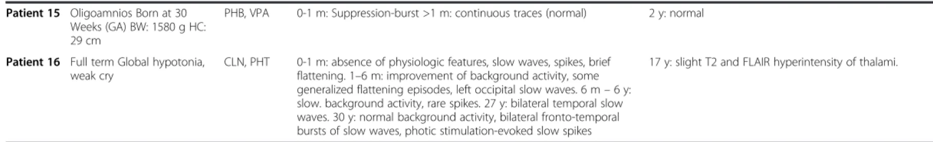
Table 2 Data on initial evaluation and treatment, EEG evolution and brain MRI (Continued)
Patient 15 Oligoamnios Born at 30 Weeks (GA) BW: 1580 g HC: 29 cm
PHB, VPA 0-1 m: Suppression-burst >1 m: continuous traces (normal) 2 y: normal
Patient 16 Full term Global hypotonia, weak cry
CLN, PHT 0-1 m: absence of physiologic features, slow waves, spikes, brief flattening. 1–6 m: improvement of background activity, some generalized flattening episodes, left occipital slow waves. 6 m – 6 y: slow. background activity, rare spikes. 27 y: bilateral temporal slow waves. 30 y: normal background activity, bilateral fronto-temporal bursts of slow waves, photic stimulation-evoked slow spikes
17 y: slight T2 and FLAIR hyperintensity of thalami.
GW gestational week, HC Head circumference, m month, Y year, PHB Phenobarbital, PHT phenytoine, VGB vigabatrin, TPM topiramate, CLN clonazepam, VPA Sodium Valproate, CBZ carbamazepine, CLB clobazam.
Milh et al. Orphanet Journal of Rare Diseases 2013, 8 :80 Page 7 of 8 http://ww w.ojrd.com/ content/8/1 /80
neuroradiological features [11]. The authors showed
that, while both mutations destabilized the open state of
the channel causing a reduction of the voltage
sensitiv-ity, the functional changes were more pronounced in
the “severe” mutation than in the benign one. In both
cases, the functional impairment could be fully restored
by the neuronal Kv7 activator retigabine. This study
suggested that the clinical disease severity may be
re-lated to the extent of the mutation-induced functional K
+ channel impairment and set the preclinical basis for
the potential use of Kv7 openers as a targeted
anticon-vulsant therapy to improve developmental outcome in
neonates. However, since two patients carrying the same
KCNQ2
mutation do not have the same epileptic
out-come, correlations between Im impairment and the
se-verity of the encephalopathy should be made with
caution. Other unknown factors may be involved in the
occurrence of the epileptic encephalopathy. Moreover,
the vast majority of the mutations we described here
were localised on segment S6 and should have different
consequences on Im current that those which have been
previously described, affecting segment S4 [11]. These
consequences still have to be studied. Overall, it is not
known whether ongoing brain dysfunction that is
ob-served in several patients is due to the Kv7-2
channelopathy or if it is a sequel of neonatal epilepsy.
This question would be of paramount interest.
Conclusions
KCNQ2
is frequently found mutated de novo in early onset
epileptic encephalopathies, especially if the epilepsy begins
within the first week of life. Despite relatively stereotyped
initial phenotype, the neurological and epileptic outcomes
were highly variable, overall severe.
Competing interests
The authors declare that they have no competing interests. Authors’ contributions
MM, SA, CM, NV, AR, BH, MC, AK, CA, GB, DV, MAB, DH, DG, AA, ND, TBV, JP, BI, NG, LV, IA, AJ, PC BC, GL and LV designed the study and interpreted the data. NBK, JSS and CL made the experiments. MM, CM, GL and LV drafted and revised the MS. All authors read and approved the final manuscript. Acknowledgements
This work was supported by INSERM (Contrat d'Interface pour Hospitalier to MM), Programme Hospitalier de recherche Clinique, Aix Marseille Université and Assistance Publique Hôpitaux de Marseille (Contrat Hospitalier de Recherche Translationnelle to LV). We thank the Centre de Ressources Biologiques of La Timone Children’s Hospital for access to the biological samples used in this study.
Author details
1INSERM, UMR_S 910 Faculté de médecine, Boulevard jean MOULIN F13005,
Marseille, France.2APHM, Service de neurologie pédiatrique, CHU Timone, Marseille, France.3Hospices civils de Lyon. Laboratoire de génétique, Hôpital
Edouard Herriot. Bron, Lyon, France.4APHP, Unité Fonctionnelle de Génétique Médicale, Département de Génétique, Groupe Hospitalier Pitié-Salpêtrière, Paris, France.5
Centre de Référence des Déficiences Intellectuelles de Causes Rares, Paris, France.6APHP. Service de
Neuropédiatrie, Hôpital Armand Trousseau, Paris, France.7APHP. Service de
neuropédiatrie, Hopital Robert Debré, Paris, France.8APHM. Département de
Génétique Médicale et Biologie Cellulaire CHU Timone, Marseille, France.
9
CHU Montpellier. Service de neuropédiatrie, Montpellier, France.10INSERM U1051, INM Montpellier, Montpellier, France.11APHP. Service de
neurophysiologie clinique Hôpital Necker, Paris, France.12CHU Besancon.
Service de neuropédiatrie, Besancon, France.13Hospices civils de Lyon,
Service de neuropédiatrie. HFME. Bron, Lyon, France.14CHU de Tours. Service de neuropédiatrie, Beranger, France.15APHP. Groupe hospitalier Pitié
Salpétrière. Service de neurologie, Paris, France.16CHU de Nantes. Service de
pédiatrie, Nantes, France.17CHU de Nantes. Service de génétique médicale,
Nantes, France.18CHU de Grenoble. Service d’électrophysiologie clinique, Grenoble, France.19Aix Marseille Université, Faculté de Médecine, Marseille,
France.
Received: 8 March 2013 Accepted: 15 May 2013 Published: 22 May 2013
References
1. Biervert C, Schroeder BC, Kubisch C, Berkovic SF, Propping P, Jentsch TJ, Steinlein OK: A potassium channel mutation in neonatal human epilepsy.
Science 1998, 279:403–406.
2. Charlier C, Singh NA, Ryan SG, Lewis TB, Reus BE, Leach RJ, Leppert M: A pore mutation in a novel KQT-like potassium channel gene in an idiopathic epilepsy family [see comments]. Nat Genet 1998, 18:53–55.
3. Singh NA, Charlier C, Stauffer D, DuPont BR, Leach RJ, Melis R, Ronen GM, Bjerre I, Quattlebaum T, Murphy JV, McHarg ML, Gagnon D, Rosales TO, Peiffer A, Anderson VE, Leppert M: A novel potassium channel gene, KCNQ2, is mutated in an inherited epilepsy of newborns. Nat Genet 1998, 18:25–29.
4. Plouin P: Benign familial neonatal convulsions and benign idiopathic neonatal convulsions. Epilepsy: a comprehensive textbook. Philadelphia: Lippincott-Raven; 1997:2247-2249.
5. Bellini G, Miceli F, Soldovieri MV, Miraglia Del Giudice E, Coppola G, Taglialatela M: KCNQ2 related disorders. 2010 Apr 27 [Updated 2013 Apr 11]. In GeneReviews™ [Internet]. Edited by Pagon RA, Bird TD, Dolan CR, et al. Seattle (WA): University of Washington, Seattle; 1993. Available from: http://www.ncbi. nlm.nih.gov/books/NBK32534/.
6. Dedek K, Fusco L, Teloy N, Steinlein OK: Neonatal convulsions and epileptic encephalopathy in an Italian family with a missense mutation in the fifth transmembrane region of KCNQ2. Epilepsy Res 2003, 54:21–27. 7. Weckhuysen S, Mandelstam S, Suls A, Audenaert D, Deconinck T, Claes LR,
Deprez L, Smets K, Hristova D, Yordanova I, Jordanova A, Ceulemans B, Jansen A, Hasaerts D, Roelens F, Lagae L, Yendle S, Stanley T, Heron SE, Mulley JC, Berkovic SF, Scheffer IE, de Jonghe P: KCNQ2 Encephalopathy: emerging phenotype of a neonatal epileptic encephalopathy. Ann Neurol 2012, 71:15–25.
8. Saitsu H, Kato M, Koide A, Goto T, Fujita T, Nishiyama K, Tsurusaki Y, Doi H, Miyake N, Hayasaka K, Matsumoto N: Whole exome sequencing identifies KCNQ2 mutations in ohtahara syndrome. Ann Neurol 2012, 72:298–300. 9. Saitsu H, Kato M, Mizuguchi T, Hamada K, Osaka H, Tohyama J, Uruno K,
Kumada S, Nishiyama K, Nishimura A, Okada I, Yoshimura Y, Hirai S, Kumada T, Hayasaka K, Fukuda A, Ogata K, Matsumoto N: De novo mutations in the gene encoding STXBP1 (MUNC18-1) cause early infantile epileptic encephalopathy. Nat Genet 2008, 40:782–788.
10. Kato M, Saitoh S, Kamei A, Shiraishi H, Ueda Y, Akasaka M, Tohyama J, Akasaka N, Hayasaka K: A longer polyalanine expansion mutation in the ARX gene causes early infantile epileptic encephalopathy with suppression-burst pattern (ohtahara syndrome). Am J Hum Genet 2007, 81:361–366.
11. Miceli F, Soldovieri MV, Ambrosino P, Barrese V, Migliore M, Cilio MR, Taglialatela M: Genotype-phenotype correlations in neonatal epilepsies caused by mutations in the voltage sensor of Kv7.2 potassium channel subunits. Proc Natl Acad Sci USA 2013, 110:4386–91.
doi:10.1186/1750-1172-8-80
Cite this article as: Milh et al.: Similar early characteristics but variable neurological outcome of patients with a de novo mutation of KCNQ2.
Orphanet Journal of Rare Diseases 2013 8:80.
Milh et al. Orphanet Journal of Rare Diseases 2013, 8:80 Page 8 of 8