HAL Id: hal-03155846
https://hal.sorbonne-universite.fr/hal-03155846
Submitted on 2 Mar 2021HAL is a multi-disciplinary open access archive for the deposit and dissemination of sci-entific research documents, whether they are pub-lished or not. The documents may come from teaching and research institutions in France or abroad, or from public or private research centers.
L’archive ouverte pluridisciplinaire HAL, est destinée au dépôt et à la diffusion de documents scientifiques de niveau recherche, publiés ou non, émanant des établissements d’enseignement et de recherche français ou étrangers, des laboratoires publics ou privés.
Clinical, biological and radiological features, 4-week
outcomes and prognostic factors in COVID-19 elderly
inpatients
R. Palich, Y. Wakim, O. Itani, O. Paccoud, S. Boussouar, M. Lévy-Soussan,
C. Soulie, N. Godefroy, A. Bleibtreu
To cite this version:
R. Palich, Y. Wakim, O. Itani, O. Paccoud, S. Boussouar, et al.. Clinical, biological and radiological features, 4-week outcomes and prognostic factors in COVID-19 elderly inpatients. Infectious Diseases Now, Elsevier, 2021, �10.1016/j.idnow.2020.12.004�. �hal-03155846�
TITLE 1
Clinical, biological and radiological features, 4-week outcomes and prognostic factors in 2
COVID-19 elderly inpatients 3
4
SHORT TITLE 5
COVID-19 in elderly patients 6
7
AUTHORS 8
R. Palich1*, Y. Wakim1*, O. Itani1, O. Paccoud1, S. Boussouar2, M. Lévy-Soussan3, C. Soulie4,
9
N. Godefroy1, A. Bleibtreu1, on behalf of the PSL-Covid working group
10
* These authors contributed equally to the work 11
12
AFFILIATIONS 13
1. Sorbonne Université, INSERM, Pierre Louis Epidemiology and Public Health Institute 14
(iPLESP), AP-HP, Pitié-Salpêtrière Hospital, Department of Infectious Diseases, F-15
75013 Paris, France 16
2. Sorbonne Université, LIB-Laboratoire d’imagerie biomédicale, INSERM, CNRS, 17
ICAN Institute of CardioMetabolism and Nutrition, ACTION Study Group, 18
Cardiothoracic Imaging Unit, AP-HP, Pitié-Salpêtrière Hospital, F-75013 Paris, France 19
3. Sorbonne Université, AP-HP, Pitié-Salpêtrière Hospital, Department of Palliative Care, 20
F-75013 Paris, France 21
4. Sorbonne Université, INSERM, Pierre Louis Epidemiology and Public Health Institute 22
(iPLESP), AP-HP, Pitié-Salpêtrière Hospital, Department of Virology, F-75013 Paris, 23
France 24
CORRESPONDING AUTHOR 26
Dr Romain Palich 27
Service des Maladies Infectieuses et Tropicales 28
Hôpital Pitié-Salpêtrière, AP-HP 29 47-83 boulevard de l’hôpital 30 75013 Paris 31 Tel: +33 1 42 16 03 93 32 Fax: +33 1 42 16 04 45 33 Email: romain.palich@aphp.fr 34 35 KEY WORDS 36 Covid-19 37 Elderly patients 38 Sars-CoV-2 39 Prognostic factors 40 Computed tomography 41 42 WORD COUNT 43 Abstract: 207 44 Text: 2638 45
ABSTRACT 1
Objective. To describe clinical, biological, radiological presentation and W4 status in COVID-2
19 elderly patients. 3
Patients and methods. All patients ≥70 years with confirmed SARS-CoV-2 infection and 4
hospitalized in the Infectious Diseases department of the Pitié-Salpêtrière hospital, Paris, 5
France, from March 1st to April 15th 2020 were included. The primary outcome was death 4
6
weeks after hospital admission. Data on demographics, clinical features, laboratory tests, CT-7
scan findings, therapeutic management and complications was collected. 8
Results. Overall, 100 patients were analyzed, including 49 patients ≥80 years. Seventy percent 9
had ≥2 comorbidities. Respiratory features were often severe as 48% needed oxygen support 10
upon admission. Twenty-eight out of 43 patients (65%) with a CT-scan had a mild to severe 11
parenchymal impairment, and 38/43 (88%) a bilateral impairment. Thirty-two patients 12
presented a respiratory distress requiring oxygen support ≥6 liters/minute. Twenty-four deaths 13
occurred, including 21 during hospitalization in our unit, 2 among the 8 patients transferred in 14
ICU, and one at home after discharge from hospital, leading to a global mortality rate of 24% 15
at W4. Age, acute renal failure and respiratory distress were associated with mortality at W4. 16
Conclusion. Elderly COVID-19 patients with several comorbidities and severe clinical features 17
survived in a substantial proportion, which could argue against transferring the most fragile 18
patients in ICU. 19
INTRODUCTION 21
COVID-19 is an infection related to the novel coronavirus SARS-CoV-2 [1]. It started 22
spreading from Wuhan, China in December 2019, leading to a major pandemic. In early 23
September 2020, nearly 27 million COVID-19 cases were reported worldwide, associated with 24
900,000 deaths [2]. In France, almost 300,500 cases and 30,500 deaths have been documented 25
since February 2020 [3]. Elderly population bear a heavy burden, with fatality rates reaching 26
26% and 58% in patients aged over 70 and 80 years, respectively. Most cohort studies have 27
found age to be the main risk factor for mortality [4–6]. 28
On May 12th in France, people over 75 years represented 56% of inpatients, 71% of deaths
29
having occurred during hospital stay, and only 19% of patients admitted in ICU [3]. Therefore, 30
elderly COVID-19 patients represent a high proportion of inpatients in medical wards. Despite 31
frequent severe respiratory forms and accumulation of risk factors, a substantial number of 32
these patients survived the infection. However, data in this population is very limited, especially 33
among survivors after discharge from hospital. 34
Herein, we aimed to describe clinical, biological and radiological presentation of COVID-19 in 35
patients over 70 years, in the Infectious and Tropical Diseases department of the Pitié-36
Salpêtrière University hospital (Paris, France), as well as clinical outcome 4 weeks (W4) after 37
admission and factors associated with mortality. 38
39
MATERIALS AND METHODS 40
Study design and participants
41
We performed a retrospective, single-center study in the Infectious and Tropical Diseases 42
department at the Pitié-Salpêtrière University Hospital, Paris, France. We included all 43
confirmed cases of COVID-19 over 70 years old admitted from March 1st to April 15th 2020.
Patients were considered to have a confirmed infection if testing by RT-PCR from 45
nasopharyngeal swab and/or sputum came back positive. 46
47
Outcomes
48
The primary outcome was vital status (survival or death) 4 weeks after hospital admission. The 49
secondary outcomes were length of hospital stay, occurrence of acute respiratory distress and/or 50
other complications during clinical course, transfer to ICU, “do not resuscitate” order, use of 51
hypnotic and opioid drugs, place of residence (at home or retirement home, subacute care ward, 52
ICU) 4 weeks after hospital admission for survivors, and factors associated with vital status at 53 week 4 (W4). 54 55 Procedures 56
We obtained data retrospectively from electronic medical records, carefully reviewed by three 57
trained physicians. Patient data included demographics, home medications, comorbidities, 58
initial signs and symptoms, onset of signs and symptoms, triage vitals, initial laboratory tests, 59
value of cycle threshold (Ct) for SARS-CoV-2 RT-PCR, chest computed tomographic scan 60
results, treatments received during hospitalization (antivirals, antibiotics, corticosteroids and 61
oxygen support), maximum body temperature, and length of hospital stay. Charlson index was 62
used to evaluate patients’ medical state before admission [7]. NEWS-2 score was used to 63
standardize the assessment of acute-illness severity at admission [8]. The occurrence of acute 64
respiratory distress during clinical course was defined as the recourse to oxygen support ≥6 65
liters/minute to maintain blood oxygen saturation (SpO2) ≥94%. We also collected other
66
significant complications (acute renal injury, acute hepatitis, bacterial infection) and maximum 67
C-reactive protein (CRP) during hospital stay. 68
Nasopharyngeal swab and sputum samples were collected and tested by RT-PCR with the 69
Cobas® SARS-CoV-2 kit (Roche), as recommended by the manufacturer, after neutralization 70
with an equal amount of Cobas lysis buffer for the SARS-CoV-2 (400/400 µL). Three 71
categories of Ct were retained: Ct<20 (high SARS-CoV-2 viral load), 20 <Ct ≤30 (mild viral 72
load) and Ct >30 (low viral load). 73
All scans were performed on a dedicated scanner (Siemens Somaton Edge) either without 74
iodine injection or after IV of 60 mL iodinated contrast agent (Iomeprol 400 Mg I/mL, Bracco 75
Imaging, Milan, IT) in cases of suspected pulmonary embolism. Radiologist (SB) who were 76
blinded to clinical and biological features including RT-PCR results, reviewed all chest CT 77
images on a PACS workstation (Carestream Health, Rochester, NY) and classified the chest 78
CT as positive or negative for COVID-19 according to CT features previously described [9– 79
11]. The radiologist also described main CT features (ground-glass opacity, consolidation, 80
reticulation/thickened interlobular septa, nodules, emphysema, pleural effusion), and lesion 81
distribution (left, right or bilateral lungs). A semi-quantitative score was assigned (0% 82
involvement; less than 25%; 25% to less than 50%; 50% to less than 75%; 75% or greater). All 83
images were viewed on both lung and mediastinal settings. 84
We reported for each patient whether a transfer to ICU had been considered during clinical 85
course, a “do not resuscitate” order had been collectively decided, and whether hypnotic or 86
opioid drugs were used. 87
Patients or family were contacted by a physician 4 weeks after admission to collect vital status 88
and place of residence at W4. 89
90
Statistics
91
We analyzed the vital status 4 weeks after hospital admission using a logistic regression model. 92
Factors investigated were: sex of patients, age, place of residence before hospital admission, 93
number of comorbidities, Charlson’s score, time from onset of symptoms and hospital 94
admission, NEW-2 score, Oxygen support need at admission, lymphocyte count at admission, 95
SARS-CoV-2 RT-PCR Ct, total lesion extension, presence of bilateral lesion, duration of 96
hospital stay and respiratory distress (requiring oxygen support ≥6 liters/minute). For all these 97
factors, missing values were defined as “unknown”. A descriptive analysis of the dependent 98
and independent variables was performed. A univariate analysis was conducted, and all factors 99
associated with death 4 weeks after hospital admission were entered into the multivariate 100
model. We decided to exclude from the analysis the lost to follow-up. The vital status 4 weeks 101
after hospital admission was also compared between patients aged 70 to 79, 80 to 89 and >90 102
year-old using a chi-square test. Survival curve was showed using Kaplan-Meier estimates. All 103
analyses were performed using R studio Version 1.2.5033 (© 2009-2019 RStudio, Inc.) and a 104
p value<0.05 was considered as significant. 105
106
Ethics
107
All discharged inpatients were included in the prospective COVID-PSL cohort, for the 4-week 108
telephonic follow-up. They signed a written informed consent. This cohort was approved by 109
the Ethics Committee Ile-De-France X (n°47-2020, NCT 04402905). 110
111
RESULTS 112
Demographics and medical background
113
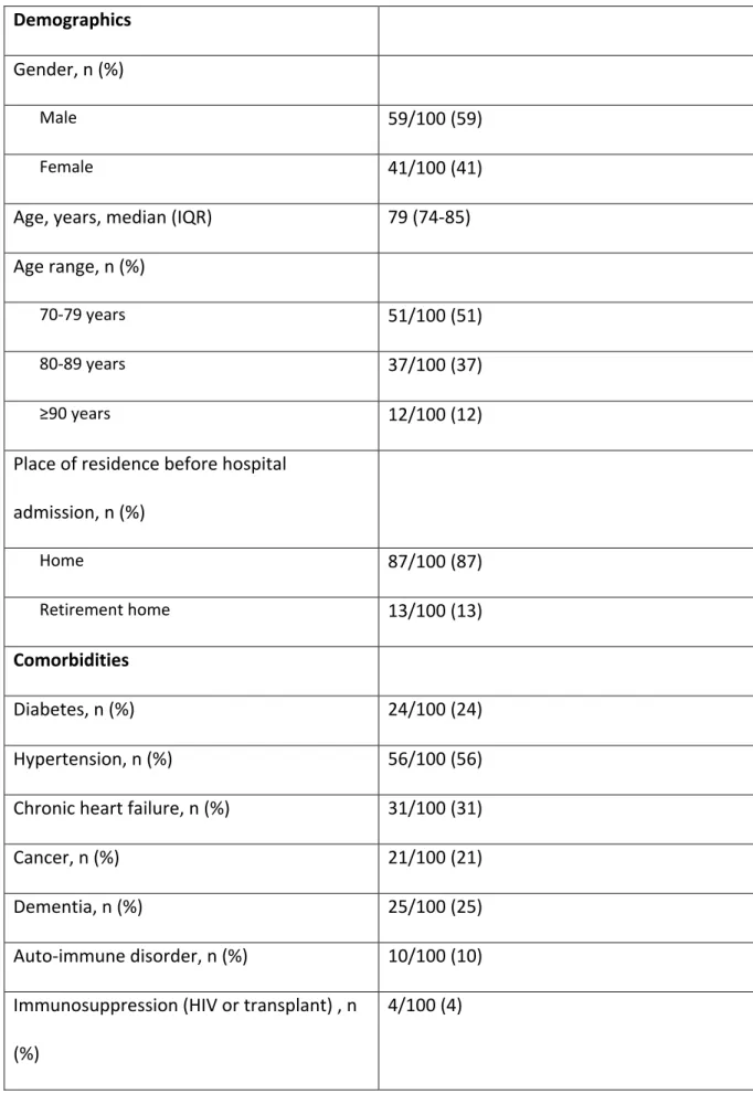
Overall, 100 patients were included for analysis, including 59 men (Table 1). Median age was 114
79 years (IQR 74-85). Fifty-one patients were between 70 and 79 years, 37 between 80 and 89 115
years, and 12 over 90 years. Eighty-nine lived independently at home and 11 in retirement home 116
before being admitted to hospital. 117
Fifty-six of them had a Charlson index ≥5 and 71 had at least 2 comorbidities (Table 1). The 118
most prevalent comorbidities were high blood pressure (56%), chronic heart failure (31%), 119
dementia (25%), diabetes (24%) and/or active cancer (21%). Eleven patients had a long-term 120
immunosuppressive and/or corticosteroid therapy. 121
122
Clinical and biological feature at hospital admission
123
Median time between the onset of symptoms and hospital admission was 4 days (IQR 2-7). 124
Seventy percent of patients were admitted to hospital before the 7th day.
125
All SARS-CoV-2 infections were confirmed by RT-PCR from nasopharyngeal swab and/or 126
sputum, with a low Ct (≤20) in 21% of cases and a mild Ct (21-35) in 28% of cases. 127
The most prevalent symptoms reported at admission were fever (69%), cough (50%), dyspnea 128
(36%), diarrhea (16%) and confusion (14%) (Table 1). Fifty-five patients had a NEWS-2 score 129
≥3 and 22% needed oxygen therapy ≥3 liters/minute upon admission. 130
Regarding biological parameters, 80% of patients had lymphopenia, 23% had thrombopenia, 131
32% had acute renal failure and 35% had hepatic cytolysis. 132
133
Thoracic CT-scan outcomes
134
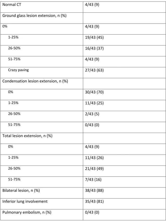
46 CT-scans were analyzed from 43 patients (3 patients had 2 CT-scans). 39 (91%) patients 135
were with abnormal lung changes: 91% had ground class opacities (GGO), 63% had crazy 136
paving (GGO associated with superimposed intralobular reticulations) and 30% had 137
condensations. 29 patients (65%) had a mild to severe parenchymal impairment, 38 patients 138
(88%) had a bilateral impairment, and the lesions were mainly distributed in the lower lungs in 139
35 patients (81%) (Table 2). 6 patients had pleural effusion. 11 patients were with common 140
accompanying diseases of which emphysema (n=9), bronchiectasis (n=1), and pleural sequelae 141
(n=1). Associated pulmonary oedema was detected in one patient. CT scan were injected in 142
42% of cases, with no pulmonary embolism detected. 143
144
Therapeutic management and clinical course during hospitalization
145
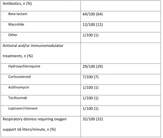
Sixty-five patients received antibiotics, including a beta-lactam in 97% of cases and/or a 146
macrolide in 98% of cases (Table 3). Thirty percent of patients received hydroxychloroquine 147
for a median duration of 9 days (IQR 2-10). No cardiac side effects were observed in patients 148
treated by hydroxychloroquine. Seven percent of patients received short-term corticosteroid 149
therapy (2-5 days). 150
Thirty-two patients presented a clinical deterioration with respiratory distress requiring oxygen 151
support ≥6 liters/minute. Respiratory distress occurred after a median time of 10 days (IQR 4-152
13 after the onset of symptoms. Among these 32 patients, 8 (25%) were transferred to ICU 153
(Figure 1). All were <80 years. For the 24 others, a “do not resuscitate” order was decided. 154
Hypnotic and opioid drugs were used in 17 of them. 155
156
Mortality rate and place of residence at W4
157
Overall, 24 deaths occurred from hospital admission to W4, including 21 deaths during 158
hospitalization in our unit, 2 deaths in ICU and one death at home after discharge from hospital, 159
leading to a global mortality rate of 24% at W4 (Figure 2a). The mortality rate in 70-79, 80-89 160
and >90 year-old patients was 10/47 (21%), 10/36 (28%) and 4/11 (36%), respectively. The 161
duration of hospital stay was significantly shorter in deceased patients than in discharged 162
patients (7.1 vs. 11.1 days, OR 0.70, 95%CI 0.47-0.88, p=0.017). Among deceased patients, 163
death occurred 9 days (IQR 13-16) in median after the onset of symptoms. 164
Overall, among surviving patients, 46/67 (69%) had returned home or to their institution and 165
21/67 (31%) were still hospitalized in a subacute care ward. 166
167
Factors associated to death at W4
168
The older the age was, the greater was the risk of dying (OR 1.15, 95%CI 1.05-1.27, p=0.005); 169
in other words, 10 more years equated to 15% more risk of dying due to COVID-19. 170
The occurrence of a respiratory distress was also associated with death (OR 6.53, 95%CI 1.20-171
45.6, p=0.038). Interestingly, 13 patients survived despite acute respiratory distress, without 172
mechanical ventilation and having received sustained oxygen support (6, 9 and 15 liters/minute 173
for 4, 3 and 6 patients, respectively). 174
Except age and respiratory distress, only acute renal failure at admission was strongly 175
associated with mortality at W4 (OR 73.8, 95%CI 5.7-3721.2, p=0.006). 176
177
DISCUSSION 178
In our study, patients aged ≥65 years with COVID-19 presented severe clinical course and 179
frequently required respiratory support. The high mortality rate (24%) is consistent with similar 180
findings in other countries such as China, Korea or USA [12–14]. In a recent cohort conducted 181
in French acute care geriatric wards, this mortality rate was higher (31%), probably due to 182
higher median age (86 vs. 79 years in our population) and poorer general health conditions 183
(29% lived in institution vs. 11% in our population) [15]. 184
Although our population was characterized by a high frequency of severe comorbidities and 185
included several very old patients (half of them were ≥80 years), 76% of patients survived 186
during their hospital stay. Moreover, 70% of our patients were admitted at hospital before D7, 187
i.e. before the theorical degradation of the clinical condition. This survival rate is all the more 188
interesting as some patients presented a temporary critical condition (n=13) and few were 189
transferred to ICU (n=8). 190
Viral pneumonia is one of the most frequent complications of COVID-19, requiring oxygen 191
therapy and endo-tracheal intubation for the most severe forms [16]. Intensive care unit (ICU) 192
stays are often extended for several weeks and burdened with high mortality rates, especially 193
among older patients [17,18] In many centers, access to ICU for patients over 70 years was 194
strongly restricted, due to the poor prognosis [19]. In our study, no ICU transfer was decided 195
for patients ≥80 years. Indeed, reported mortality rates in ICU in this population up to now are 196
40 to 80%, although more robust data on short-and long-term outcomes is needed [17,20]. 197
Oxygen support was performed by nasal canula, simple mask, or non-rebreathing mask. We 198
did not have the possibility to use very high flow oxygen therapy (Optiflow system) or non-199
invasive ventilation, that could be an alternative to endo-tracheal intubation with enhancing the 200
survival. 201
In order to assess delayed mortality in elderly patients with COVID-19, we systematically 202
contacted discharged patients or their families 4 weeks after their admission in our department. 203
Before analysis, we expected a non-negligible mortality at discharge due to sequalae of the 204
disease and loss of autonomy, but we were able to authenticate the death after discharge in only 205
one patient, that could suggest a satisfactory recovery after the disease. However, a significant 206
proportion of our patients were still hospitalized in subacute care wards, despite full pre-207
admission autonomy, suggesting a slow return to health baseline. And we could not formally 208
exclude the death of the 8 lost to follow-up patients. 209
As in previous cohorts, the most frequent symptoms associated with SARS-CoV-2 infection 210
were fever, cough and dyspnea [17,21]. However, digestive disorders and confusion as isolated 211
symptoms were not rare (16% and 14%, respectively, in our population), highlighting the 212
special attention that clinicians should have in elderly patients [22]. 213
Radiological presentations were in adequation with clinical presentations, with frequent severe 214
parenchymal impairment. Previous works showed that pulmonary lesions are more extensive 215
and more often bilateral in elderly patients, in comparison with younger patients [23,24]. 216
Surprisingly, no pulmonary embolism was detected, while venous thromboembolic disease 217
could reach 20% during COVID-19 course in hospitalized patients [25]. The number of patients 218
receiving an anticoagulant treatment before the infection (23%) could partially explain this 219
result. 220
Antibiotics were widely used, and certainly over-used, due to lack of knowledge in the 221
beginning of the pandemic outbreak. We treated patients using MERS-CoV-2 management as 222
model [26]. That being said, no antibiotics side effect were reported (no allergic reactions, no 223
Clostridioides difficile colitis, no catheter infections). On the other hand, almost half of patients
224
received hydroxychloroquine. Data, including ours, has now shown lack of positive impact of 225
this antiparasitic on the SARS-CoV-2 infection [27,28] but was not yet available when 226
managing our patients. There was no significant difference on mortality between patients 227
having received or not hydroxychloroquine in our population of elderly patients. Some patients 228
received corticosteroids, according to the clinician's judgment, in case of aggravation and/or 229
arguments for inflammatory evolution of the disease. The impact of corticosteroids on the 230
COVID-19 course is currently debated, but recent data tends to show their positive effect in the 231
COVID-19 management [29]. 232
Several comorbidities such as cardiovascular and lung diseases, diabetes, dementia or cancer, 233
all which increase with age [30]. We investigated whether clinical and biological characteristics 234
were associated with death. In our population, the age and the occurrence of respiratory distress 235
were significantly associated with mortality at W4, that match the prognostic factors found in 236
other studies [6,15,31,32]. The acute renal failure at hospital admission was also associated 237
with mortality. Other works have shown that renal impairment was observed in severe forms 238
of COVID-19, and was an independent factor associated with mortality [33,34]. Several other 239
factors such as comorbidities, severe initial clinical presentation (reflected by the NEWS-2 240
score) or biological abnormalities have been reported to be poor prognostic factors [6,15,31,32], 241
but not in our population. We assume that the characteristics of our patient sample were too 242
homogeneous to detect significant differences according to the final outcome. Indeed, all 243
patients were hospitalized in the same medicine department, almost all had a significant number 244
of comorbidities as medical background, and almost all faced severe SARS-CoV-2 infection. 245
This study has several limitations. First, it was a retrospective study with a relatively small 246
number of patients and some unavailable biological data, limiting the field of our analysis with 247
possible confounding. Second, our study subjects consisted of hospitalized patients. Thus, those 248
deemed sufficiently fit for home isolation were not included, which could explain the high 249
mortality and severity observed. Therefore, our results are not generalizable to mild cases. 250
Finally, we did not have standardized geriatric scales (i.e. ADL, IALD) to accurately assess the 251
overall progress of our elderly patients. 252
In conclusion, although being the most vulnerable population to COVID-19, a high proportion 253
of elderly patients with several comorbidities and severe respiratory feature survived the 254
infection without mechanical ventilation. In view of the extremely poor prognosis of elderly 255
patients in ICU, our findings argue for maximum and optimized care in medical departments 256
for this population. 257
258
ACKNOWLEDGMENTS 259
PSL-Covid working group: Eric Caumes, Valérie Pourcher, Christine Katlama, Gentiane 260
Monsel, Gianpierro Tebano, Alexandre Bleibtreu, Elise Klément, Roland Tubiana, Marc-261
Antoine Valantin, Luminita Schneider, Romain Palich, Antoine Fayçal, Baptiste Sellem, 262
Olivier Paccoud, Sophie Seang, Oula Itani, Nagisa Godefroy, Agathe Nouchi, Basma Abdi, 263
Cathia Soulie, Anne-Geneviève Marcelin, Vincent Calvez. 264
FUNDING 266
This study was supported by internal funding. 267
268
TRANSPARENCY DECLARATION 269
No authors have any conflicts of interest to declare. 270
REFERENCES 272
1. Li Q, Guan X, Wu P, Wang X, Zhou L, Tong Y, et al. Early Transmission Dynamics in 273
Wuhan, China, of Novel Coronavirus-Infected Pneumonia. N Engl J Med. 2020;382:1199–207. 274
2. World Health Organization (WHO). COVID-19 weekly surveillance report, data for the week 275
of 27 April-3 May 2020 [Internet]. 2020 [cited 2020 May 18]. Available from: 276
http://www.euro.who.int/__data/assets/pdf_file/0008/442808/week18-covid19-surveillance-277
report-eng-.PDF?ua=1 278
3. Santé Publique France. COVID-19 French national and regional weekly review [French] 279
[Internet]. 2020 [cited 2020 May 18]. Available from: 280
https://www.santepubliquefrance.fr/content/download/252588/2603686 281
4. Dudley JP, Lee NT. Disparities in Age-Specific Morbidity and Mortality from SARS-CoV-282
2 in China and the Republic of Korea. Clin Infect Dis Off Publ Infect Dis Soc Am. 2020; 283
5. Leung C. Risk factors for predicting mortality in elderly patients with COVID-19: A review 284
of clinical data in China. Mech Ageing Dev. 2020;188:111255. 285
6. Zheng Z, Peng F, Xu B, Zhao J, Liu H, Peng J, et al. Risk factors of critical & mortal COVID-286
19 cases: A systematic literature review and meta-analysis. J Infect. 2020; 287
7. Charlson ME, Pompei P, Ales KL, MacKenzie CR. A new method of classifying prognostic 288
comorbidity in longitudinal studies: development and validation. J Chronic Dis. 1987;40:373– 289
83. 290
8. Royal College of Physicians. National Early Warning Score (NEWS) 2: Standardising the 291
assessment of acute-illness severity in the NHS. 2017. 292
9. Dai H, Zhang X, Xia J, Zhang T, Shang Y, Huang R, et al. High-resolution Chest CT Features 293
and Clinical Characteristics of Patients Infected with COVID-19 in Jiangsu, China. Int J Infect 294
Dis IJID Off Publ Int Soc Infect Dis. 2020;95:106–12. 295
10. Sverzellati N, Milanese G, Milone F, Balbi M, Ledda RE, Silva M. Integrated Radiologic 296
Algorithm for COVID-19 Pandemic. J Thorac Imaging. 2020;35:228–33. 297
11. Pan F, Ye T, Sun P, Gui S, Liang B, Li L, et al. Time Course of Lung Changes at Chest CT 298
during Recovery from Coronavirus Disease 2019 (COVID-19). Radiology. 2020;295:715–21. 299
12. Wang L, He W, Yu X, Hu D, Bao M, Liu H, et al. Coronavirus disease 2019 in elderly 300
patients: Characteristics and prognostic factors based on 4-week follow-up. J Infect. 301
2020;80:639–45. 302
13. Lee JY, Kim HA, Huh K, Hyun M, Rhee JY, Jang S, et al. Risk Factors for Mortality and 303
Respiratory Support in Elderly Patients Hospitalized with COVID-19 in Korea. J Korean Med 304
Sci. 2020;35:e223. 305
14. CDC COVID-19 Response Team. Severe Outcomes Among Patients with Coronavirus 306
Disease 2019 (COVID-19) - United States, February 12-March 16, 2020. MMWR Morb Mortal 307
Wkly Rep. 2020;69:343–6. 308
15. Zerah L, Baudouin É, Pépin M, Mary M, Krypciak S, Bianco C, et al. Clinical 309
Characteristics and Outcomes of 821 Older Patients with SARS-Cov-2 Infection Admitted to 310
Acute Care Geriatric Wards. J Gerontol A Biol Sci Med Sci. 2020; 311
16. Chen T, Wu D, Chen H, Yan W, Yang D, Chen G, et al. Clinical characteristics of 113 312
deceased patients with coronavirus disease 2019: retrospective study. BMJ. 2020;368:m1091. 313
17. Grasselli G, Zangrillo A, Zanella A, Antonelli M, Cabrini L, Castelli A, et al. Baseline 314
Characteristics and Outcomes of 1591 Patients Infected With SARS-CoV-2 Admitted to ICUs 315
of the Lombardy Region, Italy. JAMA. 2020; 316
18. Bhatraju PK, Ghassemieh BJ, Nichols M, Kim R, Jerome KR, Nalla AK, et al. Covid-19 in 317
Critically Ill Patients in the Seattle Region - Case Series. N Engl J Med. 2020; 318
19. Haas LEM, de Lange DW, van Dijk D, van Delden JJM. Should we deny ICU admission 319
to the elderly? Ethical considerations in times of COVID-19. Crit Care Lond Engl. 320
2020;24:321. 321
20. Yang X, Yu Y, Xu J, Shu H, Xia J, Liu H, et al. Clinical course and outcomes of critically 322
ill patients with SARS-CoV-2 pneumonia in Wuhan, China: a single-centered, retrospective, 323
observational study. Lancet Respir Med. 2020;8:475–81. 324
21. Zhou F, Yu T, Du R, Fan G, Liu Y, Liu Z, et al. Clinical course and risk factors for mortality 325
of adult inpatients with COVID-19 in Wuhan, China: a retrospective cohort study. Lancet Lond 326
Engl. 2020;395:1054–62. 327
22. Lauretani F, Ravazzoni G, Roberti MF, Longobucco Y, Adorni E, Grossi M, et al. 328
Assessment and treatment of older individuals with COVID 19 multi-system disease: Clinical 329
and ethical implications. Acta Bio-Medica Atenei Parm. 2020;91:150–68. 330
23. Wang J, Zhu X, Xu Z, Yang G, Mao G, Jia Y, et al. Clinical and CT findings of COVID-331
19: differences among three age groups. BMC Infect Dis. 2020;20:434. 332
24. Liu K, Chen Y, Lin R, Han K. Clinical features of COVID-19 in elderly patients: A 333
comparison with young and middle-aged patients. J Infect. 2020;80:e14–8. 334
25. Bompard F, Monnier H, Saab I, Tordjman M, Abdoul H, Fournier L, et al. Pulmonary 335
embolism in patients with COVID-19 pneumonia. Eur Respir J. 2020;56. 336
26. Bleibtreu A, Jaureguiberry S, Houhou N, Boutolleau D, Guillot H, Vallois D, et al. Clinical 337
management of respiratory syndrome in patients hospitalized for suspected Middle East 338
respiratory syndrome coronavirus infection in the Paris area from 2013 to 2016. BMC Infect 339
Dis. 2018;18:331. 340
27. Geleris J, Sun Y, Platt J, Zucker J, Baldwin M, Hripcsak G, et al. Observational Study of 341
Hydroxychloroquine in Hospitalized Patients with Covid-19. N Engl J Med. 2020;382:2411– 342
8. 343
28. Paccoud O, Tubach F, Baptiste A, Bleibtreu A, Hajage D, Monsel G, et al. Compassionate 344
use of hydroxychloroquine in clinical practice for patients with mild to severe Covid-19 in a 345
French university hospital. Clin Infect Dis Off Publ Infect Dis Soc Am. 2020; 346
29. WHO Rapid Evidence Appraisal for COVID-19 Therapies (REACT) Working Group, 347
Sterne JAC, Murthy S, Diaz JV, Slutsky AS, Villar J, et al. Association Between Administration 348
of Systemic Corticosteroids and Mortality Among Critically Ill Patients With COVID-19: A 349
Meta-analysis. JAMA. 2020; 350
30. Guan W-J, Liang W-H, Zhao Y, Liang H-R, Chen Z-S, Li Y-M, et al. Comorbidity and its 351
impact on 1590 patients with COVID-19 in China: a nationwide analysis. Eur Respir J. 2020;55. 352
31. Zhang J, Wang X, Jia X, Li J, Hu K, Chen G, et al. Risk factors for disease severity, 353
unimprovement, and mortality in COVID-19 patients in Wuhan, China. Clin Microbiol Infect 354
Off Publ Eur Soc Clin Microbiol Infect Dis. 2020;26:767–72. 355
32. Du R-H, Liang L-R, Yang C-Q, Wang W, Cao T-Z, Li M, et al. Predictors of mortality for 356
patients with COVID-19 pneumonia caused by SARS-CoV-2: a prospective cohort study. Eur 357
Respir J. 2020;55. 358
33. Cheng Y, Luo R, Wang K, Zhang M, Wang Z, Dong L, et al. Kidney disease is associated 359
with in-hospital death of patients with COVID-19. Kidney Int. 2020;97:829–38. 360
34. Martinez-Rojas MA, Vega-Vega O, Bobadilla NA. Is the kidney a target of SARS-CoV-2? 361
Am J Physiol Renal Physiol. 2020;318:F1454–62. 362
FIGURE 1. Study flow chart and W4 outcomes. 364
365
FIGURE 2. Survival curve of study patients. 366
Table 1. Patient’s characteristics at study entry (n=100). Demographics
Gender, n (%)
Male 59/100 (59)
Female 41/100 (41)
Age, years, median (IQR) 79 (74-85) Age range, n (%)
70-79 years 51/100 (51)
80-89 years 37/100 (37)
≥90 years 12/100 (12)
Place of residence before hospital admission, n (%) Home 87/100 (87) Retirement home 13/100 (13) Comorbidities Diabetes, n (%) 24/100 (24) Hypertension, n (%) 56/100 (56) Chronic heart failure, n (%) 31/100 (31)
Cancer, n (%) 21/100 (21)
Dementia, n (%) 25/100 (25)
Auto-immune disorder, n (%) 10/100 (10) Immunosuppression (HIV or transplant) , n
(%)
COPD or asthma, n (%) 16/100 (16) Chronic kidney disease, n (%) 13/100 (13) Obesity (BMI ≥30 kg/m2) 6/100 (6) Current smoking, n (%) 6/100 (6) Number of comorbidities, n (%) 0-1 29/100 (29) 2-3 47/100 (47) ≥4 24/100 (24) Charlson’s score, n (%) 0-4 34/100 (34) ≥5 66/100 (66) Treatments ACE inhibitor, n (%) 14/100 (14) ARB treatment, n (%) 9/100 (9) Other antihypertensive treatment, n (%) 57/100 (57) Oral antidiabetic agent, n (%) 13/100 (13)
Insulin, n (%) 12/100 (12) Corticosteroid, n (%) 7/100 (7) Immunosuppressive therapy, n (%) 5/100 (5) Statin, n (%) 28/100 (28) Anticoagulant, n (%) 23/100 (23) Antiplatelet agent, n (%) 21/100 (21) Proton pump inhibitor, n (%) 29/100 (29)
Psychotropic treatment, n (%) 36/100 (36) Bronchodilator, n (%) 12/100 (12) Symptoms
Fever, n (%) 69/100 (69)
Cough and sputum, n (%) 50/100 (50) Chest tightness, difficulty breathing, n (%) 36/100 (36)
Headache, n (%) 8/100 (8)
Diarrhea, n (%) 16/100 (16)
Loss of smell and/or taste, n (%) 5/100 (5) Confusion, n (%) 14/100 (14) Hospital admission
Time from onset of symptoms, days, median (IQR)
4 (2-7)
NEW-2 score, n (%)
0-2 45/100 (45)
≥3 55/100 (55)
Oxygen support need, n (%)
No 52/100 (52)
1-2 L/min 26/100 (26)
≥3 L/min 22/100 (22)
Biological parameters
Hemoglobin (g/L), median (IQR) 12.5 (11.75-13)
Platelets (x109/L), median (IQR) 188 (151.5-245)
Thrombopenia, n (%) 22/95 (23) Total white blood cell count (x109/L),
median (IQR)
4.81 (2.63-6.50)
Lymphocyte count (x109/L), median (IQR) 86 (60 – 122)
Lymphopenia, n (%) 72/90 (80) Neutrophil count (x109/L), median (IQR) 349 (205 – 496)
Serum ALAT (U/L), median (IQR) 26 (20 -35) Hepatic cytolysis, n (%) 25/72 (35) Serum creatinine (µmol/L), median (IQR) 85 (71-106) Acute renal failure, n (%) 30/93 (32) SARS-CoV-2 RT-PCR
Ct <20 15/72 (21)
20 <Ct ≤30 37/72 (51)
Table 2. Pulmonary CT-scan outcomes (n=43).
Normal CT 4/43 (9)
Ground glass lesion extension, n (%)
0% 4/43 (9)
1-25% 19/43 (45)
26-50% 16/43 (37)
51-75% 4/43 (9)
Crazy paving 27/43 (63)
Condensation lesion extension, n (%)
0% 30/43 (70)
1-25% 11/43 (25)
26-50% 2/43 (5)
51-75% 0/43 (0)
Total lesion extension, n (%)
0% 4/43 (9)
1-25% 11/43 (26)
26-50% 21/43 (49)
51-75% 7/43 (16)
Bilateral lesion, n (%) 38/43 (88) Inferior lung involvement 35/43 (81) Pulmonary embolism, n (%) 0/43 (0)
Table 3. Therapeutic management during hospitalization (n=100). Antibiotics, n (%)
Beta-lactam 64/100 (64)
Macrolide 12/100 (12)
Other 1/100 (1)
Antiviral and/or immunomodulator treatments, n (%) Hydroxychloroquine 29/100 (29) Corticosteroid 7/100 (7) Azithromycin 1/100 (1) Tocilizumab 1/100 (1) Lopinavir/ritonavir 1/100 (1)
Respiratory distress requiring oxygen support ≥6 liters/minute, n (%)