HAL Id: inserm-00781478
https://www.hal.inserm.fr/inserm-00781478
Submitted on 4 Jun 2013
HAL is a multi-disciplinary open access
archive for the deposit and dissemination of
sci-entific research documents, whether they are
pub-lished or not. The documents may come from
teaching and research institutions in France or
abroad, or from public or private research centers.
L’archive ouverte pluridisciplinaire HAL, est
destinée au dépôt et à la diffusion de documents
scientifiques de niveau recherche, publiés ou non,
émanant des établissements d’enseignement et de
recherche français ou étrangers, des laboratoires
publics ou privés.
Multiple Sclerosis Lesions Evolution in Patients with
Clinically Isolated Syndrome
Alessandro Crimi, Olivier Commowick, Jean-Christophe Ferré, Adil Maarouf,
Gilles Edan, Christian Barillot
To cite this version:
Alessandro Crimi, Olivier Commowick, Jean-Christophe Ferré, Adil Maarouf, Gilles Edan, et al..
Multiple Sclerosis Lesions Evolution in Patients with Clinically Isolated Syndrome. SPIE Medical
Imaging 2013: Image Processing, Feb 2013, United States. pp.86690I-86690I-8. �inserm-00781478�
Multiple Sclerosis Lesions Evolution in Patients with
Clinically Isolated Syndrome
A.Crimi
a, O.Commowick
a, J.C. Ferre
b, A.Maarouf
c,d, G.Edan
band C.Barillot
aa
INRIA, 263 Av. du G. Leclerc, Rennes, France;
bCHU, 2 Rue H. le Guilloux, Rennes, France;
cNeurology Departement, University hospital Reims, France;
d
CRMBM, Aix-Marseille University, CNRS, Marseille, France;
ABSTRACT
Multiple sclerosis (MS) is a disease with heterogeneous evolution among the patients. Some classifications have been carried out according to either the clinical course or the immunopathological profiles. Epidemiological data and imaging are showing that MS is a two-phase neurodegenerative inflammatory disease. At the early stage it is dominated by focal inflammation of the white matter (WM), and at a latter stage it is dominated by diffuse lesions of the grey matter and spinal cord. A Clinically Isolated Syndrome (CIS) is a first neurological episode caused by inflammation/demyelination in the central nervous system which may lead to MS. Few studies have been carried out so far about this initial stage. Better understanding of the disease at its onset will lead to a better discovery of pathogenic mechanisms, allowing suitable therapies at an early stage.
We propose a new data processing framework able to provide an early characterization of CIS patients according to lesion patterns, and more specifically according to the nature of the inflammatory patterns of these lesions. The method is based on a two layers classification. Initially, the spatio-temporal lesion patterns are classified using a tensor-like representation. The discovered lesion patterns are then used to identify group of patients and their correlation to 15 months follow-up total lesion loads (TLL), which is so far the only image-based figure that can potentially infer future evolution of the pathology.
We expect that the proposed framework can infer new prospective figures from the earliest imaging sign of MS since it can provide a classification of different types of lesion across patients.
Keywords: MS, clinically isolated syndrome, spectral clustering
1. DESCRIPTION OF PURPOSE
Multiple Sclerosis (MS) is an acquired inflammatory, demyelinating disease of the central nervous system. MS is a major cause of disability in young adults very common in the northern hemisphere. The natural evolution of MS is highly variable and it is still not fully understood, though its nature seems related to a two-phase autoimmune response1 . At the moment it is impossible to predict accurately the severity, nature and progression for each
patient. The most known MS categorization is related to the disease course. In a Relapsing-remitting MS (RRMS) a patient experiences repetitively some symptoms for a short period of time and subsequently experiences no new signs of disease activity. More than half of RRMS patients eventually decline to a more chronic state called secondary-progressive MS (SPMS). SPMS is characterized by gradual neurological deterioration. If the disease progression is constant from the beginning, it is called primary-progressive MS (PPMS)1. Another classification
is given by the so called Lassman patterns2 , which are patterns related to the response of the immune system
discovered at microscopic level from biopsies and autopsies of MS patients. No neat correlation between Lassman patterns, MRI analysis, and the clinical course classification has emerged so far.
In acute MS, macrophages act more strongly than T-cells3 . Hence, studying the early deposits of inflamed
macrophages using specific markers is expected to lead to a better understanding of the pathology and of its evolution. This can be done using a novel contrast agent, called Ultrasmall Superparamagnetic Iron Oxide (USPIO) which is sensitive to macrophages activity and using patients at the onset of the disease. USPIO
shows different spatio-temporal patterns of enhancement compared to the traditional Gadolinium (Gd) due to its nature of highlighting macrophages4 .
The MS onset can be studied on CIS patients, which are patients who experienced a first attack suggestive of demyelination, but do not fulfill yet the criteria for multiple sclerosis even if they are very likely to develop it1. To our knowledge, no previous study has been carried out using USPIO on CIS patients. The overall challenge is to propose a new clinical and data processing paradigm in order to study different forms of MS at the onset and to classify them, leading to a better understanding of the early pathogenic mechanisms in MS, and allowing suitable therapeutic strategies at an early stage. To do so, we employ an unsupervised clustering method for discovering spatio-temporal patterns in CIS patients, using MRI volumes enhanced by USPIO and by Gd. The focus is on the longitudinal evolution of lesions enhanced by different contrast agents. The validation of these clusters is given by a statistical analysis, and a correlation with future TLL (enhanced and unenhanced lesions) by 15 months follow up. The prediction of TLL is relevant since it correlates with long term disabilities5, 6 .
2. METHODS
The proposed approach involved 25 CIS patients from different centers, which were assessed RRMS by follow-up. The MRI volumes were acquired by different hospitals using either a Verio 3T Siemens or an Achieva 3T Philips scanner according to a multi-center protocol. The volumes are isotropic at millimetric scale on both scanners. For each patient, at m time points, we acquired different MRI volumes with an interval of three months: a T1 Gd-enhanced, a T1 USPIO-enhanced, and a T2. The USPIO-enhanced MRI were acquired after 24 hours delay to let the macrophages go in the inflammatory area, though the intravenous injection of USPIO was done on the same day of the Gd-enhanced acquisition. Gd and USPIO do not study the same phenomenon: Gd studies the blood brain barrier breakdown whereas USPIO studies the inflammatory process related to macrophages. The follow-up TLLs are assessed on T2 volumes after 15 months from the last time point m. A trained technician performed the manual delineation of the enhanced lesions on T1 and of all the lesions on T2 volumes. The following subsections describe the representation of lesions in time and the clustering.
2.1 Lesion patterns features
Once the enhanced lesions are manually delineated for all m time points (two in our case), they are aligned according to their position respectively for each time point. Several features have been considered to represent the shape evolution of the lesions, such as the volume of each lesion, cell-inspired behavior7, or more advanced shape descriptor such as the Laplace-Beltrami operator8 , the volume features seems to be a too oversimplified description of the shape.
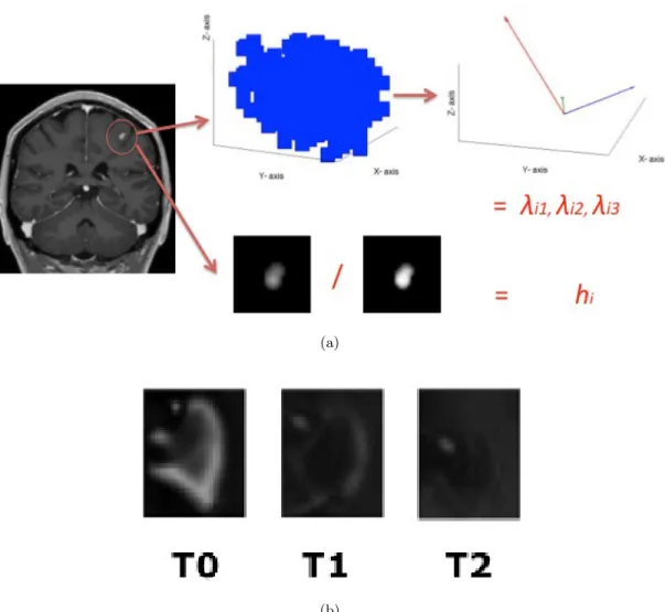
In the end, a tensor-like representation combined with a hollowness index was used. Figure 1(a) depicts the process for a single lesion, and Figure 1(b) depicts an example of Gd-enhanced lesion pattern for different time-points. Practically, the lesions are represented as 3D spatial tensors and by an hollowness index hi (see
equation (1)). This representation is also chosen because 75% of the lesions have ellipsoidal shapes or can be well approximated by an ellipsoid. The remaining 25% have a ringing shape similar to a torus9 . It can be demonstrated that a tensor-like representation of a torus produces the same eigenvectors as if the central holes would have been filled but slightly larger eigenvalues. To uniform the representation, the ring-like shape has been considered as filled and the hollowness is measured and concatenated to this information. We refer to the final section for further discussion about this aspect. The hollowness is given by the ratio
hi=
Adelineatedi
Af ulli
, (1)
where Adelineated is the lesion area directly obtained by the manual segmentation, and Af ull is the same area
with filled holes, this ratio should be 1 if there is no hole and becoming smaller than 1 if a hole is present. The ordered eigenvalues of these 3D tensors give a rotationally invariant feature representation: For each lesion at each time point, the coordinates of the n voxels belonging to a specific lesion are collected in the data matrix X = [(x1− µx), (y1− µy), (z1− µz)|(x2− µx), (y2− µy), (z2− µz)| . . . |(xn− µx), (yn− µy), (zn− µz)] regardless
are used for defining a covariance matrix Σ = n−11 XXT, which can be decomposed in its eigenvalues Λ and eigenvectors V :
Σ = V ΛVT. (2)
The eigenvectors are neglected since they represent the main directions of the lesions while the eigenvalues can represent the size of the lesions. All the three eigenvalues λ contained in the diagonal of Λ and the hollowness index for all m time points are the features representing the temporal evolution of the lesions. This means that for each single lesion, there is a feature vector fGdand fU SP IO given by
f = [λ01, λ02, λ03, h0, λ11, λ12, λ13, h1], (3)
containing the values respectively for the MRI volume Gd-enhanced and USPIO-enhanced, in decreasing order for each time point to be orientation independent, with λi1 ≥ λi2 ≥ λi3 for each of the m time point. Once the
lesions are represented for both contrast agent, the vectors fGdand fU SP IO of each lesion are concatenated in
a vector gGd−U SP IO. If one lesion has only either fGd or fU SP IO, the absence of the feature is represented
by zeros. The same lesion present in different time points m, is aligned to the other time representation of itself. This is performed registering all the manual delineation volume for the same patient to the volume at the first time point. If there is an overlap between the lesion of the different time point, then they are assessed automatically to be the same lesion and collected into the same g vector.
2.2 Patient features
The patient feature vector is given by all the cardinalities of the lesion pattern clusters nCi present in a patient
as
q = [nC1, nC2, nC3], (4)
e.g. nCi = 2 if a lesion pattern is present twice or nCi= 0 if it is not present in a specific patient. The following
subsection describes how the lesion and final patient clusters are obtained.
2.3 Clustering the lesion evolutions
The proposed framework is based on a two layers classification. Initially lesion patterns are identified, then the discovered patterns are used to identify group of patients. These two steps are performed by the same unsupervised clustering algorithm using the output of the first layer as the input of the second, i.e. the detected lesion patterns q are used as features for a second layer clustering at the patient level. Well-known unsupervised clustering algorithm are K-means, hierarchical clustering and GMM10 . These approaches generally work, but
they tend to fail when the nature of the data has a complicated structure or the cluster different sizes. Another possibility is to use spectral clustering11 , which is based on the point-to-point similarity matrix instead of the
estimate of an explicit data model. Hence, spectral clustering is adopted as clustering algorithm in the proposed framework.
Even with spectral clustering the first hurdle is the choice of parameters such as the cluster number. Since we are investigating the existence of possible patterns, we have no prior knowledge of the number of clusters and we rely on some validity indices. Many validity indices have been proposed, the reader can refer to12 for a comparison of some of them. The choice of the index depends on the nature of the data, on the subsequent used algorithm and on the number of available samples. We investigated the use of spectral-clustering and we estimated the cluster number with the algorithm proposed by Manor and Perona11related to spectral clustering.
2.4 Patient classification
Once the clustering of the lesions patterns is constructed, the detected q vectors representing the number of each detected lesion-based classes can be used to identify specific patients. Although the aim is not to predict TLL, the correlation between lesion patterns in patients and TLL is given to relate the patient classification to their disease severity aiming at the prediction of the most severe cases.
A regression model13 was used to examine the correlation between the number of detected lesion patterns -as described in section 2.1- and the future lesion load:
/ X. axis
-- A
I
/1i3 X.TO
Ti
T2
(a) (b)Figure 1. (a)The feature extraction process for a single time point. First all the lesions are delineated, then all the identified voxels are considered for computing the hallow index h and for’ building the covariance matrix and finally the eigenvalues are obtained from this covariance matrix. The process is repeated for all time points and the lesions which match in the different time point (b) are ordered in the same feature vector.
the coefficient β0 can be neglected because by construction of the model there will be at least one parameter
nCi non zero. The model examines the influence of the detected number of lesion patterns, since it is believed
that some lesion patterns are more indicative of the severity of the disease. The parameters βi were estimated
minimizing the Least-Squares linear regression given byP ||QB − T ||2, where B is the vector containing all the
βi, Q is the matrix containing all the number of patterns per patient q and T is vector containing the relative
total lesion load.
3. RESULTS
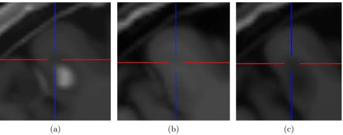
Comparing the enhancement for both contrast agents for the same lesions, it is noted that the USPIO-enhanced lesions show a different behavior than the Gd-lesions and are often visible as a mild ringing around the Gd-lesions as depicted in Figure 2. The lesions highlighted by USPIO are highlighted by Gd as well. Our pipeline results
(a) (b) (c)
Figure 2. The same lesion at the same time point: (a) Gd, (b) USPIO and (c) pre-contrast.
highlight that the same patient can have multiple lesions belonging to different lesion clusters Ci, C1 is the less
specific while the other two are more specific:
• C1comprises lesions of different dimensions (small, average, big) appearing at the first time point and then
disappearing, and generally only Gd.
• C2includes relatively average and big lesions present at both the first two time points, and with co-presence
of USPIO and Gd, where the Gd-enhanced lesions are focal and the USPIO-enhanced lesions have a ringing behavior.
• C3 comprises relatively average lesions present mainly at the first time point where the USPIO and Gd
lesion were both non-focal (ringing or non complete focal) and approximatively of the same size..
The statistical significance is assessed computing the cluster separation14, which is defined as the average distance
between each cluster centroids and the other centroids. Rank analysis over 1000000 resampled permutations of cluster separation led to the p-values < 0.0001.
The goodness of a regression model can be assessed through the R2 coefficient (also known as Coefficient
of determination)13 . The R2 coefficient measures of how well the regression line approximates the real data
points. An R2of 1.0 indicates that the regression line perfectly fits the data. The most general definition of the
coefficient is
R2= 1 − SSerr SSerr+ SSreg
, (6)
where SSerr is the the squared sum difference between the predicted value ˆT and the reference T . SSreg is the
squared sum difference between the predicted value and the mean value of T . Also this measurement is repeated in a leave one patient out cross-validation manner.The mean and variance of all the obtained R2coefficients for the features gGd−U SP IO was 0.68 ± 0.19.
Patient ID lesion clusters (and multiplicity) TLL in cm3 at m18 6 C2(×1), C3(×2), C1(×37) 28.9 11 C1(×1) 9.70 9 C2(×1) 6.72 10 C2(×1), C1(×9) 5.70 16 C3(×1) 4.70
24 no active lesions in the first 2 time points 4.63
4 C2(×1), C1(×2) 4.29 13 C1(×2) 3.54 21 C1(×2) 3.50 18 C1(×4) 3.40 25 C1(×2) 3.32 12 C1(×6) 2.10 7 C1(×9) 2.10 2 C1(×1) 1.90 19 C1(×1) 1.73 5 C1(×2) 1.70 22 C1(×4) 1.27
1 no active lesions in the first 2 time points 1.18
17 C1(×1) 1.18
8 C1(×1) 1.14
3 no active lesions in the first 2 time points 0.98
15 no active lesions in the first 2 time points 0.68
20 no active lesions in the first 2 time points 0.54
23 no active lesions in the first 2 time points 0.49
14 no active lesions in the first 2 time points 0.29
Table 1. List of patients with the relative detected patterns, and TLL in cm3 by follow-up (15 months after the m3).
The colors highlights the different group of patients. The patients highlighted in red have at least one lesion pattern belonging either to the cluster C2 or to the cluster C3, which it is considered an indication of severe condition. The
patients highlighted in blue have only one lesion pattern C1, this is the mildest condition which is clearly related to an
overall moderate TLL. The patients highlighted in green are the patients which are excluded from the clustering since they do not present active lesions at the first two time points. The other cases are simply colored in black. It was noted that except patient 11 and 24, all patients correlate with their relative TLL. The lesion of patient 9 to be correctly clustered required a tuning of the neighborhood parameter for the graph construction of the spectral clustering, while the other lesions were less sensitive to this parameter.
Table 1 shows the heterogenous distribution of patterns automatically obtained, which correlates with the future total lesion load (second layer), only patient 11 and 24 seem not correlating properly. The lesion of patient 9 to be correctly clustered required a tuning of the neighborhood parameter for the graph construction of the spectral clustering, while the other lesions were less sensitive to this parameter. The lesion load is reported as unnormalized measure in cm3. The patients are assigned by the algorithm to groups which can represent
different degrees of disease severity.
4. NEW OR BREAKTHROUGH WORK TO BE PRESENTED
A novel paradigm for spatio-temporal analysis of MS lesions has been proposed. It has been applied to a novel contrast agent USPIO that appears to give complementary information compared to the traditional Gd. We devised a two layers classification, first lesion patterns are discovered and then patients are grouped according to the lesion patterns. This final patient classification - performed at the very early stages- correlates with the future TLL after 15 months from the last used time point, this might indicate an even further evolution5 . This
5. CONCLUSIONS
The framework is able to classify lesions and MS patients from the very early stages (2 first MR scans), this classification seems to be well aligned to the TLL by follow-up, which is so far the only image-based figure that can potentially correlate to future evolution of the pathology5, 6. A further cluster is given by the lesions which
are not detected since they do not absorb any contrast agent at the first two time points. USPIO-enhanced lesions show different behaviors than Gd-enhanced lesions which are more pronounced in some lesions rather than others correlating with future lesion loads. This leads to the hypothesis that USPIO can help to mark lesions with high risk to decline to a more severe course. Although as suggested in,9USPIO could also highlight healing process, the presence of either lesion pattern C2or C3 is a sign of intense activity.
In fact, this characterization suggests as well that belonging to a specific group of patients has an incidence on the future evolution of the disease. Although promising, the predictive value of USPIO needs to be validated on larger populations.
Lesion segmentation can be automated with a segmentation method15 . The tensor-like representation for
lesions is motivated by the rotationally invariant eigenvalues and the analogy to PCA. This representation can oversimplify the complexity of the lesion enhancement even if still informative. Using the eigenvalues of the Laplace-Beltrami operator8as features can solve the issue of the hole in the ringing lesions. However, preliminary
experiments showed that this method would require a very high dimensional representation which is unfeasible with the current dataset. Although, dimensionality reduction has been recently investigated with regard to the Laplace-Beltrami operator16 , a correct feature selection with the current settings does not seem appropriate.
Future works are also given by different validity indices for clustering and by the inclusion of other MRI quantitative measurements (such as Magnetization Transfer, Diffusion MRI or relaxometry), and by comparing the classification to the clinical data available. In summary, the framework can predict future TLL and can provide an additional classification of the different type of lesions across patients that an actual lesion load cannot provide.
REFERENCES
[1] Leray, E., Yaouanq, J., Page, E. L., Coustans, M., Laplaud, D., Oger, J., and Edan, G., “Evidence for a two-stage disability progression in multiple sclerosis,” Brain 133, 1900–1913 (2010).
[2] Lucchinetti, C., Brueck, W., Parisi, J., Scheithauer, B., Rodriguez, M., and Lassman, H., “Heterogeneity of multiple sclerosis lesions: Implications for the pathogenesis of demyelination,” Annals of Neurology 47, 707–717 (2000).
[3] Bitsch, A., Schuchardt, J., S.Bunkowski, Kuhlmann, T., and Brueck, W., “Acute axonal injury in multiple sclerosis,” Brain 123, 1174–1183 (2000).
[4] Dousset, V., B.Doche, Petry, K., Brochet, B., Delalande, C., and Caille, J., “Correlation between clinical status and macrophage activity imaging in the central nervous system of rats,” Academic Radiology 9, 156–9 (2002).
[5] Brex, P., Ciccarelli, O., O’Riordan, J., Sailer, M., Thompson, A., and Miller, D., “A longitudinal study of abnormalities on MRI and disability from multiple sclerosis,” New England Journal of Medicine 346, 158–164 (2002).
[6] Filippi, M., Horsfield, M., Morrissey, S., MacManus, D., Rudge, P., McDonald, W., and Miller, D., “Quan-titative brain lesion load predicts the course of CIS,” Neurology 44, 635–641 (1994).
[7] Haermae, V., Virtanen, J., Maekelae, R., Happonen, A., Mpindi, J., Knuuttila, M., Kohonen, P., Loetjoenen, J., Kallioniemi, O., and Nees, M., “A comprehensive panel of three-dimensional models for studies of prostate cancer growth, invasion and drug responses,” PLoS One 5, 245–267 (2010).
[8] Reuter, M., Wolter, F., and Peinecke, N., “Laplace-Beltrami spectra as shape-dna of surfaces and solids,” CAD 38, 342–366 (2006).
[9] Vellinga, M., Oude-Engberink, R., Seewann, A., Pouwels, P., Wattjes, M., van-der Pol, S., Pering, C., Polman, C., de Vries, H., Geurts, J., and Barkhof, F., “Pluriformity of inflammation in multiple sclerosis shown by ultra-small iron oxide particle enhancement,” Brain 131, 800–807 (2008).
[11] Zelnik-Manor, L. and Perona, P., “Self-tuning spectral clustering,” in [NIPS ], 17 (2004).
[12] Maulik, U. and Bandyopadhyay, S., “Performance evaluation of clustering algorithms,” IEEE PAMI 24, 1650–1654 (2002).
[13] Steel, R. and Torrie, J., [Principles and Procedures of Statistics ], vol. 70, New York: McGraw-Hill (1960). [14] Gokcay, E. and Principe, J., “Information theoretic clustering,” IEEE PAMI 24, 158–71 (2002).
[15] Karimaghaloo, Z., Shah, M., Francis, S., Arnold, D., Collins, D., and Arbel, T., “Automatic detection of Gd-enhancing multiple sclerosis lesions in brain MRI using conditional random fields,” IEEE TMI 31, 1181–1194 (2012).
[16] Bernardis, E., Konukoglu, E., Ou, Y., Metaxas, D., Desjardins, B., and Pohl, K. M., “Temporal shape analysis via the spectral signature,” in [15th International Conference on Medical Image Computing and Computer Assisted Intervention ], (2012).