HAL Id: hal-01387994
https://hal.archives-ouvertes.fr/hal-01387994
Submitted on 27 Sep 2017HAL is a multi-disciplinary open access
archive for the deposit and dissemination of sci-entific research documents, whether they are pub-lished or not. The documents may come from teaching and research institutions in France or abroad, or from public or private research centers.
L’archive ouverte pluridisciplinaire HAL, est destinée au dépôt et à la diffusion de documents scientifiques de niveau recherche, publiés ou non, émanant des établissements d’enseignement et de recherche français ou étrangers, des laboratoires publics ou privés.
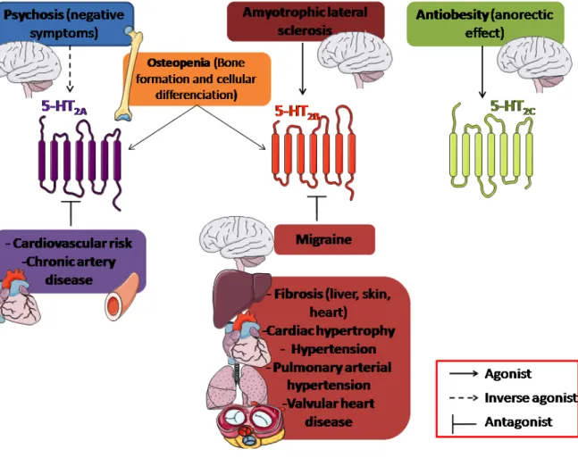
New therapeutic opportunities for 5-HT2 receptor
ligands
Luc Maroteaux, Estelle Ayme-Dietrich, Gaëlle Aubertin-Kirch, Sophie Banas,
Emily Quentin, Roland Lawson, Laurent Monassier
To cite this version:
Luc Maroteaux, Estelle Ayme-Dietrich, Gaëlle Aubertin-Kirch, Sophie Banas, Emily Quentin, et al.. New therapeutic opportunities for 5-HT2 receptor ligands. Pharmacology and Therapeutics, Elsevier, 2016, 170, pp.14 - 36. �10.1016/j.pharmthera.2016.10.008�. �hal-01387994�
New therapeutic opportunities for 5-HT2 receptor ligands
Luc Maroteauxb, Estelle Ayme-Dietricha, Gaëlle Aubertin-Kircha, Sophie Banasb, Emily Quentinb, Roland Lawsona, Laurent Monassiera, *
aLaboratoire de Neurobiologie et Pharmacologie Cardiovasculaire EA7296, Faculté de
Médecine, Fédération de Médecine Translationnelle de Strasbourg, Université et Centre Hospitalier de Strasbourg, Strasbourg, France
bINSERM UMR S-839, Institut du Fer à Moulin, Université Pierre et Marie Curie, 17 rue du
Fer à Moulin 75005 Paris, France
Short title: 5-HT2 receptors, therapeutic
Luc Maroteaux and Estelle Ayme-Dietrich contributed equally to this review and are considered as co-authors.
* Author for correspondence and reprint requests: Laurent Monassier MD, PhD
Laboratoire de Neurobiologie et Pharmacologie Cardiovasculaire, Faculté de Médecine, 11 rue Humann, 67085, Strasbourg Cedex
Tel. : +33368853392 Fax : +33368853388
e-mail : [email protected]
Keywords: 5-HT2 receptors, selectivity, pathological function, therapeutic track ABSTRACT
Serotonergic dysfunction is mainly associated with neuropsychiatric and cardiovascular disorders but has also been linked with many other pathological situations. Serotonin (5-hydroxytryptamine, 5-HT) mediates numerous physiological functions in the brain and the periphery by activating a variety of receptors. 5-HT receptors are divided in four classes, three of them belonging to the G-protein coupled receptor family. This review provides an overview of the recent pharmacological developments involving the Gq-coupled 5-HT2
receptors subfamily as well as the pathological implications of this receptor subfamily with regard to fibrosis, the central nervous system, cardiovascular disorders and cancer. The final section highlights new therapeutic opportunities and emerging research revealing unexplored medical opportunities for this class of 5-HT receptors. The development of biased 5-HT2
receptor ligands appears to be an interesting topic in various areas. In the light of recent discoveries, the need for the development of new and safer drugs should take into account the risk of cardiovascular side effects such as pulmonary hypertension and heart valve disease.
Contents
1. Introduction
2. 5-HT2 receptors: structure, coupling, oligomerization, selective ligands, allosteric
modulators, biased agonists 3. Pathologies and 5-HT2 receptors
4. Emerging research and new therapeutic opportunities 5. Conclusions and prospects
Conflict of interest Acknowledgments References
1. Introduction
This review focuses on three serotonin (5-hydroxytryptamine, 5-HT) receptors belonging to the 5-HT2 receptor subfamily: the 5-HT2A, 5-HT2B and 5-HT2C subtypes. Although the work by
D. Julius et al.,(1988) was the first reporting the cloning of a full-length functional serotonin receptor from rat, the 5-HT1c receptor, this publication was shortly followed by considerable
efforts from several groups that cloned other unidentified 5-HT receptors. The classical 5-HT2
receptor described by Peroutka et al.,(1981) was cloned in rats slightly later in 1988 (Pritchett, et al., 1988) followed by human analogue (Branchek, et al., 1990; Saltzman, et al., 1991) and was renamed 5-HT2A. The 5-HT1c receptor was renamed 5-HT2C because of its
structural similarity to the other 5-HT2 receptor, identical second messenger pathways, and
similar pharmacological properties. Pharmacological studies attempting to characterize the contractile serotonergic receptor in the rat stomach fundus initially documented its similarity to the 5-HT2C receptor. Despite the absence of detectable 5-HT2C receptor mRNA in the rat
stomach fundus, only homology cloning permitted the identification of a new receptor in 1992 in rat and mouse that was named 5-HT2B (Foguet, Hoyer, et al., 1992; Foguet, Nguyen, et al.,
1992; Kursar, et al., 1992; Loric, et al., 1992; Wainscott, et al., 1993) and in 1994 in humans (Choi, et al., 1994; Kursar, et al., 1994; Schmuck, et al., 1994; Wainscott, et al., 1996).
The investigation on the contribution of these three 5-HT2 receptors in mammalian
physiology leads to a large amount of reports in nearly all functions and organs. Some selective compounds stimulating or blocking these receptors provided an opportunity to explore various areas of human diseases. In this review, we will emphasize some important aspects of the cellular and molecular biology of these receptors and highlight some clinical situations in which these receptors appear as pathophysiological cornerstones.
2. 5-HT2 receptors: structure, coupling, oligomerization, selective ligands, allosteric
modulators, biased agonists
The closely related 5-HT2 receptors are members of the rhodopsin family of G protein–
coupled receptors (GPCRs) that all activate multiple intracellular signaling networks. The classical signal transduction pathway for this subfamily is Gq/11-coupled activation of phospholipase C (PLC) although these 5-HT2 receptors can also activate phospholipase D
and phospholipase A2 by interacting with additional pathways. These 5-HT2A, 5-HT2B and
5-HT2C receptors are post-transcriptionally modified by alternative RNA splicing, a common
mechanism for achieving protein diversity. RNA editing, on the other hand, is a less common process for generating molecular diversity. In fact, the 5-HT2C receptor is one of the few
GPCRs known to be edited. RNA editing of the 5-HT2C receptor generates functionally
distinct protein variants by altering the genetic code at the mRNA level.
2.1. Structure
5-HT2 receptors are 7 transmembrane domain receptors, with fairly long extracellular
N-terminal loops ranging from 55 amino acids for the human 5-HT2B and 5-HT2C receptors to 75
amino acids for the human 5-HT2A receptors and an intracellular C-terminus ranging from 85
amino acids for the human 5-HT2B receptors to 75 amino acids for the human 5-HT2A and
5-HT2C receptors. A new and unanticipated role of the 5-HT2B receptor N-terminus as a
negative modulator, affecting both constitutive and agonist-stimulated activity of the receptor has been shown (Belmer, et al., 2014). The recently published crystal structure of the 5-HT2B
receptor bound to ergotamine showed that this receptor exhibits conformational characteristics in both the active and inactive states: an active-like state in the helix VII conformation of the 5-HT2B receptor but only partial changes in helix VI. The differential
signaling patterns were also mirrored in the crystal structures, which showed features of a β-arrestin-biased activation state for the 5-HT2B receptor (Wacker, et al., 2013; Wang, et al.,
2013). A likely structural explanation for the distinct conformational features and biased pharmacology of ergotamine for 5-HT2B receptors can be found in the region of the
extracellular loop 2 (ECL2) junction with helix V (E212-R213-F214), which forms an additional helical turn stabilized by a structured water molecule at the extracellular tip of helix
V. The segment of ECL2 connecting helices III and V via the conserved disulfide bond is shortened in the 5-HT2B receptor, and creates a conformational constraint on the position of
the extracellular tip of helix V (Martí-Solano, et al., 2014). However, this structured water molecule involved in ECL2 junction with helix V has been challenged since differential interactions of ergotamine with the top of helices V and VI could determine the rotational freedom of helix VI (Liu, et al., 2013). No crystal structures have reported yet for 5-HT2A or
5-HT2C receptor.
More work is needed to precisely understand the structure and function of these receptors and their specific properties.
2.2. Selective agonists
There is virtually no highly selective agonist for a particular 5-HT2 receptor:
- BW723C86: 1-methyl-2- [5-(2-thienylmethoxy)-1H-indole-3-yl] ethylamine hydrochloride, has been reported to have 10-fold selectivity over the human 5-HT2C and 100-fold selectivity
over the 5-HT2A receptors (Cussac, et al., 2008; Jerman, et al., 2001; Knight, et al., 2004;
Porter, et al., 1999). Lorcaserine [(1R)-8-chloro- 2,3,4,5-tetrahydro-1-methyl-1H-3 benzazepine] has approximately 10-fold higher affinity for 5-HT2C receptor (Thomsen, et al.,
2008) over 5-HT2A and 5-HT2B receptors.
- Nor-dexfenfluramine (metabolite of dexfenfluramine), methylergonovine (metabolite of methysergide), and Ro 60-0175: 2(S)-1-(6-chloro-5-fluoro-1H-indol-1-yl)-2-propanamine fumarate are all preferential 5-HT2B agonists with about 10-fold selectivity over 5-HT2C
receptor (Cussac, et al., 2002).
- 2,5-dimethoxy-4-iodoamphetamine (DOI) is a non-selective nearly full agonist at 5-HT2
receptors with similar affinity to 5-HT2A 5-HT2B and 5-HT2C receptors (Cussac, et al., 2008;
Jerman, et al., 2001; Knight, et al., 2004; Porter, et al., 1999).
- alpha-methyl-5-HT is a non-selective nearly full agonist at 5-HT2 receptors with similar
affinity to 5-HT2A 5-HT2B and 5-HT2C receptors (Jerman, et al., 2001; Knight, et al., 2004;
Porter, et al., 1999).
2.3. Selective antagonists
A few selective antagonists are available for 5-HT2 receptor subtypes:
- The first highly selective 5-HT2A receptor antagonist reported was MDL100907
[(R)-(+)-α-(2,3- dimethoxyphenyl)-1-[2-(4-fluorophenylethyl)]-4-piperidine- methanol] (Knight, et al., 2004). Sarpogrelate [Succinic acid mono-(1-dimethylaminomethyl-2-(2-[2-(3-methoxyphenyl) ethyl] phenoxy) ethyl) ester hydrochloride], SR46349B [4-((3Z)-3-(2-dimethylaminoethyl)oxyimino-3-(2-fluorophenyl)propen-1-yl)phenol hemifumarate], and ketanserin [3-[2-[4-(4-fluorobenzoyl)piperidin-1-yl]ethyl]quinazoline-2,4(1H,3H)-dione] are preferential 5-HT2A receptor antagonists with a 10-fold higher affinity over the 5-HT2C and/or
5-HT2B sites.
- The first highly selective 5-HT2B receptor antagonist reported was LY266097:
1-(2-chloro-3,4- dimethoxybenzyl)- 6-methyl- 1,2,1-(2-chloro-3,4-tetrahydro- 9Hpyrido [1-(2-chloro-3,4-b]indole hydrochloride with a pKi of 9.7 for the human cloned 5-HT2B receptor and a 100-fold greater selectivity over
human 5-HT2C and 5-HT2A sites (Audia, et al., 1996). SB204741:
N-(1-methyl-5-indolyl)-N'-(3-methyl-5-isothiazolyl)urea has been reported as a selective 5-HT2B receptor antagonist with
approximately 100-fold selectivity over the 5-HT2C and 5-HT2A receptors but with a low
potency (Ki around 100 nM) (Bonhaus, et al., 1995). The tetrahydro-β-carboline, LY272015 [6-chloro-5-methyl -N-(5-quinolinyl) -2,3-dihydro -1H-indole-1-carboxamide] is also a fairly selective and highly potent antagonist (Cohen, et al., 1996). RS127445 [2-amino-4-(4-fluoronaphth-1-yl)-6-isopropylpyrimidine] was found to have sub-nanomolar affinity for the 5-HT2B receptor (pKi = 9.5) and 1,000 fold selectivity for this receptor as compared to
numerous other receptor and ion channel binding sites and appears as the most selective, high affinity 5-HT2B receptor antagonist available now (Bonhaus, et al., 1999). SB215505
[6-chloro-5-methyl-N-(5-quinolinyl)-2,3-dihydro-1H-indole-1-carboxamide] behaves as a high affinity and preferential inverse agonist at 5-HT2B receptors (Reavill, et al., 1999).
- SB242084 [6-chloro-5-methyl-1-[6- (2-methylpyridin-3-yloxy)pyridin-3-yl-carbamoyl]indoline] and RS-102221 [N-[5-[5-(2,5-dioxo- spiro[imidazolidine-4,4’-piperidin]-1’-yl)pentanoyl]-2,4-dimethoxy- phenyl]-4-(trifluoromethyl)benzenesulfonamide]are selective 5-HT2C
antagonists(Bonhaus, et al., 1997; Knight, et al., 2004).
- SB206553 [5-methyl-N-(3-pyridyl)-1,2,3,5-tetrahydrobenzo[1,2-b:4,5-b']dipyrrole-1-carboxamide] is a mixed 5-HT2C/5-HT2B receptor antagonist. It has been reported as a
selective 5-HT2C/2B receptor inverse agonist with 50- to 100-fold lower affinity for the 5-HT2A
and other sites (Kennett, et al., 1996; Knight, et al., 2004).
Non-selective 5-HT2 receptor antagonists such as ritanserin and mesulergine block
5-HT2 receptor-mediated effects. Atypical antipsychotics including clozapine, asenapine, or
cariprazine also have fairly high affinity for all 5-HT2 receptors (Kiss, et al., 2010; Millan, et
al., 2003; Shahid, et al., 2009; Wainscott, et al., 1996). Aripiprazole (OPC-14597) is a novel atypical antipsychotic drug, which has higher antagonist affinity (EC50 = 11 nM) for the
human 5-HT2B receptor than at the 5-HT2A or 5-HT2C receptors (Shapiro, et al., 2003).
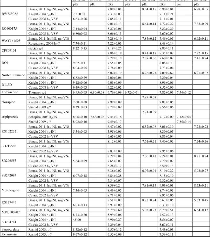
See table 1. and PDSP database, http://kidbdev.med.unc.edu/databases/pdsp.php
2.4. Coupling
Intracellular signals are inherently challenging targets because they are often ubiquitous; indeed, drug vectorization comes from GPCRs, not transduction pathways. Nonetheless, multi-target drugs acting at two key nodes in a signaling network offer one answer. Another approach to this concept is the manipulation of interactions between 5-HT receptors and their protein partners. One good example is the use of small peptides to decouple 5-HT2C
receptors from their PDZ partners, which mimics the desensitization elicited by antidepressants (Gavarini, et al., 2006). Conversely, blocking the interactions between 5-HT2C receptors and the phosphatase PTEN (phosphatase with tensin homology) reproduces
the 5-HT2C agonist-induced inhibition of the excitation of mesolimbic dopaminergic neurons
by cannabinoids, thereby preventing their rewarding effects (Ji, et al., 2006). Hence, interference with the association between 5-HT2C receptors and PTEN might be an
interesting way to counteract drug addiction.
β-Arrestins direct the agonist-induced internalization of 5-HT receptors; however, agonist-independent association with β-arrestins has been reported for non-edited (and partially edited) 5-HT2C receptors (Marion, et al., 2004). This interaction leads to constitutive
internalization (Marion, et al., 2004), an effect prevented by inverse agonists (Chanrion, et al., 2008). Calmodulin binds to the proximal region of the 5-HT2C receptor C-terminus upon
receptor activation by HT. Mutation of this motif inhibits both β-arrestin recruitment to the 5-HT2C receptor and receptor-operated ERK1,2 signaling in HEK-293 cells, which is
independent of G proteins and dependent on β-arrestins. Expression of the calmodulin mutant also prevents receptor-mediated ERK1,2 phosphorylation in cultured cortical neurons and choroid plexus epithelial cells that endogenously express 5-HT2C receptors (Labasque,
et al., 2008). Intriguingly, although β-arrestins are implicated in hallucinogenic effects mediated by 5-HT2A receptors, their trafficking is independent (Bhatnagar, et al., 2001). The
β-Arrestin-2 directs also agonist-induced internalization of 5-HT2B receptors (Janoshazi, et al.,
2007). For instance, β-arrestins contribute to activation of ERK by 5-HT2A, 5-HT2B, and
5-HT2C receptors.
PDZ (postsynaptic-density-95/disc-large/zonulla-occludens-1)-domain-containing proteins profoundly influence internalization of 5-HT2A,2B,2C receptors, and PDZ proteins are
essential for targeting 5-HT2A receptors to dendrites in cortical neurons (Xia, Hufeisen, et al.,
2003). PDZ partners are both receptor and function specific. Thus, the specific sets of PDZ proteins that interact with 5-HT2A, 5-HT2B, or 5-HT2C receptors differ. Although PSD-95
(postsynaptic-density-95) prevents 5-HT2A receptor internalization (Xia, Gray, et al., 2003), it
favors constitutive and agonist-dependent endocytosis of 5-HT2C receptors (Gavarini, et al.,
2006). Conversely, MPP3 (membrane protein palmitoylated 3) stabilizes 5-HT2C receptors at
the plasma membrane (Gavarini, et al., 2006). The 5-HT2B receptor shares the C-terminal
E-X-V/I-S-X-V sequence with 5-HT2C receptors and also binds MUPP1-PDZ domains in-vitro
C-terminus of the 5-HT2C receptor. Moreover, 5-HT2A and 5-HT2B receptors sharing the
C-terminal -E-X-V/I-S-X-V sequence with 5-HT2C receptors also bind MUPP1-PDZ domains in-vitro (Becamel, et al., 2001). The PDZ motif at the C-terminus of the 5-HT2B receptor was
also found necessary for the recruitment of the constitutive NO synthase (cNOS-NOS3) (Manivet, et al., 2000). In addition, stimulation of the 5-HT2B receptor triggered intracellular
cGMP production through dual activation of NOS3and inducible NOS (iNOS-NOS2). The group I PDZ motif at the carboxy terminus of the 5-HT2B receptor was shown to be required
for recruitment of the NOS3 transduction pathways, and NOS2 stimulation was under control of the Gα13 pathways (Manivet, et al., 2000).
The human polyomavirus JCV causes the fatal demyelinating disease progressive multifocal leukoencephalopathy in immuno-compromised patients. Elphick et al. (2004) found that the serotonergic receptor 5-HT2A could act as the cellular receptor for JCV on human
glial cells. 5-HT2A receptor antagonists inhibited JCV infection, and monoclonal antibodies
directed at 5-HT2A receptors blocked infection of glial cells by JCV, but not by SV40.
Transfection of 5-HT2A receptor-negative HeLa cells with a 5-HT2A receptor rescued virus
infection, and this infection was blocked by antibody targeting the 5-HT2A receptor. A tagged
5-HT2A receptor colocalized with labeled JCV in an endosomal compartment following
internalization. Later, it was observed that endothelial cells not expressing 5-HT2A receptor
could be infected. Following this observation, it was reported that virus entry into HEK293A cells was specifically observed when any 5-HT2A,2B,2C receptors were expressed. Recent data
confirmed that virus internalization into HEK293A cells was significantly and specifically allowed by either 5-HT2A,2B,2C serotonin receptors in a way somewhat similar to CCR5
chemokine receptor, which acts as a co-receptor for HIV-1 viral entry. This work shows that 5-HT2A,2B,2C serotonin receptors contribute to JCPyV infection by facilitating viral entry
(Assetta, et al., 2013).
A better understanding of virus/5-HT2interactions may lead to new antiviral
opportunities.
2.5. Oligomerization
Oligomeric associations of GPCRs often comprise signaling units, and 5-HT2A, 5-HT2B, and
5-HT2C receptors may all form homo- or heterodimers (Brea, et al., 2009; Herrick-Davis, et al.,
2004; Jaffre, et al., 2009). In contrast to some classes of GPCRs, agonist binding does not greatly influence formation of 5-HT-receptor dimers, indicating constitutive assembly before membrane insertion. For example, 5-HT2C receptors generate dimers in the endoplasmic
reticulum and Golgi of living cells (Herrick-Davis, et al., 2005). 5-HT2C receptor dimers
possess an interface between transmembrane helices IV and V, and dimer proximity is increased and decreased by agonists and inverse agonists, respectively (Mancia, et al., 2008). Furthermore, analysis of functionally compensating, co-expressed mutant 5-HT2C
receptors linked to Gq (fusion proteins) indicates that the dimer is asymmetric versus Gq, with both subunits binding to 5-HT and having distinct roles during signaling(Herrick-Davis, et al., 2005; Mancia, et al., 2008).
The 5-HT2A and metabotropic glutamate 2 (mGlu2) receptors assemble into
heterodimers via a transmembrane-IV and -V linking domain (Gonzalez-Maeso, et al., 2008). Intriguingly, mGlu2 receptor agonists blunt heterodimer coupling to Gi providing one substrate for their anti-hallucinogenic properties (Fribourg, et al., 2011; Gonzalez-Maeso, et al., 2008). Direct evidence has been acquired for mGlu2-5-HT2A heterodimers in the human
brain and for a reduced density in schizophrenics (Gonzalez-Maeso, et al., 2008). Nonetheless, this remains one of the few demonstrations of heterodimers in tissues. Of particular interest are ligands specific for 5-HT2A-mGlu2 complexes, or other putative
heterodimers possessing distinctive binding and coupling profiles (Gonzalez-Maeso, et al., 2008). Evidence for a functional crosstalk between 5-HT2A and D2 receptor were reported in
HEK293 cells. D2 receptor activation increases the hallucinogenic agonist affinity for 5-HT2A
receptor and decreases the inositol phosphate production. Co-immunoprecipitation studies show that the two receptors can physically interact in HEK293 cells and raise the possibility that a receptor heterocomplex mediates the crosstalk observed. In vivo, 5-HT2A receptor
expression is necessary for the full effects of D2 antagonist on MK-801-induced locomotor
activity (Albizu, et al., 2011). Behavioral studies carried out in mice lacking 5-HT2A receptors
revealed also a remarkable 5-HT2A receptor-dependent dissociation in the beneficial
antinociceptive effects of cannabinoid CB1 receptors agonist delta9-tetrahydrocannabinol (THC) and its detrimental amnesic properties. Biochemical studies showed that CB1 and 5-HT2A receptors form heteromers that are expressed and functionally active in specific brain
regions involved in memory impairment. Remarkably, costimulation of both receptors by agonists reduces cell signaling, antagonist binding to one receptor blocks signaling of the interacting receptor, and heteromer formation leads to a switch in G-protein coupling for 5-HT2A receptor from Gq to Gi proteins (Vinals, et al., 2015).
In cardiac fibroblasts, AT1 angiotensin and 5-HT2B receptors have been reported to
share common signaling pathways, which support a possible direct interaction between 5-HT2B and AT1 receptors. Using co-immunolocalization and a pull-down assay, the two
receptors were shown to interact together, which suggested that these receptors could exist in heterodimeric complexes (Jaffre, et al., 2009), but in vivo experimental confirmation is still lacking. Ghrelin, an orexigenic peptide present in the stomach, has gastroprokinetic properties. In vivo studies have shown that the ghrelin receptor (GHS-R1a) antagonist D-Lys(3)-GHRP-6 reduces food intake and delays gastric emptying in rodents but these effects are at variance with the normal phenotype of the ghrelin knockout mice. D-Lys(3)-GHRP-6, induced a pronounced contraction of stomach strips that is blocked by the 5-HT2 receptor
antagonists methysergide and yohimbine resulting from smooth muscle contractions and suggesting the possibility of direct interactions with 5-HT2B receptors (Depoortere, et al.,
2006).
A possibility for GHS-R1a/5-HT2C dimer-induced attenuation of calcium signaling was
also observed. Flow cytometry fluorescence resonance energy transfer (fcFRET) assays confirmed the direct interaction between the GHS-R1a receptor and 5-HT2C receptor.
Colocalized expression of the 5-HT2Cand GHS-R1a receptor in cultured primary
hypothalamic and hippocampal rat neurons further supports the biological relevance of such physiological interaction. When 5-HT2C receptor signaling is blocked, ghrelin's orexigenic
effect is potentiated in vivo. In contrast, the 5-HT2C receptor preferential agonist lorcaserin
attenuates ghrelin-induced food intake (Schellekens, et al., 2015). Physical associations of
the melatonin MT2 and 5-HT2C receptors as functional heteromers were also found by
co-immunoprecipitation, bioluminescence resonance energy transfer, and
pharmacological techniques both in transfected cells and in cells from human cortex
and hippocampus. MT2/5-HT2C heteromers amplify the 5-HT-mediated
Gq/phospholipase C response and trigger melatonin-induced unidirectional transactivation of the 5-HT2C protomer of MT2/5-HT2C heteromers. Pharmacological
studies reveal distinct functional properties for agomelatine, which shows "biased signaling" (Kamal, et al., 2015).
Future studies must focus on the putative heterodimerization of native 5-HT receptors and on their pharmacological profiles in the hope of identifying novel targets for therapeutic intervention.
2.6. Allosteric modulators
Positive allosteric modulators (PAMs) represent alternative approaches to orthosteric agonists (i.e., compounds that interact with the native ligand-binding site). PAMs can increase the affinity and/or efficacy of the orthosteric agonist for its target receptor by acting at a site other than the native ligand-binding site (allosteric). Importantly, so-called pure GPCR PAMs, which lack intrinsic agonist activity within a specific signaling pathway, have been described. These compounds modulate the basal tone of the endogenous ligand in a manner that conserves spatial and temporal elements of native neurotransmission (Christopoulos & Kenakin, 2002). Indeed, multiple PAMs have been identified for GPCRs and may circumvent the challenges of orthosteric agonists. First, PAMs would amplify endogenous signaling through the 5-HT2 receptors, likely resulting in a more physiologically
of a generally higher sequence divergence in allosteric sites relative to the conserved orthosteric domain, PAMs could potentially achieve higher receptor selectivity than orthosteric agonists. Indeed, some 5-HT2C receptor PAMs have been reported although as
yet the pharmacological profiles of these compounds have not been widely reported (Ding, et al., 2012). Ergotamine has been shown to occupy two distinct sites in 5-HT2B receptors, the
orthosteric site, where the indole nucleus of ergotamine resides, and the extended binding site, where the tripeptide portion is engaged. The allosteric site in the muscarinic M2 receptor is the same extracellular region as that interacting with the tripeptide portion of ergotamine. These similarities in both the M2 and 5-HT2B receptors suggest that the location of the
extracellular allosteric site for Class A GPCRs is quite similar, and in fact, argue that ergotamine likely functions as a bitopic ligand; that is it occupies both the orthosteric and putative extracellular allosteric site in the 5-HT2B receptor. It is now thought that a sodium ion
allosterically alters the binding pocket to dampen G-protein signaling, leaving β-arrestin recruitment intact. Recent structural consideration support that this sodium pocket is collapsed in the 5-HT2B receptor structure, (McCorvy & Roth, 2015).
The identification of specific PAMs at 5-HT2 receptors may conceivably lead to
improved therapeutics.
2.7. Biased agonists
Another area for 5-HT2 receptors agonist development might emerge from compounds
so-called biased agonists sharing a functional selectivity for specific intracellular signaling pathways (Kenakin, et al., 2012). 5-HT2 receptors couple to multiple intracellular pathways
including PLC and PLA2 and pharmacological evidences using recombinant cell-based
systems suggest that non-selective 5-HT2 agonists such as mCPP and quipazine may
differentially activate these signaling pathways downstream from the 5-HT2C receptor (Berg,
et al., 1998). LSD and ERG displayed bias for β-arrestin signaling at 5-HT2B receptors, as
well as other ergolines such as dihydroergotamine, methylergonovine, pergolide, and cabergoline. ERG and DHE, both of which contain a large tripeptide moiety substitution at the amide scaffold, displayed more extreme signaling bias at the 5-HT2B receptor compared
to LSD (Wacker, et al., 2013). A further approach is the identification of compounds with 5-HT2C receptor agonist activity combined with antagonist activity at the 5-HT2A receptor.
Experimental evidence supports a potential synergy between these two pharmacological properties, raising the theoretical possibility that a single drug possessing both characteristics may be a superior therapeutic compared to either alone (Booth, et al., 2009; Canal, et al., 2014; Cunningham, et al., 2013; Pockros, et al., 2012).
Currently, it is unknown whether this example of functional selectivity could be translated into any therapeutic gain, although this does open up an interesting opportunity for future drug discovery.
3. Pathologies and 5-HT2 receptors
3.1. Fibrosis and inflammation
Healing is the process of the restoration of health in an unbalanced, diseased or damaged organism. Fibrogenesis is a critical process in wound repair that generates scar tissue. It helps to protect an injured organ until damaged or lost cells are regenerated. Where an injury naturally resolves, the fibrogenic response is usually limited and the temporary scar tissue replaced by healthy functional cells (Mann & Oakley, 2013). States of chronic tissue infection or damage, ageing, tumors, feeding habits and/or exposure to drugs that generate microenvironments in which the inflammatory and fibrogenic phases of wound healing fail to resolve, cause a state of overactive wound healing and loss of the normal regenerative process (Kapetanaki, et al., 2013; Mann & Oakley, 2013). This latter state can lead to the development of progressive fibrotic disease, in which normal tissue is gradually replaced by scar tissue. Such a fibrotic disease progression can occur as a consequence of uncontrolled repair processes in many organs in response to a wide variety of chronic insults. Unless the underlying injury process is effectively managed, the spread of fibrotic matrix ultimately
impairs the architecture and functioning of the organ. In fact, fibrosis is the final common pathological outcome of many chronic and inflammatory diseases that subsequently leads to permanent scarring, organ malfunction and, ultimately, death, as exemplified by end-stage liver disease, chronic kidney disease, idiopathic pulmonary fibrosis and heart failure. Fibrosis is also a major pathological feature of many chronic autoimmune diseases like scleroderma, rheumatoid arthritis, Crohn’s disease, ulcerative colitis, glomerulonephritis and myelofibrosis. Pathologic fibrosis can affect a single organ or be systemic when it affects numerous organs and its incidence increases with low exercise or high fat high sugar diet and with age and thus with the ageing of the population. However, the causative agents triggering the development of fibrosis are often unknown. The penetrance varies among gender, the female: male ratio is 3:1 for systemic sclerosis or lung fibrosis and nearly the opposite for kidney, liver, or heart fibrosis.
Over the last decade, several investigations revealed the fact that the 5-HT system is activated during the early phases of wound repair and has a major influence on fibrogenesis. Modulating the activities of specific 5-HT receptors that trigger the activation of fibrogenic signal transduction seems a promising way to control pathological fibrosis. Mechanistic links between fibrosis and 5-HT were first reported in the 1960s for the condition called carcinoid syndrome, caused by tumors of the neuroendocrine enterochromaffin cells synthesizing 5-HT in the gut (carcinoid tumors that secrete vast quantities of 5-HT). While there is still much to be learned about the way by which 5-HT and its different receptors combine in various target cell types to regulate tissue fibrotic repair, initial evidences were produced about the implication of 5-HT2 receptor subtypes in different pathological fibrotic tissues, including skin
(Dees, et al., 2011), lung (Launay, et al., 2002), heart (Jaffre, et al., 2009; Pavone, et al., 2012), valves (Ayme-Dietrich, et al., 2012), and liver (Ebrahimkhani, et al., 2011). In addition to fibroblasts, these same receptors have been identified in hematopoietic stem cells (Amireault, et al., 2011; Launay, et al., 2012) and immune cells (de Las Casas-Engel, et al., 2013) that also contribute to fibrosis.
However, there is still a complete lack of therapeutic compounds targeting a specific 5-HT receptor and being selective for fibrotic diseases.
3.1.1. Lung fibrosis
In mouse lung homogenates, 5-HT concentrations increase significantly over the time course of bleomycin-induced fibrosis, with a maximum at day seven, together with the expression of 5-HT receptors 5-HT2A and 5-HT2B(Königshoff, et al., 2010). Pharmacological blockade of
either 5-HT2A or 5-HT2B receptors reduces bleomycin-induced lung fibrosis, as demonstrated
by reduced lung collagen content and reduced procollagen 1 and procollagen 3 mRNA expressions. Serotonin antagonists promote an antifibrotic environment by decreasing the lung mRNA levels of TGF-β1, connective tissue growth factor and plasminogen activator inhibitor-1 and JunD mRNA. Interestingly, the 5-HT2B receptor is strongly overexpressed by
fibroblasts in the fibroblastic foci in human idiopathic pulmonary fibrosis samples and in bleomycin-induced pulmonary fibrosis in rodents (Fabre, et al., 2008).
3.1.2. Liver fibrosis
In the liver, fibrogenic hepatic stellate cells (HSC), which are negative regulators of hepatocyte regeneration, are known to express 5-HT2A and 5-HT2B receptors that regulate
TGF-β1 and the downstream signaling Smads (Li, et al., 2006). HSCs are key cellular components of hepatic wound healing and fibrosis. After HSC activation, expression of 5-HT2A and 5-HT2B receptors is 100- and 50-fold that of quiescent cells, respectively. Treatment
of HSCs with 5-HT2 receptor antagonists suppresses proliferation and elevates their rate of
apoptosis. Serotonin synergizes with platelet-derived growth factor to stimulate increased HSC proliferation (Ruddell, et al., 2006). Distinct from quiescent cells, activated HSCs exhibit [Ca2+]i transients following treatment with 5-HT that are blocked by 5-HT2 receptor
antagonists (Park, et al., 2011). Stimulation of 5-HT2B receptors on HSC by 5-HT was
recently shown to activate expression of TGF-β1 (a powerful suppressor of hepatocyte proliferation) via ERK/JunD signaling. Selective antagonism of 5-HT2B receptors enhanced
hepatocyte growth in models of acute and chronic liver injury. Similar effects are observed in mice lacking 5-HT2B or JunD, and when HSC are selectively depleted. Antagonism of
5-HT2Battenuates CCl4-induced liver fibrogenesis and improves liver function in disease
models in which fibrosis is pre-established and progressive (Ebrahimkhani, et al., 2011).
3.1.3. Skin fibrosis
In skin of systemic sclerosis (SSc), expression of 5-HT2B receptors was strongly increased in
fibrotic tissue of patients and almost all fibroblasts stained positive for 5-HT2B receptors.
Dermal fibrosis is reduced in Htr2B-/- mice using both inducible and genetic models of fibrosis.
Pharmacologic inactivation of 5-HT2B receptor also effectively prevents the onset of
experimental fibrosis and ameliorates established fibrosis by decreasing mRNA levels of TGF-β1, connective tissue growth factor, plasminogen activator inhibitor-1 and Smad-3(Dees, et al., 2011). Moreover, inhibition of platelet activation prevents fibrosis in different rodent models (including bleomycin) of skin fibrosis. Consistently, mice deficient for TPH1, the rate-limiting enzyme for 5-HT production outside the central nervous system, show reduced experimental skin fibrosis (Dees, et al., 2011).
3.1.4. Heart fibrosis
In rats, treatment of neonatal cardiac fibroblasts with 5-HT increases the expression of smooth muscle α-actin, a marker of fibroblast differentiation into myofibroblasts, stimulates their migration, and enhances secretion of TGF-β1 and expression of MMPs, which seem to be mediated through 5-HT2A receptors (Yabanoglu, et al., 2009). Independently, 5-HT- or
AngII-stimulated cytokine release in adult cardiac fibroblasts is sensitive to 5-HT2B receptor
blockade, including secretion of IL-6, IL-1β, TNF-α, and TGF-β1. Treatments with epidermal growth factor receptor (EGFR, ErbB1/4)-selective inhibitors or with selective inhibitors of MMPs also abolish AngII- and 5-HT-induced cytokine release. Finally, the use of HB-EGF-/- cardiac fibroblasts confirms that EGFR transactivation is absolutely required for AngII- and 5-HT-dependent cytokine release. Collectively, these results reveal that convergent actions of AngII and 5-HT via interactions between AT1 and 5-HT2B receptors coexpressed by
non-cardiomyocytes are limiting key events in cardiac fibroblast activation (Jaffre, et al., 2009). Embryonic morphogenesis of cardiac valves and fibrotic events is a critical event linked to endothelial-mesenchymal transformation (EMT). Inducers of EMT during valvulogenesis include VEGF, TGF-β1, and Wnt/β-catenin, which are regulated in a spatiotemporal manner. Serotonin can initiate TGF-βsignaling, which in turn has been strongly implicated in fibrosis. Recent evidence suggests that degenerative valvular disease may be mediated by developmental pathways including bone morphogenic protein (BMP), Wnt and Notch signaling, nitric oxide, and angiotensin II (Orton, et al., 2012). Wnt2 acts as an angiogenic factor for endothelium in vitro and in vivo whose target genes undergo complex regulation by the tissue microenvironment (Klein, et al., 2009). By gene profiling, 5-HT2B was identified as a down-regulated target gene of Wnt2 signaling in HUVEC. The
existence of valve interstitial cells derived at different times and from different origins (i.e., embryonic epicardium and endocardial cushions and the adult bone marrow) raises the interesting possibility that these populations of fibroblasts are functionally different and, thus, differ in their susceptibility to and/or participation in fibrotic pathological processes (Visconti, et al., 2006).
If correct, deciphering the contribution of 5-HT2 receptors in these events should
advance our understanding of fibrosis and lead to new antifibrotic compounds.
3.2. Central nervous system (depression, psychosis, addiction, feeding, impulsivity) 3.2.1. Feeding and anorexigens
Maintenance of energy balance requires regulation of the amount and timing of food intake. Eating disorders are an important health problem in developed countries (Leibowitz & Alexander, 1998; Vickers & Dourish, 2004) see (http://www.cdc.gov/obesity/data/facts.html). It is now well established that in the central nervous system, 5-HT is one major neurotransmitter that controls numerous physiological processes affecting food intake. The
most extensive characterization of the serotonergic influences on energy balance pathways relates to the modulation of the arcuate nucleus POMC and NPY/AgRP neuronal populations. Previous studies have established that released 5-HT (i) hyperpolarizes and inhibits AgRP/NPY neurons and decreases an inhibitory drive onto POMC cells by activation of 5-HT1B receptors and (ii) activates POMC/CART neurons via stimulation of 5-HT2C
receptors (Heisler, et al., 2006). This leads to reciprocal increases in α-MSH release and decreases in AgRP release at melanocortin 4 receptors in target sites. Subsequent increased 5-HT neurotransmission has also been shown to regulate the hypothalamic-pituitary-adrenal (HPA) axis upstream corticotropin-releasing hormone among others (Heisler, et al., 2007). Many studies have suggested that 5-HT1B receptors inhibit neurons
that promote hunger, while 5-HT2C receptors activate neurons that promote satiety in the
hypothalamic nuclei (Heisler, et al., 2006; Lam, et al., 2008; Nonogaki, et al., 2007). However, activation of these receptors is not sufficient to fully explain the modulatory effects of 5-HT in feeding behavior. Other 5-HT receptors, such as 5-HT4 or 5-HT6, have also been
suggested to participate in the control of energy intake (Conductier, et al., 2005; Jean, et al., 2007; Vickers & Dourish, 2004).
Dexfenfluramine (d-fenfluramine, DF) is an amphetamine congener that has been utilized therapeutically as a highly efficient anorectic molecule for the treatment of obesity (Garfield & Heisler, 2009). Previously, DF was used in the treatment of obesity as well as having potential for the treatment of bulimia. However, clinical use of DF has been associated with several unacceptable side effects, including primary pulmonary hypertension and valvular heart disease (Fitzgerald, et al., 2000; Launay, et al., 2002; Rothman, et al., 2000) and this anorexigen was withdrawn from the market in 1997. Mennini and colleagues, in early 1980s, have performed pioneering work describing initially the effect of DF and derivatives in the release of 5-HT into nerve terminals by targeting the serotonin transporter (SERT) (Garattini, et al., 1986). Administration of DF suppresses food intake in both animals and humans. Animal studies have reported either a complete or partial blockade of DF-induced hypophagia by the 5-HT2 antagonist ritanserin (Goodall, et al., 1993; Neill & Cooper,
1989), the 5-HT2B/2C antagonist SB-200646 (Bourson, et al., 1996) or the 5-HT2C antagonist
SB242084 (Clifton, et al., 2000). Thus, the anorectic effect of DF has been proposed to be mediated by activation of 5-HT2C receptors (Vickers, et al., 1999; Vickers, et al., 2001), while
5-HT2B receptors have been shown to participate in the DF-induced pulmonary hypertension
(Launay, et al., 2002) and valvulopathy (Setola, et al., 2005). Since the hypophagic effect of DF persisted in 5-HT2C receptor knockout (Htr2C-/-) mice (Vickers, et al., 1999), other 5-HT
receptor subtypes must be involved in DF-induced hypophagia.
The 5-HT2B receptor has been proposed to play also a role in the regulation of food
intake (De Vry & Schreiber, 2000), and an early study showed an orexigenic effect of the preferential 5-HT2B receptor agonist BW723C86 (Kennett, et al., 1997). Furthermore, it has
been reported that 5-HT regulates appetite possibly via 5-HT2B receptors on hypothalamic
neurons (Yadav, et al., 2009). In particular, POMC-specific Htr2B-/-mice show mild hypophagia
and a reduction in fat pad mass. Like all 5-HT2 receptors, 5-HT2B receptor is Gq-coupled and
therefore excitatory, while POMC neurons have a well-established anorexigenic function. The mechanisms through which 5-HT2B receptors on POMC neurons may produce
orexigenic effects have not been established. Reports that 5-HT2B receptors on POMC
neurons mediate orexigenic effects are especially puzzling since a series of studies recently reported that expression of 5-HT2C receptor on POMC neurons has a critical anorexigenic
function (Xu, et al., 2008).
Whether and how multiple types of 5-HT receptors might be working at cross-purposes in the same or different population of POMC neurons are open questions, which require additional investigation.
Interestingly, the hypophagic response to the anorexigen and 5-HT releaser, DF, observed in wild-type (WT) mice was eliminated in Htr2B-/- mice or in WT mice treated with the
highly selective 5-HT2B receptor antagonist, RS127445. Using microdialysis, the DF-induced
compared with WT. Moreover, the strong 5-HT release observed upon DF stimulation of a synaptosomal preparation from WT was not observed in synaptosomes from Htr2B-/- mice
(Banas, et al., 2011). A 5-HT2B receptor-dependent phosphorylation of SERT (Launay, et al.,
2006) may explain the requirement for 5-HT2B receptors in the releaser action. These
findings strongly support that activation of 5-HT2B receptors is a limiting step in the SERT
dependent-releasing effect of DF, whereas other 5-HT receptors may act downstream with respect to feeding behavior. Results using the 5-HT2C receptor agonist WAY-161503
(Rosenzweig-Lipson, et al., 2006) in Htr2B-/- mice confirm the participation of 5-HT2C receptors
in feeding behavior(Banas, et al., 2011) as observed in humans during the recent clinical trial for another 5-HT2C receptor preferential agonist lorcaserin (Smith, et al., 2010; Thomsen, et
al., 2008). Moreover, it has been reported that 5-HT2C receptor-expressing POMC neurons
are required to control energy and glucose homeostasis (Berglund, et al., 2013). Nevertheless, postsynaptic receptors including 5-HT1B, 5-HT2C and possibly 5-HT2B receptors
seem to be indirectly activated via DF-induced SERT- and 5-HT2B receptor-dependent 5-HT
release. Central 5-HT neurons have recently been reported to play a major role in regulating glucose and lipid homeostasis, through recruitment and metabolic activation of brown and beige adipocytes (McGlashon, et al., 2015).
The interplay between central and peripheral 5-HT regulation of feeding behavior and energy homeostasis are not yet solved.
3.2.2. Raphe neurons and depression
In the raphe nuclei, neurotransmission by 5-HT is tightly regulated by autoreceptors that fine-tune serotonergic neurotransmission through negative feedback inhibition at the cell bodies (predominantly 5-HT1A) or at the axon terminals (predominantly 5-HT1B); however, different
roles for 5-HT2B receptors have also been detected (McDevitt & Neumaier, 2011). The
therapeutic effects induced by serotonin-selective reuptake inhibitor (SSRI) antidepressants are initially triggered by blocking the SERT and rely on long-term adaptations of pre- and post-synaptic receptors.
The 5-HT2 receptor agonist DOI decreases the firing rate of 5-HT neurons in the
dorsal raphe (DR) nucleus of WT anesthetized mice. This inhibitory response persists in
Htr2C-/- but is completely blunted in Htr2A-/- mutant mice. Moreover, the reducing effect of DOI
on DR 5-HT neuronal activity in WT mice can be attenuated by a loss of norepinephrine (NE) neurons. In WT mice, pharmacological inactivation of 5-HT2A receptors by the selective
antagonist MDL100907 reverses escitalopram-induced decrease in DR 5-HT neuronal activity. In microdialysis experiments, a single injection of escitalopram increases cortical extracellular 5-HT, but not NE, levels in awake WT mice. Although the addition of MDL100907 does not potentiate 5-HT neurotransmission, it allows escitalopram to increase cortical NE outflow and consequently to elicit an increase in swim time in the forced swimming test. Blockade of the 5-HT2A receptor may strengthen the antidepressant-like
effect of escitalopram by facilitating the enhancement of the brain NE transmission(Quesseveur, et al., 2013).
These results provide support for the use of atypical antipsychotics, which target 5-HT2 receptors, with SSRIs as a relevant antidepressant augmentation strategy.
Both short-term and long-term behavioral and neurogenic SSRI effects were abolished after either genetic or pharmacologic inactivation of 5-HT2B receptors (Diaz, et al.,
2012). Conversely, direct agonist stimulation by the preferential 5-HT2B receptor agonist,
BW723C86, induced an SSRI-like response in acute behavioral and chronic neurogenic assays. The 5-HT2B receptor is expressed by raphe serotonergic neurons, as shown by
single cell PCR. The SSRI-induced increase in hippocampal extracellular 5-HT concentration was strongly reduced in the absence of functional 5-HT2B receptors. These results support a
positive regulation of serotonergic neurons by 5-HT2B receptors (Diaz, et al., 2012).
The 5-HT2B receptor appears, therefore, to positively modulate serotonergic activity
cannot be used as therapeutic option, unless biased agonist without harmful cardiopulmonary effect could be developed.
Evidence from various sources indicates alterations in 5-HT2C receptor functions in
anxiety, depression, suicide, and other stress-related disorders treated with antidepressant drugs. Although the notion of a 5-HT2C receptor desensitization following antidepressant
treatments is rather well anchored in the literature, this concept is mainly based on in vitro assays and/or behavioral assays (hypolocomotion, hyperthermia) that have poor relevance to anxio-depressive disorders. Various serotonergic projections to distinct 5-HT2C receptor
populations exert complex modulations. Targeting 5-HT2C receptor in specific brain areas
rather than activating or blocking them in the whole brain would be the most rational therapeutic strategy (Martin, et al., 2014). Nevertheless, agomelatine, which is a potent melatonin receptor agonist, is an effective antidepressant and a potent 5-HT2B/2C receptor
antagonist as well (Millan, et al., 2003). Administration of melatonin twice daily increases the number of spontaneously active dopamine (DA) neurons but leaves the firing of NE neurons unaltered. Long-term administration of melatonin and the 5-HT2C receptor antagonist,
SB242084, have, by themselves, no effect on the firing rate and burst parameters of dorsal raphe 5-HT and ventral tegmental area DA neurons. The combination of both, however, enhances only the number of spontaneously active DA neurons, while leaving the firing of 5-HT neurons unchanged. The addition of the selective 5-5-HT2B receptor antagonist LY266097,
which by itself is devoid of effect, to the previous regimen increases for DA neurons the number of bursts per minute and the percentage of spikes occurring in bursts (Chena, et al., 2014). In conclusion, the combination of melatonin receptor activation and 5-HT2C receptor
blockade results in a disinhibit ion of DA neurons; when 5-HT2B receptors are also blocked,
the firing and the bursting activity of DA neurons are both enhanced, thus reproducing the antidepressant effect of agomelatine, supporting an effect of these receptors on DA neurons.
3.2.3. Drugs of abuse
Dopaminergic projections to the striatum inhibit the medium spiny neurons (MSN) in the striatopallidal (indirect) pathway and excite MSNs in the striatonigral (direct) pathway. There are dense 5-HT projections to the striatum from the dorsal raphe nucleus and it is known that increased 5-HT in the striatum facilitates DA release from terminals. The direct pathway excites various cortical nuclei and some of these nuclei send inhibitory projections to the DRN (dorsal raphe nuclei). Among drugs of abuse, 1) cocaine blocks all 3 monoamine transporters at similar concentrations2) amphetamine and methamphetamine are most potent at norepinephrine transporter (NET), while being 5- to 9-fold less potent at dopamine transporter (DAT), and 200- to 500-fold less potent at SERT; 3) The 3,4-methylenedioxymethamphetamine (MDMA or 'ecstasy') has higher affinity for SERT than for DAT and NET (Han & Gu, 2006).
3.2.3.1. MDMA and its metabolite MDA
The amphetamine derivative MDMA is a psychostimulant drug, widely used recreationally among young people in Europe and North America. MDMA is metabolized into N-demethylated metabolite 3,4-methylenedioxyamphetamine (MDA). The serotonergic system appears crucial for MDMA reinforcing properties. MDMA binds preferentially to, and reverses, the activity of the SERT, causing also a release of 5-HT stores from nerve terminals. Subsequent activation of postsynaptic 5-HT receptors by released 5-HT had been shown to be critical for the unique psychostimulatory effects of MDMA.
Current evidences indicate that 5-HT2A receptors modulate mesolimbic DA activity
and several behavioral responses related to the addictive properties of psychostimulants. A study evaluating the role of 5-HT2A receptors in MDMA-induced reinforcement,
hyperlocomotion and the reinstatement of MDMA-seeking behavior investigated basal and MDMA-stimulated extracellular levels of DA in the nucleus accumbens (NAcc) and 5-HT and NE in the prefrontal cortex. Self-administration of MDMA is blunted in Htr2A-/- mice compared
-/-than in WT mice. DA outflow in the NAcc is lower in Htr2A-/- compared to WT mice under
basal conditions and after MDMA challenge. In WT mice, priming does not reinstate MDMA-seeking behavior, while cue-induced reinstatement is prominent. This cue-induced reinstatement is blocked by administration of the preferential5-HT2A receptor antagonist,
SR46349B (eplivanserin). 5-HT2A receptors are crucial for MDMA-induced reinforcement and
cue-induced reinstatement of MDMA-seeking behavior. These effects are probably due to the modulation of mesolimbic dopaminergic activity (Orejarena, et al., 2011). An increase in the functionality of cortical 5-HT2A receptors was observed in mice pretreated with MDMA
compared with mice pretreated with saline, but this activation was significantly greater in mice pretreated in the locomotor environment. In contrast, the functional activity of striatal D2 receptors is significantly decreased only in mice pretreated with MDMA. These results reveal neuroadaptations in cortical 5-HT2A and striatal D2 receptors after MDMA-induced behavioral
sensitization in mice (Varela, et al., 2011). Using in vivo microdialysis and locomotor activity monitoring, repeated injections of MDMA induce a long-term sensitization of noradrenergic and serotonergic neurons, which correlates with behavioral sensitization. The development of this phenomenon, which lasts for at least 1 month after withdrawal, requires repeated stimulation of α1B-adrenergic and 5-HT2A receptors. Moreover, behavioral and
neuroendocrine assays indicate that hyper-reactivity of noradrenergic and serotonergic networks is associated with a persistent desensitization of somatodendritic α2A-adrenergic,
5-HT2A, and 5-HT1Areceptors. Repeated MDMA exposure causes strong neural and behavioral
adaptations and inhibitory feedback mediated by α2A-adrenergic and 5-HT1A autoreceptors
has an important role in the physiopathology of this addictive behavior(Lanteri, et al., 2014). The central 5-HT2B receptor has been only recently considered as an interesting
pharmacological target to treat drug addiction. Acute pharmacological inhibition or genetic ablation of the 5-HT2B receptor in mice completely abolishes MDMA-induced
hyperlocomotion and HT release in NAcc and ventral tegmental area. Furthermore, the 5-HT2B receptor dependence of MDMA-stimulated release of endogenous 5-HT from
superfused midbrain synaptosomes suggests that 5-HT2B receptors act, to favor
MDMA-stimulated 5-HT release. Thus, the 5-HT2B receptor is a regulatory component in the actions
of MDMA (Doly, et al., 2008). Htr2B-/- mice did not exhibit behavioral sensitization or
conditioned place preference following MDMA injections. In addition, MDMA-induced reinstatement of conditioned place preference after extinction and locomotor sensitization development are each abolished by RS127445 in WT mice. Accordingly, MDMA-induced dopamine D1 receptor-dependent phosphorylation of extracellular regulated kinase in NAcc is abolished in mice lacking functional 5-HT2B receptors. These results underpin the
importance of 5-HT2B receptors in the reinforcing properties of MDMA (Doly, et al., 2009).
The selective 5-HT2C receptor antagonist RS102221 can suppress MDMA-induced
hyperlocomotion. These findings provide evidence that the inactivation of 5-HT2C receptors
may reduce motor response to MDMA (Conductier, et al., 2005). A role has been ascribed to the inhibitory effects of 5-HT2C receptor activation on physiology and behavior mediated by
the mesolimbic dopaminergic pathway, particularly in the terminal region of the NAcc. The influence of this receptor subtype on functions mediated by the nigrostriatal dopaminergic pathway is less clear.
Therefore, the contribution of 5-HT2 receptors to MDMA psychostimulant effects is
complex, 5-HT2A and 5-HT2B receptors being responsible for enhanced effects, whereas
5-HT2C receptor lowering MDMA effects. 3.2.3.2. Amphetamine
Although locomotor response to d-Amphetamine is mediated by an increased release of DA and NE, blockade of either α1B-adrenergic or 5-HT2A receptors almost completely inhibits
d-amphetamine-induced locomotor response in mice. In agreement, mice lacking α1B
-adrenergic receptors hardly respond to d-amphetamine. Paradoxically, mice lacking 5-HT2A
receptors (Htr2A-/-) exhibit a twofold higher locomotor response to d-amphetamine than WT
littermates. Repeating amphetamine injections still increases Htr2A-/- mice locomotor
-adrenergicantagonist, at 1 mg/kg completely blocks d-amphetamine-induced locomotor response in Htr2A-/- naïve animals but 3 mg/kg is necessary in sensitized Htr2A-/-mice.
Because naïve Htr2A-/- mice exhibit an increased cortical noradrenergic response to
d-amphetamine, these data confirmed that repeated d-amphetamine modifies noradrenergic transmission in Htr2A-/- mice. Stimulation of 5-HT2A receptors would inhibit noradrenergic
neurons. Dramatic decrease in 5-HT2A receptor antagonist efficiency in sensitized WT mice
indicates that a disruption of the regulating role of 5-HT2A receptors on noradrenergic
transmission occurs during sensitization and thus represents a putative physiological basis of behavioral sensitization to d-Amphetamine (Salomon, et al., 2007).
Acute exposure to the selective 5-HT2B receptor antagonist LY266097 significantly
diminished the increase in DA outflow induced by d-amphetamine in the ventral striatum/NAcc, but not in the dorsal striatum (DSt) (Auclair, et al., 2010). The locomotor response of Htr2B-/- mice to d-amphetamine was found significantly enhanced as compared
with Htr2B+/+ mice (Pitychoutis, et al., 2015) supporting the participation of this receptor in the
control of DA/NE pathways. The Htr2C-/-mice showed marked alterations in the activity and
functional output of DA pathway. Htr2C-/- mice displayed increased activity of substantia nigra
pars compacta (SNc) DA neurons, elevated baseline extracellular DA concentrations in the DSt, alterations in grooming behavior, and enhanced sensitivity to the stereotypic behavioral effects of d-amphetamine and DAT blocker GBR 12909. These psychostimulant responses occurred in the absence of phenotypic differences in drug-induced extracellular DA concentration, suggesting a phenotypic alteration in behavioral responses to released DA. This was further suggested by enhanced behavioral responses of Htr2C-/- mice to the D1
receptor agonist SKF 81297, also observed inHtr2B-/- mice (Bevilacqua, et al., 2010).
Differences in DSt D1 or D2 receptor expression were not found, nor were differences in medium spiny neuron firing patterns or intrinsic membrane properties following DA stimulation (Abdallah, et al., 2009).
Thus 5-HT2B/2C receptors regulate nigrostriatal dopaminergic activity and function both
at SNc dopaminergic neurons and at a locus downstream of the DSt and 5-HT2A receptors
act at NE neurons.
3.2.3.3. Cocaine
Several studies indicate that acute treatment with 5-HT2A receptor antagonists attenuates the
reinstatement of cocaine-maintained behavior but not cocaine self-administration in rodents. Investigation of the effects of the selective 5-HT2A receptor antagonist MDL100907 on
intravenous cocaine self-administration and drug and cue-primed reinstatement was performed in rhesus macaques. The role of 5-HT2A receptors in cocaine-induced DA overflow
in the NAcc and the caudate nucleus/DSt was evaluated using in vivo microdialysis. MDL100907 significantly attenuates drug- and cue-induced reinstatement but had no significant effects on cocaine self-administration across a range of maintenance doses. Importantly, MDL100907 attenuates cocaine-induced DA overflow in the DSt but not in the NAcc (Murnane, et al., 2013).
This work revealed that 5-HT2A receptors differentially contribute to abuse-related
effects of cocaine and cocaine-induced nigrostriatal and mesolimbic DA overflow.
The peripheral administration of selective 5-HT2B receptor antagonists RS127445 or
LY266097 significantly reduced basal DA outflow in the NAcc shell, but had no effect on cocaine-induced DA outflow in this brain region. Also, RS 127445 failed to modify both basal and cocaine-induced DA outflow in the NAcc core and the DSt. This interaction may take place downstream to DA neurons and could involve an action at the level of DSt and/or NAcc DA transmission, in keeping with the importance of these brain regions in the behavioral responses of cocaine. A regulatory control may thus be exerted by the 5-HT2B receptor on
ascending DA pathways (Devroye, et al., 2015).
Thus 5-HT2B receptors may also exert, in addition to serotonergic neurons, a
facilitatory control on mesoaccumbens DA pathway activity.
The Htr2C-/- mice devoid of 5-HT2C receptors display enhanced exploration of a novel
operant intravenous self-administration model under a progressive ratio schedule of reinforcement, Htr2C-/- mice display elevated levels of lever pressing for cocaine injections,
indicating that the drug is more reinforcing in these mice. Moreover, Htr2C-/- mice exhibit
enhanced cocaine-induced elevations of DA levels in the NAcc, a brain region implicated in the stimulant and rewarding properties of cocaine. In contrast, phenotypic differences in DSt DA levels were not produced by cocaine treatment. These findings strongly implicate 5-HT2C
receptors in the serotonergic suppression of DA-mediated behavioral responses to cocaine (Rocha, et al., 2002). Depleting forebrain 5-HT induces compulsive cocaine seeking in rats with a limited cocaine taking history; this can be reversed by systemic treatment with a 5-HT2C receptor agonist and mimicked by systemic treatment with a 5-HT2C receptor antagonist
in intact animals (Pelloux, et al., 2012).
These results indicate the causal involvement of reduced serotoninergic transmission in the emergence of compulsive drug seeking after a long cocaine-taking history depending on 5-HT2C receptors.
3.2.4. Impulsivity
A feature of multiple neuropsychiatric disorders is impulsivity. Recent studies have implicated 5-HT systems in medial prefrontal cortex (mPFC) in mediating individual differences in motor impulsivity, notably the 5-HT2 receptor. High and low impulsive rats were identified in a
1-choice serial reaction time task (1-CSRTT). In western blots, protein levels of the 5-HT2A and
5-HT2C receptors predict the intensity of motor impulsivity and their ratio in mPFC positively
correlates with levels of premature responses in individual outbred rats. High phenotypic motor impulsivity is associated with diminished mPFC synaptosomal 5-HT2A:5-HT2C receptor
co-immunoprecipitations. The shRNA knockdown of mPFC 5-HT2C receptor results in
increased motor impulsivity and triggers a functional disruption of the local 5-HT2A:5-HT2C
receptor balance as evidenced by a compensatory upregulation of 5-HT2A receptor protein
expression and a leftward shift in the potency of M100907 to suppress impulsive behavior. There seem to be a direct relationship between the mPFC 5-HT2A and 5-HT2C receptors, and
their imbalance may be a functionally relevant mechanism in motor impulsivity (Anastasio, et al., 2015).
A functional polymorphism in the human HTR2B gene introducing a stop codon after
20 amino-acids (Q20*) was found to enhance impulsive behavior (Bevilacqua, et al., 2010). Especially under conditions where control was impaired, the carriers of the stop codon being more vulnerable to alcohol were more impulsive if they drank. Similarly, Htr2B-/- mice
displayed more impulsive choice in delayed discounting tasks, sought novelty, and were more active after receiving a D1 dopamine receptor agonist (Bevilacqua, et al., 2010). The phenotype of Htr2B−/− mice results from a combination of both the direct absence of 5-HT2B
receptor signaling and the neural adaptations triggered by the permanent lack during development of this receptor.
In operant-based behavioral paradigms, Htr2C-/- mice display deficits in executive
functions. Htr2C-/- mice are impaired in the acquisition of a visiospatial attention task as
assessed in the 5-CSRTT. In this task, Htr2C-/- mice exhibit marked impairment of attentional
processes, with normal response inhibition. By in vivo microdialysis, elevated extracellular DA concentrations in the NAcc of Htr2C-/- mice, during task performance, indicate that 5-HT2C
receptors impact DA homeostasis during a visiospatial attention task. The disinhibition of mesolimbic DA pathways may contribute to impaired attention and perturbed task performance in Htr2C-/- mice. Additionally, in a spatial reversal-learning task, Htr2C-/- mice failed
to improve their performance over a series of reversals, indicating that intact 5-HT2C receptor
signaling is required to accurately respond to repeated changes in reward contingencies. In contrast to the Htr2C-/- mice phenotype in the 5-CSRTT, WT mice treated with the 5-HT2C
receptor antagonist SB242084 exhibited diminished response inhibition, suggesting different effects of acute pharmacological blockade and constitutive loss of 5-HT2C receptor activity
(Pennanen, et al., 2013). These findings suggest that impaired 5-HT2C receptor signaling