HAL Id: tel-01330942
https://tel.archives-ouvertes.fr/tel-01330942
Submitted on 13 Jun 2016
HAL is a multi-disciplinary open access
archive for the deposit and dissemination of sci-entific research documents, whether they are pub-lished or not. The documents may come from teaching and research institutions in France or abroad, or from public or private research centers.
L’archive ouverte pluridisciplinaire HAL, est destinée au dépôt et à la diffusion de documents scientifiques de niveau recherche, publiés ou non, émanant des établissements d’enseignement et de recherche français ou étrangers, des laboratoires publics ou privés.
Sensorimotor integration in the moving spinal cord
Steven Knafo
To cite this version:
Steven Knafo. Sensorimotor integration in the moving spinal cord. Neurons and Cognition [q-bio.NC]. Université Pierre et Marie Curie - Paris VI, 2015. English. �NNT : 2015PA066559�. �tel-01330942�
!
Université Pierre et Marie Curie
Ecole doctorale Cerveau Cognition Comportement (ED3C)
Institut du Cerveau et de la Moelle épinière (ICM) Equipe du Dr. C. Wyart
Steven KNAFO
Thèse de doctorat de Neurosciences
Présentée et soutenue publiquement le 29 septembre 2015
Directeur de thèse : Pr Hugues Pascal-Mousselard (PU-PH, UPMC) Chef d’équipe : Dr Claire Wyart (CR1, Inserm)
Rapporteur : Dr Florian Engert (Professor, Harvard University)
Rapporteur : Dr Wenchang Li (Research fellow, University of St Andrews) Examinateur : Pr Fabrice Parker (PU-PH, Université Paris Sud)
Examinateur : Pr Philippe Cornu (PU-PH, UPMC)
Sensorimotor integration in the moving spinal cord
Remerciements / Acknowledgments
Au terme de ce travail, je souhaiterais remercier tous ceux qui ont permis sa réalisation, et en premier lieu le Pr Hugues Pascal-Mousselard et le Dr Claire Wyart qui ont soutenu et accompagné ce projet depuis le départ. Merci Hugues pour ta curiosité, ta simplicité, pour toujours trouver un moment pour discuter malgré tes responsabilités hospitalières. Claire, ton dévouement au quotidien pour ton équipe, ta bonne humeur et ta capacité à gérer tant de choses à la fois m’impressionnent chaque jour : bravo d’avoir réussi à construire ce labo où chacun peut s’épanouir, c’est un plaisir de voir chaque projet éclore au fur et à mesure…
I would also like to thank the members of my jury, and in particular Dr Florian Engert and Dr Wenchang Li for accepting to report on the thesis and to come to Paris for the defense: your research papers have been a source of inspiration and challenge throughout my PhD journey. Je remercie aussi les Pr Fabrice Parker et Philippe Cornu pour avoir encouragé mes projets de recherche tout au long de mon internat de neurochirurgie, pour leur ouverture d’esprit et leur écoute attentive.
Merci à tous les membres du labo qui ont rendu ce travail possible en y contribuant de près ou de plus loin : Charlie, for your dedication from the very first day until the last, I’m sure you’ll make a great doctor and researcher! – Alexandre, pour ton dynamisme, ta curiosité, et tes bonnes idées – Kevin, pour tes conseils avisés et pour m’avoir laissé gagner au Killer ;-) – Andy, for your molecular biology recipes but even more for being patient enough to walk me through them (several times!) – Urs, for your amazing geeky skills and for being the perfect bedmate (several times!) – Lydia, pour ton écoute, ta patience et ton t-shirt Bob l’éponge – Kris, for your permanent joy of life and for making me watch GoT – Jeff, for demonstrating that one can love guns and be so relax – Jenna, for putting Chopin in the spinning room instead of Kevin’s Maroon 5 – Pierre-Luc, pour écouter France Culture en compagnie de bons fromages – Laura, pour toujours savoir où ça se trouve dans le freezer – Sophie, pour ta boite à kiwi et ton étui à banane – Natalia et Bogdan, pour décapsuler avec le sourire malgré l’odeur – et à tous ceux qui rendent l’ICM tellement plus accueillant que le reste de l’hôpital…
Enfin, merci à ma famille de m’avoir accompagné à chaque instant : à mes grands-parents pour leur modestie et gentillesse, pour m’avoir demandé « comment vont tes petits poissons » chaque semaine, à mes parents pour m’avoir éduqué dans le plaisir du travail, à ma sœur et mes frères pour leur sagacité de tous les instants.
À Delphine, surtout, qui réalise chaque jour notre promesse d’avenir de la plus belle des façons.
Résumé
Certaines observations suggèrent que les afférences méchano-sensorielles peuvent moduler l’activité des générateurs centraux du rythme locomoteur (ou Central Pattern Generators, CPGs). Cependant, il est impossible d’explorer les circuits neuronaux sous-jacents chez l’animal en mouvement à l’aide d’enregistrements électrophysiologiques lors d’expériences de locomotion dite « fictive ». Dans cette étude, nous avons enregistré de façon sélective et non-invasive les neurones moteurs et sensoriels dans la moelle épinière pendant la locomotion active en ciblant génétiquement le senseur bioluminescent GFP-Aequorin chez la larve de poisson zèbre. En utilisant l’imagerie calcique à l’échelle des neurones individuels, nous confirmons que les signaux de bioluminescence reflètent bien le recrutement différentiel des groupes de motoneurones spinaux durant la locomotion active. La diminution importante de ces signaux chez des animaux paralysés ou des mutants immobiles démontre que le retour méchano-sensoriel augmente le recrutement des motoneurones spinaux pendant la locomotion active. En accord avec cette observation, nous montrons que les neurones méchano-sensoriels spinaux sont en effet recrutés chez les animaux en mouvement, et que leur inhibition affecte les réflexes d’échappement chez des larves nageant librement. L’ensemble de ces résultats met en lumière la contribution du retour méchano-sensoriel sur la production locomotrice et les différences qui en résultent entre les locomotions active et fictive.
Mots-clés :
GFP-Aequorin, bioluminescence, intégration sensori-motrice, locomotion, moelle épinière, poisson-zèbre
Abstract
There is converging evidence that mechanosensory feedback modulates the activity of spinal central pattern generators underlying vertebrate locomotion. However, probing the underlying circuits in behaving animals is not possible in “fictive” locomotion electrophysiological recordings. Here, we achieve selective and non-invasive monitoring of spinal motor and sensory neurons during active locomotion by genetically targeting the bioluminescent sensor GFP-Aequorin in larval zebrafish. Using GCaMP imaging of individual neurons, we confirm that bioluminescence signals reflect the differential recruitment of motor pools during motion. Their significant reduction in paralyzed animals and immotile mutants demonstrates that mechanosensory feedback enhances the recruitment of spinal motor neurons during active locomotion. Accordingly, we show that spinal mechanosensory neurons are recruited in moving animals and that their silencing impairs escapes in freely behaving larvae. Altogether, these results shed light on the contribution of mechanosensory feedback to motor output and the resulting differences between active and fictive locomotion.
Keywords:
GFP-Aequorin, bioluminescence, sensorimotor integration, locomotion, spinal cord, zebrafish
Outline
Part A. Sensorimotor integration in the spinal cord, from behaviors to circuits: new tools to close the loop? ………p.7
Abstract
1 A closed-loop approach to sensorimotor behaviors………p.9 1.1. Defining sensorimotor behaviors
1.1.1. Eliciting sensory input 1.1.2. Measuring motor output 1.2. Modulating sensorimotor behaviors
1.2.1. Sensory feedback 1.2.2. Neuromodulation
1.3. Modeling sensorimotor behaviors 1.3.1. Behavioral computations 1.3.2. Circuits computations
2 An open-loop access: sensorimotor circuits in the spinal cord across
vertebrates………..p.17 2.1. Extrinsic inputs to spinal sensorimotor circuits
2.1.1. Descending motor control 2.1.2. Ascending sensory feedback 2.2. Intrinsic spinal sensorimotor circuitry
2.2.1. Sensorimotor interneuronal networks 2.2.2. Spinal central pattern generator 2.3. Dynamic spinal sensorimotor interactions
2.3.1. Modulation of spinal circuitry from extrinsic inputs 2.3.2. Implications for plasticity after spinal cord injury
3 Closing the loop? Optogenetic manipulation of spinal sensorimotor circuits in zebrafish………p.31 3.1. Genetic targeting of spinal sensorimotor circuits in zebrafish
3.1.1. Identified sensorimotor neurons in the zebrafish spinal cord 3.1.2. A genetic toolbox for targeting populations of neurons 3.2. Optogenetic tools for monitoring and breaking neural circuits
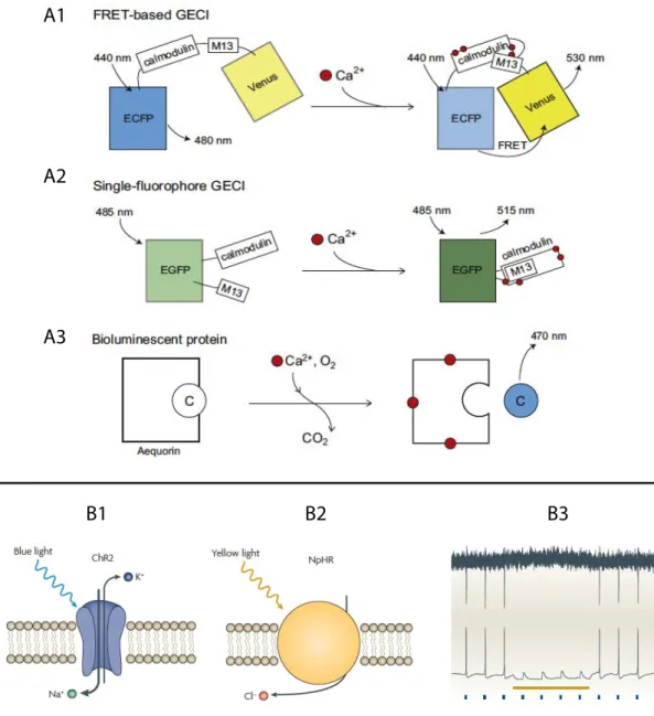
3.2.1. Reporters: monitoring neural circuits 3.2.2. Actuators: breaking neural circuits
3.3. The escape response as a model for sensorimotor integration 3.3.1. The escape response and its supraspinal control 3.3.2. Monitoring spinal neurons during active locomotion
Part B. Mechanosensory neurons enhance motor output in the zebrafish spinal cord during active locomotion………...……….p.51
Abstract
1 Introduction……….p.52
2 Results……….p.53 2.1. Bioluminescence signals reflect the level of recruitment of motor neurons
during movement
2.2. Spinal motor neurons recruitment is enhanced in the presence of mechanosensory feedback
2.3. Mechanosensory neurons are recruited during active but not fictive locomotion
2.4. Silencing mechanosensory neurons impairs escape responses
3 Discussion………...…p.66 3.1. Investigating sensorimotor integration in the spinal cord during ongoing
locomotion
3.2. Non-invasive bioluminescence monitoring of genetically targeted neurons in motion
3.3. A closed-loop circuit within the spinal cord for mechanosensory integration
4.1. Zebrafish care and strains 4.2. Generation of transgenic lines
4.3. Immunohistochemistry for GFP-Aequorin and quantification of muscle fibers 4.4. Monitoring of neuronal activity with GFP-Aequorin bioluminescence
4.5. High-speed behavior recording 4.6. Bioluminescence analysis 4.7. Kinematics analysis
4.8. Calcium imaging of spinal motor neurons 4.9. Ventral nerve root recording (VNR)
4.10. Calcium imaging of spinal sensory neurons
4.11. Behavioral analysis of freely moving BoTxLCB larvae 4.12. Statistical analysis
Part C. From spatial to genetic targeting: a paradigm shift for
neurosurgery...p.77
Abstract
1 Introduction……….p.78
2 How we moved to genetically targeted neurosciences………p.78 2.1. From morphological to genetic identification of neurons
2.2. Genetic targeting of neurons in tractable animal models 2.3. A toolbox for manipulating genetically identified neurons
3 Moving toward genetically targeted neurosurgery………p.83 3.1. Candidate diseases for genetically targeted neurosurgery
3.2. Genetic identification and cellular targeting in the human brain 3.3. Genetically targeted neuromodulation and neuroablation in patients
4 Two challenges for a paradigm shift………...p.88
Figures
Part A.
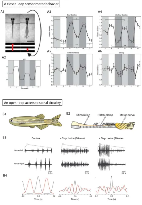
Figure 1. Closed-loop sensorimotor behaviors versus open-loop access to neural circuitry
Figure 2. Descending and ascending inputs to spinal circuits involved in sensorimotor reflexes
Figure 3. Neural substrates of spinal sensorimotor integration across vertebrates Figure 4. Monitoring and breaking neural circuits with genetically encoded reporters and actuators
Figure 5. Monitoring the activity of spinal neurons during active escape responses in zebrafish
Part B.
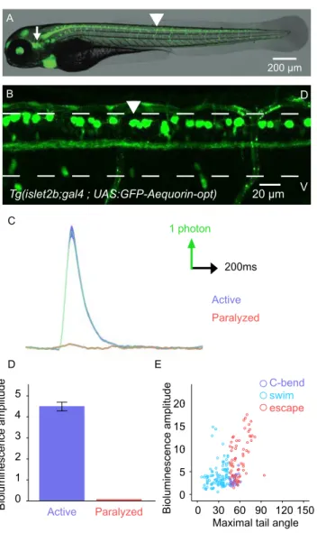
Figure 1. Bioluminescence monitoring of spinal motor neurons during active locomotion discriminates distinct behaviors
Figure 2. Calcium imaging of spinal motor neurons during fictive locomotion reveals specific patterns of recruitment for escape and slow swims
Figure 3. Spinal motor neurons recruitment is enhanced in the presence of mechanosensory feedback
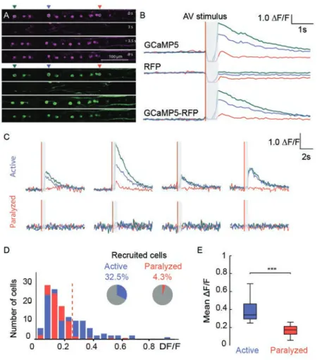
Figure 4. Mechanosensory neurons are recruited during active but not fictive locomotion
Figure 5. Calcium imaging of spinal mechanosensory neurons shows enhanced activation during motion
Figure 6. Silencing mechanosensory neurons impairs escape responses
Supp. Figure 1. Bioluminescence and kinematic parameters measured during active locomotion.
Supp. Figure 2. Calcium imaging of spinal motor neurons and fictive locomotion of ventral nerve root recording
Supp. Figure 3. Calcium imaging of trigeminal neurons in active and paralyzed
Tg(Isl2b:Gal4, cmlc2:eGFP, UAS:mRFP, UAS:GCaMP5) larvae
Part C.
Figure 1. Genetic targeting of neurons in tractable animal models. Figure 2. A toolbox for manipulating genetically identified neurons. Figure 3. Moving toward genetically targeted neurosurgery.
Part A.
Sensorimotor integration in the spinal cord, from behaviors to circuits: new tools to close the loop?
Abstract
Sensorimotor behaviors are by definition “closed-loop” processes in which sensory feedback modulates behavioral output. Sensory feedback can be provided by visual, auditory and vestibular inputs or direct proprioceptive inputs from muscle contraction. Although sensory feedback is not necessary for oscillation underlying locomotion to occur, there is evidence in the cat that sensory feedback can initiate locomotion (Lundberg, 1979) or reset the rhythm (Schomburg et al., 1998). The contribution of sensory feedback to active locomotion is however difficult to estimate for technical reasons. Indeed most physiological studies of spinal circuits involved in sensorimotor integration rely on preparations where muscles are paralyzed or dissected out, and are therefore deprived of sensory feedback. In this chapter, we will first explain closed-loop processes, and we will review the precious information obtained using “open-loop” experimental paradigms on how spinal neurons generate the neural rhythms that are at the basis of locomotion (Grillner et al., 2008). Optical and genetics techniques offer today alternatives to electrophysiology for monitoring neuronal activity from genetically defined populations of spinal neurons. We will then discuss how innovative tools for monitoring and manipulating neural activity, together with conducting sophisticated behavioral analysis, have provided exciting opportunities for “closing the loop” in genetically accessible model organisms with a special emphasis on zebrafish.
Introduction
The transformation of a sensory input into a motor output is a fundamental computation process, which is carried out by the brain and the spinal cord itself. Sensorimotor integration occurs when a set of neurons detect a sensory stimulus and process it to generate a behavioral output. Classic physiological approaches aim to record neurons specifically activated during sensorimotor integration, and to dissect the causative links by manipulating the activity of these neurons.
Sensorimotor behaviors are by definition “closed-loop” processes in which sensory feedback modulate the behavioral output. “Circuit dissection” experiment requires the experimenter to elicit a given sensory input and to quantitatively assess its behavioral output. In addition, it requires the determination of modulatory components such as systemic and local neuromodulators, or multiple sensory inputs.
Studies of spinal sensorimotor integration are mainly based on preparations of isolated spinal cord, and therefore deprived of descending and ascending inputs to the spinal cord. Such “open-loop” experimental paradigms have proved extremely valuable in understanding how spinal neurons generate neural rhythms that are at the basis of locomotion (Grillner et al., 2008). They might however not be optimal for studying spinal sensorimotor integration in a dynamic fashion.
In recent years, innovative tools for monitoring and manipulating neural activity in genetically accessible model organisms has provided alternative opportunities for “closing the loop”. By monitoring targeted populations of spinal neurons while the animal is fictively or actively performing a sensorimotor task, optogenetics offer new means to selectively study the role of a given class of neurons, without discarding sensory or neuromodulatory inputs.
Probing neural activity of targeted sensorimotor circuits during active locomotion will also open new paths to study how spinal circuits can reconfigure after removal of supraspinal inputs. This could shed light on intriguing results observed in spinal cord injured rodents (Edgerton et al., 2008), and eventually provide new means to exploit
plasticity of spinal sensorimotor circuits in pathological conditions.
1 A closed-loop approach to sensorimotor behaviors
1.1. Defining sensorimotor behaviors
1.1.1. Eliciting sensory input
Even a behavior as simple as a fly approaching an odorant fruit while flying is nothing but a trivial sensorimotor task (Seelig and Jayaraman, 2011): the fly must first detect the odor (Budick and Dickinson, 2006), extract information regarding its environmental relevance, and adapt its locomotor course to approach the fruit. All those steps have to be achieved while the animal is moving, thus adjusting its locomotor output to a changing visual, olfactory and mechanosensory feedback (Frye, 2010). Combining multiple sensory modalities (visual, olfactory, mechanosensory), and their closed-loop feedbacks, is critical to adapt to a noisy sensory environment and enhances the robustness of the behavioral output (Frye, 2010). Multisensory processing relies on interdependent sensory signals, allowing for increased efficiency during sensorimotor tasks compared to unimodal sensory stimuli (Loquet, 2013).
In mammals, it has long been demonstrated that “high-level” cortical areas, such as parietal and prefrontal cortices, were able to integrate multiple sensory modalities, increasing evidence suggests that multisensory integration also occurs in “low-level” cortices that were previously thought to be unisensory (Ghazanfar and Schroeder, 2006; Schroeder and Foxe, 2005). Studying sensorimotor integration, even at a relatively low-level, thus requires to reproduce a behaviorally relevant multisensory environment. However, experimental conditions often allow investigating only one sensory modality at a time.
One solution proposed by the field of neuroethology (Dickinson and Moss, 2012) is to consider that neural circuits can be experimentally understood in the context of the animal’s natural behavior. By focusing on innate behaviors in which the animal extracts critical sensory inputs to produce a behaviorally meaningful
locomotor output, neuroethology has provided important models for sensorimotor integration. For instance, escape behaviors, by which an animal escapes from its predator, are the perfect example of sensorimotor tasks that are crucial for the animal survival, and therefore are stereotyped. Interestingly, escape responses can be found in many species, including drosophila (Card, 2012), C. elegans (Pirri and Alkema, 2012) and several fish species (Schuster, 2012), allowing for comparative studies of sensorimotor integration across taxa.
Determining the sensory stimulus to control experimentally is a critical step of sensorimotor studies. We cannot reproduce the highly variable and multidimensional sensory inputs from the animal’s natural environment, but we should at least choose a stimulus that replicates the minimum set of sensory cues necessary to elicit a behaviorally relevant and consistent motor output (Clark et al., 2013). We also need to reliably record and quantify the locomotor output elicited by this sensory input.
1.1.2. Measuring motor output
The behavioral output of a sensorimotor transformation can be measured at different spatial and temporal scales: from the migration of an entire population of animals over several days to the analysis of single muscle fibers at millisecond timescale (Clark et al., 2013). Choosing the right scale for addressing the sensorimotor process of interest is not trivial.
At one extremity of this scale, “Taxis” behaviors, such as chemotaxis in drosophila (Gao et al., 2013) or rheotaxis in zebrafish (Suli et al., 2012), examine the cumulative change in spatial position of a group of animals over a relatively long period of time. It is also possible to look at the level of an individual in order to identify sequences of stereotyped behaviors such as mating in C. elegans (Liu and Sternberg, 1994). Sequential analysis of canonical behaviors allow the constitution of a detailed locomotor repertoire for a given specie, such as zebrafish (Budick and O'Malley, 2000). Lastly, a more detailed kinematics analysis could measure the
movements of individual joints, coupling this analysis with muscle activity recordings in rodents (Courtine et al., 2009a).
With the refinement of locomotor analysis, and the increasing set of kinematics parameters measured simultaneously, automated tracking programs have become crucial to reliably quantify behaviors. Such programs have been successfully applied to track individuals and classify behaviors in C. elegans (Baek et al., 2002), drosophila (Fry et al., 2008) or zebrafish (Mirat et al., 2013). Interestingly, such automated tracking programs can also be used to identify interactions between populations of multiple animals (Branson et al., 2009; Mirat et al., 2013), characterize mutants behaviors and build behavioral phenotypes databases (Yemini et al., 2013), and might lead to high-throughput drug screening (Mirat et al., 2013).
Analyzing complex datasets with multiple levels of kinematics parameters per animal and several animals interacting simultaneously raises important technical challenges. Reducing the dimensionality of the behavioral dataset can be achieved either by arbitrarily focusing on a restricted number of kinematic parameters or though statistical dimensionality reduction as in principal component analysis (PCA) (Musienko et al., 2011). The main issue with dataset reduction is to determine and preserve the behavioral output related to the sensory stimulus of interest. This can be achieved by computing the level of prediction or correlation between the sensory input and motor output (Briggman et al., 2006).
Although sensory input and motor output are the two ends and most accessible parts of a sensorimotor circuit, they are not sufficient to apprehend sensorimotor neural computation. Modulating inputs related to “top-down” afferents or “bottom-up” feedback also heavily influence sensorimotor processing.
1.2. Modulating sensorimotor behaviors
In real world, sensorimotor integration is a dynamic process where the animal is constantly updating its sensory inputs according to their behavioral output: as the fly is approaching the fruit, odorant and visual stimuli are continuously modified, allowing the fly to adjust its flight to reach the target (Frye, 2010). In experimental setting, the animal must often be restrained or paralyzed to allow for recording of neuronal activity. Such preparations are called “open-loop” because the motor output does not influence subsequent sensory input. But one might hypothesize that neuronal activity is not the same in the absence of sensory feedback.
“Closed loop” experiments, where new sensory information is acquired as the motor output is produced, can be obtained mainly through two complementary approaches: by attaching a miniaturized device onto a freely moving animal interacting with a controlled environment, or by providing some simulated sensory inputs to a restrained animal. The developing field of brain-machine interfaces has provided numerous studies in which cortical activity is recorded through chronically implanted electrode arrays, and decoded in real-time to control a motor effector, such as prosthetic limb (Carmena et al., 2003). It has also been possible to restore tactile sensation using a “brain-machine-brain interface”, by providing a way to produce a virtual motor output and to generate the corresponding sensory feedback (O’Doherty et al., 2012) (Tabot et al., 2013).
Such tools make it possible to monitor neuronal activity while the animal is freely behaving, but they don’t provide precise control over its sensory inputs. Virtual reality environments (Dombeck and Reiser, 2012) reproduce a simulated sensory environment that is continuously updated based on the animal behavior. Besides providing a better-controlled sensory input, virtual environments most importantly enables simultaneous neural recording by allowing the animal to perform a closed loop sensorimotor task while being physically restrained.
Combined with electrophysiology or genetically encoded calcium imaging, virtual environments have been applied in mice (Harvey et al., 2010), drosophila (Seelig et al., 2010) and zebrafish (Ahrens et al., 2012; Portugues, 2011). Notably, the zebrafish studies have showed that larvae were able to quickly modify their motor
output in response to an unexpected visual feedback (Portugues, 2011), and that this adaptive behavior correlated with state-dependent neural activity in a subset of brain areas identified using brain-wide calcium imaging (Ahrens et al., 2012).
1.2.2. Neuromodulation
State-dependent sensorimotor processing, in which the activity of a given population of neurons differs according to the behavioral state of the animal, are investigated within the larger framework of neuromodulation of neural circuits.
The core hypothesis underlying the concept of “multifunctional circuits” is that a given neural circuit should not be considered as a hard-wired diagram, activated during discrete states, but rather as a distributed network that is able to switch continuously between a variety of dynamical states to produce different patterns of activity, and eventually different behaviors (Briggman and Kristan, 2008). In a multifunctional sensorimotor circuit, a given neuron can be active during multiple locomotor behaviors (Sankrithi and O'Malley, 2010), producing different patterns of activity based on its modulatory inputs (Briggman and Kristan, 2008). External parameters, such as neuromodulatory substances (Marder, 2012) or synaptic input, e.g. sensory afferents (Latorre et al., 2013), can control the transitions between these different phases.
The neuromodulatory functions of monoaminergic substances have been extensively studied in invertebrates’ sensorimotor models such as the crustacean somatogastric ganglion (STG) (Marder, 2012). The central pattern generator circuit can generate fictive locomotor patterns and is modulated by numerous substances, from neurotransmitters released locally by projecting sensory neurons to diffused hormones released at distance by secretory structures (Blitz and Nusbaum, 2011). In spinal cord injured rats, the role of monoaminergic (in particular serotoninergic and dopaminergic) substances in modulating spinal locomotor circuits have been well documented (Musienko et al., 2011). Pharmacological manipulation, together with electrical spinal cord stimulation, could restore some locomotion independently of supraspinal inputs regeneration (Courtine et al., 2009b). Such
neuromodulatory-mediated functional recovery is also phase-dependent, suggesting that different interventions facilitate distinct phases of the locomotor pattern (Edgerton et al., 2008). This observation is in line with a multifunctional framework for the spinal sensorimotor circuits driving locomotion in spinal cord injured rats.
Intrinsic sensory states, i.e. neural dynamics that are not directly affected by an external physical stimulus, can also modulate multifunctional sensorimotor networks. One interesting example is the dual role of the gravimetric organ of the mollusk Clione limacina, which can switch between two very different rhythmic patterns, and behavioral output, depending on whether the animal is under control of a “hunting neuron” (Latorre et al., 2013). Another example of intrinsic sensory modulation is the feeding behavior of the Aplysia californica, where the same neurons drive both ingestion and rejection of food, but are differentially modulated by the coupling between the mouth muscles (Ye et al., 2006).
1.3. Modeling sensorimotor behaviors
1.3.1. Behavioral computations
Analyzing sensorimotor transformations is more complicated than just correlating an observed motor output with an experimentally elicited sensory input. Therefore, computational models for sensorimotor integration have proven more and more helpful as the number of measured variables increased with the improvement of experimental techniques.
For any sensorimotor task, the underlying computation is complex and can be modeled on a coarse behavioral scale, or on a more refined circuitry scale. Both approaches are complementary and have so far mostly been developed independently. The long-term objective is to map those behavioral computations on neural circuits models.
One major issue when dealing with sensorimotor computation is that our motor system is highly nonlinear (Franklin and Wolpert, 2011). In a linear system, one can easily predict the behavioral response to a multisensory stimulus by calculating the sum of the motor outputs for each individual sensory stimulus. However, the force developed by a muscle in response to its nervous input largely depends on other variables such as muscle length, velocity, tendons, joints positions, among others. (Zajac, 1989). Similarly, multiple sensory input creates multimodal sensory representations that are more than merely the sum of unimodal sensory inputs (Green and Angelaki, 2010).
Besides nonlinearity, many other issues increase the complexity computations of sensorimotor behaviors. For instance, noise is limiting our ability to perceive accurately sensory inputs (e.g.: estimating the location of the fruit on the table for our approaching fly) and carry out motor outputs precisely (e.g.: adjusting speed by modifying wings movements to reach the target) (Rohrseitz and Fry, 2010). Other issues include redundancy, i.e. the fact that multiple combinations of motor sequences can achieve the same behavioral task; nonstationarity, i.e. the fact that sensory and motor systems are modified throughout development and aging; uncertainty arising from noise, sensory ambiguity, partial information; and even multiple and variable delays, whether due to sensory or motor processing (Franklin and Wolpert, 2011).
One approach to resolve such complex sensorimotor computations is Bayesian decision theory (Wolpert, 2007). Bayesian decision theory aims to produce, using a probabilistic reasoning, optimal inferences based on uncertain inputs by combining prior beliefs and multiple sensory modalities. Based on these inferences, decision theory is subsequently used to decide which action is more likely to achieve the task objectives (Franklin and Wolpert, 2011). In a Bayesian system, the probability of a sensory state being true (named “posterior”) is produced by combining the probability of receiving the sensory information if that state were true (named “likelihood”) with the prior probability of that state (named “prior”) (Körding and Wolpert, 2006).
Such Bayesian sensorimotor computation can be easily tested using a simple task where a subject is asked to reach a cursor in a virtual-reality environment. The
discrepancy is introduced between the subject’s actual and displayed hand position (Körding and Wolpert, 2004). The “prior” distribution can be experimentally changed by varying the size of the discrepancy, while the sensory feedback “likelihood” is adjusted by varying the degree of visual blur controls Using this approach, the authors showed that subjects combined prior statistical distribution with sensory feedback likelihood in a Bayesian manner to optimize their performance during sensorimotor learning.
1.3.2. Circuits computations
Mapping behavioral sensorimotor computations onto identified neural circuits requires knowing how do those circuits process sensory inputs to produce a motor output at a cellular scale.
One important challenge for computing sensorimotor transformations, whether on a behavioral or cellular scale, is that they are mostly nonlinear, i.e. their motor output cannot be written as the weighted sum of its sensory inputs plus a constant. Geometrically, this means that modeling any neural network underlying a sensorimotor process requires at least a three-layers transformation, with an intermediate layer (referred to as “hidden layer”) used to recode sensory inputs before they are transformed into motor output. Such non-linear transformations can be approximated using a linear combination of “basis functions” (such as sine and cosine functions in a Fourier transform) as the intermediate layer: this is a called the “basis function approach” (Pouget et al., 2002) .This basis function approach is particularly relevant in the context of sensorimotor transformations. For instance, if a subject wants to reach toward a visual target as in the previously described experiment, the basis function approach postulates that the motor command can be approximated by the weighted sum of several non-linear basis functions of the visual and postural inputs (Pouget and Snyder, 2000). On a cellular scale, this ‘intermediate layer’ would be constituted by neurons whose firing properties, or “tuning curve”, can be described as a basis function for both visual and postural sensory inputs. Such neurons whose gain is modulated by visual and postural inputs can actually be found in the parietal
(Andersen et al., 1985), occipital (Trotter and Celebrini, 1999) and prefrontal (Boussaoud et al., 1993) cortices.
Besides nonlinearity, another major concern when looking at sensorimotor transformations is variability. Indeed, most experiments whether looking at sensorimotor processes or not, rely on mean statistics calculated from populations. However, it has been repeatedly shown that multiple solutions can produce similar circuit outputs (Marder, 2011). Even the most stereotyped motor behaviors such as rhythms generated by central pattern generators can be highly variable across animals (Marder and Taylor, 2011). The variability of the behavioral output to similar sensory inputs is well known, although not always documented. Most studies describe the “typical” behavior of the system by a single model. One attempt to take into account variability in sensorimotor circuits models would be to construct of population of models reproducing the actual behavioral data rather than trying to use a single model to reproduce the generic behavior (Marder and Taylor, 2011).
2. An open-loop access: sensorimotor circuits in the spinal cord across vertebrates
In the particular case of spinal sensorimotor circuits, a great wealth of anatomical and electrophysiological data has been accumulated over the years. However, being able to elaborate broader models in order to fit those data onto observed behaviors still remains a challenge, largely due to the fact that available techniques have prevented monitoring sensory inputs concomitantly with motor outputs until recently.
2.1. Extrinsic inputs to spinal sensorimotor circuits
2.1.1. Descending motor control
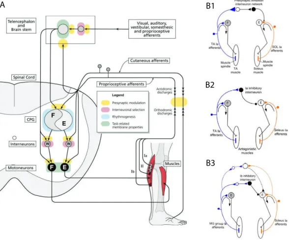
Located in the periphery of the spinal cord, white matter tracts comprise both ascending fibers, mainly located dorsally and laterally, carrying sensory information, and descending axons, mainly located ventrolaterally and laterally, carrying motor information (Figure 2A).
Descending motor tracts mainly include corticospinal tracts, which forms monosynaptic connections between motoneurons located in the primary motor cortex and spinal motoneurons located in the anterior horn of the grey matter at each segment. 80 to 90% of the corticospinal axons decussate to the contralateral side at the pyramid level in the medulla oblongata (hence the name “pyramidal tracts”), and travels in the dorsolateral funiculus (Guertin, 2013). Corticospinal tracts are mostly involved in voluntary skilled movements.
Other descending motor tracts originate mainly in subcortical nuclei in the brainstem, and in particular the reticular formation, and are called “extra-pyramidal tracts” by opposition to the corticospinal (pyramidal) tract. Extra-pyramidal tracts are composed of the rubrospinal (located along the corticospinal tract in the dorsolateral funiculus), vestibulospinal, tectospinal and reticulospinal tracts (all three located in the ventrolateral funiculus) (Bican et al., 2013) (Rossignol and Frigon, 2011).
Those descending inputs are mainly involved in autonomic functions, postural control and locomotion. More specifically: they facilitate contralateral upper limbs flexion (rubrospinal tract), neck and head motor control (tectospinal tract), autonomic functions (reticulospinal tract) and facilitating ipsilateral extensors and antigravity muscles to control tone and posture (vestibulospinal tract) (Guertin, 2013). Extra-pyramidal tracts project mainly on premotor lamina (lamina VI to VIII) of the spinal cord grey matter at each segment (Bican et al., 2013).
In particular, the role of reticulospinal pathways originating from the brainstem in the initiation and control of locomotion have been extensively studied, leading to the concept that, while the spinal central pattern generator produces the basic locomotor rhythm (see section 2.2.1), brainstem structures are necessary to activate and regulate this spinal central pattern generator (Jordan et al., 2008a; Whelan, 1996).
Numerous studies, mainly using decerebrate cat preparations, have identified several areas within the brainstem that can lead to the production of locomotion when
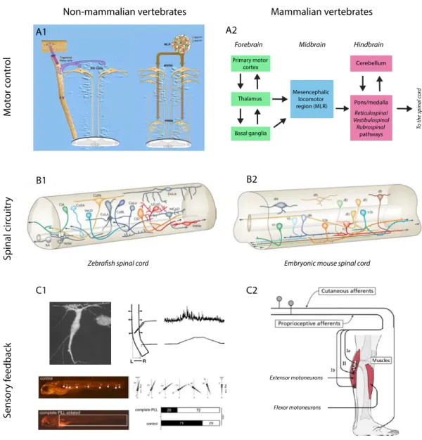
activated, whether chemically or electrically. The mesencephalic locomotor region (MLR), first identified by Shik et al (Shik et al., 1969), receives inputs from both the basal ganglia, the limbic system and the frontal cortex, and projects to neurons of the medial medullary reticular formation (MRF), and then on to interneurons in the spinal cord (Whelan, 1996). When stimulated electrically in decerebrate cats, the MLR can generate different gait patterns (walking, trotting, galloping) depending on the strength of the electrical stimulus (Rossignol et al., 2006). Interestingly, after its initial description in cats, homologous areas of the MLR have been described in many vertebrate species, including the rat (Garcia-Rill et al., 1990), lamprey (McClellan and Grillner, 1984) and monkey (Eidelberg et al., 1981).
Other areas in the midbrain, such as the medial MLR, the pontomedullary locomotor strip (PLS) or areas in the subthalamic nucleus (subthalamic locomotor region), have been shown to be involved in the control of locomotion by projecting onto spinal circuits through reticulospinal pathways in rodents (Whelan, 1996). More recently, isolated spinal cord preparations from neonatal rats and mice have allowed the identification of various neurotransmitters (N-methyl-D-aspartate, 5-hydroxytryptamine, dopamine, noradrenaline) that can elicit locomotor rhythmic activity by stimulating the spinal CPG through descending reticulospinal pathways (Jordan et al., 2008b).
In non-mammalian vertebrates, the descending control of locomotion has been particularly documented in the lamprey (Dubuc et al., 2008). Trigeminal relay cells activate reticulospinal neurons in a “all-or-nothing” fashion to elicit escape responses in response to a mechanical cutaneous stimulus (Viana Di Prisco et al., 1995). On the contrary, MLR inputs to reticulospinal neurons initiate locomotion in a graded fashion through monosynaptic cholinergic and glutamatergic inputs, with the middle rhombencephalic reticular nucleus (RRN) being activated for low intensity stimulation, and the posterior RRN being activated for as the stimulation strength increases (Wannier et al., 1998) (Figure 3A).
2.1.2. Ascending sensory feedback
While descending inputs schematically provide the motor command to spinal sensorimotor circuits, ascending afferents to the spinal cord mainly provide sensory information. In mammalian vertebrates, ascending sensory inputs include proprioceptive inputs (group Ia and II afferents from, respectively, primary and secondary endings of muscles spindles, and Ib afferents from Golgi tendon organs), cutaneous inputs (chemosensitive group III/Aδ and group IV/C fibers from nociceptive receptors). They have been extensively studied in the context of local spinal reflex pathways (Knikou, 2008; Rossignol et al., 2006) (Figure 2B).
The simplest, and fastest, somatic reflex is the monosynaptic pathway between primary sensory afferents from primary muscle spindles (Ia) and homonymous alpha motoneurons in the ventral horn of the corresponding segment grey matter. This is the basic myotatic reflex that is elicited by a muscle stretch due to a tendon tap, but is also involved in tonus and postural adjustments (Guertin, 2013). The experimental analog of the Ia reflex, the Hoffman reflex (H-reflex), where the mechanical stretch is replaced by a sub-threshold electrical stimulation of the afferent nerve, has been extensively used to investigate spinal sensorimotor circuits, and in particular presynaptic and reciprocal inhibition (Jankowska, 1992; Knikou, 2008), see (section 2.2.1).
Golgi tendon organs are force-sensitive receptors located at the muscle-tendinous junction, that are activated by passive and active muscle force. The Ib reflex arc, also known as the “inverse myotatic reflex”, is a disynaptic pathway by which group Ib sensory afferents from Golgi tendon organs inhibit alpha-motoneurons. This is the reflex arc responsible for the abrupt termination of the myotatic reflex, the well-known “clasp-knife” phenomenon (Hultborn, 2006). Although stimulating the Golgi tendon organs at rest cannot induce any movement, the Ib reflex has been suggested to be important for regulating muscle stiffness (Knikou, 2008).
While group Ib afferents from Golgi tendon organs provide information about the tension developed during muscle contraction, and group Ia afferents from primary muscle spindles inform spinal circuits about the dynamic of changes in muscle length, group II afferents from muscle spindle secondary endings provide information of muscle length itself (Jankowska and Edgley, 2010). Group Ia, Ib and II muscle afferents taken together constitute what is generally termed the “proprioception” input. Together with cutaneous afferents from nociceptors (Aδ and C fibers) and other muscle afferents (thinly myelinated group III and unmyelinated group IV fibers), group II muscle afferents constitute the flexion reflex afferents (FRA) involved in the withdrawal reflex, by which a painful stimulus lead to withdrawal of the limb through ipsilateral flexion and contralateral extension (Eccles and Lundberg, 1958). This sensorimotor reflex, more sophisticated than the “myotatic” and “inverse myotatic” reflexes, involves at least to two interneurons to either activate or inhibit the ipsilateral flexor or extensor alpha-motoneurons over several spinal segments (Guertin, 2013).
Sensory feedback pathways in non-mammalian vertebrates still remain unclear. Indeed, there is no clear equivalent to mammalian peripheral proprioceptive receptors in swimming vertebrates. However, in the lamprey, intra-spinal mechanosensitive receptors called the “edge cells” (Grillner et al., 1984) might provide movement-related sensory feedback (Di Prisco et al., 1990). Interestingly, it has recently been proposed that edge cells could be modulated by GABAergic cerebrospinal fluid contacting neurons (CSFns) (Jalalvand et al., 2014). Similar CSFns, called “Kolmer-Agduhr” cells, have been described in the zebrafish, and were able to trigger slow swim upon optical activation (Wyart et al., 2009). Another sensory feedback pathway in larvae and adult zebrafish is the lateral line system (Ghysen and Dambly-Chaudière, 2007). Mechanosensory hair cells in the lateral line neuromasts provide information about the water flow, contributing to orientating the fish against the water, a behavior called rheotaxis (Olszewski et al., 2012; Suli et al., 2012) (Figure 3C).
2.2. Intrinsic spinal sensorimotor circuitry
2.2.1. Sensorimotor interneuronal networks
Presynaptic inhibition
As we have seen, spinal circuits are continuously provided with multiple ascending sensory inputs from various sources. This sensory feedback needs to be controlled to allow for the proper execution of a motor task (Knikou, 2008). One way to control this sensory input is through presynaptic inhibition of muscle afferents on alpha-motoneurons through GABAergic axo-axonic synapses (Rudomin and Schmidt, 1999) (Figure 2B). A similar control can be achieved through primary afferent depolarization (PAD), and the two phenomena are now actually considered to be mediated by the same interneurons (Jankowska, 1992).
Initially described in relation to group Ia afferents from primary endings of muscle spindles (Frank and Fuortes, 1959), presynaptic inhibition though GABAergic interneurons has more recently also been described for group Ib and group II muscle afferents, as well as cutaneous and articular afferents (Rudomin, 2009). Although it has traditionally been considered that different subgroups of interneurons were mediating PAD of distinct muscle sensory afferents (Jankowska, 1992), it has also been demonstrated that the same interneurons, located within Rexed’s laminae VI-VII of the spinal cord grey matter (intermediate zone), could be co-excited by group Ia and group Ib afferents (Fetz et al., 1979). More surprisingly, even group Ib and group II inputs can be integrated by a common pool of interneurons, located within laminae V-VII (Bannatyne et al., 2009). These results led some authors to consider all those subpopulations of interneurons (groups Ia, Ib and II) may actually operate as a single functional population with multisensory inputs from both several types of afferents and several muscles (Jankowska and Edgley, 2010). (Figure 2 B1)
Reciprocal Ia inhibition
Considered that the same Ia muscle afferents innervate motoneurons belonging to many different motor pools, it has long been postulated that a neural pathway involving Ia afferents allowed for inhibition of alpha-motoneurons controlling antagonist muscles. The reciprocal Ia inhibition is mediated by a single glycinergic inhibitory interneuron activated by Ia afferents from a given flexor muscle, which in turn inhibits alpha-motoneurons controlling the antagonistic extensor muscle (Eccles et al., 1957a; Jankowska, 1992). As for PAD interneurons, it has later been showed that these reciprocal Ia inhibitory interneurons, located dorsomedially to the motor nuclei in the ventral horn, actually also receives convergent inputs, both excitatory and inhibitory, from multiple descending and ascending sources, including Renshaw cells (see below) (Hultborn, 1972) (Figure 2 B2).
Non-reciprocal Ib inhibition
Group Ib sensory afferents from Golgi tendon organs inhibit motoneurons projecting to synergist muscles and facilitate motoneurons projecting to antagonist muscles through di- or tri-synaptic pathways involving respectively one or two inhibitory glycinergic interneurons (Eccles et al., 1957b; Jankowska, 1992). As for Ia interneurons mediating reciprocal inhibition, Ib inhibitory interneurons exhibit a wide convergence of inputs from both descending inputs (excitatory corticospinal, rubrospinal and inhibitory reticulospinal afferents) and ascending inputs (excitatory group Ia and Ib muscle afferents, as well as cutaneous and joint afferents) (Hultborn, 2006) (Figure 2 B3).
Recurrent inhibition
Lastly, another sensorimotor interneuronal pathway involving an inhibitory interneuron is the one formed by Renshaw cells, located in the ventral horn (next to Ia reciprocal inhibitory interneurons) (Renshaw, 1946). Renshaw cells are excited by cholinergic axonal collaterals from alpha-motoneurons and provide glycinergic
recurrent inhibition to the same or synergistic muscles (Eccles et al., 1956). Again, as for other sensorimotor interneurons, Renshaw cells also receive inputs from other afferents, including ipsilateral group II and III muscle afferents, cutaneous afferents, and descending motor afferents, and project themselves not only to alpha-motoneurons but also to gamma-alpha-motoneurons, Ia reciprocal inhibitory interneurons and other Renshaw cells within the same spinal segment (Windhorst, 2007).
2.2.2. Spinal central pattern generator across vertebrates
Along with this complex interplay between sensory afferents and sensorimotor interneuronal networks, a large amount a work has converged toward the identification of a spinal network able to generate the elementary patterns and rhythms of locomotion: the spinal central pattern generator (CPG). First postulated from studies of decerebrated cats more than a century ago (Brown, 1911), extensive research in non-mammalian vertebrate species such as the lamprey (Grillner, 2003) and the Xenopus tadpole (Roberts et al., 2009) have provided many insights into the swimming CPG and its cellular mechanisms, leading to rapid advances in the understanding of the mammalian walking CPG (Kiehn, 2006).
Homology across vertebrates
Interestingly, new insights into the genetic profiles of spinal interneurons have allowed direct comparison between different classes of interneurons across all vertebrates (Goulding, 2009). Based on the dynamic expression pattern of transcription factors, five major subclasses of spinal ventral interneurons have been described, called V0, V1, V2, V3 and Hb9 interneurons (Figure 3). Each class being characterized by a specific transcription factor, such “molecular code” opens the way for functional investigation of genetically targeted, rather morphologically or electrophysiologically identified, spinal interneurons within the CPG (Figure 3B).
Several lines of evidence suggest that the rhythmogenic neurons of the CPG are glutamatergic excitatory neurons projecting ipsilaterally onto inhibitory left-right and flexor-extensor coordinating neurons at each spinal segment (Kiehn et al., 2008). Indeed, blocking inhibitory commissural or ipsilateral interneurons does not prevent rhythm generation, whether in the lamprey (Cangiano, 2005), rodent (Bonnot et al., 2002) or cat (Kato, 1987), therefore discarding the “half-center model” for CPG rhythm generation (Kiehn, 2006). Various putative candidates for the role of “pacemakers” neurons have been recently investigated (Kiehn, 2011): among them, Hb9-expressing interneurons (Tazerart et al., 2008) and V2a-Chx10 expressing interneurons (Hägglund et al., 2010) have been shown to have rhythmogenic properties in neonatal mouse models. Morphological homologs in the lamprey (Grillner, 2003) and tadpole (Li et al., 2010), and molecular homologs in zebrafish (Mclean et al., 2007) support the hypothesis of a glutamatergic ipsilateral drive to the spinal CPG.
Flexor-extensor coordination
Ipsilateral-projecting glycinergic inhibitory interneurons are known to be involved in alternation of extensor and flexor muscles activation for a long time, since flexor-extensor coordination is suppressed when glycinergic transmission is blocked but can persist in hemisected spinal preparations (Bonnot et al., 2002). Putative candidate interneurons include Ia inhibitory interneurons and Renshaw cells (see section 2.2.1), as both have been shown to fire rhythmically during locomotion and in opposing phases in respect to their flexor/extensor afferents (McCrea et al., 1980).
However, a recent study challenged this assumption (Gosgnach et al., 2006). V1-derived interneurons expressing the transcription factor Engrailed-1 (En1) are inhibitory ipsilaterally projecting interneurons that give rise to Renshaw cells and Ia inhibitory interneurons. Genetic knock out of En1-expressing neurons induced slower locomotor activity and increased step cycle, but did not suppress flexor-extensor coordination. This suggests the existence of other ipsilateral inhibitory interneurons, that might be specific to mammalian locomotor CPG (Kiehn, 2006).
Left-right coordination
Coordination of left-right activity during locomotion is mainly achieved through commissural interneurons that are crossing the midline via the ventral commissure (Kiehn, 2006). Experiments in mice have revealed a dual system for left/right coordination: 1) during alternative walking, contralateral motoneurons inhibition is achieved either through mixed glycinergic and GABAergic inhibitory commissural interneurons projecting monosynaptically to contralateral motoneurons, or excitatory commissural interneurons projecting onto contralateral inhibitory premotor interneurons; 2) during synchronous “hopping”, contralateral motoneurons excitation is achieved through glutamatergic commissural interneurons (Quinlan and Kiehn, 2007).
Candidate commissural interneurons for this left/right dual model are derived from Dbx1 positive neurons from the V0 transcription domain (Lanuza et al., 2004), in which about one third of commissural interneurons are glutamatergic (Evx-1-positive, V0V interneurons) and two thirds are inhibitory (Evx1-negative, V0D interneurons) (Moran-Rivard et al., 2001). A recent study (Talpalar et al., 2013) confirmed and further refined this hypothesis by showing that V0-ablated mice exhibited a hopping gait at all frequencies, while selective ablation of inhibitory V0 interneurons (V0D) led to a lack of left-right alternation only at low frequencies whereas selective ablation of excitatory V0 interneurons (V0V) led to similar hopping gait but only at medium and high frequencies.
Neurons participating to the left-right alternation spinal network have also been identified in non-mammalian vertebrates. In the Xenopus tadpole, inhibitory glycinergic commissural interneurons are responsible for mid-cycle reciprocal inhibition and are driven by descending glutamatergic interneurons (Roberts et al., 2009). In the lamprey, both inhibitory and excitatory commissural interneurons have been described with left-right alternating pattern of activity (Grillner, 2003). Lastly, similar glycinergic inhibitory and glutamatergic excitatory commissural interneurons have been identified in the zebrafish, sharing molecular markers with the mouse V0
neurons, although the network details have not yet been worked out (Fetcho and Mclean, 2010).
2.3. Dynamic spinal sensorimotor interactions
2.3.1. Modulation of spinal circuitry from extrinsic inputs
Both descending motor inputs and ascending sensory feedback can modulate the activity of the spinal CPG. Indeed, if the CPG is able to generate the basic locomotor patterns, dynamic sensorimotor interactions with both supraspinal and peripheral inputs continuously modulate these patterns to achieve a flexible adaptation to the environment. Such interactions take place in a phase-dependent (swing/stance) and state-dependent (forward/backward) manner, that is extrinsic inputs will result in different modulations depending on the ongoing phase of the locomotor cycle (Rossignol et al., 2006).
As discussed in section 2.1.1, supraspinal pathways, such as the MLR and its projections through the reticulospinal tract, can induce locomotion in “fictive preparations”, i.e. isolated spinal cord or decerebrated adult cat preparations. However, descending pathways, whether carrying sensory or motor information, can also modulate ongoing locomotion. Such modulation can be achieved either though modulation of brainstem command circuitry, or through direct modulation of spinal circuitry (McCrea, 2001).
Vestibular inputs (relaying information about balance and posture) modulate the activity of reticulospinal neurons with a phasic pattern during fictive locomotion in lampreys, thereby avoiding a counteractive drive from reticulospinal neurons during ongoing locomotion (Bussières and Dubuc, 1992). A recent study in zebrafish larvae suggested that vestibular inputs are able to differentially recruit dorsal and ventral premotor spinal microcircuits during postural correction, possibly prefiguring the mammalian modular organization of spinal flexor/extensor microcircuits (Bagnall and McLean, 2014). The influence of visual feedback on the control of locomotion
can be experienced on a daily basis when one needs to anticipate and adjust his gait to avoid an obstacle (Rossignol, 1996). New experimental paradigms, such as the optomotor response in zebrafish (Orger et al., 2008), have started to shed light on the neural circuitry responsible for visually induced locomotion.
Besides descending inputs, ascending sensory feedback, from either proprioceptive or cutaneous inputs, can also modulate the activity of the spinal CPG. Cutaneous inputs (C and A fibers, see section 2.1.2) are mainly involved in correcting the steps in response to external perturbations, such as an uneven floor, during the different phases of the step cycle (McCrea, 2001; Rossignol et al., 2006). Interestingly, the same cutaneous stimulus can lead to responses in flexor or extensor muscles depending on the initial position of the limb, therefore behaving as excitatory inputs to a given muscle group in one locomotor phase, and excitatory to the antagonist muscles in the opposite phase, a phenomenon termed “reversal” (Rossignol and Gauthier, 1980).
Proprioceptive feedback also has an important role in modulating ongoing locomotion, in particular by adjusting the duration of, and facilitating the switch between, the different phases of the step cycle, therefore setting the frequency of locomotion (Rossignol et al., 2006). For instance, in decerebrate cats preparations, stimulation of group Ib afferents from Golgi tendon organs of extensor muscles can reset the locomotor cycle by abruptly terminating the ongoing fictive flexor activity and initiating a new burst in the extensor recording (Conway et al., 1987). Similarly, stretch-evoked Ia inputs can increase the duration of the stance phase, but only when stimulated during flexor activity (Guertin et al., 1995).
Therefore, patterns of fictive locomotion produced by the spinal CPG should not be considered as a fixed output of a hard-wired circuit, but should be viewed rather as a dynamic multimodal process whose output is modulated by the various supraspinal and peripheral sensory inputs.
2.3.2. Implications for plasticity after spinal cord injury
The emerging concept that intrinsic spinal circuits can produce adaptive locomotion with modulation by sensory feedback, independently, at least to some extent, from supra-spinal inputs, bears important consequences for new neurorehabilitative strategies after spinal cord injury.
Experimental paradigms with adult cats walking on a treadmill have demonstrated that neither bilateral lesion of the dorsolateral spinal cord (interrupting cortico- and rubrospinal tracts) (Jiang and Drew, 1996), nor bilateral lesion of the ventrolateral spinal cord (interrupting vestibulo- and reticulospinal tracts) (Brustein and Rossignol, 1998), could permanently suppress quadrupedal locomotion. However, after unilateral complete hemisection at the lower thoracic (T13) level, interrupting both dorsal and ventral descending pathways, cats showed a complete paralysis of the ipsilateral hindlimb during the first three days, followed by a progressive recovery over the following three weeks (Rossignol and Frigon, 2011). Interestingly, this recovery was accompanied by a modification of the step cycle, forelimb/hindlimb and left/right coordination (Martinez et al., 2012). These results suggest that the intrinsic spinal circuitry is able to produce locomotion even after removal of all supraspinal inputs, and that this recovery is underpinned by extensive reorganization of the spinal sensorimotor network (Martinez and Rossignol, 2013). They also suggest that treadmill-induced locomotor training, by providing sensory feedback, is crucial to drive the reorganization of spinal circuits (Rossignol and Frigon, 2011).
To test this hypothesis of a plastic spinal CPG, Rossignol et al. designed a dual-lesion paradigm in which a first hemisection performed at the T10/T11 spinal level is followed, after several weeks of locomotor training and complete recovery, by a complete spinal transection at the T13 level (Barrière et al., 2008; Martinez and Rossignol, 2013). The major finding was that cats regained full locomotor performance after only 24 hours, without any training of pharmacological intervention (Barrière et al., 2008), therefore indicating that intrinsic changes within the spinal
CPG had indeed occurred during the rehabilitation period, and could be retained after the complete removal of supraspinal inputs.
Similar results have been obtained recently in rodents (Edgerton et al., 2008), in which recovery of coordinated hindlimbs locomotion on a treadmill could be achieved only one-week after complete thoracic (T7) spinal transection when combined lumbosacral electrical epidural stimulation (EES) and systemic application of serotoninergic agonists were applied (Courtine et al., 2009a). Interestingly, removing peripheral sensory inputs by unilateral dorsal rhizotomy prevented EES-facilitated locomotor recovery after complete spinal transection, but only on the deafferented side, thereby confirming the hypothesis that sensory feedback drives the reorganization of intrinsic spinal circuitry (Lavrov et al., 2008).
However, those results only concerned treadmill-induced “automatic” locomotion. To which extent can we exploit the plasticity of spinal sensorimotor circuits to induce restoration of voluntary locomotion? This question was investigated by a recent study (Brand et al., 2012), in which the authors used a simultaneous dual hemisection paradigm in adult rats together with a so-called “electrochemical neuroprosthesis” (i.e. the combination of lumbosacral epidural electrical stimulation together with systematic administration of a cocktail of monoaminergic agonists). They observed that rats trained with a robotic postural interface encouraging supra-spinally mediated locomotion could regain voluntary control through remodeling of corticospinal projections. A similar approach have even been used successfully in a paraplegic human subject, who could regain some voluntary control of one of his lower extremity after intensive rehabilitation and electrical epidural stimulation, although this recovery was very limited and observed in a single individual only (Harkema et al., 2011).
These results have raised hopes that clinically significant locomotor recovery can be achieved through reorganization of intrinsic sensorimotor circuitry, facilitated by intensive training and electrical and/or chemical manipulation. However, one major issue of such studies is that they can probe changes in spinal circuitry only in a very indirect manner.
Indeed, until now, one had to choose whether to be able to access spinal circuitry in open-loop “fictive” preparations, discarding the sensory feedback but being able to identify and record from neurons within the spinal cord, or to preserve active locomotion and sensory feedback but having only a limited and indirect access to spinal circuits. However, new tools and animal models might change this conundrum in a near future.
3. Closing the loop? Optogenetic manipulation of spinal sensorimotor circuits in zebrafish
3.1. Genetic targeting of spinal sensorimotor circuits in zebrafish
3.1.1. Identified sensorimotor neurons in the zebrafish spinal cord
As in any other vertebrates, neurons in the zebrafish spinal cord can be broadly divided between motoneurons, sensory neurons, and interneurons (Lewis and Eisen, 2003). Recent developments of genetic tools allowing specific targeting of subtypes of neurons has allowed marked progress in our understanding of their functional roles, and has led to a refined classification.
Sensory neurons within the spinal cord mainly include mechanosensitive Rohon-Beard neurons, of which homologs can be found in most anamniote vertebrate, such as Xenopus tadpoles and lampreys (Reyes et al., 2003). Rohon-Beard neurons are derived from the same neural plate domain that generates neural crest cells, and presumably die during development to be replaced by dorsal root ganglion cells in adult zebrafish (Lewis and Eisen, 2003). When stimulated optically, Rohon-Beard neurons are able to trigger escape responses (Douglass et al., 2008; Wyart et al., 2009), through either direct excitation of reticulospinal cells (Douglass et al., 2008) or activation of CoPA interneurons (Pietri et al., 2009).
In larvae, both primary and secondary motoneurons (together with oligodendrocytes) are derived from the pMN transcription domain in the ventral
spinal cord, are positive for olig2 expression and persist through adulthood (Kimmel et al., 1994; Lewis and Eisen, 2003). Primary motoneurons are located more dorsally (with subtypes according to their position from caudal to rostral: CaP, MiP, RoP), innervate fast muscles, and are involved in fast swimming and startle response, while secondary motoneurons, located more ventrally, innervate both slow and fast muscles, and are also involved in slow swim (Lewis and Eisen, 2003).
To explore the differences between slow swim and escape spinal networks, Ritter et al. used a head-embedded preparation in which they could elicit either slow swim by illuminating the head with a fiber optic, or escapes by tapping the head with a piezoelectric actuator. They simultaneously monitored the activity of morphologically identified interneurons in the embedded part of the tail using calcium imaging, and recorded the movements of the caudal tail using a high-speed camera (Ritter et al., 2001). They showed that “circumferential ipsilateral descending” (CiDs) interneurons were activated during escapes but not during slow swim movements, while excitatory glutamatergic “multipolar commissural and descending” (MCoDs) interneurons were, on the contrary, activated during swimming but not during escapes (Ritter et al., 2001).
A subsequent study from the same group (Bhatt et al., 2007) combining calcium imaging and paired patch recording, confirmed that CiDs were responsible for motoneurons excitation during escapes, and showed that stronger escapes elicited by a head tap were associated with the recruitment of a larger number of CiDs than delayed escapes elicited by a tail tap, thereby apparently contradicting previous results about differential descending control from the hindbrain (Bhatt et al., 2007; Liu and Fetcho, 1999). Interestingly, the same authors also demonstrated that reinervation of CiDs by regenerating Mauthner axon, following injection of cAMP, was associated with improved locomotor performances (Bhatt et al., 2004).
Using isolated spinal cord from larvae zebrafish, a “topographic map” of recruitment for premotor interneurons has been documented (Mclean et al., 2007; 2008). MCoDs interneurons, located in the ventral spinal cord, provided a phasic drive to a subset of ventral contralateral motoneurons during slow swimming patterns.