HAL Id: hal-03193228
https://hal.archives-ouvertes.fr/hal-03193228
Submitted on 15 Apr 2021HAL is a multi-disciplinary open access archive for the deposit and dissemination of sci-entific research documents, whether they are pub-lished or not. The documents may come from teaching and research institutions in France or abroad, or from public or private research centers.
L’archive ouverte pluridisciplinaire HAL, est destinée au dépôt et à la diffusion de documents scientifiques de niveau recherche, publiés ou non, émanant des établissements d’enseignement et de recherche français ou étrangers, des laboratoires publics ou privés.
Ultrasound and microbubble-assisted gene delivery:
recent advances and ongoing challenges
Anthony Delalande, Michiel Postema, Nathalie Mignet, Patrick Midoux,
Chantal Pichon
To cite this version:
Anthony Delalande, Michiel Postema, Nathalie Mignet, Patrick Midoux, Chantal Pichon. Ultrasound and microbubble-assisted gene delivery: recent advances and ongoing challenges. Therapeutic Deliv-ery, Future Science Ltd, 2012, 3 (10), pp.1199-1215. �10.4155/TDE.12.100�. �hal-03193228�
Ultrasound and microbubble-assisted gene delivery: recent
1advances and ongoing challenges
23
Anthony Delalande, Michiel Postema, Nathalie Mignet, Patrick Midoux, Chantal Pichon 4
5
Abstract 6
Having first been developed for ultrasound imaging, microbubbles are nowadays proposed as 7
tools for ultrasound-assisted gene delivery, too. Their behaviour during ultrasound exposure 8
causes transient membrane permeability of surrounding cells, facilitating targeted local 9
delivery. The increased cell uptake of extracellular compounds by ultrasound in the presence 10
of microbubbles is attributed to a phenomenon called sonoporation. Sonoporation has been 11
successfully applied to deliver nucleic acids in vitro and in vivo in a variety of therapeutic 12
applications. However, the biological and physical mechanisms of sonoporation are still not 13
fully understood. 14
In this review, we discuss recent data concerning microbubble-cell interactions leading to 15
sonoporation and we report on the progress on ultrasound-assisted therapeutic gene delivery 16
in different organs. In addition, we outline ongoing challenges of this novel delivery method 17
for its clinical use. 18
20
Key terms 21
22
Sonoporation: phenomenon by which ultrasonic stimulation transiently alters the 23
permeability of cell plasma membrane, thereby allowing the uptake of extracellular 24
molecules. This phenomenon is amplified by the presence of microbubbles. 25
Microbubbles: encapsulated gas bubbles with diameters up to 10 micrometres. Firstly 26
developed as contrast agents for ultrasonic medical imaging, they might be used as carrier 27
vehicles to deliver drugs or genes, aided by sonoporation. 28
Acoustic cavitation: formation, oscillation, and collapse of bubbles under sonication. 29
Ultrasonic and microbubbles-assisted gene delivery: external-triggered method of plasmid 30
DNA transfer based on sonoporation. 31
Introduction 33
34
Gene therapy aims at introducing genetic material into mammalian cells to cure 35
genetic deficiencies and a large variety of acquired diseases. So far, viruses have 36
demonstrated the feasibility of gene therapy and remain the best vehicles to introduce genes 37
into cells. But severe fatal adverse events of viral vectors including acute immune response 38
and insertion mutagenesis observed in gene therapy clinical trials have raised serious safety 39
concerns about their use [1,2]. 40
Therefore, clinical developments still require the use of alternative approaches of 41
high safety, low immunogenicity and easy manufacture. Moreover, the size limitation 42
capacity, weakness of cell targeting and manufacturing issues has boosted efforts to search for 43
non-viral options. During the last decade, many efforts have been carried out to design 44
synthetic or chemical gene delivery systems that incorporate viral-like features required to 45
transfect efficiently cells [3-5]. The final goal of these systems is to deliver a plasmid 46
encoding a therapeutic protein into target cells upon systemic administration. This challenge 47
could be achieved if several extracellular barriers are overcome to reach the target cells where 48
the therapeutic gene has to be efficiently expressed. Tremendous efforts have been made to 49
improve the efficacy of different types of “synthetic viruses” and elegant strategies have been 50
proposed for that as reported in recent reviews [6-8]. 51
However, achieving optimal targeted gene delivery systems based on chemical 52
compounds is still highly challenging. Narrow therapeutic indices and lack of selectivity 53
towards target tissues have been observed. The absence of specificity that causes severe 54
toxicity often hampers the efficacy at the target site. In this respect, innovative delivery 55
strategies that could selectively target tissues of interest are ongoing both in experimental and 56
in clinical research. It is obvious that designing an efficient targeted delivery strategy is the 57
next step to further improve delivery systems and to reduce side effects. Among the delivery 58
systems found are several physical methods starting from a simple naked DNA injection to 59
much more sophisticated systems such as electroporation and ultrasound-mediated delivery 60
(USMD). 61
Ultrasound is used for diagnostic clinical applications and for treatment either by 62
itself or in combination with drugs [9,10]. Among the variety of imaging modalities, 63
ultrasonic imaging is a well-established and reliable technique for diagnosis.The development 64
of real-time imaging has allowed for visualizing a variety of anatomical structures non-65
invasively. These last years, therapeutic applications of ultrasound have gained new interests 66
thanks to the exploitation of ultrasound for delivery. The effect of ultrasound on biological 67
systems was already reported in 1927 [11], but it took until 1987 until the first study on 68
ultrasound-assisted gene delivery was published [12]. Depending on the acoustic settings, 69
ultrasound can induce either thermal or non-thermal effects, each having its own applications. 70
The thermal effects are obtained using high ultrasound intensities and are due to the 71
absorption of acoustic energy by tissue leading to heating. This property of ultrasound is used 72
in techniques like high-intensity focused ultrasound (HIFU) for focussed ultrasound surgery 73
(FUS) or physiotherapy by ultrasound. One of the first applications of HIFU was 74
sonothrombolysis for ischemic stroke treatment. It has been shown that the introduction of 75
microbubbles in the blood pool could act as cavitation nuclei and decrease the energy 76
threshold for thrombolysis by a third [13]. The thermal effects can present a risk on sonicated 77
tissues; the World Federation for Ultrasound in Medicine and Biology Temperature state that 78
an elevation of 1.5°C is considered safe while an elevation of 4-5°C during 5 minutes is 79
potentially dangerous [14]. The non-thermal effects are obtained using lower ultrasound 80
intensities. The main non-thermal effects are inertial cavitation and acoustic streaming. These 81
effects can induce some benefits such as tissue healing or ultrasound-mediated drug or gene 82
delivery. 83
The use of ultrasound as an external trigger is one approach that has proven to be 84
effective for drug delivery. The drugs to be delivered can be chemotherapeutic agents for 85
cancer therapy applications or oligonucleotides for gene therapy applications. Ultrasound 86
enables to control both the drug release and the release location by use of ultrasound [15-21]. 87
1. Ultrasound
89
Ultrasound is a form of mechanical vibration of matter with a frequency beyond human 90
audible range, i.e., above 20 kHz. The speed of sound is dependent of the medium: roughly 91
340m/sec in air, 1480 m/sec in pure water, 1550 m/sec in soft tissue and 3700m/sec in bone 92
[22,23]. The particle excursion in a sound wave is related to the instantaneous local pressure 93
through the wave equation [24]. The wave is damped with propagation distance due to tissue 94
absorption and geometric effects [24]. This behaviour induces mechanical effects in the 95
medium which are amplified when inertial cavitation occurs. Inertial cavitation is the process 96
of formation and subsequent collapse of bubbles driven by an acoustic field [25]. Acoustic 97
cavitation takes place when the peak-negative pressure amplitude of ultrasound waves at 98
given frequency exceeds the so-called cavitation threshold [24]. If so-called cavitation nuclei 99
brought by impurities in the medium are present, the cavitation threshold is lowered. 100
Preformed microbubbles serve as cavitation nuclei and are exploited in ultrasonic contrast 101
imaging. These so-called ultrasound contrast agents consist of micron-size gas-filled bubbles 102
(typical diameter ranged is 1-10 µm) encapsulated by an elastic shell. They oscillate during 103
ultrasound exposure (stable cavitation) and demonstrate highly nonlinear behaviour that 104
ameliorates their detection. Oscillating microbubbles introduced in the blood stream produce 105
a sound field themselves and therefore indicate regions of tissue perfusion [26,27]. The 106
acoustic backscattering of these agents is several orders of magnitude higher than the 107
surrounding tissues. 108
It has been proposed that microbubble activity plays a key role in therapeutic 109
applications [9]. Stable cavitation refers to the (nonviolent) regime of microbubble oscillation, 110
whereas inertial or transient cavitation corresponds to microbubble oscillation with 111
increasingly large amplitudes until collapsing violently [28]. In the nonviolent regime, fluid 112
streaming around oscillating microbubbles may create enough shears to (transiently) open up 113
cell membranes. Furthermore, the microbubbles themselves may interact with, or even 114
translate through cell membranes [29]. In the inertial regime, jets from collapsing 115
microbubbles have been observed to penetrate cells, inducing permanent damage [30]. 116
2. Mechanisms of ultrasound-mediated delivery (USMD)
118
Several studies have been conducted these last years to delineate mechanisms 119
involved in USMD [31-35]. Many experimental data have shown that exposure to ultrasound 120
increases the cellular uptake of extracellular molecules. The uptake is significantly higher if 121
an ultrasound contrast agent is added [36,37]. However, it is under discussion exactly how 122
cells that are subjected to ultrasound internalize extracellular compounds, and which cellular 123
responses they evoke. 124
Sonoporation is the ultrasound-assisted transient permeabilization of cell 125
membranes. This permeabilization allows for the transfer of molecules between the intra- and 126
extracellular medium. Several sonoporation mechanisms have been identified. The main 127
hypotheses of microbubble interaction with cells are the push and pull mechanisms, micro-128
jetting, micro-streaming, and, more recently, translation of microbubbles through cells [29]. It 129
has been proposed that the attraction of oscillating microbubbles by cells is caused by 130
sonophores, tiny cavities inside cell membranes [38].All these sonoporation mechanisms have 131
been demonstrated with the aid of high-speed photography. These mechanisms of membrane 132
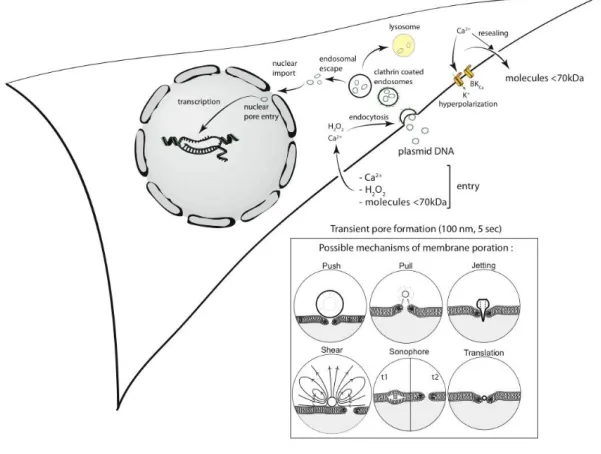
poration have been summarized in Figure 1 (inset). 133
If a microbubble is touching a cell, the microbubble oscillations induce a cellular 134
massage [39]. This cellular massage mechanically stimulates the plasma membrane. It has 135
been hypothesised, that during the expansion phase of the microbubble the plasma membrane 136
is pushed inward and that during the compression phase the microbubble pulls the plasma 137
membrane outward [31].Repeated push and pull on the plasma membrane creates weaknesses 138
that might lead to the formation of a pore. In a high-mechanical-index regime, the formation 139
of a microjet can occur. A microjet is a fluid protrusion towards a surface during inertial 140
bubble collapse. Jetting has been observed through cells with laser-generated cavities [30]. 141
However, this mechanism induces high cell toxicity. Several studies conducted in silico, in 142
vitro, and in cellulo suggest that microbubble jetting behaviour is less likely to be the
143
dominant sonoporation mechanism [40]. 144
Another possibility of pore formation mechanism is the rupture of the cell membrane by the 145
streaming flow created by an oscillating microbubble. Indeed, when an oscillating 146
microbubble is attached to a cell membrane the shear forces induced are strong enough to 147
deform and rupture the cell membrane locally [41,42]. 148
Recently a hypothesis has been presented for cell membrane pore formation after 149
ultrasonic stimulation in the absence of microbubbles[38], which involves bilayer sonophores. 150
In electron microscopy images of fish skin after sonication[43], holes in the inter-cellular 151
space were observed, attributed to cavitation between membrane layers (plasma membrane, 152
nucleus and mitochondria). The hypothesis states that cavitation nuclei could be present 153
between the membrane layers inducing a membrane swelling during the rarefaction phase of 154
ultrasound. This deformation could lead to the formation of a pore. This is consistent with the 155
theory on ultrasound-stimulated liposome membranes[21]. 156
A new mechanism has been discovered recently by observing microbubble 157
interactions with cells at a low mechanical index (MI). It appears that the microbubbles can 158
enter into the cell straight through the membrane. This translation phenomenon has been 159
proven by the use of fluorescent labelled microbubbles[44]. The fluorescence of the 160
microbubble has been found in the cell after the observation of a translating microbubble. The 161
microbubble loses (part of) its shell during translation and dissolves quickly after entering the 162
cell. This new mechanism may trigger new drug delivery strategies. 163
Size of the pore 164
The formation of a pore during sonication is well-established. Several groups have 165
detected the presence of pores on cell membranes by electron microscopy after sonoporation 166
[34,45,46]. Mehier-Humbertet al. were the first to study the poration size. They showed the 167
presence of 100 nm-sized pores in the cell membrane by electron microscopy. The use of 168
fluorescence nanospheres of specific sizes allowing for the measuring of the uptake in 169
MATBIII cells by flow cytometry. They concluded that sonoporation induced a poration of 170
the cell membrane facilitating the transfer of 75-nm-sized spheres. However this study has 171
been done with a specific type of microbubble making the comparison with other 172
studiesdifficult. More recently, Zhou et al. used an electro-diffusion model to measure the 173
size of the pore during sonoporation [47]. This technique based on transmembrane current 174
measurements by voltage clamp [48] permitted to evaluate the size of the pore in live during 175
sonoporation, the pore size measured was 110±40 nm. This value is consistent with the results 176
obtained by electron microscopy. Although several studies have shown the presence of a pore 177
after sonication, we still do not know if these pores are responsible of the drug entry. The cell 178
membrane permeabilization and viability is highly dependent on the ultrasound parameters 179
[49]. Juffermans et al. have reviewed the cellular effects of ultrasound [50]. Ultrasound has 180
been shown to induce a transient intracellular calcium entrance only in the presence of 181
microbubbles [48,51-56]. This calcium entrance could promote pore closure [48] and 182
endocytosis [57,58]. The closure time of the pore has been estimated at 5 seconds after 183
ultrasound stimulation [34,47]. It has been reported that sonoporation induces the production 184
of hydrogen peroxide (H2O2), which plays an important role in calcium entrance and
185
consequently in pore formation [59]. This production of H2O2 has been shown by others and
186
is thought to be related to inertial cavitation [55,60,61]. A hyperpolarisation of sonicated cells 187
has been recorded on different cellular types (Xenopus oocyte, MDA-MB-231, NIH3T3, 188
H9c2) in the presence of microbubbles[48,62,63]and in absence of microbubbles [55]. This 189
phenomenon is directly related to a mechanical stress of the cell membrane. A similar 190
hyperpolarization was observed applying a mechanical pressure on the membrane using a 191
glass probe [64]. The hyperpolarization is attributed to the opening of the BKCa channels
192
[62,63]. The hyperpolarization and the calcium signaling could increase the uptake of 193
macromolecules by endocytosis or macropinocytosis.Moreover, we have found sonoporation 194
can also induce an outward transport of small intracellular molecules that likely due to 195
membrane destabilization [33]. The permeabilization engenders a transient release of small 196
molecules such as enhanced green fluorescent protein (eGFP) from the cytosol of HeLa cells 197
stably expressing eGFP gene while preserving cell viability. These results reinforce the theory 198
that the pore formation is a transient mechanism. Active sonoporation is not likely to be the 199
only mechanism for increased uptake, because endocytosis mechanism might also be 200
involved [56]. It is still not clear if the type of mechanism(s) involved could be both 201
dependent on the microbubble chemical composition and on the type of tissue. 202
Importance of the size of the cargo and endocytosis 203
The presence of a pore during sonoporation allows for the passive transfer of 204
molecules between the extracellular and the intracellular medium. Hence, the size of the pore 205
limits the size of the cargo to deliver. The use of fluororescent dextran has shown a 206
completely different localization profile depending of the molecular size of dextran after 207
sonoporation [56]. A small dextran molecule (3 kDa) would be localized and diffused inside 208
the cytoplasm and in the nucleus, a medium-sized dextran molecule (70 kDa) would be 209
present in the cytoplasma only, and large dextran molecule (150 and 500 kDa) would be 210
found aspatchy structures in the cytoplasm. Endocytosis plays an important role in the USMD 211
process, indeed it has been shown that endocytosis inhibitors could decrease the drug delivery 212
efficiency by sonoporation [55,56]. In these studies, the authors presented a colocalization of 213
the drug to deliver and the coating endosomes protein, clathrin. Meijering et al. used dextrans 214
as reporter cargos. They observed that the inhibition of endocytosis pathways (clathrin, 215
caveolin and macropinocytosis) inhibited the delivery. The size of pDNA corresponds to the 216
MDa weight range (a 6 kb plasmid average molecular weight would be 3.6 MDa). The 217
important size of plasmid DNA (even folded) implies that the entrance of the plasmid in the 218
cell through a pore is not the main event. Paula et al.have observed a colocalization between 219
pDNA and clathrin suggesting that pDNA can be routed to the clathrin-mediated endocytosis 220
machinery [56]. The link between endocytosis and drug delivery by sonoporation seems to be 221
wellestablished.Yet, when the plasmids reach the endocytosis machinery, they have to escape 222
from the endosomes in order to avoid lysosomal degradation and be available for the import 223
to the nucleus for gene expression. To our knowledge, the trafficking of the plasmid DNA 224
after sonoporation is still ill known.The mechanisms of gene uptake after sonoporation are 225
summarized in Figure 1. 226
The gene transfer efficiency is dependent on acoustic pressure, frequency, duty cycle, 227
sonication time, the concentration of microbubbles and local plasmid DNA (pDNA), and cell 228
type used [33,49,65]. The use of luciferase reporter plasmid has permitted to identify the 229
optimal parameters for an efficient genedeliveryin vitro. Withour experimental setup,the 230
optimal parameters found were 1 MHz, 150kPa, 40% duty cycle (ratio of run time to total 231
cycle time), 60 sec of sonication time and 0.3% (v:v) of microbubble. Under these conditions, 232
less than 10% of cytotoxicity has been measured (Delalande et al., unpublished results). 233
234
3. Microbubbles features for improved efficiency in gene delivery
235
Most microbubbles used as commercial ultrasound contrast agents have a 236
biodegradable shell composed of phospholipids and a heavy gas core, to prevent them from 237
rapid dissolution. Their diameters are typically below 10 µm, to allow them to pass through 238
the capillary system. 239
The usual production techniques are based on emulsion by shaking or sonication. 240
They induce a wide size distribution of the microbubble size. Moreover, the use of ultrasound 241
in this technique generates high pressures and temperatures which could lead to a degradation 242
of the shell components. Getting a narrow distribution of the microbubble population with 243
these methods is challenging and requires the use of centrifugation and filtration steps. 244
However other microbubble production methods using microfluidic T-junction chambers or 245
coaxial electrohydrodynamic atomisation are under development and could be able to produce 246
microbubbles of a specific diameter size [66]. 247
While the size distributions of microbubbles used is appropriate for imaging, they are 248
nevertheless too wide and present a polydispersity unsuitable for therapeutic applications 249
[67], for extensive reviews see [21,36,37,68]. The thickness and the elasticity of the shell 250
determine microbubbles stability. A microbubble having a very soft shell would be disrupted 251
at small pressure variations. However, a microbubble with a very hard shell would not be able 252
to oscillate. The most elastic shells are made of phospholipids while the stiffer shells are 253
made of polymers or proteins. To reduce the softness of the phospholipid-based microbubble 254
shell, polymers are usually added. Custom microbubbles are mainly made with classical 255
phospholipids, perfluorocarbon gas and pegylated lipids [69-72]. The presence of the 256
polyethylene glycol (PEG) on the lipid plays the role of emulsifying agent [73]. The 257
development of acoustically active microbubbles has been optimized (for a review [68]) but 258
efforts have been mainly focused to get microbubbles with an optimized acoustic response. 259
These microbubbles are highly echogenic but again they are made with classical 260
phospholipids without any specific feature. Recently, more complexes molecular architecture 261
of liposome-based microbubble have been described: liposomes bubbles consisting of PEG-262
modified liposomes that encapsulate perfluoropropane gas enclosed in PEG-lipid micelles 263
[74,75]and a hybrid particle made with microbubble loaded with liposomes that are made of 264
thousands of small unilamellar biotinylated liposomes attached via avidin molecule to 265
biotinylated microbubble[76]. Of note, the big size, the complexity of architecture and the use 266
of biotin and avidin in those systems are not convenient for in vivo use. 267
To achieve improvements in sonoporation, the development of new microbubbles 268
able to reach specifically the target and deliver the nucleic acid locally is needed. Targeted 269
microbubbles like Target-Ready Micromarker® (VisualSonicsTM) can be found on the 270
market, the targeting consists of the conjugation of antibodies linked to the microbubble by 271
biotin-streptavidin interaction (Figure 2). Targeted-microbubbles with antibodies can interact 272
with a specific antigen present on a cell membrane allowing the binding of the targeted 273
microbubble even when they are in flow [77]. For example, microbubbles can be targeted to 274
the P-selectin to analyze the endothelium inflammation [78] or to the scVEGF have been 275
developed to analyze tumor angiogenesis [79]. These microbubbles are mainly used in 276
molecular imaging, their development was only based on the acoustic properties and they are 277
not able to carry nucleic acids. 278
Two main strategies have been proposed to prepare microbubbles able to bind 279
nucleic acids: (i) the use of cationic lipids (DOPE, DOTAP) in the composition of the 280
microbubble shell allowing electrostatic interactions between the nucleic acid and the 281
microbubble, (ii) Compacting the nucleic acid using polymers or liposomes linked to the 282
microbubble by biotin-streptavidin interaction (Figure 2).The use of cationic microbubbles to 283
directly complex pDNAis being more and more reported [80-84]. In these studies, gas-filled 284
cationic liposomes were made with neutral classical (DMPC or DSPC) and cationic lipids as 285
(1,2-distearoyl-3-trimethylammoniumpropane). There are different manners to couple 286
liposomes or polymers on microbubbles. Amongst them are the deposition of single [85]or ii) 287
multi-layers [86]of cationic polymer on the microbubble shell to complex pDNA and a 288
covalent linking of nucleic acids-nanoparticle carriers [87]. These strategies offer the 289
possibility of having the DNA complexes and the microbubble at the same location and likely 290
at the same time. As an example, Sirsiet al. have coupled PEI on the surface of lipid-based 291
microbubbles via polyethylene glycol lipid [88]. 292
Despite a good acoustic response of these microbubbles, the level of in vivo gene transfer 293
obtained with them was rather low may be due to different limitations of the use of large 294
bubbles size (limited or no extravasation). These last years, Maruyama and colleagues have 295
developed bubble liposomes (BLs) which have smaller size (< 1µm) than conventional 296
microbubbles. These structures combine the liposomes features and the acoustic activity of 297
microbubbles. They are composed of DSPC and DSPE-PEG2000-OMe phospholipids filled 298
with perfluoropropane gas [89]. BLs were efficient to deliver gene in vitro and in vivo. The 299
optimal parameters used for in vitro delivery are 2 MHz ultrasound frequency, duty cycle of 300
50%, 2.5 W/cm2 of intensity and 10 sec of exposure time. Whilst for in vivo (solid or ascites 301
tumors), the best parameters are 1 MHz ultrasound frequency, duty cycle of 50%, 1 W/cm2 of 302
intensity and 1 to 2 min of exposure time [90-93]. Taking into account the particular features 303
of these BLs (small size, gas particles embedded in liposomes). It will be of interest to know 304
if the different proposed events leading to pore formation can be applied for such structures 305
(figure 1). 306
307
4. Applications of US-mediated delivery for in vivo gene delivery
308
The potentiality of US-mediated delivery (USMD) applied for gene transfer is attested 309
by the growing number of related publications for both in vitro and in vivo use [94-96]. The 310
minimal invasiveness and the high targeting capacity of USMD render the technique potential 311
for clinical transfer. Efficient gene transfer by sonoporation has been obtained when 312
transmitted ultrasound frequencies used are close to those used clinically and extend from 0.5 313
to 4 MHz. Molecules having variable molecular weights ranging from plasmids [35], 314
oligonucleotide [97] to radioactive tracers [39] were successfully incorporated into cells by 315
sonoporation. In the first studies, pDNA were used alone in presence of ultrasound without 316
microbubbles. Significant results have been obtained in vitro as well as in vivo with focused 317
ultrasound [98-100]. Microbbubles have been added to enhance the sonoporation efficiency. 318
However, the level of gene expression is still not higher than that obtained with chemical 319
vectors even though it can be 1 or 2 orders of magnitude more than the level obtained with 320
pDNA alone [94,95]. One of the main difficulties in the field of ultrasound-assisted gene 321
delivery is the lack of homogeneity in sonication set-ups and acoustic conditions used 322
yielding comparisons rather hard to make. Indeed, ultrasound parameters applied are variable 323
in terms of frequency (from 1 to 4 MHz), of acoustic power (from to 0.5 to 5 W/cm2) of pulse 324
mode (from 10% to continuous wave) and of stimulation time (from 10 sec to 30 min). The 325
association of ultrasound with gas microbubbles is necessary to get an optimal transfection 326
even though application of ultrasound alone induces a weak molecule transfer into cells 327
[46].The majority of studies conducted on USMD has been performed in vitro with a variety 328
of cell types including primary cells [95]. The main conclusion that can be drawn is the 329
necessity to optimize ultrasound parameters, microbubble and pDNA concentrations for every 330
cell types. Concerning the type of microbubbles, one critical feature is their stability 331
considering the positive correlation between microbubble lifetime and the sonoporation 332
efficiency [101]. Moreover, the stability of gas microbubbles is dependent on both its shell 333
and gas composition [65,101]. 334
These last years, more and more studies report the successful use of ultrasound-335
enhanced gene transfer in vivo both with reporter and therapeutic genes. In the next step, we 336
will focus more on advances in USMD for in vivo gene delivery applications and especially 337
for cardiovascular applications following systemic injection. Then we will describe studies 338
relative to USMD application following local injection of pDNA in musculoskeletal tissues. 339
Next, some recent significant data obtained by combining USMD and others delivery systems 340
will be reported. 341
Cardiovascular applications 343
It is not surprising that one of the first application of ultrasound and microbubble for 344
gene delivery concerns cardiovascular imaging system. Since, imaging of cardiovascular 345
system has been highly improved by contrast enhanced ultrasound thanks to the wide 346
development of microbubbles in order to detect cardiovascular perfusion pathologies. 347
Combination of the visualization of heart structures and gene delivery has been obtained by 348
exploiting USMD in rats. Commercial and custom microbubbles were tested to deliver pDNA 349
encoding luciferase in the left ventricle [102]. Triggered 1.3 MHz insonation applied every 4 350
heartbeat gave higher gene expression compared to insonation under continuous mode. The 351
organ specificity is validated by the restriction of gene expression mainly to the heart. The 352
absence of toxicity and the safety of the method (no significant modification in host genes 353
regulation) permit a repeated treatment enhancing the duration of gene expression [103]. 354
Taniyama and colleagues have reported a gene therapy trial on rat restenosis after 355
angioplasty model [46]. A long-term benefit effect of angioplasty is limited by restenosis 356
phenomenon occurring in 40% of patients. This process is produced by an abnormal smooth 357
muscle cell proliferation of intima. The delivery of pDNA encoding p53 anti-oncogene 358
protein in the presence of ultrasound (2.5 W/cm2) and Optison™ microbubbles has produced 359
an overexpression of p53 in smooth muscle cells leading to an inhibition of intima cells 360
proliferation on rat carotid artery [46]. 361
USMD applications for therapeutic angiogenesis have been widely investigated in 362
myocardial infarction and in hind limb ischemia models in rodents. In a rabbit hind limb 363
ischemic model, the transfer of pDNA encoding the Hepatocyte Growth Factor (HGF) by 364
ultrasound associated with Optison™ microbubbles has allowed to get a better angiogenesis 365
compared to transfer of pDNA alone or pDNA with ultrasound [46]. In a rat model of 366
infarction, a treatment combining ultrasound and microbubbles with pDNA encoding the 367
HGF reduced significantly the scar and left ventricle weight with an increased number of 368
capillaries compared to control groups comprising treatment with pDNA alone or pDNA with 369
ultrasound and untreated rats [104]. More recently, the efficiency of USMD was investigated 370
in a severe chronic ischemia model [105]. The treatment consisting of VEGF-165 gene 371
delivery was performed 2 weeks after the induction of unilateral hind limb ischemia in rats. 372
Upon intravenous injection, of 500 µg pDNA encoding VEGF-165 coupled to these 373
microbubbles were injected intravenously (IV), the muscle blood flow assed by contrast-374
enhanced ultrasound and the vessel density investigated by fluorescent microangiography 375
were improved in ischemic adductor muscles exposed to ultrasound and with minimal 376
changes in control groups. The expression of VEGF persisted for 4 weeks. Even though the 377
amount of pDNA used was quite high, this study demonstrates the potentiality of USMD for 378
gene delivery since it was expected that microbubbles would have a low transit through 379
ischemic skeletal muscle. The same research group has compared the efficacy of the VEGF 380
gene delivery by USMD after IV injection to intramuscular (IM) injection [106]. They found 381
that the best increase of the microvascular blood flow and volume was obtained with USMD 382
despite the low transfection efficiency. This superiority could be attributed to the wider 383
localization of transgene expression in the vascular endothelium of capillaries and arterioles 384
of ischemic adductor muscle whilst it was mainly localized to perivascular regions and 385
myocytes of injection site area after IM injection. Along the same line, Fuji and colleagues 386
have delivered VEGF gene or Stem Cell Factor gene, another angiogenic gene, to the murine 387
myocardium seven days after coronary artery ligation [107]. DNA delivery was performed in 388
presence of Definity® (Lantheus medical imaging) MB and US at 8 MHz with a mechanical 389
index of 1.6 directed to the heart during 20 min with an intermittent mode of 1 burst every 390
500 msec. Two weeks post-treatment, an improved capillary and arterial density, myocardial 391
function and infarct morphometry was obtained in treated animals. The cardiac repair proved 392
by echocardiography and myocardial perfusion, was further improved when multiple 393
treatments were performed (1, 3 or 6 at 2 days interval) [108]. 394
Ultrasound application away from injection site of pDNA and microbubbles can also 395
induce a sufficient gene delivery. The injection of pDNA encoding the luciferase combined 396
with cationic lipid microbubbles by intra-muscular (IM), intra-venous (IV) or intra-arterial 397
(IA) routes and with an insonation of rat hind limb skeletal muscles has also been assessed 398
[82]. Luciferase activity detected in limb muscles following IA injection was similar to that 399
obtained following IM injection and was 200-fold greater than achieved after IV 400
administration. Overall, these studies demonstrate the strength of this method for future 401
cardiovascular disease therapies. 402
Bone, muscle, intervertebral discs and tendon 403
Osteoinduction is required in the field of orthopedics when there are high bone 404
defects caused by fracture, joint related surgery and congenital anomalies. The improved 405
knowledge of genes involved in bone formation has facilitated the development of new 406
therapeutic applications for bone repair and bone regeneration [109]. Amongst them, Bone 407
Morphogenetic Proteins (BMPs) are known for their ability to induce bone formation. BMP11 408
transfection has been successfully achieved in canine teeth in vivo by sonoporation, this 409
transfection has allowed a reparative dentin formation [110]. In 2007, a pilot study made by 410
Gazit’s group showed that sonoporation is able to induce bone formation in hindlimb muscle 411
of mice [111]. Acoustic parameters consisting of 1 MHz ultrasoundat5 W/cm2, 50%duty 412
cycle and 10 min exposure time combined with injection of 50 µg pDNA encoding rhBMP-9 413
mixed to 5% Optison™ gave the highest efficiency for ectopic bone formation. It is 414
interesting to note that compared to electroporation, the volume of bone formed using 415
sonoporation in muscles was lower but more dense. This difference in the volume density 416
obtained by sonoporation could be attributed to the therapeutic benefit effect of ultrasound. 417
Indeed, it is known that low-intensity pulsed ultrasound (LIPUS) has a positive effect on bone 418
regeneration [112]. 419
Another study related to the BMP-2 gene transfer by repeated sonoporation 420
treatments confirmed the promise of USMD for bone induction [113]. BMP-2 has the ability 421
to induce the differentiation of non-osteogenic cells into osteoblasts [114]. Sonoporation has 422
been done at 1 MHz, 4 W/cm2 and a 50% duty cycle in presence of 75 µg pDNA and 423
SonoVue® microbubbles. X-ray imaging, histochemical analysis and biochemical evaluation 424
were performed to assess the osteo-induction. Data show that when the transcutaneous 425
sonoporationwas repeated 7 times with a 24-hour interval, a cartilage and immature bone 426
were detected in the treated area after 14 days. On day 21, radiographies show denser 427
opacities than on day14 and muscle fibers revealed the presence of bone matrix with bone 428
marrow and many osteoblasts concomitantly with high level of biochemical markers (calcium 429
and alkaline phosphatase). 430
These last decades, the aging population led to an upsurge of age-related pathologies 431
as degenerative diseases. In many spinal disorders, degeneration of the intervertebral discs is 432
an underlying etiology that causes pain and morbidity [115,116].Nishida et al. have been the 433
first who reported the application of USMD on intervertebral discs [117]. They have mixed 434
microbubbles with pDNA encoding the GFP and the firefly luciferase before local injection 435
into rat coccygeal intervertebral discs followed by ultrasound exposure on the surface of 436
injected discs. Ultrasound application induced an improvement of the gene transfer efficiency 437
by 11-fold over pDNA alone. The method has been also successful to transfer siRNA in 438
intervertebral discs [118]. Local injection of siRNA (20pmol) mixed with Optison™ followed 439
by ultrasound exposure (1 MHz, 2 W/cm2 and 60 sec ultrasound exposure time) has resulted 440
in long-term expression lasting up to 24 weeks. This expression led to long-term down-441
regulation of exogenous reporter gene in rat discs in vivo. This unusual long period of RNA 442
inhibition in intervertebral disc in vivo may be due to the long quiescent state of highly 443
differentiated disc cells. 444
We have also observed such lasting expression in Achilles tendons. Tendons are also 445
composed of tenocytes that divide very slowly. Our recent data show remarkably that it is 446
possible to get an efficient localized gene expression in Achilles tendons which is sustained 447
up to 100 days by using ultrasound and BR14 lipid shelled microbubbles[119,120]. 448
Optimized gene transfer was obtained with 1 MHz ultrasound frequency, 200kPa and 40% 449
duty cycle in the presence of 10µgpDNA and 5×10⁵BR14 microbubbles. The level of gene 450
transfer was 130-fold more efficient than that obtained with naked pDNA. Note that this 451
sustained gene expression is relied on the presence of microbubbles since no effect of 452
ultrasound or microbubble alone was obtained. The level of gene expression obtained here 453
was as good as with adenoviral vectors in tendons highlighting the potential of this system 454
[121]. Most importantly, this approach has permitted the restoration of fibromodulin gene 455
expression fibromodulin KO mice. Ultrastructural analysis of these tendons revealed that 456
collagen fibrils diameter distribution and circularity were similar that of wild type mice 457
indicating that the fibromodulin expression was enough to restore the collagen fibrils 458
phenotype one week post-transfection. 459
The effectiveness of this method in tendons could be due to the presence of standing wave 460
which is produced when the reflected ultrasound beam from any sort of interface and the 461
progressive ultrasound beam merge together [150]. Tendons being structures close to bone, a 462
standing wave can be created. Therefore, the exact acoustic power that exactly applied to 463
tendons is hard to know because of the standing wave. This could explain the need of long 464
exposure time as 10 min to achieve high and sustained tendon gene transfer. 465
466 467
Cancer gene therapy 468
The USMD feasibility has also been examined for cancer gene therapy. Several 469
studies have been performed to evaluate its efficiency on different cancer cells in vitro (for a 470
review see [94,95,122]). Intra-tumoral injection of anti-tumor gene or systemic injection gene 471
delivery followed by a percutaneous application of ultrasound on the tumor region has also 472
been reported [122-125]. One recent example concerns the production of interleukin (IL)-12 473
protein through the injection of pDNA encoding IL-12 either in skeletal muscular fibers or 474
directly delivered to the targeted tumor or tissue by blood vessels close to it [92]. Intra-tumor 475
administration is still the best route giving efficient gene expression and in most cases 476
repeated administration is required to reduce the tumor growth [126-128]. One explanation 477
could be the high dilution of pDNA after injection in the blood pool. 478
From these studies, one can conclude that efficient acoustic conditions have been obtained 479
with 1 MHz with an output of 2 to 5W/cm2 and 20 to 50% duty cycle with various exposure 480
times that seems to be dependent on the microbubbles used. In most case, in vivo ultrasound-481
mediated gene therapy resulted in a 55% cure rate in tumor-bearing animals. This efficacy is 482
comparable to that obtained with an electrotransfer-based approach but with the benefit of the 483
non-invasiveness of the method. 484
Combination of USMD and other delivery systems 485
USMD has also been used in combination with viral or non-viral vectors in order to 486
enhance gene transfer efficiency. The benefit of this combination is the potentiality to 487
enhance the delivery of the gene in a vicinity of ultrasound and microbubble action. 488
An improvement of viral adenoviral gene transfer has been reported in rat cardiomyocytes in 489
vivo following heart exposure with ultrasound. The treatment was consisting of viral particles 490
injection into apical myocardium of the ventricle and application of 1 MHz ultrasound, 1.5 491
W/cm2, continuous wave during 5 min before and after injection. This condition has enhanced 492
the percentage of transfected myocytes from 1.7% to 13.2%[129]. Another study reported by 493
Horward et al .concerns the encapsulation of adenoviral vectors inside Imagent® microbubble 494
injected via tail vein [130]. This has enhanced the specificity of the transgene expression in 495
vitro as well as in vivo to the target organs. Their results elegantly demonstrate that when 496
Imagent® microbubbles were reconstituted in presence of adenoviral vectors, it allows 497
preventing their destruction from complement system activation. The improved transduction 498
in the targeted area was generated upon application of acoustic pressures less than 500kPa. 499
More recently, a combination of adenoviral vector (Ad5 serotype) that encodes for MDR-1 500
associated with Albumin-coated perfluoropropane gas microbubbles filled with fluorocarbon 501
has been found to be efficient to transduce rabbit bone marrow mononuclear cells [131]. The 502
improvement of gene expression observed was almost 3-fold (8.5% with Ad-5 alone versus 503
24.5% with Ad-5 combined with ultrasound and microbubble) with no alteration of the cell 504
viability. The presence of microbubbles did not enhance significantly the gene transfer 505
efficiency compared to ultrasound alone. This might be due to the stability of the microbubble 506
used. Indeed, it has been found that microbubble stability is one of the requirements for 507
optimal sonoporation efficiency. A comparison between Optison™, SonoVue® and 508
Sonazoid® has shown that stability, more than size and shell, crucially influenced gene 509
therapy (Alter et al., 2009). Optison™ and Sonazoid® have a similar efficiency in heart that 510
is superior to that of SonoVue®, this latter being less stable. 511
Combining viral vectors with ultrasound and microbubble gives the possibility to 512
downscale by at least one order of magnitude the amount of viral vectors required for an 513
efficient transduction as shown by Muller et al.[132]. In their work, AAV vectors 514
encapsulated in liposomes-based bubbles upon systemic injected have been efficiently 515
targeted into rat hearts upon ultrasound application which is a noninvasive technique in 516
contrast to direct intramyocardial or intracoronary. Of note, all studies have demonstrated that 517
no relevant cardiac adverse events occurred following these treatments [133,134]. 518
Recombinant adeno-associated virus (rAAV) vectors thought being demonstrated as 519
useful tool for gene delivery into eye[135], are hard to scale up rendering their use for clinical 520
applications limited [136]. The easy accessibility of ocular surface has opened the use of 521
physical method as electroporation to transfer therapeutically genes [137]. USMD can deliver 522
pDNA into retinal ganglion cells safely and effectively in vitro and in vivo. An enhancement 523
of rAAV transduction into retinal ganglion cells of rats was obtained after intravitreal 524
injection. Recombinant AAV serotype 2 infection combined with ultrasound has led to an 525
efficient, stable and safe transfection of the retina [138]. Gene transfer in retina using USMD 526
has been tested in vitro and in vivo. The rAAV serotype 2 was chosen to assess the feasibility, 527
efficiency, and safety of the transfection of rAAV2 into RGCs in vivo by USMD. When 528
examining the retinal flat mounts, data prove that eGFP expression in the AAV2-eGFP and 529
USMD-treated group was the strongest and the number of transfected retinal ganglion cells 530
was higher (19.48% versus 3.23%) compared to control groups. Gene transfer into retinal 531
ganglion cells has a tremendous application because of its potentiality to treat glaucomatous 532
optic neuroprotection by preventing apoptosis [139]. 533
A combination of USMD and chemical vectors has been also proposed in different 534
recent studies. Sonoporation has been found to enhance the efficiency of polyethyleneimine 535
(PEI) in vitro [140-142]; as well as in vivo[143,144]. Deshpande and Prausnitz have reported 536
that the combination of ultrasound and PEI has a synergistic effect to increase pDNA 537
transfection efficiency[141]. They examined the influence of ultrasound and PEI:pDNA 538
complexation on transfection of human aortic smooth muscle cells and human prostatic 539
carcinoma DU145 cells with GFP and luciferase reporter genes. Ultrasound stimulation 540
improves the transfection by up to 18-fold relative to naked DNA and by 90-fold when this 541
latter is complexed with PEI. The combination of ultrasound and Optison™ microbubbles 542
with PEI/pDNA complexes increased the transfection up to 200-fold resulting in the 543
transfection of 34% of the cells. Qiu and colleagues have assessed the correlation between 544
acoustic cavitation and sonoporation in ultrasound-mediated gene transfection with PEI in 545
vitro [142]. The study was done on MCF-7 cells and with 1MHz ultrasound frequency in the
546
presence of microbubbles. Data indicate that the transfection efficiency initially increased 547
linearly with the acoustic cavitation, reaching saturation when the acoustic cavitation is too 548
high. There was a high correlation between the measured acoustic cavitation, the sonoporation 549
pore size assessed by electron microscopy, and the cell viability. Xenariou et al. have 550
evaluated the effect of sonoporation on gene delivery mediated by a cationic lipid GL67 551
complexed with pDNA and PEI/pDNA in lungs [145]. Upon nasal instillation of 100µg of 552
pDNA in mice, their chests were exposed with ultrasound. Despite the possible ultrasound 553
waves attenuation when travelling through air, sonoporation(1 MHz, 3 W/cm2, 20% duty 554
cycle and 20 min of exposure time) increases the transfer of naked pDNA when mixed with 555
microbubbles (Optison™), but no enhancement of the transfection efficiency of GL67/pDNA 556
or PEI/pDNA occurred. Moreover, lung hemorrhages were observed with the optimal 557
acoustic parameters. These data suggest the necessity of optimizing the acoustic conditions 558
and the type of microbubbles used to prevent ultrasound bio-effects without altering the 559
benefit on the gene delivery. The main aim will be to develop original microbubbles for safe, 560
non-invasive and target controlled drug delivery by sonoporation in order to enhance the 561
delivery of their payload at a specific location. 562
563
5. Considerations and future perspectives
564
Exciting results from recent clinical trials demonstrate without doubt the promise of 565
gene therapy. The advances made shows that the field is now surely moving from the 566
conceptual technology to more and more clinical translational. The majority of these advances 567
have been obtained with viral vectors. Therefore, there is still room for non viral methods to 568
be developed since they are more secure in terms of safety. But, improving their efficacy is 569
mandatory for a widespread use. The combination of the ultrasound trigger effect with 570
targeted gas microbubbles as drug/gene carrier holds great promise by offering a double 571
targeting controlling both gene release and gene transfer location [15-21]. The non-572
invasiveness of this system renders it superior to other physical methods as electroporation. 573
However, some challenges must be overcome to ensure its efficiency and data consistency. 574
Below are some points that could be considered: 575
Rigorous characterization of acoustic fields and description of the experimental 576
configurations: One has noticed that there is lack of consistency regarding the ultrasound 577
exposure configurations in many studies. Researchers in the field are encouraged to describe 578
the set-up used and to make such measurements. This will make the comparison between 579
different studies easier and will help to the identification of critical points which in turn will 580
allow to fast forwarding the technology. Recently, the Safety Committee of the British 581
Medical Ultrasound Society has published a useful set of recommendations that could be 582
easily followed [146]. 583
Combining a judicious choice of microbubbles composition and plasmid DNA 584
sequence with regard to gene delivery in a specific cell type: It is know well admitted that the 585
gene transfer efficacy of any non viral system is highly dependent on the cell or organ type. 586
So far in the case of USMD, cationic microbubbles that complex pDNA did not improve the 587
efficiency as much as expected. This could be due to the fact that if an endocytosis process 588
occurs during sonoporation, internalized pDNA has to overcome cellular barriers similar to 589
those encountered by chemical vectors [147]. Therefore, pDNA escape from endosomes and 590
its diffusion and entry inside the nucleus must be improved. This could be tackled by fine 591
tuning microbubble composition and reducing their size. 592
The composition of pDNA backbone and its length has to be chosen with care depending on 593
the type of gene expression needed (transient or long-term expression). Today, enough 594
experience has been gained in the field of gene delivery to define pDNA constructs that bear 595
specific sequences facilitating nuclear import and long-term gene expression [147-149]. 596
An improved knowledge of microbubble-cell interactions and how they affect the cell 597
and the impact on the gene delivery is still required to exploit this method in a safe and 598
efficient way. A better understanding of underlying mechanisms that are induced at the 599
plasma membrane and inside the cells during the sonoporation would establish a rational 600
determination of ultrasound exposure conditions. Compilation of new observations and 601
identifying molecular signaling mechanisms induced by sonoporation could help to optimize 602
rationally this delivery method. 603
The development of ultrasound platform that allow monitoring in real time a 604
concomitant imagine of gene delivery could be useful. In this case, it would be necessary to 605
design original microbubbles that could serve as platforms to develop targeted microbubble 606
for both imaging and delivery applications. 607
608 609
Executive summary: 610
Gene therapy potentiality to cure inherited and acquired diseases is well established. Non-611
viral methods offer a good alternative for gene therapy due to safety reasons. Unlike other 612
physical delivery methods, ultrasound-mediated delivery allows to combine the possibility of 613
reaching deeper organs with a non-invasive manner and to restrict the delivery at a specific 614
area. 615
Sonoporation mechanism
616
- Sonoporation uses ultrasound activation of microbubbles to increase the permeability of 617
plasma membrane of cells. 618
-Gas microbubbles oscillations under ultrasound activation induce membrane alteration 619
including formation of transient pores and endocytosis process. 620
Applications for gene delivery
621
- Ultrasound and microbubbles-assisted delivery can be exploited to efficiently deliver genes 622
in vitro as well as in vivo in several organs after systemic or local administration. The optimal
623
ultrasound parameters used are highly dependent on the microbubble, the type of cell or the 624
tissue. 625
- In most case, efficient acoustic conditions have been obtained with 1 MHz with an output 626
ranging from 2 to 5W/cm2 and from 20 to 50% duty cycle with various exposure times that 627
seems to be dependent on the type of microbubbles used and insonified tissues. 628
- Ultrasound and microbubble-assisted delivery combined with viral or chemical vectors has a 629
synergetic effect to improve the gene delivery in the vicinity of ultrasound and microbubble 630
action. 631
Future challenges
632
-Designing targeted microbubbles that are able to carry the gene of interest could also reduce 633
the amount of nucleic acid needed. 634
- More knowledge on the impact of microbubbles-cell interactions and the intracellular 635
routing of microbubbles and pDNA has to be gained to fully exploit this methodology. This 636
will allow to fine tune microbubbles composition and to design adequate equipment 637
according to the target organs and to translate the technology for clinical use. 638
Figures
640
641 642
Figure 1.Schematic description of mechanisms occurring during sonoporation. 643
Upon ultrasound exposure, the microbubble is interacting with cell membrane leading to the 644
formation of a pore. Here are summarized the six possible mechanisms of membrane poration 645
(inset): the pushing or pulling of the membrane by a microbubble; the microbubble jetting 646
toward the cell membrane; the mechanical shear force; the sonophore formation within the 647
membrane layers and the translation of a microbubble to a cell. The membrane poration is a 648
transient mechanism (5 sec) leading to the passive entry/exit of ions and small molecules. 649
Calcium and H2O2entry enhances the endocytosis process allowing extracellular molecule
650
uptake. Hyperpolarization produced by BKCa channels activation caused by calcium entry
651
could positively impact the endocytosis. The pDNA was found mainly uptaken via clathrin-652
mediated pathway. The pDNA has to escape from endosomes to avoid lysosomes and it must 653
655
656 657
Figure 2. Representation of two possible strategies for microbubble targeting and 658
functionalization for gene delivery. 659
661
References 662
Papers of special note have been highlighted as: 663 * of interest 664 ** of considerable interest 665 666
1. Raper SE, Chirmule N, Lee FS et al. Fatal systemic inflammatory response syndrome 667
in a ornithine transcarbamylase deficient patient following adenoviral gene transfer. 668
Mol Genet Metab, 80(1-2), 148-158 (2003).
669
2. Hacein-Bey-Abina S, von Kalle C, Schmidt M et al. A serious adverse event after 670
successful gene therapy for X-linked severe combined immunodeficiency. N Engl J 671
Med, 348(3), 255-256 (2003).
672
3. Mahato RI. Non-viral peptide-based approaches to gene delivery. Journal of drug 673
targeting, 7(4), 249-268 (1999).
674
4. Midoux P, Pichon C, Yaouanc JJ, Jaffres PA. Chemical vectors for gene delivery: a 675
current review on polymers, peptides and lipids containing histidine or imidazole as 676
nucleic acids carriers. British journal of pharmacology, 157(2), 166-178 (2009). 677
5. Wagner E, Ogris M, Zauner W. Polylysine-based transfection systems utilizing 678
receptor-mediated delivery. Adv Drug Deliv Rev, 30(1-3), 97-113 (1998). 679
6. Glover DJ. Artificial Viruses: Exploiting Viral Trafficking for Therapeutics. Infect 680
Disord Drug Targets, (2011).
681
7. Miyata K, Nishiyama N, Kataoka K. Rational design of smart supramolecular 682
assemblies for gene delivery: chemical challenges in the creation of artificial viruses. 683
Chem Soc Rev, 41(7), 2562-2574 (2012).
684
8. Wagner E. Polymers for siRNA Delivery: Inspired by Viruses to be Targeted, 685
Dynamic, and Precise. Acc Chem Res, (2011). 686
9. Mitragotri S. Healing sound: the use of ultrasound in drug delivery and other 687
therapeutic applications. Nat Rev Drug Discov, 4(3), 255-260 (2005). 688
10. Lindner JR. Molecular imaging with contrast ultrasound and targeted microbubbles. J 689
Nucl Cardiol, 11(2), 215-221 (2004).
690
11. Wood RW, Loomis AL. The physical and biological effects of high-frequency sound-691
waves of great intensity. Philos. Mag., 4(22), 417-436 (1927). 692
12. Fechheimer M, Boylan JF, Parker S, Sisken JE, Patel GL, Zimmer SG. Transfection of 693
mammalian cells with plasmid DNA by scrape loading and sonication loading. Proc 694
Natl Acad Sci U S A, 84(23), 8463-8467 (1987).
695
13. Tachibana K, Tachibana S. Albumin microbubble echo-contrast material as an 696
enhancer for ultrasound accelerated thrombolysis. Circulation, 92(5), 1148-1150 697
(1995). 698
14. Barnett SB, Ter Haar GR, Ziskin MC, Rott HD, Duck FA, Maeda K. International 699
recommendations and guidelines for the safe use of diagnostic ultrasound in medicine. 700
Ultrasound Med. Biol., 26(3), 355-366 (2000).
701
15. Kinoshita M, McDannold N, Jolesz FA, Hynynen K. Targeted delivery of antibodies 702
through the blood-brain barrier by MRI-guided focused ultrasound. Biochem Biophys 703
Res Commun, 340(4), 1085-1090 (2006).
704
16. Frenkel V. Ultrasound mediated delivery of drugs and genes to solid tumors. Adv 705
Drug Deliv Rev, 60(10), 1193-1208 (2008).
706
17. Kost J, Leong K, Langer R. Ultrasound-enhanced polymer degradation and release of 707
incorporated substances. Proc Natl Acad Sci U S A, 86(20), 7663-7666 (1989). 708
18. O'Neill BE, Li KC. Augmentation of targeted delivery with pulsed high intensity 709
focused ultrasound. Int J Hyperthermia, 24(6), 506-520 (2008). 710
19. Rapoport N, Gao Z, Kennedy A. Multifunctional nanoparticles for combining 711
ultrasonic tumor imaging and targeted chemotherapy. J Natl Cancer Inst, 99(14), 712
1095-1106 (2007). 713
20. Schroeder A, Avnir Y, Weisman S et al. Controlling liposomal drug release with low 714
frequency ultrasound: mechanism and feasibility. Langmuir, 23(7), 4019-4025 (2007). 715
21. Schroeder A, Kost J, Barenholz Y. Ultrasound, liposomes, and drug delivery: 716
principles for using ultrasound to control the release of drugs from liposomes. Chem 717
Phys Lipids, 162(1-2), 1-16 (2009).
718
22. Lide DR. Speed of Sound in Various Media. In: CRC Handbook of Chemistry and 719
Physics, 84th Edition. (CRC Press, Boca Raton, Florida, 2003)
720
23. Goss SA, Frizzell LA, Dunn F. Ultrasonic absorption and attenuation in mammalian 721
tissues. Ultrasound Med Biol, 5(2), 181-186 (1979). 722
24. Postema M. Fundamentals of Medical Ultrasonics (Spon Press, London, 2011). 723
25. Postema M, Gilja OH. Contrast-enhanced and targeted ultrasound. World J 724
Gastroenterol, 17(1), 28-41 (2011).
725
26. Morgan KE, Allen JS, Dayton PA, Chomas JE, Klibaov AL, Ferrara KW. 726
Experimental and theoretical evaluation of microbubble behavior: effect of transmitted 727
phase and bubble size. IEEE Trans Ultrason Ferroelectr Freq Control, 47(6), 1494-728
1509 (2000). 729
27. Dayton PA, Morgan KE, Klibanov AL, Brandenburger GH, Ferrara KW. Optical and 730
acoustical observations of the effects of ultrasound on contrast agents. IEEE Trans 731
Ultrason Ferroelectr Freq Control, 46(1), 220-232 (1999).
732
* One of the first studies reporting the behavior of microbubbles under ultrasound exposure.
733 734
28. Wu J, Nyborg WL. Ultrasound, cavitation bubbles and their interaction with cells. Adv 735
Drug Deliv Rev, 60(10), 1103-1116 (2008).
736
29. Postema M, Kotopoulis S, Delalande A, Gilja OH. Sonoporation: why microbubbles 737
create pores. Ultraschall in der Medizin, 33(1), 97-98 (2012). 738
30. Prentice P, Cuschierp A, Dholakia K, Prausnitz M, Campbell P. Membrane disruption 739
by optically controlled microbubble cavitation. Nat. Phys., 1(2), 107-110 (2005). 740
* This study elegantly shows the pushing and pulling behaviour of a single microbubble next
741
to a cell under ultrasound has been shown by high speed imaging.
742 743
31. van Wamel A, Kooiman K, Harteveld M et al. Vibrating microbubbles poking 744
individual cells: drug transfer into cells via sonoporation. J Control Release, 112(2), 745
149-155 (2006). 746
32. Duvshani-Eshet M, Machluf M. Therapeutic ultrasound optimization for gene 747
delivery: a key factor achieving nuclear DNA localization. J Control Release, 108(2-748
3), 513-528 (2005). 749
33. Kaddur K, Lebegue L, Tranquart F, Midoux P, Pichon C, Bouakaz A. Transient 750
transmembrane release of green fluorescent proteins with sonoporation. IEEE Trans 751
Ultrason Ferroelectr Freq Control, 57(7), 1558-1567 (2010).
752
34. Mehier-Humbert S, Bettinger T, Yan F, Guy RH. Plasma membrane poration induced 753
by ultrasound exposure: implication for drug delivery. J Control Release, 104(1), 213-754
222 (2005). 755
* This study demonstrates the membrane poration during sonoporation and its role for drug
756
delivery.
757 758

![[PDF] Cours ASP : Création d'un User Control | Formation informatique](data:image/gif;base64,R0lGODlhAQABAIAAAP///wAAACH5BAEAAAAALAAAAAABAAEAAAICRAEAOw==)