HAL Id: hal-02972759
https://hal.univ-grenoble-alpes.fr/hal-02972759
Submitted on 30 Nov 2020HAL is a multi-disciplinary open access archive for the deposit and dissemination of sci-entific research documents, whether they are pub-lished or not. The documents may come from teaching and research institutions in France or abroad, or from public or private research centers.
L’archive ouverte pluridisciplinaire HAL, est destinée au dépôt et à la diffusion de documents scientifiques de niveau recherche, publiés ou non, émanant des établissements d’enseignement et de recherche français ou étrangers, des laboratoires publics ou privés.
3D structure of three jumbo phage heads
Emmanuelle Neumann, Takeru Kawasaki, Grégory Effantin, Leandro Estrozi,
Orawan Chatchawankanphanich, Takashi Yamada, Guy Schoehn
To cite this version:
Emmanuelle Neumann, Takeru Kawasaki, Grégory Effantin, Leandro Estrozi, Orawan Chatchawankanphanich, et al.. 3D structure of three jumbo phage heads. Journal of General Virology, Microbiology Society, 2020, �10.1099/jgv.0.001487�. �hal-02972759�
3D structure of three jumbo phage heads
Emmanuelle Neumann1, Takeru Kawasaki2, Grégory Effantin1, Leandro F. Estrozi1, Orawan
Chatchawankanphanich3, Takashi Yamada2,4,*, Guy Schoehn1,*
1Université Grenoble Alpes, CNRS, CEA, Institute for Structural Biology (IBS), F-38000,
Grenoble, France.
2 Department of Molecular Biotechnology, Graduate School of Advanced Sciences of Matter,
Hiroshima University, Higashi-Hiroshima 739-8530, Japan.
3 Plant Research Laboratory, National Center for Genetic Engineering and Biotechnology,
NSTDA, Pathum Thani, Thailand
4 Hiroshima Study Center, The Open University of Japan, Hiroshima 730-0053, Japan
Emmanuelle Neumann : 0000-0003-4100-5054 Takeru Kawasaki : 0000-0001-6581-8573 Grégory Effantin : 0000-0002-6957-0875 Leandro Estrozi : 0000-0003-2548-2547 Orawan Chatchawankanphanich : 0000-0003-4676-7904 Takashi Yamada : 0000-0002-3225-4182 Guy Schoehn : 0000-0002-1459-3201 *Corresponding authors:
Dr Guy Schoehn. Tel: 00-33-4-57-42-85-68 Email: guy.schoehn@ibs.fr
Dr Takashi Yamada Tel: 00-81-82-424-7752 Email : tayamad@hiroshima-u.ac.jp
Running title: 3D structure of three jumbo phage heads
Keywords : jumbo phage, structure, head, electron microscopy 1 2 3 4 5 6 7 8 9 10 11 12 13 14 15 16 17 18 19 20 21 22 23 24 25 26 27 28 29 30 31 32 33
ABSTRACT
Jumbo phages are bacteriophages that carry more than 200 kbp of DNA. In this study we characterized two jumbo phages (RSL2 and XacN1) and one semi-jumbo phage (RP13) at the structural level by cryo-electron microscopy. Focusing on their capsids, three-dimensional structures of the heads at resolutions ranging from 16 Å to 9 Å were calculated. Based on these structures we determined the geometrical basis on which the icosahedral capsids of these phages are constructed, which includes the accessory and decorative proteins that complement them. A triangulation number novel to Myoviridae (RSL2; T=21) was discovered as well as two others which are more common for jumbo phages (T=27 and T=28). Based on one of the structures we also provide evidence that accessory or decorative proteins are not a prerequisite for maintaining the structural integrity of very large capsids. 34 35 36 37 38 39 40 41 42 43 44 45 46
Introduction
Bacteriophages are viruses that infect bacteria. They are extremely numerous and the number of known bacteriophages has increased at a rate of approximately 100 per year for decades [1]. An increasing number of studies have suggested that bacteriophages are an attractive option for alternatives to antibiotics[2]. Bacteriophages can be polyhedral, filamentous, or pleomorphic and may have either a long, short, contractile, or flexible tail. Some are even tail-less. They can contain either single- or double-stranded DNA or RNA. Caudal bacteriophages
represent the vast majority of known bacterial viruses and have been classified in different families according to their tail morphology which include Siphoviridae (long flexible tail),
Myoviridae (long contractile tail), and Podoviridiae (short tail). Bacteriophages carrying more
than 200 kbp of DNA are commonly known as “jumbo phages” [3, 4].
The capsids of phages have icosahedral symmetry and are constructed from a basic brick which, until now, has always been based on the canonical structure of the HK97 major capsid protein. The number of currently known jumbo phages is around 100 [5] with the dimension of these phages varying from 100 to 160 nm in diameter for the head and a triangulation number between 19 and 52 [6]. Only a few of the known jumbo phages have been characterized structurally, including kZ [7], RSL1 [8], M12 [9] and N3, Pau, PBS1, 121Q, and G [6]. The highest resolution of their three-dimensional structures has been limited to 9 Å. Particularly within the jumbo phage family and more generally in the bacteriophage world, viruses use different types of accessory or decorating proteins. Because of the large size of their genome, they also exhibit large heads with high triangulation numbers (usually higher than 20). The larger the genome is, the larger the capsid has to be. This is a general rule in the virus world with a correlation between genome length and capsid size[10]. However there are deviation to the rule: for the characterized jumbo phages, the average density of packed DNA has been measured experimentally to be between 0.39 and 0.55 bp/nm3 which represent a variation of 40% [6].
Towards the aim of contributing to a better understanding of the structural characteristics of jumbo phages including DNA packing, we examined the structures of two large jumbo phages (RSL2 with a medium-sized genome of 224 kbp [11] and XacN1with a large genome of 385 kbp [12]) and one smaller semi-jumbo phage (RP13) carrying also a smaller genome of about 180 kbp (phages with a genome smaller than 200 kbp but close to 200 kbp are defined as semi-jumbo phages). Here we present three new three-dimensional structures of jumbo 47 48 49 50 51 52 53 54 55 56 57 58 59 60 61 62 63 64 65 66 67 68 69 70 71 72 73 74 75 76 77 78 79 80
phage heads: two of them representing new examples of known triangulation numbers (T=27 and T=28) and one new type of geometry (T=21) never described for a Caudovirales. Two of phage heads are decorated with different proteins (on the outside and also on the inside of the capsid) but XacN1 appears to be naked, with the capsid built only by one type of protein (with the exception of the vertices). The DNA packing density is in the common range of 0.39 and 0.55 bp/nm3 for XacN1 and RP13 but much lower for RSL2 (0.29). All together this is showing that there is no rule that can be applied for phages neither in the capsid composition nor in the genome-capsid size correlation.
Materials and Methods
Bacteriophage production and purification
Ralstonia phages RSL2 [11] and RP13 [13] were isolated from Japan and Thailand, respectively. They were propagated with Ralstonia solanacearum MAFF 106603 as the host. Host bacterial cells were cultured in CPG medium containing 0.1% (w/v) casamino acids, 1.0% (w/v) peptone, and 0.5% (w/v) glucose [14] at 28˚C with shaking at 200-300 rpm. When the cultures reached an OD600 of 0.05, each bacteriophage was added at a multiplicity of
infection (MOI) of 0.1. After culturing for a further 12-24 h, the cells were removed by centrifugation at 5,000 x g for 15 min at 4˚C in a R12A2 rotor in a Hitachi himac CR21E centrifuge. The supernatant was membrane-filtered (0.45-µm pore; Steradisc, Kurabo Co. Ltd., Osaka, Japan), and the pellet was dissolved in SM buffer (50 mM Tris-HCl at pH 7.5, 100 mM NaCl, 10 mM MgSO4, and 0.01% gelatin) after centrifugation at 15,000 × g for 1h at
4˚C.For further purification, the phage suspension was layered on a 20-60% sucrose gradient and centrifuged with a P28S rotor in a Hitachi CP100ultracentrifuge at 40,000 × g for 1 h. The purified phages were stored at 4˚C. Xanthomonas phage XacN1 [12] was isolated in Japan and propagated with Xanthomonas citri MAFF 301080 as the host. Xanthomonas cells were cultured in NB medium (Difco, BBLBD, Cockkeysville, MD, USA) at 28˚C with shaking at 220 rpm. When cultures reached an OD600 of 0.03, XacN1was added at a MOI of
0.1. After culturing for a further 12-24 h, the cells were removed by centrifugation at 5,000 x g for 15 min at 4˚C and the supernatant was membrane-filtered as above. XacN1 was pelleted by centrifugation at 15,000 × g for 1h at 4˚C and dissolved in SM buffer as above. For further purification, the phage suspension was layered on a 20-60% sucrose gradient and centrifuged at 40,000 × g for 1 h as above.
81 82 83 84 85 86 87 88 89 90 91 92 93 94 95 96 97 98 99 100 101 102 103 104 105 106 107 108 109 110 111 112 113 114
Negative staining electron microscopy
Negative-stain grids were prepared using the mica-carbon flotation technique[15]. Briefly, samples were adsorbed on the clean side of a carbon film previously evaporated on mica and then stained using 2% (w/v) Ammonium Molybdate pH 7.5 for 30 s. The sample/carbon ensemble is then transferred to a grid and air-dried. Images were acquired under low dose conditions (<30 e−/Å2) on a Tecnai 12 FEI electron microscope operated at 120 kV using a
Gatan ORIUS SC1000 camera (Gatan, Inc., Pleasanton, CA).
Cryo-EM
3.5 μl of concentrated sample were applied to glow discharged (25 mA, 40 s) R3.5/1 quantifoil copper grids (Quantifoil Micro Tools). The excess of solution was blotted using a Vitrobot (20°C, 100% humidity, 2-s blotting time, and blot force 1) and subsequently flash-frozen in liquid ethane.
The grids were transferred to a Tecnai F30 Polara electron microscope working at 300 kV. Movies (40 frames of 0.1 s and a dose of 1 electron/Å2 per frame) were recorded manually on
a K2 summit direct electron detector using the low dose module in the GMS3 software (Gatan) software at a nominal magnification of ×12,000 in super resolution mode (1.64 Å per pixel at the sample level for RSL2) and x20,000 in counting mode (1.94 Å per pixel at the sample level for XacN1 and RP13).
Image Analyses
For the RSL2 dataset, the re-alignment of the frames has been performed automatically using the Latitude S software. For the two other datasets, Motioncor2 has been used excluding frames 1 and 2[16]. CTF parameters were determined using GCTF [17].
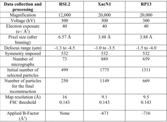
RSL2
The images have been binned four times (final pixel size of 6.57 Å). The initial 3D model of full RSL2 capsids has been calculated with the RIco software [18]. Thereafter all image analyses and capsid reconstructions have been performed using the Relion software [19] imposing icosahedral symmetry. The final reconstruction includes 250 particles out of 499 for a resolution of 16 Å (FSC determined using the gold-standard method implemented in Relion [19] at 0.143 threshold; Supplementary Figure 1).
XacN1 and RP13 115 116 117 118 119 120 121 122 123 124 125 126 127 128 129 130 131 132 133 134 135 136 137 138 139 140 141 142 143 144 145 146 147 148
The images have been binned two times (final pixel size of 3.88 Å). All the image analysis including generation of an initial model have been performed using the Relion software [19]. The final reconstruction of XacN1 and RP13 respectively includes 1149 and 669 particles for a resolution of 9.1 Å and 9.4 Å (FSC determined using the gold-standard method implemented in Relion [19] at 0.143 threshold; Supplementary Figure 1).
Reducing the binning to 2 for RSL2 and no binning for XacN1 and RP13 did not improve the resolution of the corresponding map.
Figures were generated using Chimera[20]. All the statistics are summarized in Table 1.
Fitting of the bacteriophage HK97 MCP into the EM map
The X-rays structure of HK97 MCP (pdb 2FT1) was fitted into the XacN1 and RP13 map using Chimera[20]. Briefly the entire structure (7 monomers) was first roughly placed by hand in the EM map and in a second step only one monomer was used. For this second step, the long alpha helix of HK97 monomer, which is easily recognizable was used as a landmark. Final refinement of the fitting was performed using the Chimera function “fit in map”.
Results
Two of the bacteriophages described here (RSL2[11] and RP13 [13]) were isolated from the phytopathogen Ralstonia solanacearum. The third one (XacN1 [12]) also a jumbo phage, was isolated from the phytopathogen Xanthomonas citri. The three phages belong to the Caudovirales order and exhibit the typical Myoviridae morphology with a contractile tail and an isometric head (Figure 1). Negative staining images of RSL2 (Figure 1A) clearly show that the tail is decorated by fibres at two different levels (arrows). For XacN1, an annular structure of unknown function is present around the tail (Figure 1B, arrow). RP13 differs from the other two phages because it exhibits a double-layered baseplate like the Twort-like phage 812 [21]. The [tail length]:[capsid diameter] ratios are quite different among the different viruses (Table 2). For this study we mainly focused our structural analyses on the virus head.
RSL2
Cryo-electron microscopy images show that the sample was a mixture between bacteriophages with a head full of DNA and others which have released their DNA (appearing as light shades in the images). Only capsids full of DNA were selected to perform icosahedral image analysis. The resulting structure shows a capsid having a diameter of 139 nm from 149 150 151 152 153 154 155 156 157 158 159 160 161 162 163 164 165 166 167 168 169 170 171 172 173 174 175 176 177 178 179 180 181 182
vertex to vertex (5-fold axis) and 128 nm along the 2-fold axis. The triangulation number, which determines the number of protein copies forming the capsid, is T=27 and was deduced from the hexagonal lattice present on the surface. It was calculated using the formula T=h2+hk+k2 where h and k are the number of local symmetry axes to be crossed to go from
one 5-fold axis to the next [22]. For RSL2, the observed numbers were h=3 and k=3 (Figure 2A).
The resolution obtained for RSL2 was limited to 16 Å due to a limited number of particles (250 particles; Figure 2A and Table 1). Each facet of the capsid is flat and composed of 13 hexamers. These hexamers as well as the pentamers are most probably made of the same protein: the major capsid protein (MCP) which, for this virus, is encoded by ORF117 (predicted size of 82,440 Da but observed size of 70 kDa according to SDS-PAGE and LC-MS/MS analyses as described before[11]). This is the largest known MCP. An icosahedral capsid with a triangulation number of T=27 is assembled from 27x60 asymmetric units (or 20x13 hexamers plus 12 pentamers). In the case of a caudal bacteriophage, a pentamer must be removed because one of the vertices is occupied by the portal. The total number of MCPs in this capsid is therefore 1615 since it appears at this resolution that the 11 vertices that do not bind the tail are composed of the same protein.
A hollow tube can be found at the centre of each hexamer with a diameter of 40 Å and a length of 70 Å. The 260 cylindrical structures project outward from the capsid and appear to cross the capsid and slightly extend out from the inner face of the hexamer (Figure 3C). The stoichiometry of this protein is difficult to assess at this resolution as it lacks recognisable features. The outside of the capsid is further decorated with another cylindrical protein (70 Å in length and 30 Å in diameter) that is bound to the periphery of the hexamers and lying parallel to the capsid surface. It associates in 810 dimers and forms bridges between neighbouring hexamers/pentamers (Figure 3B). The structure and organisation of the dimer is reminiscent from that observed in the KZ[7] and PBS1 capsids[6] which have the same triangulation number. It is interesting to note the presence of extra densities on the inside of the capsid, at the level of the 5-fold axis (Figure 3A, black arrows). This kind of structure is different from the one observed in RSL1 which exhibits a much more complex structure made of a trimer and a dimer [8]. Isosurface visualisation of these densities from the inside of the capsid (Figure 3D) shows that they are directly connected to the capsid at the 5-fold axis level (central globular structures) but also to the base of the cylindrical structures present at 183 184 185 186 187 188 189 190 191 192 193 194 195 196 197 198 199 200 201 202 203 204 205 206 207 208 209 210 211 212 213 214 215 216
the centres of each hexamer. The nature of these densities is unknown, but they may correspond to proteins or DNA/protein complexes that link the DNA to the capsid and allow the DNA to be organised.
Focusing on the inside of the capsid, one can note the absence of DNA organised in concentric layers which is probably not due to the lack of resolution as it becomes visible at about a 20 Å resolution. It is possible to distinguish a hexagonal mono domain organisation of DNA with a cylindrical rod spanning the interior of the head and oriented along the tail axis (dotted rectangle at bottom of Figure 1A; Supplementary Figure 2C). When RSL2 was exposed to a very high dose of electrons (>100 e-/Å2), an “inner body” similar to the one
observed for KZ [23] and 121Q [6] can be visualized. However, this bubblegram is slightly different from that observed with KZ as it has an arch at one end of the cylinder that is close to the tail. This arch is reminiscent of the structures visible under the 5-fold axis in Figure 3D.
XacN1
1149 particles have been used out of 1775 to obtain a 9 Å resolution three-dimensional map of the XacN1 head. Only particles loaded with DNA were analysed. The diameter of the final reconstructed full capsid is 139.5 nm from vertex to vertex and 116 nm along the 2-fold axis (Figures 1B and 2B). The triangulation number of this head is T=28 (h=4, k=2). The major capsid protein of XacN1is 463 aa in length and about 49 kDa in mass as described before[12]. Even with the medium resolution of this reconstruction, it was possible, due to the presence of a long “spinal” alpha helix, to unambiguously fit the X-ray structure of HK97 [24] into the cryo-electron microscopy map (Supplementary Figure 3A). The handedness of the structure is therefore most likely to be dextro (T=28,d). There are only two other known capsids from the Caudavirales order where three-dimensional reconstructions show such symmetry: 121Q [6] and PhAPEC6 [25].
The structure of the external and internal faces of the capsid is very smooth compared to other phages (Figures 2B, 3F, and 3G). This is especially true if compared with 121Q which harbours two types of decoration proteins at the periphery and on the middle of the hexamer. The only protruding components are located at the 5-fold axis with the presence of a turret-like structure (dimensions of 38 Å in height and 60 Å in diameter; Figure 3F). This kind of extension is quite common in the bacteriophage world [8, 9, 26]. Inspection of the central 217 218 219 220 221 222 223 224 225 226 227 228 229 230 231 232 233 234 235 236 237 238 239 240 241 242 243 244 245 246 247 248 249 250
slice of the three-dimensional reconstruction clearly shows that there are at least six concentric layers of DNA separated by 23.6 Å (Figure 3E). When irradiated at a high electron dose, the XacN1 head did not exhibit any bubblegram-type structure (data not shown).
RP13
Image analysis of the RP13 head started with 1311 particles. The best 669 particles yielded a three-dimensional reconstruction image at 9 Å resolution. This capsid shows a triangulation number of T=21, which was the smallest triangulation (and dimensions) of the three jumbo phages analysed here. The diameter of the particle from vertex to vertex is only 114 nm and between two opposed two-fold axes it is 97 nm. The obtained resolution enabled fitting of the HK97 X-ray structure into the RP13 capsid reconstruction leading to the assumption that the capsid handedness is T=21 laevo (supplementary Figure 3B). The RP13 phage is the first HK97-related phage to exhibit this kind of triangulation number. Only lipidic phages like PM2 [27], FLiP (Flavobacterium-infecting, lipid-containing phage [28]), or P23-77 [29] have shown an organization with similar geometry but with a pseudo T=21 dextro triangulation number. These types of phages do not use the canonical HK97 hexameric structure to build up their capsid but rather incorporate an adenovirus-like trimeric structure. Based on the three-dimensional structure it is clear that the vertices of the capsid are built by the same protein as the facet (Figures 3I and 3J, pentamer and hexamer). The capsid is therefore composed of 1255 copies of the major capsid protein (20x10 hexamers per facet plus 11 pentamers).
Decoration proteins can be found on the top of the major capsid protein in a position crossing local two-fold axes somewhat similar to what was observed in RSL2. The shape of the dimeric decoration proteins surrounding the hexamer is more globular in case of RSL2 compared to that of RP13. The RP13 decoration protein also appears to be hollow. On the inside of the particle, in the middle of each hexamer, one can also find a globular extra density (Figure 3J, right, arrow). On the DNA level, it is possible to distinguish at least five concentric layers of DNA separated by 26.3 Å. Like XacN1, an irradiation-sensitive inner body was not detected for this virus (data not shown).
Discussion 251 252 253 254 255 256 257 258 259 260 261 262 263 264 265 266 267 268 269 270 271 272 273 274 275 276 277 278 279 280 281 282 283
We determined the icosahedral capsid structures of three jumbo phages. Two of these phages are representatives of known triangulation number groups (T=27 and T=28), whereas the third one has a triangulation symmetry number that was not previously known to exist for caudal bacteriophages (T=21,l). Surprisingly, XacN1 has a smaller size compared to RSL2 even though it has a higher triangulation number, but this may be due to the difference in size of their respective MCPs (46 Da vs 70 kDa). The distance between the centre of two adjacent hexamers is slightly higher for RSL2 compared to XacN1 and RP13 (120 Å vs 113 Å, respectively). Different decoration proteins have been visualized on the outer portion of RSL2 and within its inner area. In contrast, XacN1 has the most basic capsid of the three phages studied here as no decoration proteins were observed on the exterior of the capsid. This proves that these accessory proteins are not essential to ensure the solidity of bacteriophage capsids, even when faced with the enormous internal pressures required to compact up to 400 kbp of DNA. For RSL2, because its MCP is much larger compared to the other two phages, one cannot be completely sure if the dimer present at the periphery of the hexamer is an extra protein or part of the MCP itself. However, it is likely that this dimer consists of accessory proteins since the same kind of dimer is present in KZ and PBS1 and because the MCPs are much smaller in these phages. A new kind of phage decoration protein that forms a hollow tube was also found in the RSL2 capsid. One would need to study this at higher resolution to determine what role this protein may have.
RSL2 carries only 224 kbp of DNA but it has the largest capsid. RSL2 is also the only phage in this study that has an inner body that does not exhibit concentric organisation of DNA layers within its three-dimensional structure. According to the structure we determined, large protein extensions are present on the inner part of the capsid and the DNA appears to be connected to the lower part of the hollow tube present at the centre of each hexamer. All of this together suggests that there are different types of DNA organisation in the jumbo phage world: one organised around an inner body with DNA connected to the capsid through dedicated structures near the 5-fold axes of the capsid; and another one as classical toroidal structures.
Two out of the three phages exhibits a “classical” DNA packing density of 0.49 and 0.39 whereas the third one has as very low one: RSL2 (0.29). This shows that the exceptions still exist and also shows the value of continuing extensive structural studies on viruses.
284 285 286 287 288 289 290 291 292 293 294 295 296 297 298 299 300 301 302 303 304 305 306 307 308 309 310 311 312 313 314 315 316
The RP13 capsid protects 180 kbp of DNA: less than most jumbo phages but more than classical bacteriophages. N3 DNA is 207 kbp for T=19,l and with a capsid diameter of 120 nm (vertex to vertex) compared to 114 nm for RP13 [6]. Because the capsid dimensions and the DNA size are in the same range for N3 and RP13, and even if RP13 does not technically meet the jumbo phage criteria (> 200 kbp), one can say that RP13 belongs to a new “semi-jumbo” category.
Conclusion
We found a new triangulation number symmetry to add to the bacteriophage morphology catalogue as well as a new kind of decoration protein. Many triangulation numbers are observed in nature. RP13 exhibits a higher T number than N3 but with DNA size that is too small for jumbo phage classification. We propose thatRP13 belongs to a new group of semi-jumbo phages. It seems that the distinction between classical myophages and jumbo phages is not clearly defined and that there is a continuum in the DNA sizes carried by
Myoviridae. The diversity of accessory/decoration proteins is enormous. One can imagine that
during evolution each phage has dipped into a common well to create its own combination of decorative/accessory proteins. Extensive study of these accessory proteins would help to discover novel protein properties (e.g. recognition of ligand).
Data availability
Cryo-EM maps have been deposited in the Electron Microscopy Data Bank: - RSL2 capsid map EMDB 11178
- XacN1 capsid map EMDB 11180 - RP13 capsid map EMDB 11179
Genome sequence accession numbers for the different bacteriophages are:
- RSL2 : AP014693 - XacN1 : AP018399 - RP13 : LC554890
Authorship Confirmation Statement
317 318 319 320 321 322 323 324 325 326 327 328 329 330 331 332 333 334 335 336 337 338 339 340 341 342 343 344 345 346 347 348 349 350
T.K. produced and purified the three bacteriophages. O.C. isolated RP13 in Thailand. G.S and E.N. prepared cryo-EM grids. E.N. and G.S. collected cryo-EM data on a FEI Polara EM. E.N. performed cryo-EM image processing and cryo-EM 3D reconstructions with the help of GE and LFE. The manuscript was written by G.S., E.N. and T.Y. with input from all authors. G.S. was responsible for the conception and direction of the work, analysing and interpreting data and revising the final drafts of the manuscript. All co-authors have reviewed and approved of the manuscript before submission and agree to be accountable for all aspects of the work. This manuscript has been submitted solely to this journal and is not published, in press, or submitted elsewhere.
Acknowledgments
We thank Maria Bacia-Verloop for technical assistance; Aymeric Peuch for help with the usage of the EM computing cluster.
This work used the platforms of the Grenoble Instruct-ERIC centre (ISBG ; UMS 3518 CNRS-CEA-UGA-EMBL) within the Grenoble Partnership for Structural Biology (PSB), supported by FRISBI (ANR-10-INBS-05-02) and GRAL, financed within the University Grenoble Alpes graduate school (Ecoles Universitaires de Recherche) CBH-EUR-GS (ANR-17-EURE-0003).The electron microscope facility is supported by the Auvergne-Rhône-Alpes Region, the Fondation Recherche Medicale (FRM), the fonds FEDER and the GIS-Infrastructures en Biologie Sante et Agronomie (IBISA).
Conflicts of interest
The authors declare that there are no conflicts of interest.
Funding information
This work received no specific grant from any funding agency 351 352 353 354 355 356 357 358 359 360 361 362 363 364 365 366 367 368 369 370 371 372 373 374 375 376 377 378 379
Figures legend:
Figure 1: Electron microscopy of bacteriophages RSL2, XacN1 and RP13.
A - RSL2
Top : Negative staining image of RSL2. The black arrows indicate decoration of the phage tail with fibrillary structures.
Bottom: Cryo electron microscopy image of the jumbo phage. The inner electron-dense body is highlighted by a rectangle or an arrow. The white arrow indicates some free DNA released from the bacteriophage.
B – XacN1
Top : Negative staining image of XacN1. The arrows highlight an annular density decorating the phage tail.
Bottom : Cryo electron microscopy image of the jumbo phage. The arrow points to the same structure as the one highlighted in negative staining.
C – RP13
Top : Negative staining image of RP13. The double arrow highlights the presence of a double layered baseplate in the virus. The inset show the bacteriophage in a contracted state with the inner tube of the tail sticking out.
Bottom : Cryo-EM image of RP13. The double arrow points to the double baseplate.
The scale bar represents 100 nm.
Figure 2: Three-dimensional reconstruction of the three jumbo phages obtained from cryo-EM images.
Three-dimensional reconstruction of the three jumbo phages represented as isosurface on the top of the figure. One facet is highlighted by a black triangle. The structures are color coded according to the radius of the particle as indicated in D.
380 381 382 383 384 385 386 387 388 389 390 391 392 393 394 395 396 397 398 399 400 401 402 403 404 405 406 407 408 409 410 411 412
A diagram showing the organization of the asymmetric units in one of the facet of the icosahedron is drawn for each virus. The organization of the hexamers in this facet makes it possible to determine the triangulation number that characterizes each of the bacteriophages. The different decoration proteins are also shown. The scale bars represent 20 nm.
A – RSL2. The bacteriophage head has a triangulation number T=27 (h=3; k= 3 ; T=h2 + hk
+ k2).
B - XacN1. The bacteriophage head has a triangulation number T=28, dextro ( h=4; k= 2 ; T=h2 + hk + k2).
C - RP13. The bacteriophage head has a triangulation number T=21, laevo ( h=4; k= 1 ; T=h2 + hk + k2).
D – Color code used in A-C to color the capsids according to their radius (in nm).
Figure 3 : Central section and decoration protein
RSL2
A – Half of the central section of the RSL2 density map. The protein density is in white. Black arrows are highlighting densities on the inner part of the capsid. The white asterisk is highlighting the cylindrical spike and the white arrow the dimer around the hexamers. The scale bar represents 20 nm.
B - Schematic view (left) and enlarged isosurface view (center) of the 5-fold axis showing that the vertex is surrounded by dimers. The vertex is also prominent compared to the rest of the capsid.
C - Schematic view (left) and enlarged isosurface view (center) of one pseudo 6-fold axis showing that the hexamers are surrounded by dimers and that there is a hollow cylindrical density sticking out from its center. On the right, isosurface view of a hexamer seen from the inside of the capsid. The arrow is pointing to the inner part of the spike present in the center of the hexamer. 413 414 415 416 417 418 419 420 421 422 423 424 425 426 427 428 429 430 431 432 433 434 435 436 437 438 439 440 441 442 443 444 445 446
D - Detailed view of the inside of the capsid along the 5-fold axis. The blue densities are corresponding to the extra densities indicated in A by the arrows. They represent probably nucleoprotein complexes involved in the DNA organization.
XacN1
E - Half of the central section of the PhXacn1 density map showing that the capsid is roughly smooth. Turret-like structures are only seen at the 5-fold axes, and some extra densities are also visible under the 5-fold axes (arrow). It is possible to distinguish at least 6 concentric layers of DNA. The protein/DNA densities are in white. The scale bar represents 20 nm.
F - Schematic view (left) and enlarged isosurface view (center) of the 5-fold axis showing the vertex composition. The right panel is a side view of the vertex showing the extra protein forming the turret-like structure.
G - Schematic view (left) and enlarged isosurface view (center) of one pseudo 6-fold axis showing that the capsid is quite smooth and probably only composed by the major capsid protein.
RP13
H - Half of the central section of the RP13 density map showing that the capsid is decorated by dimers (black arrow). No extra-densities are visible neither on the outside nor on the inside of the 5-fold axes. It is possible to distinguish at least 5 concentric layers of DNA. The protein/DNA densities are in white. The scale bar represents 20 nm.
I - Schematic view (left) and enlarged isosurface view (center) of the 5-fold axis showing that the vertex is surrounded by dimers. The protein forming the vertex has, at this resolution, the same shape as the one forming the hexamers (the major capsid protein) (see J).
J - Schematic view (left) and enlarged isosurface view (center) of one pseudo 6-fold axis showing that the hexamer formed by the MCPs is surrounded by dimers. The right panel is showing an isosurface view of a hexamer seen from the inside of the capsid. The arrow is pointing to an extra density which is probably a non-identified protein.
447 448 449 450 451 452 453 454 455 456 457 458 459 460 461 462 463 464 465 466 467 468 469 470 471 472 473 474 475 476 477 478 479 480
Table 1 : Electron microscopy statistics for the image analysis of the three bacteriophages.
Table 2 : Dimensions of the different parts of the three bacteriophages (capsid, tail and DNA). The inner volume of the capsid has been measured using UCSF Chimera as described
in [6]. 481 482 483 484 485 486 487 488 489 490 491 492
References
1 - Ackermann HW. 5500 Phages examined in the electron microscope. Arch Virol. 2007;152(2):227–243. doi:10.1007/s00705-006-0849-1
2 - Nikolich MP, Filippov AA. Bacteriophage Therapy: Developments and Directions.
Antibiotics (Basel). 2020;9(3):E135. Published 2020 Mar 24. doi:10.3390/antibiotics9030135
3 - Hendrix RW. Jumbo bacteriophages. Curr Top Microbiol Immunol. 2009;328:229-240. doi: 10.1007/978-3-540-68618-7_7
4 - Yuan Y, and Gao M, Jumbo bacteriophages:an overview. Front Microbiol. 2017;8:403. doi: 10.3389/fmicb.2017.00403
5 - Saad AM, Soliman AM, Kawasaki T, et al. Systemic method to isolate large bacteriophages for use in biocontrol of a wide-range of pathogenic bacteria. J Biosci Bioeng. 2019;127(1):73–78. doi:10.1016/j.jbiosc.2018.07.001
6 - Hua J, Huet A, Lopez CA, et al. Capsids and Genomes of Jumbo-Sized Bacteriophages Reveal the Evolutionary Reach of the HK97 Fold. mBio. 2017;8(5):e01579-17. . doi:10.1128/ mBio.01579-17
7 - Fokine A, Kostyuchenko VA, Efimov AV, et al. A three-dimensional cryo-electron microscopy structure of the bacteriophage KZ head. J Mol Biol. 2005;352(1):117–124. doi:10.1016/j.jmb.2005.07.018
8 - Effantin G, Hamasaki R, Kawasaki T, et al. Cryo-electron microscopy three-dimensional structure of the jumbo phage ΦRSL1 infecting the phytopathogen Ralstonia solanacearum.
Structure. 2013;21(2):298–305. doi:10.1016/j.str.2012.12.017
9 - Stroupe ME, Brewer TE, Sousa DR, Jones KM. The structure of Sinorhizobium meliloti phage ΦM12, which has a novel T=19l triangulation number and is the founder of a new group of T4-superfamily phages. Virology. 2014;450-451:205–212. doi:10.1016/j.virol.2013.11.019 493 494 495 496 497 498 499 500 501 502 503 504 505 506 507 508 509 510 511 512 513 514 515 516 517 518 519 520 521 522 523 524 525 526
10 - Cui J, Schlub TE, Holmes EC. An allometric relationship between the genome length and virion volume of viruses. J. Virol. 2014 ;88(11) :6403-6410.
11 - Bhunchoth A, Blanc-Mathieu R, Mihara T, et al. Two asian jumbo phages, ϕRSL2 and ϕRSF1, infect Ralstonia solanacearum and show common features of ϕKZ-related phages.
Virology. 2016;494:56–66. doi:10.1016/j.virol.2016.03.028
12 - Yoshikawa G, Askora A, Blanc-Mathieu R, et al. Xanthomonas citri jumbo phage XacN1 exhibits a wide host range and high complement of tRNA genes. Sci Rep. 2018;8(1):4486. doi:10.1038/s41598-018-22239-3
13 - Bhunchoth A, Phironrit N, Leksomboon C, et al. Isolation and characterization of bacteriophages that infect Ralstonia solanacearum in Thailand. ISHS Acta Horticulturae 1207 2018 ; V International Symposium on Tomato Diseases: Perspectives and Future Directions in Tomato Protection. pp. 155-162 doi:10.17660/ActaHortic.2018.1207.20
14 - Horita M, and Tsuchiya K. Causal agent of bacterial wilt disease Ralstonia
solanacearum. In: National Institute of Agriculture Sciences (Ed.), MAAF Microorganism
Genetic Resources Manual No.12. 2002 National Institute of Agricultural Sciences, Tsukuba, Japan, pp. 5-8.
15 - Valentine R, Shapiro B, and Stadtman E. Biochemistry. 1968;7, 2143-2152
16 - Zheng SQ, Palovcak E, Armache JP, Verba KA, Cheng Y, Agard DA. MotionCor2: anisotropic correction of beam-induced motion for improved cryo-electron microscopy. Nat Methods. 2017;14(4):331–332. doi:10.1038/nmeth.4193
17 - Zhang K. Gctf: Real-time CTF determination and correction. J Struct Biol. 2016;193(1):1–12. doi:10.1016/j.jsb.2015.11.003
18 - Estrozi LF, Navaza J. Ab initio high-resolution single-particle 3D reconstructions: the symmetry adapted functions way. J Struct Biol. 2010;172(3):253–260. doi:10.1016/j.jsb.2010.06.023 527 528 529 530 531 532 533 534 535 536 537 538 539 540 541 542 543 544 545 546 547 548 549 550 551 552 553 554 555 556 557 558 559 560
19 - Scheres SH. RELION: implementation of a Bayesian approach to cryo-EM structure determination. J Struct Biol. 2012;180(3):519–530. doi:10.1016/j.jsb.2012.09.006
20 - Pettersen EF, Goddard TD, Huang CC, et al. UCSF Chimera--a visualization system for exploratory research and analysis. J Comput Chem. 2004;25(13):1605–1612. doi:10.1002/jcc.20084
21 - Nováček J, Šiborová M, Benešík M, Pantůček R, Doškař J, Plevka P. Structure and genome release of Twort-like Myoviridae phage with a double-layered baseplate. Proc Natl
Acad Sci U S A. 2016;113(33):9351–9356. doi:10.1073/pnas.1605883113
22 - Caspar D. and Klug A. Physical principles in the construction of regular viruses. Cold Spring Harbor Symp. Quant. Biol. 1962 27:1-24. doi: 10.1101/sqb.1962.027.001.005
23 - Wu W, Thomas JA, Cheng N, Black LW, Steven AC. Bubblegrams reveal the inner body of bacteriophage φKZ. Science. 2012;335(6065):182. doi:10.1126/science.1214120
24 - Helgstrand C, Wikoff WR, Duda RL, Hendrix RW, Johnson JE, Liljas L. The refined structure of a protein catenane: the HK97 bacteriophage capsid at 3.44 A resolution. J Mol
Biol. 2003;334(5):885–899. doi:10.1016/j.jmb.2003.09.035
25 - Wagemans J, Tsonos J, Holtappels D, Fortuna K, Hernalsteens JP, Greve H, Estrozi LF, Bacia-Verloop M, Moriscot C, Noben JP, Schoehn G, Lavigne R. Structural Analysis of Jumbo Coliphage phAPEC6. Int J Mol Sci. 2020;21(9):E3119. doi: 10.3390/ijms21093119
26 - Lander GC, Baudoux AC, Azam F, Potter CS, Carragher B, Johnson JE. Capsomer dynamics and stabilization in the T = 12 marine bacteriophage SIO-2 and its procapsid studied by CryoEM. Structure. 2012;20(3):498–503. doi:10.1016/j.str.2012.01.007
27 - Huiskonen J, Kivelä H, Bamford D, et al. The PM2 virion has a novel organization with an internal membrane and pentameric receptor binding spikes. Nat Struct Mol Biol 2004;11:850–856. https://doi.org/10.1038/nsmb807 561 562 563 564 565 566 567 568 569 570 571 572 573 574 575 576 577 578 579 580 581 582 583 584 585 586 587 588 589 590 591 592 593 594 595
28 - Laanto E, Mäntynen S, De Colibus L, et al. Virus found in a boreal lake links ssDNA and dsDNA viruses. Proc Natl Acad Sci U S A. 2017;114(31):8378–8383. doi:10.1073/pnas.1703834114
29 - Rissanen I, Grimes JM, Pawlowski A et al. Bacteriophage P23-77 Capsid Protein Structures Reveal the Archetype of an Ancient Branch from a Major Virus Lineage Structure. 2013; 21(5): 718–726. doi: 10.1016/j.str.2013.02.026 596 597 598 599 600 601 602 603 604 605 606
Data collection and processing RSL2 XacN1 RP13 Magnification 12,000 20,000 20,000 Voltage (kV) 300 300 300 Electron exposure (e-/ Å2) 40 40 40
Pixel size (after binning) 6.57 Å 3.88 Å 3.88 Å Defocus range (mm) -1.3 to -4.5 -1.0 to -3.5 -1.5 to -4.0 Symmetry imposed 532 532 532 Number of micrographs 73 889 659 Initial number of selected particles 499 1775 1311 Number of particles for the final reconstruction 250 1149 669 Map resolution (Å) FSC threshold 0.14316 0.1439.1 0.1439.5 Applied B-Factor (Å2) None -671 -716
RSL2 XacN1 RP13 Capsid Capsid diameter 2-fold axis 128 nm 115 nm 97 nm Capsid diameter vertex to vertex 139 nm 134 nm 114 nm Internal diameter of the capsid 2-fold axis 108 nm 108 nm 89.5 nm Capsid volume (x 103 nm3) 830 790 460 Capsid thickness 42 Å 38 Å 40 Å T Number ; h ; k T=27 h=3, k=3 T=28,d h=4, k=2 T=21,l h=4, k=1 Decoration protein Number of dimers 810 0 630 Decoration proteins Number of spikes (6-fold) 260 0 0 Tail Tail Length 1650 Å 1180 Å 1010 Å Number of repeats 44 31 28 Pitch 37.5 Å 38 Å 36 Å DNA DNA (kbp) 224 kbp 385 kbp 180 kbp DNA spacing 23.6 Å 26.3 Å Avg density of packaged DNA (bp/nm3) 0.29 0.49 0.39