HAL Id: hal-03126622
https://hal.archives-ouvertes.fr/hal-03126622
Submitted on 31 Jan 2021
HAL is a multi-disciplinary open access
archive for the deposit and dissemination of
sci-entific research documents, whether they are
pub-lished or not. The documents may come from
teaching and research institutions in France or
abroad, or from public or private research centers.
L’archive ouverte pluridisciplinaire HAL, est
destinée au dépôt et à la diffusion de documents
scientifiques de niveau recherche, publiés ou non,
émanant des établissements d’enseignement et de
recherche français ou étrangers, des laboratoires
publics ou privés.
Linking pollution and cancer in aquatic environments: A
review
Ciara Baines, Adelaide Lerebours, Frédéric Thomas, Jérôme Fort, Randel
Kreitsberg, Sophie Gentes, Richard Meitern, Lauri Saks, Beata Ujvari,
Mathieu Giraudeau, et al.
To cite this version:
Ciara Baines, Adelaide Lerebours, Frédéric Thomas, Jérôme Fort, Randel Kreitsberg, et al.. Linking
pollution and cancer in aquatic environments: A review. Environment International, Elsevier, 2021,
149, pp.106391. �10.1016/j.envint.2021.106391�. �hal-03126622�
Environment International 149 (2021) 106391
Available online 27 January 2021
0160-4120/© 2021 The Author(s). Published by Elsevier Ltd. This is an open access article under the CC BY-NC-ND license
(http://creativecommons.org/licenses/by-nc-nd/4.0/).
Review article
Linking pollution and cancer in aquatic environments: A review
Ciara Baines
a,*, Adelaide Lerebours
b, Frederic Thomas
c,d, Jerome Fort
b, Randel Kreitsberg
a,
Sophie Gentes
b, Richard Meitern
a, Lauri Saks
e, Beata Ujvari
f, Mathieu Giraudeau
b,c,d,1,
Tuul Sepp
a,1aInstitute of Ecology and Earth Sciences, University of Tartu, Vanemuise 46, 51014 Tartu, Estonia
bLIttoral, ENvironnement et Soci´et´es (LIENSs), UMR7266, CNRS Universit´e de La Rochelle, 2 rue Olympe de Gouges, 17042 La Rochelle Cedex, France cCREEC/CREES, 911 Avenue Agropolis, BP 6450134394 Montpellier Cedex 5, France
dMIVEGEC, UMR IRD/CNRS/UM 5290, 911 Avenue Agropolis, BP 6450134394 Montpellier Cedex 5, France eEstonian Marine Institute, Universty of Tartu, M¨aealuse 14, 12618 Tallinn, Harju County, Estonia
fSchool of Life and Environmental Sciences, Centre for Integrative Ecology, Deakin University, Waurn Ponds, VIC, Australia
A R T I C L E I N F O
Handling Editor: Frederic Coulon
Keywords: Cancer Pollution Aquatic animals Marine ecosystems Freshwater ecosystems Oncogenic contaminants A B S T R A C T
Due to the interconnectedness of aquatic ecosystems through the highly effective marine and atmospheric transport routes, all aquatic ecosystems are potentially vulnerable to pollution. Whilst links between pollution and increased mortality of wild animals have now been firmly established, the next steps should be to focus on specific physiological pathways and pathologies that link pollution to wildlife health deterioration. One of the pollution-induced pathologies that should be at the centre of attention in ecological and evolutionary research is cancer, as anthropogenic contamination has resulted in a rapid increase of oncogenic substances in natural habitats. Whilst wildlife cancer research is an emerging research topic, systematic reviews of the many case studies published over the recent decades are scarce. This research direction would (1) provide a better un-derstanding of the physiological mechanisms connecting anthropogenic pollution to oncogenic processes in non- model organisms (reducing the current bias towards human and lab-animal studies in cancer research), and (2) allow us to better predict the vulnerability of different wild populations to oncogenic contamination. This article combines the information available within the scientific literature about cancer occurrences in aquatic and semi- aquatic species. For the first aim, we use available knowledge from aquatic species to suggest physiological mechanisms that link pollution and cancer, including main metabolic detoxification pathways, oxidative damage effects, infections, and changes to the microbiome. For the second aim, we determine which types of aquatic animals are more vulnerable to pollution-induced cancer, which types of pollution are mainly associated with cancer in aquatic ecosystems, and which types of cancer pollution causes. We also discuss the role of migration in exposing aquatic and semi-aquatic animals to different oncogenic pollutants. Finally, we suggest novel research avenues, including experimental approaches, analysis of the effects of pollutant cocktails and long-term chronic exposure to lower levels of pollutants, and the use of already published databases of gene expression levels in animals from differently polluted habitats.
1. Introduction
Pollution, together with climate change and over-exploitation, is one of the most severe consequences of the anthropocene for both marine and freshwater ecosystems (H¨ader et al., 2020). Toxic substances such as persistent organic pollutants, pesticides, and heavy metals, but also pharmaceuticals and microplastics, have been shown to negatively
affect the health and survival of aquatic organisms (Carolin et al., 2017; Duis & Coors, 2016; Morrissey et al., 2015; Li, 2014; Zhou et al., 2010). Due to the interconnectedness of aquatic ecosystems through the highly effective marine and atmospheric transport routes, all aquatic ecosys-tems are potentially vulnerable to pollution (H¨ader et al., 2020), and therefore, the effects of pollution on the health of aquatic organisms requires urgent attention from the scientific community.
* Corresponding author.
E-mail address: ciara.baines@ut.ee (C. Baines).
1 Shared last authorship.
Contents lists available at ScienceDirect
Environment International
journal homepage: www.elsevier.com/locate/envint
https://doi.org/10.1016/j.envint.2021.106391
As the negative impact of anthropogenic pollution on aquatic and semiaquatic organisms has been widely acknowledged, the next step should be to focus on specific physiological pathways and pathologies linking pollution to wildlife mortality. Whilst the release of large quantities of pollutants can cause an acute impact, measured by large- scale sudden mortalities of aquatic organisms, lower levels of contami-nation and the mixture of several contaminants may be more difficult to track but can result in pathologies from long term accumulation in the tissues of aquatic animals (Austin, 1998). A better understanding of specific pathological effects of pollutants enables us to pinpoint species and ecosystems that are most vulnerable to contamination and to distinguish pollutants that pose the greatest threats to the health of wild animals. A systematic focus on a specific physiological pathway from pollution to deterioration of the health of an organism might reveal similar patterns in a wider range of species and populations in contact with aquatic contaminants. For that, we need comprehensive reviews of the studies available on specific pathologies caused by contamination in aquatic ecosystems.
One of the pollution-induced pathologies that should be at the centre of attention in ecological and evolutionary research is cancer. Anthro-pogenic contamination has resulted in a rapid increase of both evolu-tionarily familiar and novel oncogenic substances in natural habitats, and presented animals inhabiting these environments with novel evolutionary pressures (McAloose & Newton, 2009; Giraudeau et al., 2018; Vittecoq et al., 2018). Whilst some oncogenic contaminants originating from natural sources have always been present in aquatic environments (Menzie et al., 1992), new substances have been added to this toxic cocktail, and the concentrations of oncogenic pollutants have reached unprecedented levels in many human-impacted habitats (e.g.
Froehner et al., 2018). There are numerous examples of high mortality resulting from cancer in animals living in polluted aquatic environments such as, the high prevalence of intestinal adenocarcinomas in beluga whales (Delphinapterus leucas) from the Saint Lawrence Estuary (a highly polluted habitat due to effluents from aluminium smelting facilities,
Martineau et al., 2002), the linking of cancer occurrence with organo-chlorine contaminants in California sea lions (Zalophus californianus,
Randhawa et al., 2015), and the numerous discoveries of different types of fish tumours in polluted habitats (Brown et al., 1973; Black & Bau-mann, 1991). From a conservation perspective, understanding the leading causes for high rates of cancer within some wild populations allows us to better predict the vulnerabilities of wild populations to oncogenic contamination (McAloose & Newton, 2009). Some of the more prevalent aquatic contaminants with known oncogenic effects include persistent organic pollutants (for example polycyclic aromatic hydrocarbons (PAHs)), dioxins, polychlorinated biphenyls (PCBs), pes-ticides such as dichlorodiphenyltrichloroethane (DDT, Ashraf, 2017) and heavy metals (Tchounwou et al., 2012). The oncogenic effects of named pollutants might be further amplified by other characteristics of the Anthropocene, such as high levels of disturbance, climate change, introduction of novel pathogens, changes in food items, or genetic di-versity level (Giraudeau et al., 2018). Considering the numerous ex-amples of pollution-induced oncogenic processes in aquatic animals, in addition to other environmental and ecological processes that trigger neoplastic development (Thomas et al., 2017), we can expect that cancer is an underestimated cause of disease and mortality in many aquatic and semi-aquatic organisms living within human-impacted habitats. From the perspective of human health, studying the mechanistic links be-tween pollution and cancer in non-model organisms, in an ecological context, provides a novel perspective for understanding eco-evolu-tionary processes linked to cancer at the cellular and organismal levels (Hamede et al., 2020).
A better understanding of the links between pollution and cancer in aquatic animals allows using wild species as sentinels for the contami-nation of aquatic environments with oncogenic chemicals for preventing an impact on human health, but also for preventing the negative effects of contamination on wild populations and ecosystems. Especially when
we consider that although mode and level of contamination may differ, similar mechanisms for cancer regulation and pathways for cancer initiation have evolved between vertebrate species, whether aquatic or terrestrial (for example: Schug et al., 2016; Dujon et al., 2020). This article combines the information available in the scientific literature about pollution and cancer in aquatic environments (see Tables 1 and
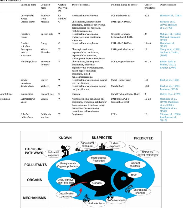
S1) with the aim of addressing the following questions and proposing new research avenues. Using the available data, we predict which types of cancer aquatic pollution is more likely to cause and analyse the types of pollution currently associated with cancer in aquatic environments. We discuss the physiological mechanisms linking pollution and cancer, including main metabolic detoxification pathways, oxidative damage effects, infections, and changes in microbiome. Based on available studies, we describe which species are more vulnerable to pollution- induced cancer in aquatic environments and discuss which other spe-cies could be vulnerable based on analogy. We discuss the role played by pathogens and migration in exposing aquatic and semi-aquatic animals to oncogenic pollution. Finally, we suggest novel research avenues (Fig. 1), including the use of already published databases of gene expression levels in animals from differently polluted habitats. 2. What types of cancer does aquatic pollution cause?
Knowing where to look for cancer is one of the first challenges in measuring the extent to which populations are affected by pollution- induced cancer and understanding the mechanistic link between pollu-tion and cancer. Humans are regularly exposed to oncogenic pollupollu-tion, for example, through the air that they breathe. As a result, lungs are an organ that is commonly affected by pollution-induced cancer in humans (Kim et al., 2018). Similarly, in fish, organic pollutants are often absorbed through the gills and skin, and accumulate in lipid-rich tissues, such as liver, brain, gonads, and hypodermal lipid storages. Studies that link pollution and cancer in aquatic habitats indicate that skin cancer is a frequent outcome of living in contaminated waters, especially in several fish species (Table 1). For example, as a result of exposure to contami-nants, freshwater drums (Aplodinotus grunniens) develop chromato-phoromas (Black, 1983), white suckers (Catostomus commersonii) and brown bullheads (Ameiurus nebulosus) develop papillomas (Sonstegard, 1977; Baumann et al., 1991), common dab (Limanda limanda) develop hypodermal lipomas and papillomas (Vethaak et al., 2009) and walleyes (Sander vitreus) dermal ossifying fibromas (Black et al., 1982).
The natural sources of oncogenic contaminants (Menzie et al., 1992), which have existed for hundreds of millions of years, have allowed fish and other organisms to develop various pathways to transform (like cytochrome p450 systems) or reduce (metallothioneins) the effects of these pollutants (Livingstone, 1998). Whilst xenobiotic metabolism protects the organism effectively from an array of lipid-soluble con-taminants, in the case of some pollutants, such as PAHs, this process is responsible for the production of the highly reactive and genotoxic metabolites that lead to cancer development (Gao et al., 2018). In addition, chronic exposure combined with adverse environmental con-ditions can overwhelm these systems, causing severe malfunctions, including tumours. Accordingly, next to skin cancer, cancers of the liver, that assist in processing contaminants, are very common. Hepatic tu-mours have been described in many fish species including white suckers, brown bullheads, common dab, Atlantic tomcod (Microgadus tomcod), hagfish (Myxine glutinosa), English sole (Parophrys vetulus), winter flounders (Pseudopleuronectes americanus), and walleyes (references in
Table S1). Similarly, other organs exposed to high concentrations of contaminants such as bile duct and kidneys are often affected by ma-lignant processes, as seen in studies of white suckers (i.e. Blazer et al., 2017) and rainbow trout (Oncorhynchus mykiss, Shelton et al., 1983; Groff, 2004; Dale et al., 2009).
Interestingly, malignant tumours in lipid-rich brain tissues, which could be a promising future study direction (Fig. 1), are often under- described and under-studied throughout the literature. It is likely that
Table 1
Studies that link pollution to cancer in aquatic and semi-aquatic species. For an overview of all the studies published on cancer in aquatic and semi-aquatic species, please see Table S1. Scientific names have been updated with accepted names from WoRMS (WoRMS Editorial Board, 2020).
Scientific name Common
name Captive (C)/Wild
(W)
Type of neoplasia Pollution linked to cancer Cancer
prevalence Other reference
Platyhelminthes Bdellocepha-la
brunnea planaria C Undetermined neoplasia Benzo(a)pyrene Hoshina & Teshirogi, (1991)
Girardia
dorotoceph-ala planaria C Undetermined neoplasia Cadmium sulphate 3.4–14.7 Hall et al., (1986) Girardia tigrina Dugesia
tigrina C Undetermined neoplasia Cadmium sulphate and 12–0- tetradecanoylphorbol-13-
acetate (PMA)
30–90 Voura et al., (2017)
Mollusca Corbicula
fluminea Asian clam C Undetermined neoplasia in digestive gland Bisphenol A Benjamin, Competente & de Guzman, (2019)
Crassostrea
virginica Eastern oyster W/C Neoplasms in kindney, gastrointestinal tract, gonadal, gill, heart and neural elements and included carcinomas
PCB’s, hydrocarbons (inc. PAH’s), heavy metals (As, Cd, Cr, Cu, Pb, Mn, Ni)
7–14 Gardner & Yevich,
(1988)
Mya arenaria Soft-shell
clam W/C Disseminated neoplasia, gonadal neoplasia, haemocytic leukemia, seminomas and dysgerminomas
PCB’s, pesticides (ß- endosulfan and
α-endosulfan), hydrocarbons (oil), PAH’s, heavy metals
68–90 Reinisch et al., (1984); Muttray et al., (2012); Brown, (1980); Gardner et al., (1991); B¨ottger et al., (2013)
Mytilus edulis Blue mussel W/
Farmed Disseminated neoplasia/ haemocytic leukemia Sewage, pulpmill effluents, metals 23–56 Charles et al., (2020); St-Jean et al., (2005)
Mytilus
galloprovin-cialis Mediterr- anean mussel Farmed Gonadal neoplasia/ germinoma Heavy metals, PCB’s, PAH’s 15–60 Ruiz et al., (2013)
Fish Ameiurus
nebulosus Brown bullhead W Skin/liver neoplasms, cholangioma, hepatoademona, hepatocarcinoma,
cholangiocarcinoma,
PCB, PAH 22–41 Baumann &
Harshbarger, (1995); Baumann, (1998); Baumann & Harshbarger, (1998), Baumann et al., (1991)
Anguilla anguilla European eel W Undetermined neoplasia in liver
and spleen Pesticides, heavy metals, PAH’s 30 Ribeiro et al., (2005)
Aplodinotus
grunniens Freshwater drum W Chromatophoroma, neurolemmoma, carcinoma PAH 16.67 Black, (1983)
Ameiurus melas Black
bullhead W Oral papillomas Chlorine 73 Grizzle, Melius & Strength (1984)
Catostomus
commersonii White sucker W Hepatic neoplasm, hepatocellular carcinoma, bile duct carcinoma, papillomas,
Industrial pollution, PAH’s ~60 Hayes et al., (1990);
Sonstegard, (1977)
Danio rerio Zebrafish C Malignant neoplasms of the intestine, epidermal papillomas, Chondroma or chondrosarcoma, hepatic neoplasia, hemangioma, peripheral nerve sheath tumor, hemangiosarcoma, hepatocellular adenoma, hepatocellular carcinoma, biliary adenoma, biliary carcinoma, seminomas, adenocarcinoma, adenomas, leiomyosarcoma, rhabdomyosarcoma PAH (7,12-Diniethylbenz[a] anthracene), Ethlynitrosourea, N-methyl- N’-nitro-N-nitrosoguanidine (MNNG) 16–100 Beckwith et al., (2000); Spitsbergen et al. (2000a, 2000b) Fundulus heteroclitus heteroclitus
Mummichog W/C Hepatoblastoma, hepatocellular
adenoma, hepatocellular carcinoma, cholangiocellular proliferative lesions
PAH’s inc. BaP 35 Wills et al., (2010);
Vogelbein et al., (1990)
Hoplias
malabaricus Trahira W Pancreatic neoplasia Persistent organic pollutants, Miranda et al., (2008)
Limanda
limanda Common dab W Papillomas, hepatocellular adenoma, hepatocellular carcinoma
Cadmium, PCB’s 20 Lerebours et al.,
(2014); Vethaak et al., (2009)
Microgadus
tomcod Atlantic tomcod W Hepatic neoplasm; hepatocellular carcinomas PAH/PCB/pesticides 44.3–92.9 Dey et al., (1993)
Myxine glutinosa Hagfish W Hepatocellular adenoma, hepatocellular carcinoma, cholangiocellular carcinoma, cholangiocellular adenofibrosis, mixed hepato- and cholangio- cellular tumours
PCB’s 5.8 Falkmer et al., (1978)
Nibea mitsukurii Croaker W Chromatophoroma Nifurpirinol 71–100 Kimura et al., (1989) (continued on next page)
in wild populations, tumour diagnosis is limited by specific types of analyses (e.g. histopathology from tissues most likely affected), causing bias in published studies. New molecular methods (e.g. biomarkers that are either specific or nonspecific to a certain tissue) could resolve this imbalance.
3. Which types of pollution are mainly associated with cancer in aquatic environments?
Increase in cancer prevalence can and has been linked to various types of pollution (Table 1), including organic pollutants (e.g. muta-genic PAHs, genotoxic persistent organic pollutants, POPs) and non- essential metallic trace elements (e.g. cadmium, arsenic). However, it Table 1 (continued)
Scientific name Common
name Captive (C)/Wild
(W)
Type of neoplasia Pollution linked to cancer Cancer
prevalence Other reference
Oncorhynchus
mykiss Rainbow trout C/ Farmed Hepatocellular carcinoma PCB’s/aflatoxin B1 40.2 Shelton et al., (1983)
Oryzias latipes Medaka C Cholangioma, hepatocellular carcinoma, hemangiopericytoma, perisinusoidal cell neoplasm, rhabdomyosarcoma
PAH’s (BaP, DMBA) Fabacher et al.,
(1991); Hawkins et al., (1990)
Parophrys
vetulus English sole W Hepatocellular carcinoma, cholangiocellular carcinoma, adenomas
Creosote (aromatic
hydrocarbons), PAH’s 27 Malins et al., (1985); Malins & Haimanot, (1990)
Poecilia
reticulata Guppy C Hepatocellular neoplasms PAH’s (BaP, DMBA) 19–46 Hawkins et al., (1990)
Pseudopleu- ronectes americanus
Winter
Flounder W Cholangiocarcinomas, hepatocellular carcinomas, hepatocellular adenoma, cholangioma, hepatic neoplasms
PAH/pesticides/metals 18 Chang et al., (1998),
Gardner & Yevich, (1988)
Platichthys flesus European
flounder W Cholangioma, hemangioma, carcinomas, adenomas, angiosarcoma, hepatoblastoma, mixed hepato-cholangio carcinoma, mixed hepatoangiosarcoma
PCB’s, organochlorines 24–72 K¨ohler, Wahl &
S¨offker, (2002); Koehler, (2004)
Sander
canadensis Sauger W Hepatocellular carcinomas, dermal ossifying fibroma Metal (copper ores) 100 Black et al., (1982)
Sander vitreus Walleye W Hepatocellular carcinoma, dermal
ossifying fibroma Metals/PAH
<30 Black et al., (1982);
Baumann, (1998)
Amphibians Rana pipiens Leopard frog C Sarcoma 3-methylcholanthrene (PAH) 9 Outzen et al., (1976)
Mammals Delphinapterus
leucas Beluga W Adenocarcinoma, squamous cell carcinoma, granulaosa cell tumour, dysgerminoma, lymphosarcoma, neuroendocrine carcinoma, transitional cell carcinoma
PAH (BaP), PCB’s
(organohalogens) 18–24 Martineau et al., (1994); Martineau
et al., (2002); Martineau et al., (1988)
Zalophus
californianus California sea lion W Carcinoma PCB’s 18 Ylitalo et al., (2005); Randhawa et al., (2015)
has been estimated that 100,000 new substances currently used on the European market can potentially end up in the environment and should therefore be considered as emerging pollutants (Brack et al., 2018). Developing our understanding of how this wide variety of pollutants contributes to cancer in aquatic species is paramount in understanding both how populations are likely to be impacted in polluted habitats and the biological mechanisms that initiate oncogenic processes with expo-sure (Table 2). However, the wide spectrum and low concentrations of these substances in the environment present challenges for under-standing their effects on the health of aquatic organisms, especially, when such linkages between tumour prevalence and presence of potentially mutagenic toxicants are not always causal.
One finds the substances that one looks for – therefore it is important for (evolutionary) ecotoxicologists to assess the effects of groups of toxicants rather than limit interpretation to a specific chemical group, as is often the case in older studies. For example, the two most prominent groups studied (Table 1, Fig. 1) are PAHs and heavy metals, which have been widely described to co-occur in environments affected by anthro-pogenic pollution (e.g. Hsu et al., 2019; Monaco et al., 2017; Dai et al., 2020). These chemical ‘cocktails’ that are present in wild environments are known to have additive, synergistic or antagonistic effects (Laetz et al., 2009; Ansari et al., 2004). This synergism and antagonism can be further exacerbated when considering the effect of other environmental stressors resulting from climate change (see Crain et al., 2008). In addition, abiotic environmental stressors and physiological factors, such as ocean warming, that modify the bioavailability of pollutants (Parmesan & Yohe, 2003), or physiological responses to reproduction periods, that alter metabolism, could increase species sensitivity to mutagenic toxicants. Furthermore, some marine debris, specifically microplastic particles, are known to form biofilms, which could poten-tially act as a vector for increased oncogenic viral transmission (Cole et al., 2011) and can accumulate various aquatic contaminants (Ziccardi et al., 2016). Understanding the pathways that expose different species to a pollutant or group of pollutants is important in understanding whether certain species, for example benthic species that are exposed through sediment contact and trophic transfer (Sakurai et al., 2009; Cope et al., 2008), are more at risk from oncogenic contaminants (see
Section 5). The bioaccumulation or biomagnification of pollutants and pollutant cocktails varies, not only between the type of contaminants but also, between the species, habitat and feeding habits of the animal exposed (Bakir et al., 2016).
However, there is still relatively little known about cocktail effects and the impacts of multiple environmental stressors, in relation to cancer development in natural environments. Based on the studies published so far, aquatic ecosystems in contact with organic pollutants or heavy metals should be the focus for the study of wildlife cancer, however, emerging contaminants should not be overlooked. Whilst there is currently only one study linking cancer in aquatic animals to pesticide exposure (Chang et al., 1998), this scarcity is probably not an indicator of a missing link, but rather of a study gap. Experimental studies would be necessary for disentangling the effects of individual pollutants on cancer development in aquatic species and in addition, longitudinal studies following the clean-up of polluted aquatic
environments, could be an underused opportunity for understanding which types of pollutants are mainly responsible for cancer in wild animals.
Cancer is a multifactoral disease and many pollutants do not directly cause cancer but alter biological systems that affect natural cancer defence mechanisms (eg. Schug et al., 2016). Whilst the mechanisms behind cancer initiation following pollution exposure (such as the for-mation of DNA adducts and the activation of specific genes), have been explored in some studies (Table 2) we suggest that this is an important research area that needs further attention in the future.
4. Physiological mechanisms that link pollution and cancer
4.1. Metabolic pathways connecting pollution and cancer
The precise mechanistic cause and effect relationship connecting environmental pollution and cancer, specifically at the subcellular/ molecular level, is still uncharacterized (Lerebours et al., 2014). Therefore, studies exploring this concept would provide a background knowledge of the metabolic processes that, with exposure to pollution, could increase cancer risk within aquatic populations. In the context of toxicological effects, aryl hydrocarbon receptor (AHR, AhR, AH-r, or Ah- r as used in different publications) expression is probably one of the most researched pathways that plays a pivotal role in detoxification processes in aquatic animals (e.g. Zhou et al., 2010). The expression of AHR, a ligand-activated nuclear transcription factor, can be altered through exposure to a variety of environmental contaminants such as POPs (including PAHs and halogenated aromatic hydrocarbons, HAHs, Zhou et al., 2010). A recent study suggests that the desensitization of AHR pathway genes in gulf killifish (Fundulus grandis) can help individuals cope with heavy urban pollution load (PAHs and HAHs) in Galveston Bay, USA (Oziolor et al., 2019). We hypothesise that there are similar downregulation mechanisms in other aquatic species to potentially protect cells from oncogenic contaminants.
The induction of synthesis of heat shock proteins (HSPs) as a response to any kind of environmental stress (temperature, salinity, hypoxia, chemicals, etc.) has been demonstrated in all organisms examined to date, from prokaryotes to humans, and could constitute a link with cancer. For example, both histopathological alterations and a significant increase in HSP70 expression in milk fish (Chanos chanos) were found at a site polluted with heavy metals when compared to a ‘less’ polluted site in India (Rajeshkumar & Munuswamy, 2011). Evi-dence supports that HSPs are likely to have anti-apoptotic properties and are actively involved in tumour cell proliferation, invasion, differenti-ation, metastases and death. Whilst there have been some studies sug-gesting increased expression of HSP’s in fishes with exposure to pollution, more research is needed to understand the links between HSP expression and cancer in aquatic animals.
Endocrine disruption has been evidenced to occur as a result of exposure to some environmental pollutants (eg. pesticides) (Soto & Sonnenschein, 2010). These chemicals bind to endocrine receptors and alter the hormonal response which are known to control cancer initia-tion in humans (Schug et al., 2016). The endocrine system controls a Table 2
Studies that have linked aquatic pollutants and cancer in wild animals through a specific physiological mechanism.
Pollutant type Suggested mechanism Species References
PAH (BaP) EROD activity/Mitochondrial DNA damage Mummichog (Fundulus heteroclitus) Wills et al., 2010
PAH/PCB/pesticides Activation of K-ras oncogene/DNA lesions Atlantic tomcod (Microgadus tomcod) Wirgin et al., 1989; Cormier
et al., 1989
PAH Genotoxicity Winter Flounder (Pseudopleuronectes
americanus) Baumann 1998,
PAH (BaP), PCB’s
(organohalogens) Immunosuppression, DNA adducts Beluga (Delphinapterus leucas) Martineau et al., 1994
Cadmium Pollutant-induced mutational activation/inactivation of key
number of biological processes in all vertebrates and whilst disruption has been linked to cancer in humans and rodents, it is likely that these chemicals also play an underestimated role in cancer initiation in aquatic animals (Schug et al., 2016)
4.2. Cancer induction by oxidative stress and DNA damage pathways
As aquatic animals are arguably exposed to a greater quantity and variety of pollutants than terrestrial animals, they present good oppor-tunities to understand the link between pollution and cancer at the cellular level. A broad spectrum of pollutants can compromise the genomic integrity of cells by inducing DNA damage or by generating oxidative stress (Rastogi et al., 2010; Lerebours et al., 2013; Rhee et al., 2013). DNA damage plays a key role in mutagenesis and the develop-ment of cancer (Rotchell et al., 2001; Du Corbier et al., 2005; Lerebours et al., 2014, 2017). Certain PAHs have the capacity to enter redox cycles and induce the production of reactive oxygen species (ROS), thereby causing oxidative stress (An et al., 2011). Some benzo(a)pyrene (BaP) metabolites can undergo redox-cycling, producing ROS thereby leading to DNA damage (Chatgilialoglu and O’Neill, 2001) and mutations (Bonner et al., 2005).
Considering this importance of oxidative stress and DNA damage in initiating oncogenic processes, and the impact of carcinogens on these processes, it is surprising that the link between exposure to carcinogens, oxidative stress, DNA damage and neoplasia has been scarcely addressed in aquatic organisms. A clear relationship between oxidative DNA damage and neoplasia was evidenced in feral English sole exposed to PAHs from Eagle Harbor, U.S. (Malins et al., 1985). Interestingly, an increase in antioxidants (such as superoxide dismutase, catalase and glutathione) has been observed in long-lived aquatic organisms such as in the mud clam (Arctica islandica) and the red-eared slider turtle
(Tra-chemys scripta elegans) (for a review see Wojtczyk-Miaskowska & Schlichtholz, 2018). This suggests that the antioxidant system confers cell survival by preventing ROS-induced DNA damage and subsequent cancer development.
Depending on the type of DNA damage induced, several DNA repair mechanisms are triggered to maintain genomic stability. However, some of them are more prone to errors and are often associated with cancer. PAHs, specifically BaP metabolites, can form DNA adducts that cause errors in DNA replication during DNA repair (Boelsterli, 2007; Phillips & Arlt, 2007). Exposure to PAHs has been firstly, linked to an increase in DNA adducts in the pale chub (Zacco platypus) (Lee et al., 2014), killifish (F. grandis and F. similis) (Willett et al., 1995) and Beluga whales (Mathieu et al., 1997) and secondly, correlated to DNA adduct formation and neoplastic lesions in fish such as brown bullhead (Ploch et al., 1998) and English sole (Reichert et al., 1998) and marine mammals such as harbour porpoises (Acevedo-Whitehouse et al., 2018) and beluga whales (Poirier et al., 2019). In order to further explore these cause-effect re-lationships, studies should consider oxidative stress and DNA damage/ repair biomarkers, histopathological conditions and a high number of carcinogens (including metabolites) together in environmental sce-narios, where aquatic organisms are often exposed to a mixture of pollutants.
4.3. Interactions between the gut microbiota and pollutants and consequences for cancer development.
The microbiome is essential for major biological functions, including the biotransformation of xenobiotics (Sousa et al., 2008; Jandhyala et al., 2015) and the control of cancerous processes (Elinav et al., 2019). A disruption of the microbiota balance, named dysbiosis, leads to the alteration of the functioning of the gut microbiome and could result in cancers (Scanlan et al., 2008; Meng et al., 2018, Fig. 1). Many pollutants such as metals (cadmium, lead, arsenic), pesticides (DDT, chlorpyrifos, glyphosate) and PCBs have demonstrated their ability to cause quick microbiome dysbiosis, affecting both the microbiota composition and
also the microbiota metabolic functions, mainly in vertebrates and through trophic transfer (Flandroy et al., 2018).
Microbiota also has, in some cases, the capacity to influence pollutant behaviour and ultimately their molecular and physiological effects (Dietert & Silbergeld, 2015). Gut microbiota has been known for a long time to be involved in biotransformations of xenobiotics through various mechanisms (Scheline, 1973; Sousa et al., 2008; Kang et al., 2013; Claus et al., 2016). However, the few enzymes that have been recognized to play a role in these transformations have mainly been studied in humans (Eriksson & Gustafsson, 1970; Williams et al., 1970; Bakke & Gustafsson, 1984; Rafil et al., 1991; Rold´an et al., 2008). Many carcinogenic pollutants metabolized by gut microbiota, mainly in ver-tebrates, include PAHs (benzo(a)pyrene), nitrated PAHs, nitrotoluenes, pesticides (DDT), PCBs and metals (mercury, arsenic) (Claus et al. 2016).
Hence, microbiota can on one hand interact with environmental pollutants (microbiota can modify the cycle and thus the toxicity of pollutants that in turn affect the microbiota composition) and on the other hand modulate cancer development. Thus, one can predict that gut microbiota could be a major driver in the processes leading to cancer emergence in aquatic organisms that have been chronically exposed to a cocktail of contaminants. Therefore, microbiota could be considered as a promising biological indicator of the organism’s health status and, accordingly, more attention should be focused on the microbiota in future wildlife cancer studies.
5. Evidence of cancer in animals living in (polluted) aquatic environments
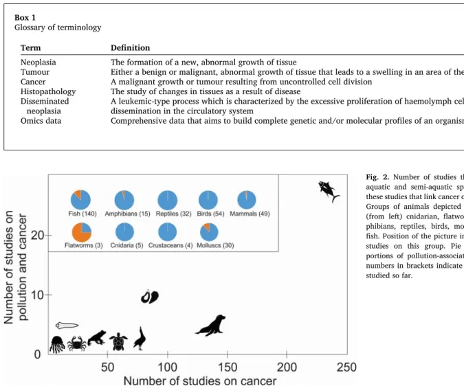
Understanding the reasons for cross-species variation in cancer development as a result of exposure to pollution promises extremely valuable information on oncogenic processes, as the limited research conducted on non-standard model organisms has already provided tremendous insights on the natural mechanisms of cancer resistance. Despite its value, robust cancer prevalence data on animals is surpris-ingly limited to date, currently only available for 31 wild vertebrate species (Ujvari et al. 2017). The latter database indicates stunning cross- species variation of cancer rates in aquatic and semi-aquatic species with less than 1% of the sea otters (Enhydra lutris), 23% of white-fronted geese (Anser albifrons) and 64% of the gray seals (Halichoerus grypus) developing some type of cancer (Ujvari et al. 2017). To compare, the general estimates for a lifetime risk for cancer in humans is around 40% (e.g. Grunau et al. 2018). While cancer prevalence data is extremely limited, there is more information available on cancer occurrences across wild species. Considering that the representation of aquatic spe-cies in cancer studies is impressive. There have been reports of neoplasia in over 330 aquatic species and a variety of studies that link cancer to contaminant exposure (Table S1). However, there are challenges to fully understanding the role pollution plays in inducing cancer, such as the dynamic nature of cancerous cell development, multiple contributing factors to neoplasia development in natural environments and ethical concerns in cancer diagnosis in wild animals. Histopathology (Box 1), through dissection, is currently the most reliable method used to mea-sure cancer prevalence. This has likely contributed to the study bias towards fish and bivalves in pollution-induced cancer studies (Fig. 2). However, given the range of taxa that have shown susceptibility to pollution-induced neoplasia, the literature indicates that this phenom-enon might affect a much wider range of species than originally considered.
5.1. Invertebrates 5.1.1. Platyhelminthes
Three species of planaria (Dugesia tigrina, Voura et al., 2017
(accepted as Girardia tigrina, WoRMS Editorial Board, 2020), Dugesia
WoRMS Editorial Board, 2020) and Bdellocephala brunnea, Hoshina & Teshirogi, 1991) have shown evidence of pollution-induced neoplasia (Table 1). Interestingly, G. tigrina has more recently been used as a model species for cadmium oncogenesis to better understand the mechanisms leading to cancer development (Voura et al., 2017). Using planaria, with their known regeneration capabilities, could provide a deeper understanding of cancer in humans and lead to potential thera-pies to improve survival (Oviedo & Beane, 2009).
5.1.2. Cnidaria
A range of cnidaria species have shown evidence of neoplasia development from hydra to Acropora corals, however, currently there have been no conclusive studies linking neoplastic development in cnidaria to pollution within the literature. Hydractinia echinate showed evidence of induced neoplasms under experimental conditions (Millane et al., 2011) and whilst there have been several reports of tumours on coral species in natural environments (Sutherland, Porter & Torres, 2004), only two cases have diagnosed these tumours as neoplastic (Table S1). Peters, Halas & McCarty, (1986) diagnosed nodules on
Acropora palmata colonies as calicoblastic epitheliomas. The second
study diagnosed calicoblastic epitheliomas in Acropora valenciennesi and found similar tumours in the species Acropora valida, although the latter species were not diagnosed using histopathology. A study on Porites sp., that found tumour prevalence as high as 39.1% in one of the surveyed locations, found a correlation between distance from the local city and percentage of evident tumours (Kaczmarsky, 2006). The authors sug-gested coastal pollution as a possible cause, although this was not tested.
5.1.3. Molluscs
The majority of studies on neoplasia in invertebrates have focused on molluscs (Table S1, Fig. 2), potentially because many species within this phylum are widely farmed and consumed by humans. There are two main types of neoplasia reported in bivalves; disseminated neoplasia (DN, see Section 6) and gonadal neoplasia (Table S1). There is evidence that exposure to pollution can increase cancer prevalence in a range of species (Table 1). For example, exposure to PCB’s resulted in cancer rates of up to 90% of a population of soft-shell clams (Mya arenaria) (Reinisch et al., 1984). Furthermore, cancer was induced in 14% of oysters (Crassostrea virginica) exposed to contaminated sediments in a laboratory experiment (Gardner & Yevich, 1988). Cancer rates reduced to 7% in caged individuals placed in natural environments.
Intriguingly, disease prevalence can differ between farmed and wild populations of molluscs due to population density (Morley, 2010). Farmed molluscs are often in greater population densities and could therefore be at more risk of neoplastic development, especially due to the transmissible nature of cancer in some bivalve species (Morley, 2010). Most molluscs are sedentary, so aquatic pollution could pose a significant risk to a number of species. In addition, the bioaccumulation of oncogenic pollutants, such as cadmium, within the tissues of bivalves (e.g. Rumisha et al., 2012), could increase cancer development in some mollusc communities.
5.1.4. Crustaceans
Whilst neoplasia has been recorded in very few crustacean species (Table S1) and pollution-induced cancer has not been recorded, Box 1
Glossary of terminology
Term Definition
Neoplasia The formation of a new, abnormal growth of tissue
Tumour Either a benign or malignant, abnormal growth of tissue that leads to a swelling in an area of the body Cancer A malignant growth or tumour resulting from uncontrolled cell division
Histopathology The study of changes in tissues as a result of disease Disseminated
neoplasia A leukemic-type process which is characterized by the excessive proliferation of haemolymph cells and their dissemination in the circulatory system Omics data Comprehensive data that aims to build complete genetic and/or molecular profiles of an organism
Fig. 2. Number of studies that indicate cancer in aquatic and semi-aquatic species, and number of these studies that link cancer occurrence to pollution. Groups of animals depicted in this figure include (from left) cnidarian, flatworms, crustaceans, am-phibians, reptiles, birds, molluscs, mammals, and fish. Position of the picture indicates the number of studies on this group. Pie charts illustrate pro-portions of pollution-associated studies per group, numbers in brackets indicate the number of species studied so far.
histopathological cell changes, suggesting first stages of hemic neoplasia development, have been recorded in the tropical crab (Paratelphusa
hydrodromous) with exposure to cadmium chloride (Victor, 1993). This suggests that this species could be susceptible to pollution-induced cancer. However, further research is needed to determine whether these cell changes develop into neoplastic lesions and therefore whether this species, and others, are susceptible to pollution-induced cancer.
5.2. Vertebrates 5.2.1. Fish
There have been three main approaches used in fish-based oncogenic studies. Firstly, a number of studies compare the contaminant concen-tration within the tissue or bile with the rate of neoplasia. Examples of species studied include common dab, winter flounder and European eel (Anguilla anguilla). These studies have shown evidence of pollution- induced cancer resulting from exposure to contaminants, such as PAHs, pesticides and metals (Lerebours et al., 2014; Chang et al., 1998; Ribeiro et al., 2005). Secondly, studies have tested for a relationship between the levels of contaminants in the sediments and cancer preva-lence. Baumann & Harshbarger (1998) have, for example, shown that reductions in the levels of PAHs in sediments, following the closure of the local coking plant, led to a significantly lower percentage of neoplasia in brown bullheads (Ameiurus nebulosus). Thirdly, experi-mental studies in captivity have been used to determine susceptibility to carcinogens, mostly using model species. For example, zebrafish (Danio
rerio) have been exposed to known carcinogens in laboratory
experi-ments (Beckwith et al., 2000; Spitsbergen et al., 2000a; Spitsbergen et al., 2000b).
A few studies have combined the first two approaches by measuring contaminant concentrations in both sediments and fish tissue or bile samples (e.g. Baumann et al., 1991; Malins et al., 1985). This has the potential to develop our understanding of pathways of carcinogens and whether high levels of contaminants in sediments predict the concen-trations within an organism and therefore the potential increased risk of neoplasia. For example, Sakurai et al., (2009) exposed marbled sole (Pleuronectes yokohamae) to a range of contaminants in bottom sedi-ments and food sources of fish, showing that dichlorodiphenyldi-chloroethylene (DDE) is transferred in higher concentrations through the sediment whilst DDT was transferred through the food. The different pathways in chemical absorbance increase the challenge in under-standing the risk of carcinogen-induced neoplasia in aquatic species. Many of the fish species in Table 1 are benthic species and are at greater risk from contaminant uptake through both the sediment and trophic transfer (Fan et al., 2017). The bioaccumulation and biomagnification of contaminants up trophic levels could further increase the risk of demersal fish to pollution-induced neoplasia (Kelly et al., 2007).
There are many challenges in developing a clear understanding of how susceptible fishes are to pollution-induced neoplasia. For example, Atlantic tomcod showed increases in liver neoplasia from 3 to 10% at ‘clean’ sites to 44–93% at ‘polluted’ sites (Dey et al., 1993). However, no significant relationship between the levels of individual pollutants and the prevalence of neoplasia was found. This suggests that either antag-onistic or synergistic effects of contaminants (see Section 3) play a role in increasing the toxicity of individual contaminants (Hartl, 2002) or that other biological or environmental factors are causing the increase of neoplasia measured in these polluted systems. Interestingly, Atlantic tomcod have shown evidence of developing a resistance to some pol-lutants, including PCB’s, thereby reducing their cancer risk (Wirgin & Waldman, 2004). Understanding which species can adapt to anthropo-genically impacted environments, and the mechanisms for this adapta-tion, could give useful insight into oncogenic evolution (Vittecoq et al. 2018).
5.2.2. Amphibians
As many amphibians are semi-aquatic they could be susceptible to
increased neoplasia prevalence from aquatic pollution exposure. Different types of cancer have been diagnosed in amphibians, for example, carcinoma in the common frog (Rana temporaria) and lym-phosarcoma and leukaemia in the alpine newt (Ichthyosaura alpestris) (O’Brien et al., 2017) (See Table S1). In addition, Outzen et al., (1976)
induced sarcomas in leopard frogs (Rana pipiens) using the carcinogenic PAH, 3-methylcholanthrene. Furthermore, the marsh frog (Rana
rid-ibunda) showed histopathological alterations, diagnosed as early stages
of holangiofibroma development, from a polluted river in northern Greece whereas no histopathological alterations were observed in frogs from a clean river (Loumbourdis, 2007). Loumbourdis, (2007) suggested that heavy metals, such as cadmium, were involved in the histopatho-logical changes observed in frogs in polluted rivers. Therefore, it is possible that pollution in freshwater environments could increase cancer rates in frogs
5.2.3. Reptiles
Cancer in aquatic and semi-aquatic reptiles has been shown to affect a range of species including snakes, turtles, crocodiles and lizards. Studies on green turtles (Chelonia mydas) are the only species researched that suggests a link between cancer and pollution. Although it is widely accepted that fibropapillomas in turtles result from infection with che-lonid herpesvirus 5 (Jones et al. 2016), a number of studies suggest a positive association between neoplasia development and pollution (Adnyana et al., 1997; dos Santos et al., 2010; Van Houtan et al., 2014; Foley et al., 2005). For example, dos Santos et al., (2010) used an ecological index to determine water quality in an area and found an increase in fibropapillomas from 0% in clean waters to 58.8% at polluted sites. In addition, as neoplasia development is often a multifactorial problem, it is possible that aquatic pollution could be working in concert with the viral etiology, resulting in increased neoplasia prevalence (see Section 6) (Ujvari et al., 2017, Chapter 3).
5.2.4. Birds
Studies have observed that aquatic birds develop cancer (Table S1), however, there has been little focus on how aquatic pollution influences the occurrence of neoplasia. As dissection is required for tissue collec-tion for histopathology analysis, it is understandable why many aquatic bird species, which are often protected by law, have not been targeted to determine cancer prevalence across a population. Bioaccumulation and biomagnification of pollution in aquatic birds has been studied exten-sively and can impact the health of species differently depending on their ability to eliminate, metabolise and store contaminants (e.g. Tsy-gankov et al., 2016; Mwangi et al., 2016; Borgå et al., 2007; Albanis et al., 1996). Furthermore, many aquatic birds are at the top of their food chain making them susceptible to high levels of pollution through tro-phic transfer (Burger & Gochfeld, 2002; Romero-Romero et al., 2017). As aquatic birds are susceptible to cancer and the accumulation of contaminants linked to cancer prevalence in other species (Table 1), it is reasonable to assume birds inhabiting aquatic environments could be affected by pollution-induced cancer.
5.2.5. Mammals
Two species of marine mammals have shown evidence of pollution- induced neoplasia. An isolated population of Beluga whales in the St. Lawrence Estuary, Canada showed higher rates of cancer, due to benzo (a)pyrene exposure present in sediments, than Arctic populations (Martineau et al., 2002). As belugas feed on benthic organisms ( Marti-neau et al., 2002) both trophic transfer and regular contact with polluted sediments could increase their exposure to oncogenic contaminants. Cancer in Californian sea sions has been suggested to be influenced by a range of factors including genetics, environmental contaminants and infectious disease (Browning et al., 2015). Although carcinomas in Californian sea lions have a viral etiology, specifically the otarine herpesvirus-1 (Buckles et al., 2006), another study found a positive relationship between PCB concentration in sea lion blubber and
carcinoma rates (Ylitalo et al., 2005). Many persistent pollutants, such as PCBs, have been shown to bioaccumulate and bio-magnify up trophic levels, increasing the risk to marine mammals of accumulating high concentrations of organic contaminants within their tissue (Jepson & Law, 2016).
Table S1 shows that neoplasia can occur in a range of other marine mammals, therefore it is possible other species are at risk from pollution- induced cancer. However, further work is required to determine whether mammal species in coastal areas, such as the Californian sea lions and beluga whales are more vulnerable to pollution-induced can-cer than species that live further offshore, or whether current research efforts have simply been limited to these two species.
6. Characteristics of aquatic animals that may affect cancer prevalence
6.1. Vulnerability to pathogen-induced and transmissible cancers
Cancers with infectious causations have been extensively studied in humans (where 15–20% of cancers are supposed to be initiated and/or caused by infectious agents) and domestic animals but are much less understood in wild animals (Ewald & Swain Ewald, 2015). Several cases of virus-induced cancer have been detected in marine and freshwater vertebrates including, for example, those caused by papilloma viruses in sperm whale (Physeter macrocephalus, Lambertsen et al., 1987), Bur-meister’s porpoise (Phocoena spinipinnis, Van Bressem et al., 2007) and beluga whale (De Guise et al., 1994), by herpes virus in California sea lion (King et al., 2002) and green turtles (Lu et al., 2000) or by retrovirus in walleye fish, (Martineau et al., 1992) or Atlantic salmon (Salmo salar,
Paul et al., 2006). Similarly, neoplasia with infectious causation have been detected in aquatic invertebrates such as flatworm (Tar & Torok, 1964), shrimps (Lightner & Brock, 1987) or hydra (Domazet-Loˇso et al., 2014; Rathje et al., 2020).
Some pollutants have the potential to influence the dynamic of oncogenic processes caused by infectious diseases through their immune-suppressive effects but, surprisingly, only a handful of studies have investigated how environmental pollutants might influence the prevalence or severity of these cancers. The results of these studies (for example, Californian sea lions (Ylitalo et al., 2005) and green turtles (Foley et al., 2005)) show a general trend toward an increase of cancer prevalence in polluted habitats. Interestingly, this pathology has also been associated with the distribution of toxic benthic dinoflagellates (Prorocentrum sp.) of the Hawaiian Islands, through the production of a tumour-promoting toxin, okadaic acid (Landsberg et al., 1999). There is thus a need for more studies investigating the complex relationships between infection, cancer and the multitude of chemicals polluting freshwater and marine habitats. A crucial aspect to consider, that has been ignored so far, is the impact of pollution on the survival of the infectious agents and then on their transmission rate between hosts (Morley, 2010). Previous studies on pathogens, unrelated to oncogenic processes, suggested that both these essential fitness-related traits of virus biology might be impacted differently depending on the contam-inants present in the environment (Chou et al., 1998; Kocan et al., 2001). In addition, by impacting host density through their effects on survival, reproduction and host population dynamic, pollutants have the poten-tial to also influence the transmission rate of oncogenic pathogens, but this aspect has, to the best of our knowledge, never been studied.
In addition to virus-induced cancer, transmissible cancers show a similar potential to being affected by pollution. To date, nine trans-missible cancers have been detected, with most of them recorded in marine ecosystems (Metzger et al., 2015). In marine habitats, horizontal transmission of malignant cells has been demonstrated in soft-shell clams (M. arenaria), mussels (Mytilus trossulus, M. edulis, M. chilensis), cockles (Cerastoderma edule), and inter-species contagion was observed in golden carpet shell clams (Polititapes aureus) where the cancer cells derived from another species, the pullet shell clam (Venerupis corrugate)
(Metzger et al., 2016). In all these cases, transmissible cancer manifests itself in the form of DN, and has, in some cases led to the death of a significant part of the population (Dujon et al., 2020).
The importance of pollution on DN prevalence or transmission has not been deeply investigated so far, but the impact of several chemical pollutants on DN in several host species has been assessed, mostly through correlative studies where sample sizes and/or chemical mea-surements are not sufficient to draw firm conclusions. This literature (reviewed in Carballal et al., 2015) has shown mixed results and has been published before the finding that DN could be characterized as a transmissible cancer in some species. We thus call for more studies on this topic in order to predict and potentially prevent future DN outbreaks in an increasingly polluted marine habitat.
6.2. Migration and cancer
Aquatic organisms, more often as a rule than an exception, undertake migrations of some extent during their ontogeny and thus it is likely that the environmental conditions in those different habitats shape individ-ual life history. Fish species that are generally considered sedentary (e.g. coastal flatfishes) as adults, may still make movements of considerable distances. For instance, European flounders (Platichthys flesus) can un-dertake migrations up to 80–95 km with the longest recorded migration reaching approximately 700 km (Ojaveer & Drevs, 2003). Therefore, it can be expected that individual fish may encounter different contami-nant levels during their migration (sensu lato – including mass move-ments between nursery and breeding grounds but also larval drift and individual short-range feeding migrations) over patchy environments. Marine top predators, such as marine mammals and seabirds, are at risk of accumulating the highest levels of pollutants. These species travel hundreds to thousands of kilometres every year to commute between their breeding and wintering grounds, and like fish species, are thus exposed to a wide range of pollutants along their annual cycle. For instance, great skuas (Stercorarius skua), breeding in the pristine Arctic, overwinter along industrialized coasts of Europe, North Africa and North America where accumulated concentrations of POPs might affect some populations (Leat et al., 2013). Similarly, mercury concentrations accumulated by little auks (Alle alle) are up to four times higher during their winter spent off Newfoundland than when breeding in Greenland (Fort et al., 2014). Whether or not this seasonal contamination can in-crease the risk of cancer still needs to be investigated, especially in species where pollutants are already known to have dramatic impacts on individuals and populations (Desforges et al., 2018).
Migration has received most attention in the form of studies on biomarker-based biological effect assessments, where the migrations have been considered as confounding factors that lead to a decrease in the bioaccumulation and biological effects of contaminants (Cooreman et al., 1993; Kirby et al., 1999; Kirby et al., 2000) and influence the variation in prevalence of pollution-induced cancer between different regions, sexes and age groups (Vethaak et al., 2009). However, it has to be kept in mind that migrations can result both in the dilution of pol-lutants when moving to cleaner environments (e.g. Kirby et al., 2000) and in contamination of animals when moving to or through more polluted areas (Vethaak et al., 2009).
Observations of a causal link between migratory patterns, pollutants and cancer is challenging as it requires determining where individuals get contaminated during different seasons, the levels of pollutants spe-cifically accumulated within each occupied habitat and how each contributed to cancer. Therefore, for species that migrate to cleaner environments, the time elapse between exposure and capture could enable the organism to neutralize (e.g. detoxification pathways, DNA repair systems) the effects of the contaminants (Kirby et al., 2000). Whilst recent advances in the methods for describing individual mi-grations (e.g. Campana, 1999; Hussey et al., 2015; Kays et al., 2015) and potential exposure sources (e.g Carr et al., 2017) have improved, the causal links between migration patterns and exposure to contaminants,
and subsequent carcinogenic impacts still lack demonstration. For instance, in a case study on a white sucker migration route, only sex and age of individuals, and not the sampling site could be statistically linked to the prevalence of neoplasia (Hoffman et al., 2020). Thus, in future work, simultaneously utilizing methods for detailed description of in-dividual movements (e.g. otolith microchemistry, animal-borne tracking devices) and contaminant exposure (e.g. stable isotope anal-ysis) should be used to describe how individual migrations may influ-ence variations in contaminant-related cancer prevalinflu-ence in aquatic organisms.
7. Potential of “omics” databases
Ecotoxicology research has produced a considerable amount of “omics” data (Martyniuk & Simmons, 2016). Researchers exposing various model and non-model organisms to tumour inducing chemicals have only occasionally explored the data in the context of cancer. A search in the NCBI Gene Expression Omnibus (GEO) DataSets with the keywords (pollution OR toxicology) AND (fish[organism]) AND (car-cino* OR cancer OR tumor OR neoplas*) gave 5 results (4 non- redundant) while excluding the last search criteria returned 250 data-sets. An equivalent query in the EMBL-EBI ArrayExpress returned 1 and 34 matches, respectively. Even more data can be found with manual scanning of papers in the field of toxicogenomics. For example, pen-tabrominated diphenyl ether, a cancer inducing pollutant (Dunnick et al., 2018), when used in an exposure experiment on European flounder, produced a time series of gene expression data (Kuiper et al., 2008; Williams et al., 2013), that was not included in the above NCBI or ArrayExpress search results. However, these datasets could constitute a valuable resource to discover potential early signs of cancer progression. Most commonly, the detection of physiological mechanisms responsible for tumour progression has relied on pathway analysis on the set of differentially expressed genes (eg. Hummerich et al., 2006). In essence, this analysis workflow encompasses transcript assembly and annotation, pathway enrichment analysis and interpretation of the re-sults. Proper transcript annotation is beyond the scope of this article, but it should be noted that reviewing annotations for old sequencing data from non-model organisms might be needed. The first step for pathway analysis might be finding the human orthologues of the genes, as non- human pathway databases are still incomplete (Rahmati et al., 2020). One way to find orthologues is to use the Ensembl Biomart orthologue filter. For non-model organisms, tools like OrthoDB (Kriventseva et al., 2019), OrthoFinder (Emms & Kelly, 2015) or JustOrthologues (Miller et al., 2019) could be used. In addition, tools like g:Profiler (Raudvere et al., 2019), pathDIP (Rahmati et al., 2020), DAVID (Huang et al., 2009) or pathfindR (Ulgen et al., 2019) enable finding cancer related pathways in the dataset. Arguably, the most important databases for finding cancer related genes from published pollution related data are Atlas of Cancer Signaling Network ACSN (Kuperstein et al., 2015) and COSMIC Cancer Gene Census (Sondka et al., 2018). An example of using the COSMIC database on non-model organisms can be found in a recent paper on seabirds (Meitern et al., 2020). However, the biggest challenge in such database comparisons is forming sensible hypotheses about tumour evasion mechanisms and validating them experimentally. 8. Synthesis and future avenues
Apart from climate change and over-exploitation, pollution presents one of the critical challenges for marine and freshwater ecosystems (H¨ader et al., 2020). Furthermore, cancer has been linked to pollution, so far, in around 30 aquatic species (Table 1). Pollutants can suppress the host immune system and thus increase the susceptibility of organ-isms to cancer-causing pathogens and/or the long-term accumulation of pollutants and tissue damage in organs and thus, initiate oncogenic processes. Migration and position in the food chain also influence in-dividual levels of contaminant accumulation (Fig. 1).
It is evident from the literature that there has been a bias towards cancer studies in fish compared to other groups of species (Fig. 2). We suggest that this bias needs urgent addressing with future studies focusing on other species’ groups such as birds, amphibians and in-vertebrates. In order to include a wider range of species in the study of aquatic pollution and cancer, it is important to recognise the need for novel biomarkers and less invasive methods of cancer diagnoses other than histopathological analysis of carcasses (which we predict will still be the main reliable approach in the short run). The development of biomarkers such as those proposed in Table 3 could be one approach to solve this problem. These biomarkers have been linked to both cancer and pollution in a range of species, but whether they could be reliably used as a biomarker for cancer prevalence in a population, across numerous species, remains to be determined. Moreover, whilst skin and gills have been reported as the organs suffering more frequently from malignancies in aquatic species (Table S1), this could be because non- lethal sampling of the malignant tissue can be employed, compared to other lipid-rich tissues and organs related to xenobiotic metabolism, which would require sacrifice.
Freshwater environments could be an easy target for developing our understanding of how pollution induces neoplasia, by using restoration projects as natural experimental study systems. Determining cancer prevalence in a population prior to and following ecosystem restoration could provide new case studies for how species’ physiological and life- history trade-offs are impacted by cancer. A similar approach was used to show reduced neoplasia in brown bullheads following the closure of a coking plant (Baumann & Harshbarger, 1995). Therefore, we argue that utilizing restoration projects could help to bridge some of the gaps in current knowledge for pollution-induced cancer, particularly for other groups of species such as invertebrates and amphibians.
The past centuries have already seen the release of a large variety and quantity of chemical pollutants, with estimates predicting that 100,000 new substances currently used in Europe may become future pollutants, entering the food chain (Brack et al., 2018) and accumulating in high concentrations in some individuals, populations or species (e.g.
UN Environment, 2019; Jones & De Voogt, 1999). Understanding the combined impacts of environmental parameters and pollution cocktails on cancer prevalence is an area that needs urgent research. For example, various pollutants may simultaneously accumulate in the same organ-isms that might also face rising water temperatures and elevated expo-sure to oncogenic viruses/pathogens, resulting in a cascading event that could lead to cancer. Whilst the current effort has been mainly focused on highly polluted environments, future research effort should be tar-geted at understanding the health effects of an exposure to a chronic low-to-medium oncogenic pollutant (or pollutant cocktail) level, also in combination with other ecological factors. In addition, often it is not the initial pollutant that is carcinogenic to an animal but a metabolised, or degraded version (eg. BaP). It is important to understand the processes around degradation of contaminants that could lead to increased mutagenesis in aquatic animals after their release into marine or freshwater environments (Smital et al., 2004). We recommend further studies focus also on how contaminants are transformed within aquatic environments to better understand the extent of carcinogenic pollution at every stage of its degradation.
Whilst there are several direct physiological links between pollutant exposure and oncogenic processes (such as oxidative stress and pollutant metabolism by-products), changes in pollution levels could also contribute to cancer development by more frequent occurrences of dysbiosis. One of the possible indirect pathways is a change in meta-bolism rate, which has been linked to cancer formation (Dang, 2012), and this trait also plays a significant role in processing xenobiotics (Sokolova and Lannig, 2008). Rapidly changing temperatures have shown evidence of altering the uptake and metabolism of benzo(a)pyr-ene, a known carcinogen, in gulf toadfish (Opsanus beta) (Kennedy & Walsh, 1994) which could potentially affect the likelihood of developing cancer. Another pathway includes the disruption of the microbiota