HAL Id: hal-01166899
https://hal.sorbonne-universite.fr/hal-01166899
Submitted on 23 Jun 2015
HAL is a multi-disciplinary open access
archive for the deposit and dissemination of
sci-entific research documents, whether they are
pub-lished or not. The documents may come from
teaching and research institutions in France or
abroad, or from public or private research centers.
L’archive ouverte pluridisciplinaire HAL, est
destinée au dépôt et à la diffusion de documents
scientifiques de niveau recherche, publiés ou non,
émanant des établissements d’enseignement et de
recherche français ou étrangers, des laboratoires
publics ou privés.
Distributed under a Creative Commons Attribution - NonCommercial - NoDerivatives| 4.0
International License
Oxidative proteome alterations during skeletal muscle
ageing
Sofia Lourenço dos Santos, Martín A Baraibar, Staffan Lundberg, Orvar
Eeg-Olofsson, Lars Larsson, Bertrand Friguet
To cite this version:
Sofia Lourenço dos Santos, Martín A Baraibar, Staffan Lundberg, Orvar Eeg-Olofsson, Lars Larsson,
et al.. Oxidative proteome alterations during skeletal muscle ageing. Redox Biology, Elsevier, 2015,
5, pp.267-274. �10.1016/j.redox.2015.05.006�. �hal-01166899�
Research Paper
Oxidative proteome alterations during skeletal muscle ageing
So
fia Lourenço dos Santos
a,b,c, Martin A. Baraibar
a,b,c, Staffan Lundberg
d,
Orvar Eeg-Olofsson
d, Lars Larsson
e,f, Bertrand Friguet
a,b,c,na
Sorbonne Universités, UPMC Univ Paris 06, UMR 8256, Biological Adaptation and Ageing-IBPS, Paris F-75005, France
b
CNRS UMR-8256, Paris F-75005, France
c
Inserm U1164, Paris F-75005, France
dDepartment of Women's and Children's Health, Uppsala University, Uppsala SE-751 82, Sweden e
Department of Physiology and Pharmacology, Karolinska Institutet, Stockholm SE-171 77, Sweden
fDepartment of Clinical Neuroscience, Clinical Neurophysiology, Karolinska Institutet, Stockholm SE-171 77, Sweden
a r t i c l e i n f o
Article history: Received 27 April 2015 Received in revised form 21 May 2015
Accepted 29 May 2015 Available online 3 June 2015 Keywords:
Ageing
Human rectus abdominis skeletal muscle Oxidative stress
Protein carbonylation Proteomics
a b s t r a c t
Sarcopenia corresponds to the degenerative loss of skeletal muscle mass, quality, and strength associated with ageing and leads to a progressive impairment of mobility and quality of life. However, the cellular and molecular mechanisms involved in this process are not completely understood. A hallmark of cel-lular and tissular ageing is the accumulation of oxidatively modified (carbonylated) proteins, leading to a decreased quality of the cellular proteome that could directly impact on normal cellular functions. Al-though increased oxidative stress has been reported during skeletal muscle ageing, the oxidized protein targets, also referred as to the‘oxi-proteome’ or ‘carbonylome’, have not been characterized yet. To better understand the mechanisms by which these damaged proteins build up and potentially affect muscle function, proteins targeted by these modifications have been identified in human rectus abdominis muscle obtained from young and old healthy donors using a bi-dimensional gel electrophoresis-based proteomic approach coupled with immunodetection of carbonylated proteins. Among evidenced protein spots, 17 were found as increased carbonylated in biopsies from old donors comparing to young coun-terparts. These proteins are involved in key cellular functions such as cellular morphology and transport, muscle contraction and energy metabolism. Importantly, impairment of these pathways has been de-scribed in skeletal muscle during ageing. Functional decline of these proteins due to irreversible oxi-dation may therefore impact directly on the above-mentioned pathways, hence contributing to the generation of the sarcopenic phenotype.
& 2015 The Authors. Published by Elsevier B.V. This is an open access article under the CC BY-NC-ND license (http://creativecommons.org/licenses/by-nc-nd/4.0/).
Introduction
Skeletal muscles require dynamic changes in energy supply and oxygenflux for contraction, making it prone to reactive oxygen species (ROS)-mediated damage as a result of an increase in electron flux and leakage from the mitochondrial respiratory
chain. Although physiological concentrations of ROS can function as signaling molecules that regulate proliferation, growth, differ-entiation, and apoptosis [1], when ROS levels overcome the ca-pacity of cellular antioxidant systems, they become toxic, in-troducing oxidative modifications on cellular macromolecules such as nucleic acids, lipids and, in particular, proteins, inflicting alterations to normal cellular functions. In skeletal muscle, oxi-dative stress state has negative consequences on action-potential conduction, excitation–contraction coupling, satellite cell differ-entiation, muscle contraction and mitochondrial respiration[2].
Skeletal muscle ageing is associated with the gradual degen-erative loss of skeletal muscle mass, quality, and strength, a con-dition known as sarcopenia. Oxidative stress contribute at least in part to muscle atrophy[3–6], and previous studies have addressed the extent of protein oxidative damage in the development of sarcopenia in mammalian models[7–10]. Although it is believed that oxidative stress contributes to skeletal muscle dysfunction Contents lists available atScienceDirect
journal homepage:www.elsevier.com/locate/redox
Redox Biology
http://dx.doi.org/10.1016/j.redox.2015.05.006
2213-2317/& 2015 The Authors. Published by Elsevier B.V. This is an open access article under the CC BY-NC-ND license (http://creativecommons.org/licenses/by-nc-nd/4.0/). Abbreviations: 1D, one-dimensional; 2D, bi-dimensional; ATP, adenosine
tripho-sphate; CK, creatine kinase; DNP, 2–4-dinitrophenylhydrazone; DNPH, 2,4-dini-trophenylhydrazine; GAPDH, glyceraldehyde-3-phosphate dehydrogenase; GPD1, Glycerol-3-phosphate dehydrogenase [NADþ], cytoplasmic; HSP70, Heat shock 70 kDa protein; IEF, Isoelectric focusing; MM-CK, muscle-type creatine kinase; MS, mass spectrometry; MyBPC, myosin-binding protein C; PCr–CK, phosphocreatine– creatine kinase; RA, rectus abdominis; RMI, relative modification index; ROS, re-active oxygen species; ZASP, LIM domain-binding protein 3
nCorresponding author at: Sorbonne Universités, UPMC Univ Paris 06, UMR
8256, Biological adaptation and ageing-IBPS, F-75005 Paris, France. E-mail address:[email protected](B. Friguet).
through macromolecular damage, the molecular mechanisms re-main elusive. Protein post-translational modifications induced by ROS are important features of oxidative stress[11]. Among them, protein carbonyl content is by far the most commonly used marker of protein oxidation[12,13]. In fact, protein carbonylation occurs when proteins directly react with ROS, leading to the formation carbonyl groups (aldehydes and ketones) for instance on such amino acids side chains as arginine, lysine, threonine and proline. Introduction of carbonyl groups on proteins can also occur through the reaction of aldehydic products of lipid peroxidation and of dicarbonyl compounds upon glycation and glycoxidation. Since it often leads to a loss of protein function and an increased ther-mosensitivity or hydrophobicity of the targeted protein [14,15], protein carbonylation has been considered as an indicator of protein damage.
In human skeletal muscle, preliminary studies on vastus later-alis and external intercostal muscles have shown increased accu-mulation of protein carbonyls during ageing[16–19]. However, in most cases, the protein targets of these oxidative damages and their functional consequences have not been identified. Indeed, this is an essential step to get a complete view of protein oxidative modifications and to understand the mechanisms by which these oxidized proteins potentially contribute to muscle weakness and dysfunction during ageing.
Therefore, proteomic studies, including the analysis of protein abundance as well as protein carbonylation are expected to pro-vide valuable information to unravel the key molecular pathways implicated. In fact, proteomics and in particular bi-dimensional (2D) gels represent appropriate tools for the detection and iden-tification of specific carbonylated proteins in a complex mixture
[2,13,20]. The identification of such oxidatively modified proteins (i.e. the oxi-proteome components), can give some insights into the mechanisms by which these damaged proteins accumulate and potentially affect cellular and/or tissular function during ageing or in disease conditions[21]. In this paper, the occurrence and characterization of carbonylated proteins was studied in hu-man rectus abdominis muscle obtained from young and old healthy donors. Although no significant differences in global protein car-bonylation was observed at the proteome level, we have used 2D gel electrophoresis based proteomic approaches to improve the resolution of individual proteins for the quantitative analysis of their carbonylation status and further identification of these major skeletal muscle proteins that are targeted by oxidative damage during human rectus abdominis skeletal muscle ageing.
Material and methods Human biopsies
Human rectus abdominis muscle biopsies were obtained during surgery. Each biopsy used has the written consent of the volunteer donor. A total of 22 human muscle biopsies were used: 11 from healthy men individuals between 0 to 12 years old (named young samples) and 11 from healthy men individuals between 52 and 76 years old (named old samples) (Table 1). All muscle biopsies had an initial wet weight between 15 and 24 mg (Table 1). The study was approved by the Ethical Committee at the Uppsala University Hospital.
Protein extraction for proteomics analyses
Proteins extracts from skeletal muscle biopsies were obtained by physical disruption of the sample biopsies using a ULTRA-TURRAXs T25 (IKAs) at 4°C in a lysis buffer containing 10 mM Tris–HCl (pH 7.4), 8 M urea, 2 M thiourea, 4% CHAPS and 20 mM
DTT. After incubation on ice for 20 min, soluble proteins were recovered after clarification by centrifugation for 40 min at 21,000 g. Proteins were further precipitated using the 2D clean-up kit (GE Healthcare) and the resulting pellet was re-suspended into the same lysis buffer. Protein concentrations were determined by the Bradford Method[22]using the Bio-Rad Protein Kit Assay (Bio-Rad).
Protein carbonyl immunodetection after derivatization with DNPH Carbonylated proteins were derivatized with 2,4-dini-trophenylhydrazine (DNPH) to form 2–4-dinitrophenylhydrazone (DNP) proteins adducts [23]. For total carbonyl quantification, equal quantities of proteins were loaded and separated by SDS-PAGE 12% (v/v). Chemicals for SDS-SDS-PAGE were purchased from Bio-Rad. All other chemicals were of analytical grade and obtained from Sigma-Aldrich. For the detection of carbonylated proteins, gels were electrotransferred onto Hybond-C nitrocellulose mem-branes (GE Healthcare) and incubated with anti-DNP antibodies (1:5000, Sigma-Aldrich). Carbonylated proteins were revealed by a fluorescent anti-rabbit IgG 800CW (1:15,000) polyclonal antibody (LI-COR). Densitometry analyses were performed using NIH ImageJ software and the data are expressed as % volume in pixels.
For 2D gel electrophoresis, derivatization of proteins carbonyls was achieved on IPG strips after isoelectric focusing (IEF), with a 10 mM DNPH, 2 M HCl solution at room temperature (RT) as de-scribed previously[24]. 500mg of protein were diluted on a re-hydratation buffer (7 M urea, 2 M thiourea, 1% Amberlite, 4% CHAPS, 1.2% Destreak (GE Healthcare), 0.5% Pharmalyte pH 3–10 (GE Healthcare)) and loaded into 13 cm IPG strips pH 3–10 NL (GE Healthcare). Gel rehydration of the IPG strips was done overnight at RT in an Immobiline DryStrip rewelling tray. IEF was performed using an Ettan™ IPGphor™ 3 Isoelectric Focusing System (GE Healthcare) at 20°C using the following electrical profile: step, 150 V for 11 h; grad, 1000 V for 3 h; grad, 8000 V for 2 h; step, 8000 V for 1 h. Neutralization step after derivatization with DNPH was performed with 2 M Trizma-Base containing 30% of glycerol (GE Healthcare). Before SDS-PAGE, all IPG strips were equilibrated with a 6 M urea, 10% SDS and 30% Glycerol (GE Healthcare), 0.5 M Tris–HCl (pH 8.8) solution for 2 steps of 15 min: the first one with
Table 1
Characteristics of the samples biopsies. Sample no Donor age
(years)
Wet weight (mg) Protein recovery ratio (w/w in %)a
1 0 24.00 7.0 2 1 20.80 4.3 3 5 20.00 7.6 4 9 17.00 9.3 5 6.5 15.00 7.1 6 10.75 21.90 7.6 7 12 22.00 9.1 8 7.1 22.00 9.7 9 76 20.30 9.5 10 70 18.00 8.3 11 74 19.10 7.8 12 66 19.60 8.0 13 74 19.20 8.4 14 65 18.66 5.2 15 65 18.78 6.6 16 71 24.00 10.8 17 12 20.00 15.6 18 3 20.50 3.6 19 9 20.38 11.0 20 56 20.47 7.4 21 61 20.53 17.1 22 52 20.13 27.2
aProtein recovery ratio corresponds to the protein amount in mass / biopsy
mass.
S. Lourenço dos Santos et al. / Redox Biology 5 (2015) 267–274 268
equilibration solution containing 1% DTT and the second with equilibration solution containing 3% iodoacetamide. SDS-PAGE was carried out using the Protean II system (Bio-Rad). For each sample, two 12% (v/v) polyacrylamide gels were performed in parallel: one for total protein stains with Coomassie Brilliant Blue G-250 (Bio-Rad) for further mass spectrometry analysis; and the other with derivatized protein residues for electrotransfer onto nitrocellulose membranes, where total amount of proteins were stained by a Fast Green solution (Sigma-Aldrich) prior to anti-bodies incubation for loading control. Membranes were then blocked with Odyssey blocking buffer (LI-COR) overnight at 4°C. Primary anti-DNP antibodies (1:5000, Sigma-Aldrich) were in-cubated for 1 h at RT. Revelation was done by afluorescent anti-rabbit IgG 800CW (1:15,000) polyclonal antibody (LI-COR). Washing steps were done with a PBS/0.1% Tween solution. Car-bonylated proteins were revealed by the Odyssey Infrared Imaging System (LI-COR).
Proteomics data acquisition and analysis
Spot detection and quantification were carried out using the image Master 2D Platinum 7 software (GE Healthcare). For com-parison, we used the data expressed as spot % volume (%vol) in pixels, which corresponds to a normalized value of the spot vo-lume by considering the total vovo-lume of all the spots present in the membrane[25]. To take into account the total amount of loaded protein in each IPG strip, we defined a %vol per carbonylated spot (N%vol) by normalizing the %vol of each anti-DNP incubated membrane with its corresponding %vol on the Fast Green-sta-tioned membrane. The relative modification index ratio (RMI ratio) was obtained by dividing the N%vol of the old group of samples by the N%vol of the young one.
In-gel digestion and mass spectrometry
Spots of interest were manually excised from Coomassie Blue stained 12% (v/v) polyacrylamide gels and were automatically
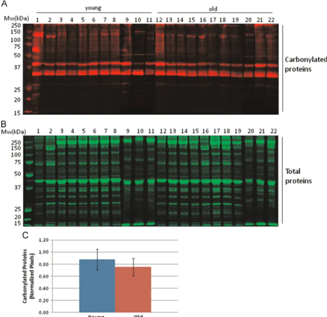
in-Fig. 1. Analysis of total carbonylated proteins of human rectus abdominis muscle biopsies. (A) Carbonylated protein profiles from young and old human biopsies after 1D gel electrophoresis. Protein carbonyl were detected by Western-blotting against protein-DNP derivatives and monitored byfluorescent secondary antibody hybridation. (B) Total protein profiles stained with colloidal Coomassie brilliant blue G after 1D gel electrophoresis. (C) Densitometric analysis of carbonylated protein western-blots. Semi-quantitative assessment of modified proteins was done using total protein staining for normalization. Relative values are expressed as mean7S.D. (n¼11) and no significant difference was found between the young and old groups.
gel digested by trypsin[26] using a robot Freedom EVO 100 di-gester/spotter robot (Tecan). Resulted peptides were then desalted for mass spectrometry (MS/MS) analysis. Results obtained were subjected to a search on the SwissProt database using the MASCOT software (Matrix Science Ltd., London, UK). The software compares the MASCOT peptide sequences derived from spectra with those in libraries for protein identification. Search parameters were as follows: database, SwissProt; taxonomy, all entries or mammalian; enzyme, trypsin; allow up to one missed cleavage; fixed mod-ifications, none; variable modifications, methionine oxidation; peptide mass tolerance, 70 ppm; and fragment mass tolerance, 500 ppm.
Results and discussion
Initial screening looking at changes at the global proteome le-vel of carbonylated proteins was performed after derivatization of protein carbonyls with DNPH followed by immunodetection of DNP protein adducts after SDS-PAGE (Fig. 1A). Densitometry ana-lysis was performed and normalized by total protein content for each sample (Fig. 1B and C). No significant differences at the global level in protein carbonyl content was detected between groups, in agreement with what was previously reported by Marzani et al. who did notfind a statistical difference on protein carbonyl con-tent during ageing in both rectus abdominis and vastus lateralis human muscles [18]. More recently, Fanò et al., using skeletal muscle biopsy samples obtained from vastus lateralis, found that protein carbonyls, showed a significant increase during ageing. However, this difference was not significant after splitting by gender[27]. Importantly, immunodetection of carbonylated pro-teins after one-dimensional (1D) electrophoresis separation has
serious limitations for the resolution of single protein bands and can provide only restricted information.
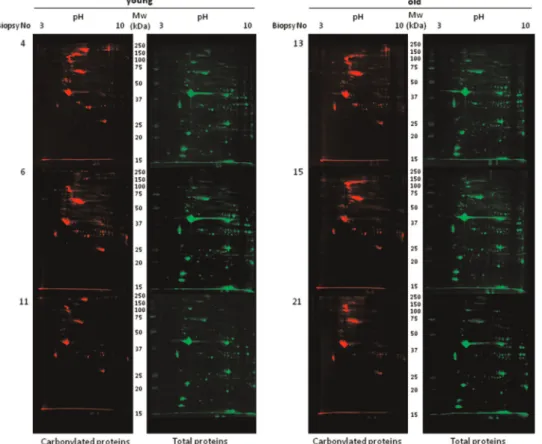
To further analyze the occurrence of protein carbonylation at the single protein level, 2D gel electrophoresis separation of pro-tein extracts was performed prior to immunodetection of carbo-nylated proteins. 2D gels are very appropriate to investigate pro-tein isoforms since many post-translational modifications (such as carbonylation of arginine or lysine residues) often leads to changes in the isoelectric point of proteins, and thus shift the position of the protein in 2D gels. After electrotransfer onto nitrocellulose membranes total protein profiles were obtained by fast-green staining (Fig. 2). All the analyzed samples displayed a similar protein migration pattern, suggesting no drastic shifts in the protein profiles at the expression level (Fig. 2, right panels) be-tween the two experimental groups. Immunodetection of carbo-nylated proteins was performed after their derivatization by DNPH as described in Material and methods. Interestingly, the pattern of the modified proteins was not superimposable with the pattern obtained for the total protein staining, indicating that certain proteins represent preferential targets for these deleterious oxi-dative modifications. Several proteins of high molecular weight were found preferentially carbonylated (Fig. 2, left panel) while certain proteins with an isoelectric point higher than 5, also ap-peared to be preferentially carbonylated.
A relative modification index (RMI) per spot was calculated in order to evidence differentially carbonylated proteins between the two experimental groups taking into account their protein ex-pression levels. Seventeen protein spots exhibited an RMI ratio consistently higher than 1.3 (increasingly carbonylated) in all biopsies from the aged group as compared with the young one. On the other hand, fourteen protein spots showed decreased carbo-nylation in the old group (RMIo0.7). Protein spots evidenced as
Fig. 2. Oxi-proteome analysis of young and old human skeletal muscle samples. Protein extracts from young (n¼3) and old (n¼3) human skeletal muscle biopsies were separated by 2D gel electrophoresis. After the second dimension, gels were electrotransferred onto nitrocellulose membranes for subsequent immune detection of DPNH-derivatized carbonylated proteins (left panels). Densitometry analysis was done by image Master 2D software (GE Healthcare) using total protein staining (right panels) as loading control.
S. Lourenço dos Santos et al. / Redox Biology 5 (2015) 267–274 270
increasingly carbonylated were excised from Coomassie Blue stained 2D gels (Fig. 3) and analyzed by MS/MS for protein iden-tification. Protein spots that were identified in the aged group are listed in Table 2. Among the identified proteins, 8 are muscle specific, 4 are ubiquitous in different tissues and organ systems, and 2 proteins belong from plasma (Table 2).
The identified skeletal muscle proteins were then analyzed and grouped by metabolic pathways and cellular functions. Major biological functions include muscle contraction, energy transduc-tion and energy metabolism (Fig. 4). Among proteins involved in muscle contraction, we have found that myosin 7, troponin T, myosin-binding protein C (MyBPC), and LIM domain-binding protein 3 (ZASP) are increasingly carbonylated in aged muscle. Interestingly, the decreased speed of contraction observed in old age at both the muscle and motor protein levels is a hallmark of skeletal muscle ageing [28–31]. Myosin is a highly conserved protein that converts chemical energy into mechanical force and a key protein for muscle contraction. Previous studies have shown increased glycation of myosin in bothfiber types of aged rats[32]. In addition, the carbonylated residues have been identified [33]. Interactions of myosin with cytoskeletal proteins such as titin, myomesin/M protein and MyBPC play an important role in thick filaments physiology. MyBPC contributes to the assembly and stabilization of thick filaments and modulates the formation of actomyosin cross-bridges, via direct interactions with both thick myosin and thin actinfilaments[34,35]. The importance of MyBPC to muscle contraction is further emphasized by the discovery that mutations in genes encoding MyBPC cause myopathies in both skeletal[36,37]and cardiac muscles[38–40]. Increased oxidation of MyBPC, together with the previously reported age-dependent decrease of myosin isoforms and regulatory proteins like myosin binding proteins C and H[41–44], may contribute to the destabi-lization of muscle fibers. In addition, perturbations in the thin sarcomerefillaments of muscle fibers have also been associated with differently expression of actin and its regulatory proteins such as troponin and tropomyosin[42,43,45,46]. Here we found
increased carbonylation levels of troponin T in the elderly. Tro-ponin inhibits the actomyosin Mg2þ-ATPase and the Ca2þrelease
from the sarcoplasmic reticulum inducing a change in the tropo-nin–tropomyosin conformation that exposes myosin binding sites on the actin filament and activates the myosin ATPase, thereby allowing muscle contraction [47,48]. Since muscle contraction depends on myofibrillar thin filament interactions[49], the oxi-dative damage to these proteins may well compromise this in-teraction and hence muscle contraction.
The motor functions of striated muscle crucially depend on the highly ordered arrays of thick myosin and thin actinfilaments in sarcomeres. Although its detailed role has not been elucidated yet, the LIM domain-binding protein 3 (ZASP), found as increasingly carbonylated in aged rectus abdominis muscle, functions as an adapter to couple protein kinase C-mediated signaling to the cy-toskeleton and to maintain Z-disc stability as well as cytoskeletal ultrastructure during contraction. Accumulation of this protein in its oxidized form may lead to protein aggregates formation and myofibril disintegration that may result in muscle weakness.
Muscle contraction depends also in high-energy fluxes where creatine kinase plays a central role. During muscle contraction, myosin hydrolyzes ATP into ADP upon filament sliding. The re-newal of ATP is achieved mainly by the phosphocreatine–creatine kinase (PCr–CK) system. Among the creatine kinase (CK) iso-enzymes, the muscle-type CK (MM-CK) specifically binds to the myofibril M-line and is associated with the action-activated myosin ATPase as an intramyofibrillar ATP regenerator [50–52]. Since this MM-CK was found irreversibly oxidized in old rectus abdominis muscle, this could alter muscle metabolism and com-promise muscle contraction in the elderly. Besides the PCr–CK system, the glycolytic network and its closer interaction between mitochondria and organelles also provide energy within muscle cells [53]. In this study, 3 glycolytic enzymes appear as highly carbonylated in old slow oxidative skeletal rectus abdominis mus-cle: the fructose-bisphosphate aldolase A, the glycerol-3-phos-phate dehydrogenase (GPD1) and the glyceraldehyde-3-phosglycerol-3-phos-phate dehydrogenase (GAPDH) (Table 2). Interestingly, several of these enzymes involved in anaerobic metabolism have been found de-creased with age in murine and human skeletal muscle[54,55].
Although slow skeletal muscles uses mainly oxidative mi-tochondrial processes to generate the levels of ATP needed to maintain contractile activity for long time without showing fatigue
[53], they also rely in glycolysis for their energy production[56]. For this reason, muscle metabolism and contraction in aged ske-letal muscle would be affected by irreversible carbonylation on glycolytic proteins, as previously reported in certain diseases when these glycolytic enzymes are not functional[57–62]. Defects in the muscle form of glycogen phosphorylase, another protein found increased carbonylated in old rectus abdominis muscle, are in-volved in type 5 glycogen storage disease, also known as McArdle disease, a myopathy also characterized by exercise intolerance, such as easy fatigability, muscle cramps and contractures as well as muscle weakness [63,64]. Intramuscular glycogen acts as a readily available source of glucose-6-phosphate for glycolysis within skeletal muscle and glycogen phosphorylase catalyses the rate-limiting step in glycogenolysis[65]. Therefore, a deficiency in glycogen phosphorylase may result in the inability to mobilize muscle glycogen during anaerobic metabolism[66].
Although it is well recognized that ageing causes changes in the proteome, the nature and targets of these changes, their sequences on skeletal muscle function and how they may con-tribute to sarcopenia have not yet been completely elucidated. Our results suggest that oxidative stress during skeletal muscle ageing targets the contractile machinery, but also structural and reg-ulatory proteins. In addition, the main mechanism of energy pro-duction, the phosphocreatine kinase system was affected by
Fig. 3. Coomassie blue staining of one representative 2D gel. The numbers and positions of the 17 selected spots identified by MS/MS correspond to those found as consistently increased on the old group (RMI41.3).
carbonylation in the elderly while other energetic metabolic pathways such as glycolysis appear to be also affected. Finally, the heat shock 70 kDa protein (HSP70), a key player in protein quality control with different identified roles in skeletal muscle[67], such as protection against oxidative stress, was also found highly car-bonylated in old skeletal muscle. Up-regulation of heat shock proteins is a well know feature of muscle ageing[46,68,69]. Fur-ther studies should address the functional status of the identified carbonylated proteins and related cellular pathways associated
with muscle dysfunction in order to reveal their role in the de-velopment of the ageing phenotype.
Acknowledgments
The authors wish to acknowledge the FP7 EU-funded MyoAge Project (No. 223576), and COST Action CM1001, coordinated by Dr. Gillian Butler-Browne and Dr. Tilman Grune, respectively, as well
Table 2
Localization of carbonylated proteins identified from old skeletal muscle biopsies. Protein name and localization Swiss-Prot
ac-cession no Protein spot noa Mascot scoreb Sequence cov-erage (%) No. of sequenced peptides Theoretical protein mass (kDa) Theoretical PI RMI ratioc Ubiquitous proteins:
Collagen alpha-1(VI) chain CO6A1_HUMAN 1 205 7 5 108 5.3 4.34 Heat shock cognate 71 kDa protein HSP7C_HUMAN 5 256 15 5 71 5.4 1.63 Glycerol-3-phosphate dehydrogenase [NADþ], cytoplasmic (GPD1) GPDA_HUMAN 13 214 22 4 38 5.8 1.52 Glyceraldehyde-3-phosphate dehy-drogenase (GAPDH) G3P_HUMAN 14 243 12 3 36 8.6 1.67 Glyceraldehyde-3-phosphate dehy-drogenase (GAPDH) G3P_HUMAN 15 424 25 5 36 8.6 1.68 Voltage-dependent anion-selective
chan-nel protein 1
VDAC1_HUMAN 16 685 39 8 31 8.6 1.88
Muscle specific proteins:
Myosin-binding protein C, slow-type (MyBPC)
MYPC1_HUMAN 2 119 3 3 128 5.8 1.54 Glycogen phosphorylase, muscle form PYGM_HUMAN 3 882 22 13 97 6.6 1.67 Myosin-7 MYH7_HUMAN 7 63 – 1 223 5.6 3.12 Creatine kinase M-type KCRM_HUMAN 8 206 9 3 43 6.8 1.44 Creatine kinase M-type KCRM_HUMAN 9 416 20 6 43 6.8 1.54 Fructose-bisphosphate aldolase A ALDOA_HUMAN 10 611 31 7 39 8.3 1.65 Troponin T, slow skeletal muscle TNNT1_HUMAN 11 512 22 6 33 5.9 1.87 Troponin T, slow skeletal muscle TNNT1_HUMAN 12 244 22 5 33 5.9 1.42 LIM domain-binding protein 3 (ZASP) LDB3_HUMAN 17 206 8 4 77 8.5 1.70 Plasma proteins:
Serotransferrin TRFE_HUMAN 4 399 17 10 77 6.8 1.31 Serum albumin ALBU_HUMAN 6 986 29 13 69 5.9 1.33 Spots of interest were identified by MALDI-TO–FTOF-MS as described under Material and methods. For each spot, different parameters clarifying protein identification by MS are indicated.
aProtein spot number refers to the numbered spots inFig. 3. b
Mascot protein scores greater than 56 are significant (Po0.05).
cRMI ratio represents the Relative Modification Index ratio.
Fig. 4. Functional grouping of muscle proteins increasingly oxidized with age. Increasingly oxidized muscle-specific proteins identified in aged rectus abdominis biopsies were grouped in three functional categories: muscle contraction, energy metabolism and energy transduction.
S. Lourenço dos Santos et al. / Redox Biology 5 (2015) 267–274 272
as the AFM-Téléthon for their support. In addition, they are very grateful to M. Cédric Broussard at the Plate-forme Protéomique Université Paris-Descartes 3P5 for performing the mass spectro-metry analyses.
References
[1] E. Barbieri, P. Sestili, Reactive oxygen species in skeletal muscle signaling, J. Signal Trans. 2012 (2012) 982794,http://dx.doi.org/10.1155/2012/982794 22175016.
[2] M.A. Baraibar, M. Gueugneau, S. Duguez, G. Butler-Browne, D. Bechet, B. Friguet, Expression and modification proteomics during skeletal muscle ageing, Biogerontology 14 (3) (2013) 339–352,http://dx.doi.org/10.1007/ s10522-013-9426-7 23624703.
[3] S.K. Powers, A.N. Kavazis, J.M. McClung, Oxidative stress and disuse muscle atrophy, J. Appl. Physiol. 102 (6) (2007) 2389–2397,http://dx.doi.org/10.1152/ japplphysiol.01202.2006 17289908.
[4] M.A. Pellegrino, J.-F. Desaphy, L. Brocca, S. Pierno, D.C. Camerino, R. Bottinelli, Redox homeostasis, oxidative stress and disuse muscle atrophy, J. Physiol. 589 (9) (2011) 2147–2160,http://dx.doi.org/10.1113/jphysiol.2010.203232 21320887.
[5] S.K. Powers, A.J. Smuder, D.S. Criswell, Mechanistic links between oxidative stress and disuse muscle atrophy, Antioxid. Redox Signal. 15 (9) (2011) 2519–2528,http://dx.doi.org/10.1089/ars.2011.3973 21457104. [6] S.K. Powers, A.J. Smuder, A.R. Judge, Oxidative stress and disuse muscle
atrophy: cause or consequence? Curr. Opin. Clinical Nutr. Metab. Care 15 (3) (2012) 240–245,http://dx.doi.org/10.1097/MCO.0b013e328352b4c2 22466926.
[7] D.C. Andersson, M.J. Betzenhauser, S. Reiken, A.C. Meli, A. Umanskaya, W. Xie, T. Shiomi, R. Zalk, A. Lacampagne, A.R. Marks, Ryanodine receptor oxidation causes intracellular calcium leak and muscle weakness in aging, Cell Metab. 14 (2) (2011) 196–207,http://dx.doi.org/10.1016/j.cmet.2011.05.014 21803290. [8] K.B. Choksi, J.E. Nuss, J.H. Deford, J. Papaconstantinou, Age-related alterations
in oxidatively damaged proteins of mouse skeletal muscle mitochondrial electron transport chain complexes, Free Radic. Biol. Med. 45 (6) (2008) 826–838,http://dx.doi.org/10.1016/j.freeradbiomed.2008.06.006 18598756. [9] L.M. Snow, N.A. Fugere, L.V. Thompson, Advanced glycation end-product
ac-cumulation and associated protein modification in Type II skeletal muscle with aging, J. Gerontol. A: Biol. Sci. Med. Sci. 62 (11) (2007) 1204–1210,http: //dx.doi.org/10.1093/gerona/62.11.1204 18000139.
[10] L.V. Thompson, D. Durand, N.A. Fugere, D.A. Ferrington, Myosin and actin expression and oxidation in aging muscle, J. Appl. Physiol. 101 (6) (2006) 1581–1587,http://dx.doi.org/10.1152/japplphysiol.00426.2006 16840579. [11] N. Breusing, T. Grune, Biomarkers of protein oxidation from a chemical,
bio-logical and medical point of view, Exp. Gerontol. 45 (10) (2010) 733–737,http: //dx.doi.org/10.1016/j.exger.2010.04.004 20403419.
[12] M.A. Baraibar, B. Friguet, Oxidative proteome modifications target specific cellular pathways during oxidative stress, cellular senescence and aging, Exp. Gerontol. 48 (7) (2013) 620–625,http://dx.doi.org/10.1016/j.exger.2012.10.007 23127722.
[13] M.A. Baraibar, R. Ladouce, B. Friguet, Proteomic quantification and identifica-tion of carbonylated proteins upon oxidative stress and during cellular aging, J. Proteomics 92 (2013) 63–70,http://dx.doi.org/10.1016/j.jprot.2013.05.008. [14] T. Grune, R. Shringarpure, N. Sitte, K. Davies, Age-related changes in protein
oxidation and proteolysis in mammalian cells, J. Gerontol. A: Biol. Sci. Med. Sci. 56 (11) (2001) B459–B467,http://dx.doi.org/10.1093/gerona/56.11.B459 11682566.
[15] M.A. Baraibar, A.G. Barbeito, B.B. Muhoberac, R. Vidal, A mutant light-chain ferritin that causes neurodegeneration has enhanced propensity toward oxi-dative damage, Free Radic. Biol. Med. 52 (9) (2012) 1692–1697,http://dx.doi. org/10.1016/j.freeradbiomed.2012.02.015 22348978.
[16] P. Gianni, K.J. Jan, M.J. Douglas, P.M. Stuart, M.A. Tarnopolsky, Oxidative stress and the mitochondrial theory of aging in human skeletal muscle, Exp. Ger-ontol. 39 (9) (2004) 1391–1400,http://dx.doi.org/10.1016/j.exger.2004.06.002 15489062.
[17] O. Pansarasa, L. Castagna, B. Colombi, J. Vecchiet, G. Felzani, F. Marzatico, Age and sex differences in human skeletal muscle: role of reactive oxygen species, Free Radic. Res. 33 (3) (2000) 287–293,http://dx.doi.org/10.1080/
10715760000301451 10993482.
[18] B. Marzani, G. Felzani, R.G. Bellomo, J. Vecchiet, F. Marzatico, Human muscle aging: ROS-mediated alterations in rectus abdominis and vastus lateralis muscles, Exp. Gerontol. 40 (12) (2005) 959–965,http://dx.doi.org/10.1016/j. exger.2005.08.010 16213688.
[19] E. Barreiro, C. Coronell, B. Laviña, A. Ramírez-Sarmiento, M. Orozco-Levi, J. Gea, PENAM Project, Aging, sex differences, and oxidative stress in human re-spiratory and limb muscles, Free Radic. Biol. Med. 41 (5) (2006) 797–809,http: //dx.doi.org/10.1016/j.freeradbiomed.2006.05.027 16895800.
[20] A. Rogowska-Wrzesinska, M.-C. Le Bihan, M. Thaysen-Andersen, P. Roepstorff, 2D gels still have a niche in proteomics, J. Proteomics 88 (2013) 4–13,http: //dx.doi.org/10.1016/j.jprot.2013.01.010 23353020.
[21] M.A. Baraibar, L. Liu, E.K. Ahmed, B. Friguet, Protein oxidative damage at the crossroads of cellular senescence, aging, and age-related diseases, Oxid. Med.
Cell. Longev. 2012 (2012) 1–8,http://dx.doi.org/10.1155/2012/919832. [22] M.M. Bradford, A rapid and sensitive method for the quantitation of
micro-gram quantities of protein utilizing the principle of protein-dye binding, Anal. Biochem. 72 (1976) 248–254, http://dx.doi.org/10.1016/0003-2697(76)90527-3 942051.
[23] R.L. Levine, D. Garland, C.N. Oliver, A. Amici, I. Climent, A.G. Lenz, B.W. Ahn, S. Shaltiel, E.R. Stadtman, Determination of carbonyl content in oxidatively modified proteins, Methods Enzymol. 186 (1990) 464–478,http://dx.doi.org/ 10.1016/0076-6879(90)86141-H 1978225.
[24] M.A. Baraibar, J. Hyzewicz, A. Rogowska-Wrzesinska, R. Ladouce, P. Roepstorff, V. Mouly, B. Friguet, Oxidative stress-induced proteome alterations target different cellular pathways in human myoblasts, Free Radic. Biol. Med. 51 (8) (2011) 1522–1532,http://dx.doi.org/10.1016/j.freeradbiomed.2011.06.032 21810466.
[25] E.K. Ahmed, A. Rogowska-Wrzesinska, P. Roepstorff, A.-L. Bulteau, B. Friguet, Protein modification and replicative senescence of WI-38 human embryonic fibroblasts, Aging Cell 9 (2) (2010) 252–272, http://dx.doi.org/10.1111/j.1474-9726.2010.00555.x 20102351.
[26] A. Shevchenko, H. Tomas, J. Havlis, J.V. Olsen, M. Mann, In-gel digestion for mass spectrometric characterization of proteins and proteomes, Nat. Protoc. 1 (6) (2006) 2856–2860,http://dx.doi.org/10.1038/nprot.2006.468 17406544. [27] G. Fanò, P. Mecocci, J. Vecchiet, S. Belia, S. Fulle, M.C. Polidori, G. Felzani,
U. Senin, L. Vecchiet, M.F. Beal, Age and sex influence on oxidative damage and functional status in human skeletal muscle, J. Muscle Res. Cell Mot. 22 (4) (2001) 345–351,http://dx.doi.org/10.1023/A:1013122805060 11808774. [28] P. Höök, X. Li, J. Sleep, S. Hughes, L. Larsson, In vitro motility speed of slow
myosin extracted from single soleusfibres from young and old rats, J. Physiol. 520 (2) (1999) 463–471,http://dx.doi.org/10.1111/j.1469-7793.1999.00463.x 10523415.
[29] X. Li, L. Larsson, Maximum shortening velocity and myosin isoforms in single musclefibers from young and old rats, Am. J. Physiol.—Cell Physiol. 270 (1996) C352–C360.
[30] L. Larsson, X. Li, W.R. Frontera, Effects of aging on shortening velocity and myosin isoform composition in single human skeletal muscle cells, Am. J. Physiol. 272 (2 1) (1997) C638–C6499124308.
[31] P. Höök, V. Sriramoju, L. Larsson, Effects of aging on actin sliding speed on myosin from single skeletal muscle cells of mice, rats, and humans, Am. J. Physiol.—Cell Physiol. 280 (4) (2001) C782–C78811245594.
[32] B. Ramamurthy, L. Larsson, Detection of an aging-related increase in advanced glycation end products in fast- and slow-twitch skeletal muscles in the rat, Biogerontology 14 (3) (2013) 293–301, http://dx.doi.org/10.1007/s10522-013-9430-y 23681254.
[33] M. Li, H. Ogilvie, J. Ochala, K. Artemenko, H. Iwamoto, N. Yagi, J. Bergquist, L. Larsson, Aberrant post-translational modifications compromise human myosin motor function in old age, Aging Cell 14 (2) (2015) 228–235,http://dx. doi.org/10.1111/acel.12307 25645586.
[34] M.A. Ackermann, A. Kontrogianni-Konstantopoulos, Myosin binding protein-C: a regulator of actomyosin interaction in striated muscle, J. Biomed. Bio-technol. 2011 (2011) 636403,http://dx.doi.org/10.1155/2011/636403 22028592.
[35] S.J. Van Dijk, K.L. Bezold, S.P. Harris, Earning stripes: myosin binding protein-C interactions with actin, Pflugers Arch. 466 (3) (2014) 445–450,http://dx.doi. org/10.1007/s00424-013-1432-8 24442149.
[36] B. Markus, G. Narkis, D. Landau, R.Z. Birk, I. Cohen, O.S. Birk, Autosomal re-cessive lethal congenital contractural syndrome type 4 (LCCS4) caused by a mutation in MYBPC1, Hum. Mutat. 33 (10) (2012) 1435–1438,http://dx.doi. org/10.1002/humu.22122 22610851.
[37] M.A. Ackermann, P.D. Patel, J. Valenti, Y. Takagi, E. Homsher, J.R. Sellers, A. Kontrogianni-Konstantopoulos, Loss of actomyosin regulation in distal ar-throgryposis myopathy due to mutant myosin binding protein-C slow, FASEB J. 27 (8) (2013) 3217–3228,http://dx.doi.org/10.1096/fj.13-228882 23657818. [38] S. Schlossarek, G. Mearini, L. Carrier, Cardiac myosin-binding protein C in
hypertrophic cardiomyopathy: mechanisms and therapeutic opportunities, J. Mol. Cell. Cardiol. 50 (4) (2011) 613–620,http://dx.doi.org/10.1016/j. yjmcc.2011.01.014 21291890.
[39] G. Bonne, L. Carrier, P. Richard, B. Hainque, K. Schwartz, Familial hypertrophic cardiomyopathy: from mutations to functional defects, Circ. Res. 83 (6) (1998) 580–593,http://dx.doi.org/10.1161/01.RES.83.6.580 9742053.
[40] S.P. Harris, R.G. Lyons, K.L. Bezold, In the thick of it: HCM-causing mutations in myosin binding proteins of the thickfilament, Circ. Res. 108 (6) (2011) 751–764,http://dx.doi.org/10.1161/CIRCRESAHA.110.231670 21415409. [41] D. Capitanio, M. Vasso, C. Fania, M. Moriggi, A. Viganò, P. Procacci,
V. Magnaghi, C. Gelfi, Comparative proteomic profile of rat sciatic nerve and gastrocnemius muscle tissues in ageing by 2-D DIGE, Proteomics 9 (7) (2009) 2004–2020,http://dx.doi.org/10.1002/pmic.200701162 19333999.
[42] J. Gannon, P. Doran, A. Kirwan, K. Ohlendieck, Drastic increase of myosin light chain MLC-2 in senescent skeletal muscle indicates fast-to-slowfibre transi-tion in sarcopenia of old age, Eur. J. Cell Biol. 88 (11) (2009) 685–700,http: //dx.doi.org/10.1016/j.ejcb.2009.06.004 19616867.
[43] C. Gelfi, A. Vigano, M. Ripamonti, A. Pontoglio, S. Begum, M.A. Pellegrino, B. Grassi, R. Bottinelli, R. Wait, P. Cerretelli, The human muscle proteome in aging, J. Proteome Res. 5 (6) (2006) 1344–1353,http://dx.doi.org/10.1021/ pr050414x 16739986.
[44] L.V. Thompson, D. Durand, N.A. Fugere, D.A. Ferrington, Myosin and actin expression and oxidation in aging muscle, J. Appl. Physiol. 101 (6) (2006) 1581–1587,http://dx.doi.org/10.1152/japplphysiol.00426.2006 16840579.
[45] P. Doran, K. O’Connell, J. Gannon, M. Kavanagh, K. Ohlendieck, Opposite pa-thobiochemical fate of pyruvate kinase and adenylate kinase in aged rat skeletal muscle as revealed by proteomic DIGE analysis, Proteomics 8 (2) (2008) 364–377,http://dx.doi.org/10.1002/pmic.200700475 18050275. [46] L. Staunton, M. Zweyer, D. Swandulla, K. Ohlendieck, Mass
spectrometry-based proteomic analysis of middle-aged vs. aged vastus lateralis reveals in-creased levels of carbonic anhydrase isoform 3 in senescent human skeletal muscle, Int. J. Mol. Med. 30 (4) (2012) 723–733,http://dx.doi.org/10.3892/ ijmm.2012.1056 22797148.
[47] B. Wei, J.-P. Jin, Troponin T isoforms and posttranscriptional modifications: evolution, regulation and function, Arch. Biochem. Biophys. 505 (2) (2011) 144–154,http://dx.doi.org/10.1016/j.abb.2010.10.013 20965144.
[48] A.V. Gomes, J.D. Potter, D. Szczesna-cordary, The role of troponins in muscle contraction. I, UBMB Life 54 (6) (2002) 323–333,http://dx.doi.org/10.1080/ 15216540290114676 12665242.
[49] W. Scott, J. Stevens, S.A. Binder-Macleod, Human skeletal musclefiber type classifications, Phys. Ther. 81 (2001) 1810–181613510739.
[50] T. Wallimann, T. Schlösser, H.M. Eppenberger, Function of M-line-bound creatine kinase as Intramyofibrillar ATP regenerator at the receiving end of the phosphorylcreatine Shuttle in muscle, J. Biol. Chem. 259 (8) (1984) 5238–5246
6143755.
[51] T. Hornemann, S. Kempa, M. Himmel, K. Hayess, D.O. Fürst, T. Wallimann, Muscle-type creatine kinase interacts with central domains of the M-band proteins myomesin and M-protein, J. Mol. Biol. 332 (4) (2003) 877–887,http: //dx.doi.org/10.1016/S0022-2836(03)00921-5 12972258.
[52] T. Hornemann, M. Stolz, T. Wallimann, Isoenzyme-specific interaction of muscle-type creatine kinase with the sarcomeric M-line is mediated by NH (2)-terminal lysine charge-clamps, J. Cell Biol. 149 (6) (2000) 1225–1234,http: //dx.doi.org/10.1083/jcb.149.6.1225 10851020.
[53] S. Schiaffino, C. Reggiani, Fiber types in mammalian skeletal muscles, Physiol. Rev. 91 (4) (2011) 1447–1531,http://dx.doi.org/10.1152/physrev.00031.2010 22013216.
[54] I. Piec, A. Listrat, J. Alliot, C. Chambon, R.G. Taylor, D. Bechet, Differential proteome analysis of aging in rat skeletal muscle, FASEB J. 19 (9) (2005) 1143–1145,http://dx.doi.org/10.1096/fj.04-3084fje 15831715.
[55] M. Gueugneau, C. Coudy-Gandilhon, O. Gourbeyre, C. Chambon, L. Combaret, C. Polge, D. Taillandier, D. Attaix, B. Friguet, A.B. Maier, G. Butler-Browne, D. Béchet, Proteomics of muscle chronological ageing in post-menopausal women, BMC Genomics 15 (1) (2014) 1165, http://dx.doi.org/10.1186/1471-2164-15-1165 25532418.
[56] A.E. Halseth, D.P. Bracy, D.H. Wasserman, Functional limitations to glucose uptake in muscles comprised of differentfiber types, Am. J. Physiol.—En-docrinol. Metab. 280 (6) (2001) E994–E99911350781.
[57] H. Kishi, T. Mukai, A. Hirono, H. Fujii, S. Miwa, K. Hori, Human aldolase A deficiency associated with a hemolytic anemia: thermolabile aldolase due to a single base mutation, Proc. Natl. Acad. Sci. USA 84 (23) (1987) 8623–8627,
http://dx.doi.org/10.1073/pnas.84.23.8623 2825199.
[58] J. Kreuder, A. Borkhardt, R. Repp, A. Pekrun, B. Göttsche, U. Gottschalk, H. Reichmann, W. Schachenmayr, K. Schlegel, F. Lampert, Brief report:
inherited metabolic myopathy and hemolysis due to a mutation in aldolase A, N. Engl. J. Med. 334 (17) (1996) 1100–1104,http://dx.doi.org/10.1056/ NEJM199604253341705 8598869.
[59] H. Nakajima, W. Amano, T. Kubo, A. Fukuhara, H. Ihara, Y.-T. Azuma, H. Tajima, T. Inui, A. Sawa, T. Takeuchi, Glyceraldehyde-3-phosphate dehydrogenase ag-gregate formation participates in oxidative stress-induced cell death, J. Biol. Chem. 284 (49) (2009) 34331–34341,http://dx.doi.org/10.1074/jbc. M109.027698 19837666.
[60] T. Sato, A. Morita, N. Mori, S. Miura, Glycerol 3-phosphate dehydrogenase 1 deficiency enhances exercise capacity due to increased lipid oxidation during strenuous exercise, Biochem. Biophys. Res. Commun. 457 (4) (2015) 1–6,http://dx.doi.org/10.1016/j.bbrc.2015.01.043 25603051.
[61] A. Vigelsø, R. Dybboe, C.N. Hansen, F. Dela, J.W. Helge, A. Guadalupe Grau, GAPDH andβ-actin protein decreases with aging, making stain-Free tech-nology a superior loading control in western blotting of human skeletal muscle, J. Appl. Physiol. 118 (3) (2015) 386–394,http://dx.doi.org/10.1152/ japplphysiol.00840.2014 25429098.
[62] M.J. MacDonald, L.K. Marshall, Mouse lacking NADþ-linked glycerol
phos-phate dehydrogenase has normal pancreatic beta cell function but abnormal metabolite pattern in skeletal muscle, Arch. Biochem. Biophys. 384 (1) (2000) 143–153,http://dx.doi.org/10.1006/abbi.2000.2107 11147825.
[63] Y. Kitaoka, D.I. Ogborn, M.I. Nilsson, N.J. Mocellin, L.G. MacNeil, M. A. Tarnopolsky, Oxidative stress and Nrf2 signaling in McArdle disease, Mol. Genet. Metab. 110 (3) (2013) 297–302,http://dx.doi.org/10.1016/j. ymgme.2013.06.022 23906480.
[64] T.A. Kohn, T.D. Noakes, D.E. Rae, J.C. Rubio, A. Santalla, G. Nogales-Gadea, T. Pinós, M.A. Martín, J. Arenas, A. Lucia, McArdle disease does not affect skeletal musclefibre type profiles in humans, Biol. Open 3 (12) (2014) 1224–1227,http://dx.doi.org/10.1242/bio.20149548 25432515. [65] A. Katz, H. Westerblad, Regulation of glycogen breakdown and its
con-sequences for skeletal muscle function after training, Mamm. Genome 25 (9– 10) (2014) 464–472,http://dx.doi.org/10.1007/s00335-014-9519-x 24777203. [66] A. Lucia, G. Nogales-Gadea, M. Pérez, M.A. Martín, A.L. Andreu, J. Arenas,
McArdle disease: what do neurologists need to know? Nat. Clin. Pract. Neurol. 4 (10) (2008) 568–577,http://dx.doi.org/10.1038/ncpneuro0913 18833216. [67] Y. Liu, L. Gampert, K. Nething, J.M. Steinacker, Response and function of
ske-letal muscle heat shock protein 70, Front. Biosci. 11 (2006) 2802–2827,http: //dx.doi.org/10.2741/2011 16720354.
[68] P. Doran, J. Gannon, K. O’Connell, K. Ohlendieck, Aging skeletal muscle shows a drastic increase in the small heat shock proteins alphaB-crystallin/HspB5 and cvHsp/HspB7, Eur. J. Cell Biol. 86 (10) (2007) 629–640,http://dx.doi.org/ 10.1016/j.ejcb.2007.07.003 17761354.
[69] A. Lombardi, E. Silvestri, F. Cioffi, R. Senese, A. Lanni, F. Goglia, P. de Lange, M. Moreno, Defining the transcriptomic and proteomic profiles of rat ageing skeletal muscle by the use of a cDNA array, 2D- and blue native-PAGE ap-proach, J. Proteomics 72 (4) (2009) 708–721,http://dx.doi.org/10.1016/j. jprot.2009.02.007 19268720.
S. Lourenço dos Santos et al. / Redox Biology 5 (2015) 267–274 274