DEVELOPMENT OF QUANTITATIVE METHODS FOR QUALITY ASSESSMENT OF ISLETS OF LANGERHANS
by
ANNA PISANIA
B.S.E. Chemical Engineering, University of Patras, Greece (2001)
M. Eng Chemical Engineering Practice, Massachusetts Institute of Technology (2003) A thesis submitted to the Department of Chemical Engineering in partial fulfillment of
the requirements for the degree of
Doctor of Philosophy in Chemical Engineering at the
MASSACHUSETTS INSTITUTE OF TECHNOLOGY
May 2007
© Massachusetts Institute of Technology 2007. All rights reserved
Author
Department of Chemical Engineering May 21st, 2007
Certified by - .. - . a, . . I ,% If
Clark K. Colton Professor of Chemical Engineering Thesis Supervisor Accepted by MASSACHUSETTS INSTITUTE OF TECHNOLOGY
JUN 11 2007
LIBRARIES
William M. Deen Professor of Chemical Engineering Chairman, Department Committee on Graduate StudentsARCHVES
ITroo
yoveig
/IOV,Zia Kai MiX6rl
1HT•6via,
yza 6a
do6a Xoovv
KLVC1Kal
aKOp•a Kavouv y1a <va.
To my parents,
Sia and Michael Pisania,
for everything they have done
and still do for me.
ABSTRACT
Development of Quantitative Methods for Quality Assessment of
Islets of Langerhans
by Anna Pisania
Submitted to the Department of Chemical Engineering on May 21, 2007, in partial fulfillment of
the requirements for the degree of Doctor of Philosophy in Chemical Engineering
Transplantation of isolated islets of Langerhans has promising potential to cure type 1 diabetes by inducing long-term normoglycemia and insulin independence. The feasibility of clinical islet transplantations has been established according to the Edmonton Protocol. However, results are variable, and it is not yet possible to predict transplantation outcome from in vitro measurements with islet preparations. Currently, islet enumeration is based on microscopic visualization after staining with a zinc-specific binding dye (dithizone, DTZ) and manual counting with normalization on the basis of an islet equivalent (IE), an islet with volume equivalent to that of a sphere with a 150 Pm diameter. Islet viability is based on a live/dead staining test with two fluorescent dyes, fluorescein diacetate (FDA) and propidium iodide (PI). These methods are operator-dependent and prone to error due to the variability in islet size and shape and are not predictive of transplantation outcome.
We developed quantitative assays that allowed reproducible evaluation of meaningful properties that affect the clinical outcome in impure human islet preparations. For purity estimation, we examined light microscopic (LM) morphological analysis of 1-jm sections to estimate the islet volume fraction and compared the results with those of electron microscopy (EM) ultrastructural analysis on the same preparations. For quantifying the total number of cells in a preparation, we developed an assay based on nuclei counting and compared three different counting methods: (1) visual counting in a hemacytometer and automatic counting by (2) aperture resistance measurements (Coulter Multisizer II) and (3) flow cytometer measurements (Guava PCA). The methods differed in the way nuclei were distinguished from fragments, accuracy, time required and range of linearity. Total amount of tissue was also quantified by DNA measurements. A theoretical framework was developed in order to combine volume fraction data from the LM analysis with the total number of cells in the tissue from nuclei measurements in order to estimate the total number of islets present in impure preparations. To evaluate tissue viability, we used oxygen consumption rate measurements (OCR), an assay of mitochondrial function. We developed a very small stirred chamber system (Micro
Oxygen Uptake System, Model FO/SYS210T, Instech Laboratories, Plymouth Meeting, PA) specifically designed for measurements with islets. OCR measurements combined with an assay of total amount of tissue quantification (nuclei counting or DNA analysis) provide a measure of the tissue fractional viability. We used the methods we developed to characterize a large number of islet preparations of different species prior to and following culture. We also studied the transient response of cells and islets to various stresses, as reflected by assays of different type. We found that membrane integrity tests are poor indicators of the fractional viability of a cell sample, while mitochondrial function assays identify cell death at its earlier stages.
We examined the predictive capability of the assays we developed through in vivo studies. Ttransplantation experiments were performed with rat islets implanted into mice and high purity fraction of human islets transplanted into mice. We found that OCR measurements combined with a measure of total amount of tissue (nuclei or DNA) are good predictors of the transplantation outcome.
Thesis Supervisor: Clark K. Colton Title: Professor of Chemical Engineering
Acknowledgements
The journey of the completion for my PhD was long and sometimes difficult, like any other journey in life. A lot of people helped make this journey fun, exciting, and worthwhile even during the rough times. I would like to take the time and thank all of them, hoping I will not forget any one.
I would like to thank my thesis committee, Dr. Susan Bonner-Weir, Dr. Klearhcos K. Papas, Dr. Greg Stephanopoulos, and Dr. Gordon Weir, for their advice, guidance, and understanding through a lot of questions that I came across during my PhD. In particular, I would like to thank my advisor, Clark K. Colton, for his support, his teaching and his persistence in excellence. Clark has been a good advisor that had helped me grow as a
scientist. He has also been there to celebrate changes in my life, academic and not.
Drs. Susan Bonner-Weir and Gordon Weir from Joslin Diabetes Center have played a very important part in this journey as well. They have been very patient in helping me, a chemical engineer, understand the biological aspects of my project and teaching me a way to think as a biologist as well. I would like to thank Dr. Susan Bonner-Weir for her patience in training me for the morphological analysis (light microscopy) performed in the studies. They have also both been very helpful with useful discussions
and great advice not only for my research but also for my career development.
I would like to thank Dr. Klearchos K. Papas (University of Minnesota) for useful discussions and some preliminary work done before I joined the lab. I would also like to thank the other faculty and staff at the Department of Chemical Engineering at MIT that have helped me along this journey with their advice and presence. I would also like to express my gratitude to a number of people at Joslin Diabetes Center that have guided me
through the biology and the mysteries of the islet world and have helped me develop my interdisciplinary skills. In particular I would like to thank Lorella Marselli, who patiently introduced me to the techniques of frozen sections, Chris Cahill and Alla Pinkwasov (Joslin DERC) that helped a lot with suggestions and advice on the preparation of plastic and frozen sections and shared their secrets on sectioning, and all the other people in the lab that offered help when I needed it. I would like to say a big thank you to the Islet Isolation Core, Vaja Tchipasvilli, Gaurav Chandra, Vassileios Kostaras, Ji Lei, Jack J. O'Neil, and Abdlukadir Omer for providing most of the islets used in this thesis and always trying to satisfy our large needs for tissue. They have all been very good colleagues and friends.
I would also like to thank the members of the Colton group for their friendship, fun conversations, and academic support. In particular, I would like to thank Daryl E. Powers and Michael J. Rappel, who joined the group the same time I did. Their friendship and help made my PhD process a lot more enjoyable and fun. They have helped a lot with some of the experiments but also with our discussions and words of encouragement throughout the years. It has been a privilege sharing my office space with them in the last five years and I can only hope to find such great colleagues and friends in my future positions. In addition to Daryl and Mike, I would also like to thank Amy S Lewis and Michelle T. Miller for help with some of the experiments and our fun conversations, wedding planning (great timing), and a lot of fun outside the lab. Finally, I would like to thank the rest of the member of the Colton group that has grown a lot since I joined for their friendship and support throughout this time. I would like to express my gratitude to Samantha Polak, an undergraduate student that worked with me for the study on stimulation with exogenous substrates, helped me become a better supervisor and teacher, and became a good friend.
In addition to the academic support from all the people mentioned above, I would like to thank all the other friends I made at MIT for their moral support and great friendships. I would like to thank my very good friends Adi and Chad Augustine that have been a very important, fun, and caring part of my life here, and I am very grateful to them. I would also like to thank Wanda Lau, Patrick Underhill for their friendship and the "European Grew"; Joanna Yu, Magali Pages, Thierry Savin, Alexander Mitsos, and Evangelia Lambidoni for making the first semester at MIT a lot easier and more fun and for their friendship that lasted throughout the years. They have all become great friends and I am looking forward to a lot more fun times with them outside MIT.
Coming to MIT from Greece, I could not have asked for a better group of Greeks to find here that made MIT feel so much like home. I have made a lot of Greek (loud, cheerful, and happy as we are) friends that have all contributed with their friendship my journey. I would like to thank, in no particular order but as they come in mind, Anna Stefanidou, Theodore Konstantakopoulos, Maria-Katerina Nikolinakou, Costas Pelekanakis, Aristos Karalis, George Constantinidis, Angelina Aisopou, Olia Galenianou, Vassileios Dendroulakis, Panagiotis Lemonidis, Ted Akiskalos, Petros Boufounos, Lia Kolokouri, Apostolos Fertis, Mariathi Liapi, Kostis Ouggrinis, Aggelos Bletsas, Yiannis Kitsopanidis, Fenia Makrydaki, Yiannis Zavoleas, Andreas Zezas, Yiannis Bertsatos for
all the fun times we spent together in coffee or lunch breaks, parties, Easter preparations, barbeques, and for making MIT feel a lot more like home.
I would also like to thank my friends from Greece for keeping up with our friendship that is so important to me, for being patient with my not so frequent emails when times were busy, and for offering their support and encouragement all this time. It has been great having them in my life and I am always looking forward to spending time with them. Thenk you so much Theodora (Loloumba), Lina, Fanetta Aggelos, Ioanna, Katerina, Katerina, Georgia, Sasa, Costas, Dionisis, and so many other friends from college, high school, guides that have been there for me.
I would finally like to thank my family. My mom and dad, Sia and Michalis Pisania, from Greece, and my brother Nikolas, who lives now in England, for their support, understanding, encouragement and most importantly their unconditional love that gave me the strength I needed to undertake such a big step. Lag cvXaptorr 7rcdpa rcoA2
yia 6da! I would also like to thank the family that I created here, my mom and dad
in-law, Marcella and Mo Haghgooie, my sister in in-law, Anna Haghgooie for their warm welcoming into their family and a lot of fun times in Michigan, Chicago, Cape Cod, Greece, and many more to come! I would also like to thank nana, Mary O'Brien, the most energetic, smart, and happy 90-year old person one could meet. It has been a great routine visiting her on Sundays afternoon to spend a nice, fun, and relaxing couple of hours away from the stress of MIT. I have been looking forward to those visits every Sunday for the past five years and will do so for more in the future. I would also like to thank the rest of the O'Briens; uncle Paul, aunt Chris, and Richard for providing a great family environment far away from home.
At last but not least, and may be the most important thank you of all, is for my husband Ramin Haghgooie. I met Ramin my first day at MIT during orientation in Kresge Auditorium. We fell in love during the first semester at MIT... It was one of those things in life that are meant to be because how else would you explain something so wonderful? And I am so extremely happy for it...he made the whole journey worth it, even when research did not go as planned. I am very grateful to him for his love and support during the happy and the tough times. I am very lucky to have him in my life and I can never tell him enough. Ki eyc o' 'cvXaptrrc6 zro26l ayd7Rt puov!
Funding for this research was provided by grants from the NIH (NCRR ICR U4Z RR 16606, RO1-DK063108, the Joslin Diabetes and Endocrinology Research Center [DERC] DK36836), the JDRF Center for Islet Transplantation at Harvard Medical School, and the Diabetes Research and Wellness Foundation.
TABLE OF CONTENTS
List of F igures... ... 17
L ist of Tables... ... 21
Chapter 1. Intoduction Into the World of slets ofLangerhans ... 23
1.1 Islet Isolation ... ... 26
1.1.1 H um an Islets ... ... 26
1.1.2 Rat and Porcine Islets ... 28
1.2 What Do We Want to Know? ... . . ... . . ... ... .. . . 29
1.3 Current Islet Assessment Methods ... 29
Chapter 2. Quantitative Analysis of Cell Composition of Human Pancreatic P rep arations ... ... 35
2.1 Materials and Methods ... ... 36
2.1.1 Islet Isolation Method... ... ... 36
2.1.2 Morphological Analysis ... 37
2.1.3 Islet Purity in a Preparation Determined by Three Methods ... 40
2.1.4 Islet and p-cell Volume in a Preparation Determined by Two Methods ....41
2.1.5 Measurement of the Void Fraction of Packed Cell Pellet ... 42
2.1.6 Islet V ital Staining... ... 43
2.1.7 DNA Determination ... 43
2.1.8 Statistics... ... 43
2.2 R esults... ... 44
2.2.1 Large Intraislet Vascular Spaces of Freshly Isolated Islets Demonstrated by Light Microscopy ... ... 44
2.2.2 Determination of Cell Composition and Number Fraction by Electron Microscopy (EM) ... 44
2.2.3 Cell Volume Fraction Determination ... ... 45
2.2.4 Consideration of Extracellular Volume ... ... 45
2.2.5 Number of Islet Cells and P Cells per IE... .... 46
2.2.6 Islet Volume Fraction (Purity) Estimated by DTZ versus EM Determination of Cell Composition ... ... 47
2.2.7 Comparison of Two Methods to Characterize Entire Islet Preparations and Estimate the Number of IE ... ... 48
2.2.8 Estimation of 0-cell Volume from Knowledge of IE Number ... 49
2.2.9 DNA Content of Tissue Pellets ... ... 50
2.2.10 Correlations Between Donor Characteristics and IE Yield...51
2.3 D iscussion ... ... 51
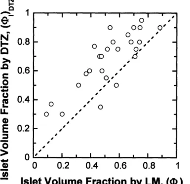
Chapter 3. Comparison of Light and Electron Microscopy for Measuring Purity of Human Pancreatic Islet Preparations ... 57
3.1 M aterials and M ethods ... ... 58
3.1.1 Islet Isolation ... ... 58
3.1.2 Volume Fraction Islets by DTZ Staining ... 59
3.1.4 Electron Microscopy ... 60
3.1.5 Light Microscopy ... ... 60
3.1.6 Interconversion Between Islet Number and Volume Fractions ... 62
3.1.7 Statistics... ... 62
3.2 R esults... ... 63
3.2.1 Vascular Void Fraction... ... 63
3.2.2 Comparison Between Islet Volume Fraction Determined by Three M ethods ... ... 63
3.3 D iscussion ... ... 69
Chapter 4. Frozen Sections: A Promising Alternative to Plastic Sections for Measuring Purity of Human Pancreatic Islet Preparations... ... 71
4.1 Materials and Methods ... 72
4.1.1 Islet Isolation ... ... 72
4.1.2 Preparation of Plastic Sections ... ... 72
4.1.3 Preparation of Frozen Sections...73
4.1.4 Volume Fraction Islets by DTZ Staining ... ... 74
4.1.5 Volume Fraction Islets by Morphological Analysis... 74
4.1.6 Volume Fraction Islets by MG/DTZ Staining... .... 75
4.2 R esults... ... 76
4.2.1 Comparison Between Staining Protocols for Frozen Sections ... 76
4.3 D iscussion ... ... 77
Chapter 5. Enumeration of slets by Nuclei Counting and Light Microscopic Analysis .. 79
5.1 Materials and Methods ... 81
5.1.1 Culture of Cell Lines and Islets ... 81
5.1.2 Human Islet Isolation. ... ... ... 81
5.1.3 Cells and Nuclei for Counting... ... 82
5.1.4 Counting Methods ... 82
5.1.5 DNA Analysis ... 84
5.1.6 Morphological Analysis ... 84
5.1.7 Number of IE from Nuclei Counting Data and LM Morphology ... 85
5.1.8 Volume Fraction Islets by DTZ Staining ... 86
5.1.9 Number of IE from Conventional DTZ Staining and Visual Counting ... 86
5.1.10 Number of IE from PCV Measurements and LM Morphology ... 87
5.1.11 Statistics... ... 87
5.2 R esults... ... 88
5.2.1 Comparison of Counting Methods ... ... 88
5.2.2 Comparison of Two Methods to Determine NIE of Rat Islets ... 95
5.2.3 Comparison of Three Methods to Determine NIE of Human Islets ... 96
5.3 D iscussion ... ... 97
Chapter 6. A Quantitative Membrane Integrity Test for Islets of Langerhans ... 103
6.1 Materials and Methods ... 104
6.1.1 Culture of Cell Lines and Islets ... 104
6.1.3 Cell Enumeration by Nuclei Counting ... ...104
6.1.4 Membrane Integrity by 7-AAD Differential Staining ... 105
6.1.5 Membrane Integrity by Trypan Blue ... 106
6.1.6 Cell Viability by MTT Assay ... 107
6.1.7 Heat-killed Cells or Islets ... 107
6.1.8 Statistics... 107
6.2 R esults... ... 108
6.2.1 Dye Selection... 108
6.2.2 Cells ... 108
6.2.3 Islets... ... 111
6.2.4 Comparison of Two Methods to Determine Fraction of Cells with Impermeable Cell Membranes ... ... 112
6.3 D iscussion ... ... 113
Chapter 7. A Stirred Microchamber for Oxygen Consumption Rate Measurements w ith P ancreatic Islets... 115
7.1 Materials and Methods ... 117
7.1.1 Culture of Cell Lines and Islets ... 117
7.1.2 Islet Enumeration by Visual Counting ... 117
7.1.3 Cell and Islet Enumeration by Nuclei Counting ... 117
7.1.4 DNA Content... 118
7.1.5 Quality Assessment Measurements ... 18
7.1.6 Statistics... ... 12 1 7.2 R esults... ... 122
7.2.1 Characterization of the OCR Apparatus and Measurements... 122
7.2.2 OCR Correlation with Measures of Viability... 125
7.2.3 OCR Measurements with Islets ... 130
7.3 D iscussion ... ... 130
Chapter 8. Characterization of Islet and Non-Islet Tissue Prior To and Following C ulture ... ... 133
8.1 M ethods ... ... 134
8.1.1 Islet Isolation and Culture ... 134
8.1.2 DNA Content... 135
8.1.3 Islet Enumeration by Nuclei Counting ... ...135
8.1.4 Oxygen Consumption Rate (OCR)... 135
8.1.5 Membrane Integrity ... 136
8.1.6 Islet Volume Fraction by Morphological Analysis ... 136
8.1.7 P3 Cell Identification by Immunocytochemical Insulin Staining ... 137
8.1.8 Statistics... 137
8.2 R esults... ... 138
8.2.1 D N A /cell... ... 138
8.2.2 DNA and Nuclei Recovery... 139
8.2.3 OCR/cell and OCR/DNA ... ... 142
8.2.4 OCR Recovery... 146
8.2.5 OCR/DNA Comparisons Between Centers... .... 146
8.2.6 Membrane Integrity by 7-AAD ... 147
8.2.7 OCR/cell with Impermeable Cell Membranes by 7-AAD ... 148
8.2.8 Purity ... ... 148
8.3 D iscussion... ... 152
Chapter 9. Dynamics of Cell Death Evaluated by Mitochondrial Function, Apoptosis, and Membrane Integrity Assays ... ... 155
9.1 M aterials and M ethods ... 156
9.1.1 Culture of Cell Lines and Islets ... 156
9.1.2 Cell Suspension Preparation... 157
9.1.3 Cell and Islet Enumeration by Nuclei Counting ... 157
9.1.4 Stresses and Mechanisms ... 157
9.1.5 Assessment Methods ... 158
9.1.6 Statistics... ... 162
9.2 R esults... ... 162
9.3 D iscussion ... ... 169
Chapter 10. Stimulation with Exogenous Substrates During Oxygen Consumption Rate Measurements ... ... ... 171
10.1 Materials and Methods ... 172
10.1.1 Islet Isolation and Culture ... 172
10.1.2 Morphological Analysis by Light Microscopy (LM) ... 172
10.1.3 Oxygen Consumption Rate (OCR)... ... 173
10.1.4 Stimulation with Exogenous Substrates During OCR Measurements ... 173
10.1.5 Statistics... 174
10.2 R esults... 174
10.2.1 Maximum Stimulation Index in Nutrient-Free Media... 174
10.2.2 Stimulation Index in Different Media (PBS, CMRL, DMEM)... 175
10.2.3 Comparison of OCR/cell Between Different Media ... 179
10.3 D iscussion ... ... 180
Chapter 11. Conclusions and Outlook ... 183
11.1 Quantitative Methods for Islet Quality Assessment ... 184
11.1.1 Purity ... ... 84
11.1.2 Quantity ... 184
11.1.3 Viability ... 184
11.1.4 Dynamics of Cell Death ... 185
11.2 Predictive Capability ... 86
11.2.1 Potency ... 186
11.3 Future Work... 186
Chapter 12. Appendices... ... 189
12.1 Appendix A. Supplemental Material for Chapter 2 ... 189
12.1.1 Number Fraction and Volume Fraction Relationships in Islet Preparations ... ... 189
12.1.2 Cells ... 189
12.1.3 Tissues ... 192
12.1.4 Parameters ... 194
12.1.5 Packed Cell Volume ... 195
12.2 Appendix B. Supplemental Material for Chapter 8 ... 203
12.3 Appendix C. Data for Rat, Human Fresh, Human Cultured/Shipped Islets and H um an N on-Islet Tissue ... 209
Chapter 13. Nomenclature ... 237
List of Figures
Figure 1.1. Scanning electron microscopy photograph of a typical isolated islet with a m ean diam eter of 150 pm ... ... 25
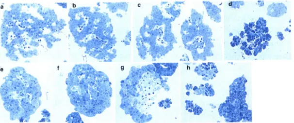
Figure 1.2. Schematic diagram of human islet isolation... ... 27 Figure 1.3. Typical rat aliquot of islet preparation stained with dihizone and viewed under a m icroscope. ... ... 30 Figure 1.4. Typical aliquot of rat islet preparation stained with FDA/PI and viewed under a m icroscope. ... 31 Figure 2.1. Toluidine blue stained 1-pm plastic sections of purified human islet
preparations... ... 38 Figure 2.2. Electron micrographs of pellets of purified islet preparations showing characteristics of the different cell types ... 39 Figure 2.3. Volume fraction (purity) data for individual islet preparations. ... 47 Figure 2.4. Comparison of two methods for counting the number of islet equivalents in an islet preparation ... ... 49 Figure 2.5. Correlation between P3-cell volume and the number of IE. ... 50 Figure 3.1. Identification between islet and non-islet tissue by light microscopy from plastic sections. ... ... 61 Figure 3.2. Frequency distribution of the vascular void volume fraction (vI by LM for 27 freshly isolated clinical preparations. ... 63 Figure 3.3. Frequency distribution of the islet volume fraction by (A) EM, (B) LM, and (C) D TZ staining... .... 66 Figure 3.4. Calculated islet volume fraction by EM is plotted against the measured islet volum e fractions by LM ... ... 67 Figure 3.5. Estimated islet volume fraction by DTZ staining is plotted against the measured islet volume fractions by LM. ... 68 Figure 4.1. Representative photograph of slide from frozen section of human islets stained with hematoxylin analyzed by LM ... 75 Figure 4.2. Representative photograph of slide from frozen section of human islets stained with MG/DTZ analyzed by LM. ... 76 Figure 5.1. Calculation of number of islet cells and islet equivalents in impure human islet preparations from nuclei counting and LM morphological analysis. ... 86 Figure 5.2. Output of each counting method for nuclei from high purity human islets. ...88 Figure 5.3. Output of each counting method for nuclei from low purity human islets. ....89 Figure 5.4. Measured versus calculated concentration of nuclei from INS-1 cells counted by all three methods and plotted on log-log coordinates. ... 90
Figure 5.5. Measured versus calculated concentration of nuclei from INS-1 cells counted by all three methods and plotted on linear coordinates for concentrations up to 2x10⁵ nuclei/m l ... 90 Figure 5.6. Measured versus calculated concentration of nuclei from INS-1 cells counted by all three methods and plotted on linear coordinates for concentrations up to
106 nuclei/m l. ... 9 1
Figure 5.7. Nuclei concentration versus cell concentration for INS-1 and PTC3 cells...92 Figure 5.8. Coefficient of variation from triplicate counts versus number of nuclei (from INS-I cells) counted by hemacytometer and flow cytometer... 93 Figure 5.9. Coefficient of variation versus the number of rat IE (estimated assuming
1560 cells per IE) sampled using three different kinds of pipette tips. ... 94 Figure 5.10. Distributions of DNA content per cell (pg DNA/cell). ... 95 Figure 5.11. Number of IE by DTZ staining/manual counting versus number of IE by LM/nuclei counting for 27 rat islet preparations. ... 96 Figure 5.12. Comparison of three methods for NIE determination for 12 research preparations of freshly isolated human islet preparations. ... 97 Figure 5.13. Average rat islet represented as a scalene ellipsoid with three half-axes a = 1.0, b = 0.82, and c = 0.60 (74) shown in four different orientations ... 101 Figure 6.1. Illustration of protocol for membrane integrity measurements with 7-AAD differential staining used in this study. ... ... 106 Figure 6.2. Normalized fraction of impermeable cells by 7-AAD differential staining measurement is plotted versus the fraction of untreated cells in mixtures with heat-treated cells. ... 109 Figure 6.3. Fraction of cells with intact membranes measured by 7-AAD differential staining versus fraction of cells with intact membranes measured by visual counting w ith trypan blue. ... 110 Figure 6.4. Relative fraction of cells with intact membranes measured by 7-AAD differential staining versus relative fraction of viable cells measured by MTT ... 111 Figure 6.5. Relative fraction of cells with intact membranes measured by 7-AAD differential staining versus relative fraction of viable cells measured by MTT...1....12 Figure 7.1. Schematic diagram of device for measuring oxygen consumption ... 120 Figure 7.2. Actual traces (individual data points) of measured pO2 versus time with rat islet sam ples ... 123 Figure 7.3. Precision of the OCR measurement. ... ... 125 Figure 7.4. Dependence of OCR on cell viability ... 126 Figure 7.5. Fraction of 3TC3 cells that were membrane impermeable after culture under normal and a variety of stressful conditions for 24 hr ... 128
Figure 7.6. Frequency distribution of OCR per viable cell for stressed and unstressed
PTC 3 cells ... ... 129
Figure 8.1. Frequency distributions of DNA content. ... 140
Figure 8.2. Frequency distributions of OCR/cell... 143
Figure 8.3. Frequency distributions of OCR/DNA ... 144
Figure 8.4. Frequency distributions of the OCR/DNA for different centers (MIT, MN). 146 Figure 8.5. Frequency distributions of the fraction of cells with impermeable cell m embranes by 7-AAD. ... 147
Figure 8.6. Frequency distributions of OCR/membrane-impermeable cell... 149
Figure 8.7. OCR/DNA versus fraction of cells with impermeable cell membranes by FDA/PI staining and 7-AAD for (A) rat islets, (B) human fresh islets from UMN and JDC, and (C) porcine islets from UMN ... ... 150
Figure 9.1. Data from Jurkat cells plotted versus time of exposure to 1 PiM cam ptothecin .... ... 165
Figure 9.2. Data from INS-1 cells plotted versus time of exposure to 5 mM streptozotocin.. ... 166
Figure 9.3. Data from islets plotted versus time of exposure to anoxia. ... 167
Figure 9.4. Data from assays of the same type averaged at similar time points and plotted versus time of stress exposure ... 168
Figure 9.5. Data from mitochondrial function and apoptotic events assays averaged at similar time points and plotted versus time of stress exposure...169
Figure 10.1. Effect of glucose concentration on the oxygen consumption rate (OCR) studied on two preparations of rat islets in PBS and DMEM ... 177
Figure 10.2. Effect of glucose concentration on the OCR studied on three preparations of shipped human islets in PBS and DMEM ... 178
Figure A- 1. Volume definitions and relationships in islet preparations ... 192
Figure A- 2. Volume and number definitions and relationships in a packed-cell pellet. 196 Figure B- 1. Conversion from islet volume measurements of fresh tissue to the beta cell volume fraction exclusive of vascular spaces ... ... 204
Figure B- 2. Conversion from islet volume measurements of fresh tissue to the number fraction beta cells using previously defined relationships ... 205
Figure B- 3. Conversion from the P cell volume fraction exclusive of vascular spaces on day 2 to the number fraction beta cells using previously defined relationships. ... 206
List of Tables
Table 2.1. Donor characteristics and processing data from 33 clinical preparations. ... 37 Table 2.2. Number fraction of cell types in 33 islet preparations determined by EM...45 Table 2.3. Volume fraction of cell types in 33 islet preparations determined by EM and the theoretical number of cells in an islet equivalent (IE) ... 46 Table 2.4. Volume and number characteristics of 33 islet preparations determined from EM and PCV m easurem ents. ... 48 Table 2.5. Comparison of literature values for p-cell content in human islets ... 54 Table 3.1. Number fraction cells by EM and islet volume fractions by EM, LM, and D TZ staining. ... ... 65 Table 8.1. DNA/cell and the fraction of original tissue (quantified by nuclei and DNA) recovered after 2 days culture ... 141 Table 8.2. OCR/cell, OCR/DNA, and fraction of original OCR recovered after 2 days culture ... ... 145
Table 8.3. Islet volume fraction,
1
cell volume fraction, number fraction of original P cells, and fraction of original nuclei recovered after 2 days culture a... 151Table 9.1 Summary of properties, site of action, and counting method of membrane integrity dyes used in the experiments ... 161 Table 10.1. Stimulation index (0-20 mM glucose) in nutrient-free medium (D-PBS)....175 Table 10.2. Stimulation index over basal OCR at 0 mM glucose for two sets of measurements obtained with rat islets in PBS and DMEM... 176 Table 10.3. Stimulation index over basal OCR at 0 mM glucose for three sets of measurements obtained with shipped human islets in PBS and DMEM ... 178 Table 10.4. Ratio of OCR/cell in DMEM over that in PBS for rat and human islets in different glucose concentrations ... 179
Table A. 1. Estimated volume (pm3) of pancreatic cells. ... 192
Table A. 2. Data from EM and LM morphological analysis and DTZ staining of 27 hum an islet preparations. ... ... 198 Table A. 3. Calculated quantities from EM and LM morphological analysis of 27 hum an islet preparations. ... 199 Table A. 4. Data and calculated quantities from packed cell volume measurements combined with LM analysis for 27 preparations ... ... 200 Table A. 5. Data and calculated quantities from packed cell volume measurements combined with EM analysis for 33 preparations. Similar format to Table A. 4...201 Table B. 1. Parameter values used to estimate fp from pxv... 208
Table C. 1. Data for Rat Islets ... 10 Table C. 2. Data for Fresh Human Islets ... ... 217 Table C. 3. Data for Cultured/Shipped Human Islets ... .... 225 Table C. 4. Data for Human Non-Islet Tissue ... ... 233
Chapter 1.
Introduction Into The World of
Islet of Langerhans
Diabetes mellitus is a very common disease nowadays. The number of people diagnosed with diabetes is constantly increasing. The World Health Organization (WHO) estimates that more than 180 million people worldwide have diabetes (1). This number is likely to more than double by 2030. Almost 7 percent of the population in the United States has diabetes mellitus, and even though 14.6 million have been diagnosed with the disease, about 6.2 million people are unaware they have the disease (2). Diabetes is a disorder of metabolism, in which the body does not produce or properly use insulin due to malfunction of the pancreas or the cells. Insulin is the hormone that controls the entry of glucose into the cells and functions in the regulation of the metabolism of carbohydrates and fats, especially the conversion of glucose to glycogen. In people with diabetes, the pancreas produces little or no insulin, or the body can not effectively use the insulin it produces. Therefore, glucose builds up in the blood and does not enter into the cells to help them grow. Finally, after it reaches a critical value, it overflows into the urine and passes out of the body. Thus, the body loses its main source of fuel even though the blood contains large amounts of glucose. The causes of diabetes continue to be a mystery, although both genetics and environmental factors (obesity, lack of exercise) appear to play important roles.
There are three main types of diabetes. Type 1 diabetes is an autoimmune disease, where the insulin-producing 03 cells in the pancreas are attacked and destroyed by the immune system. The pancreas then produces little or no insulin. People with type 1 diabetes need to take insulin daily to live. Type 2 diabetes is usually part of a metabolic
syndrome that includes obesity, elevated blood pressure, and high levels of blood lipids. In type 2 diabetes, the pancreas is either producing not enough insulin or the cells ignore it, and the body cannot use the insulin effectively, a condition called insulin resistance. Therefore, eventually, the insulin production decreases and glucose builds up in the blood and the body cannot make efficient use of its main source of fuel. Finally, gestational diabetes develops only during pregnancy. Like type 2 diabetes, it occurs more often in people with a family history of diabetes. Though it usually disappears after delivery, the mother is at increased risk of getting type 2 diabetes later in life.
A lot of research is carried out in order to cure diabetes and manage its complications. Major advances in this area include the development of quick acting insulin analogs or external insulin pumps that deliver insulin without the need of insulin injections. Pancreas transplantation also provides promising results but very strong drugs are administered in order to prevent rejection of the transplanted organ. A less invasive surgery would be transplantation of islets of Langerhans, the endocrine cells in the pancreas that produce insulin. Islet transplantation has been of interest for a long time, as summarized in a timeline (3) but it has been recent successes in the field that have made it a popular, promising option for treatment of type 1 diabetes. In early 2000, researchers at the University of Alberta in Edmonton, Canada, announced promising results with islet transplantation in seven patients with type 1 diabetes (4). At the time of the report in New
Engand Journal of Medicine, 80% of the treated patients were insulin independent at one
year after the transplantation.
Islets of Langerhans constitute only 1-2% of the pancreas volume. They are spheroidal aggregates with diameters typically ranging from 40 to 150 pm consisting of about 1600 cells (Figure 1.1). The insulin-secreting P cells comprise about 60% on a volume basis of the pancreatic islets. They are named after the German pathologist Paul Langerhans (1847-1888), who discovered them in 1869. There are four other types of cells in an islet besides the P cells: a cells that produce glucagon, which raises the level of glucose (sugar) in the blood; 5 cells that produce somatostatin which inhibits the release of numerous other hormones in the body; and pancreatic polypeptide (PP) cells and Dl cells, about which little is known.
Figure 1.1. Scanning electron microscopy photograph of a typical isolated islet with a mean diameter of 150 pm.
Following the initial report in 2000 from Edmonton of insulin independence in the first seven patients treated with a new protocol (4), nearly 500 patients with type 1 diabetes received islet transplants at 43 institutions worldwide as of mid 2005, and high rates of insulin independence were observed at 1 year in the leading transplant centers (5). Despite these very promising results, various problems currently remain to be solved, including the following.
* Initial insulin independence usually requires an average of 12000 islets/kg body weight, or about 850000 islets for a 70-kg human, which corresponds to the islet yield from two or more human cadavers (6, 7). For islet transplantation to be a widely accepted treatment modality, it is essential that the donor/recipient ratio be brought down to 1:1 (8). Although several centers report insulin independence with some single donors (6, 8, 9), success with single donors is limited by the quality of the islet preparation, which is related in part to the quality of the donor pancreata (9, 10).
* Even experienced centers can obtain clinical-grade preparations in no more than about 50% of islet isolations (11).
* Patients typically have glucose intolerance. It has been suggested that functional capacity for insulin secretion is only about 20 to 40% of that in nondiabetic patients (12, 13)
* There is a progressive loss of insulin independence over time, leaving only about 50% of patients still insulin free at 2 years and 10% at 5 years (6).
The entire process (from brain death of the donor and organ removal to the post transplant period) can last several days, during which a variety of factors and sequential insults can lead to a substantial loss in islet mass and viability (9). The quality of the original islets of the donor, itself an important factor, can only decrease. Donor brain death could exert a negative influence on islets by evoking the release of inflammatory
cytokines (14-17), but this postulate is based only on experiments in rodents with extreme brain destruction. Hypoxia, or even anoxia, occurs to varying degrees during pancreas retrieval, storage, and islet isolation. Mechanical and enzymatic disruption adds stresses during islet isolation. Hypoxia likely occurs during most conventional islet culture conditions and may be severe during islet shipment.
These stresses, in turn, lead to mitochondrial dysfunction-including excessive levels of reactive oxygen species (among other possible sources of ROS), intracellular redox imbalance, accumulation of reduced pyridine nucleotides, mitochondrial membrane depolarization, cytochrome c release, and change in energy state resulting from insufficient ATP generation. All of these are associated with activation (as cause and/or effect) of apoptotic and necrotic pathways (18, 19). Last, following transplantation islets are exposed to hypoxia until vascularization occurs and to presumed stress from the immune system. It has been hypothesized that islets are further damaged by instant blood-mediated inflammatory reaction that may be induced by inflammatory mediators such as tissue factor and MCP-1 following contact of the islets with ABO-compatible blood (8).
To move forward in development of improved methods, it is essential to have assays to assess meaningful characteristics of islet preparations. Unfortunately, the tools in clinical use today are rather blunt instruments. Currently used parameters are not capable of quantifying the "dose" of islets transplanted and are not predictive of transplantation outcome (20-22). Development of improved methods will have a number of benefits in terms of facilitating the ability of practitioners to (1) predict transplantation outcome, (2) improve islet isolation procedures, (3) make more efficient use of islets, (4) standardize test conditions in research, and (5) facilitate FDA licensing (21, 23) and insurance coverage.
Development of improved methods to characterize islet preparations prior transplantation has been the focus of this thesis. In Chapter 1, we begin with a brief introduction and description of the islet isolation process. We identify the areas of interest in the islet quality field and the islet properties we want to be able to assess prior transplantation.
1.1 Islet Isolation 1.1.1 Human Islets
The pancreas is surgically removed from the donor by a surgical team, stored in University of Wisconsin (UW) preservation solution (24) on ice and shipped to the processing facility. The pancreas may be stored by the so-called "two-layer method", in which the pancreas floats at the interface between UW solution and oxygenated perfluorocarbon (PFC)-an oxygen carrier-in order to reduce oxygen limitations and ischemia. Extensive studies have been performed on the purported advantages provided by the two-layer method (25-30) but recent measurements suggest that the current technique provides little, if any, amelioration of oxygen supply limitations in the tissue (31, 32).
In the islet isolation process (Figure 1.2), the pancreas is digested enzymatically with mechanical agitation by automatic means or hand shaking. It was first described by the laboratory of Lacy (33) in 1988, and since then several improvements have been reported. Briefly, the pancreas is dissected from the duodenum, and the main and accessory ducts are clamped and divided. An enzyme blend that includes collagenase is injected through a cannula placed at the pancreatic duct to allow the organ to distend. Collagenase was originally used as the dissociation enzyme but has been substituted in most centers by Liberase, a standardized mixture of purified enzymes (primarily two types of collagenase) from Roche Applied Sciences, because of improved islet yields from human pancreata (34-36). After distension, the pancreas is cut into a number of small pieces (typically 8 to 10) that are placed in the dissociation chamber, which is called a Ricordi chamber. The chamber consists of an upper conical part that is separated by a mesh from the lower cylindrical part, in which stainless steel spheres and the pancreatic pieces are placed. The lower part has two inlets at its base, whereas the conical part has an outlet at its top.
Distension with Collagenase/Protease Solution Ischaemic Conditions Gradient _ Exocrine fugation Tissue t ation
'V pm 1-2% original pancreas volume Figure 1.2. Schematic diagram of human islet isolation.
The pancreas is distended with an enzyme blend and placed in the Ricordi chamber.
Through enzymatic digestion and mechanical disruption, islets are liberated from the pancreas and purified with a density gradient centrifugation.
The pancreatic tissue is digested by a combination of enzymatic digestion at 370C
and mechanical agitation of the spheres provided by manual or motorized shaking of the chamber. The enzyme is circulated by a peristaltic pump through the chamber and
heating system, which allows the enzyme to be activated in the chamber. Continuous
flow allows the islets to be collected, when desired, from the top of the conical part of the chamber after they are released. Cooling and dilution of the islets prior to collection
protects them from further enzymatic action. The outlet of the chamber where islets are
collected is periodically sampled to monitor the dissociation process and to decide when ---
--to s--top the enzymatic digestion. At the end of the dissociation process, the cell aggregates are collected in separate containers, and the pancreatic ducts and vessels are retained in the cylindrical part of the chamber by the mesh.
In the final purification step, density gradient centrifugation is used to separate the small islet fraction from the non-islet fraction (exocrine: duct, acinar), usually using a specialized cell processor (COBE 2991 Cell Processor). The gradient medium used for human islet isolations is Ficoll, and the density gradients are continuous (between 1 and 1.11 g/ml). At the end of the purification step, the upper layers usually contain highly purified islets at a density of about 1.08 g/ml, whereas less pure and embedded islets are found within more dense layers. The tissue is emptied into about 14 tubes or flasks, and tubes 4 through 9 (in order of elution) contain most of the islet population. The content of individual tubes is accessible for assays and experiments. Usually, all fractions containing significant numbers of islets are combined in clinical preparations for transplantation.
1.1.2 Rat and Porcine Islets
Isolation of rat pancreatic islets was first successfully performed and described by
Lacy and Kostianovsky, and current procedures are based on their method-a two-step process involving digestion with collagenase followed by sedimentation (37). This method was later modified to include discontinuous Ficoll gradient purification (38). Some centers use different gradient media, such as Histopaque. The pancreas is dissected from the surrounding tissue and stored in supplemented Hanks Balanced Salt Solution (HBSS) or UW solution. The pancreas is distended in a similar way as for the human islet isolation, placed in a 50-ml tube, and then digested with crude collagenase or the more well-defined collagenase preparation known as LiberaseRl in a static incubation for about 20 min in a water bath at 370C. After the digestion, cold medium supplemented with serum is added to stop the digestion.
The tissue is washed in fresh medium and passed through a mesh (about 400 Atm) to remove large fibrous pieces. The collected tissue is centrifuged, washed, and resuspended in the density gradient medium with culture medium on top. Islets are collected from the interface at a density about 1.077 g/ml. Islets are washed in fresh medium two to three times and purified further with sedimentation.
Porcine islets are isolated in a manner similar to that of human islets (39-42). Pancreata are distended with a collagenase solution and digested using a modified semi automated method. The digestion takes place also at 370C and lasts 40 to 60 min. The purification step also takes place with a Ficoll gradient in a COBE 2991 Cell Processor. The density gradients are 1.11, 1.096, and 1.06 g/ml, and most of the islets are collected between 1.11 and 1.096 g/ml. Some centers follow different protocols for porcine islets, with a static incubation in a chamber similar to the Ricordi chamber. Islets isolated from rat and porcine pancreata usually contain very little contamination by other pancreatic cells. By contrast, human islet preparations that are transplanted typically consist of about 50% non-islet components on average, largely acinar and duct cells (43-46).
1.2 What Do We Want to Know?
The general areas used to characterize islet preparations prior to transplantation include (1) safety, (2) identity, (3) quantity, (4) viability, (5) potency, and (6) other. The information needed reflects the answers to the following questions.
* Safety: Is the preparation sterile? Is it free from microbial cells, mycoplasma,
endotoxins, and other adventitious agents?
* Purity and composition: What fraction of the preparations is islet (endocrine) and
non-islet (exocrine) tissue? What is the cell composition (i.e., what fraction is
0 cells, non-p islet (a, 6, PP) cells, duct, and acinar cells)?
* Quantity: How much total tissue is present? What is the tissue volume? What is
the number of cells? What is the volume and number of cells of islet and non-islet tissue? What is the volume and number fraction of the individual component cells, especially P cells?
* Viabilioty. For the total tissue and for the islet tissue, how much is viable? What
fraction of the tissue is viable? How, indeed, do we define viability? How do we determine what is alive now? How do we deal with cells that are alive now but destined to be dead later because of irreversible commitment to the cell death process?
* Potency and function: What is the insulin secretory capacity in terms of
glucose-stimulated insulin secretion? Do the islets cure diabetes when transplanted in a laboratory animal?
* Other: Can anything be learned from gene expression profiling? Are there other
parameters of interest?
In this thesis, we focus in detail on the second through fourth of these areas. The fifth area, potency and function, is also of interest but will only be briefly discussed. Glucose-stimulated insulin secretion, as an indicator of islet function, measured with freshly isolated islet preparations does not appear to correlate with transplantation outcome (46). Transplantation of varying quantities of islets into the renal capsule of an immunodeficient mouse has arguably been the standard for more than 15 years (47, 48) for predicting clinical success, but the results are apparent only after a number of days and are therefbre of retrospective value. Nonetheless, it is a potentially useful assay for correlating potency or dose to transplantation outcome and for testing in vitro islet quality assays.
1.3 Current Islet Assessment Methods
The islet field is increasingly feeling the need to develop and employ quantitative methods to assess islet quality prior to transplantation. Even so, most of the islet centers are currently using techniques developed a long time ago that do not provide meaningful information on the islet quality. We summarize the current methods employed by most islet centers to, assess purity, quantity, and viability of islet preparations.
* Purity: Purity is currently assessed visually after staining an aliquot of the islet
zinc-specific binding dye used to discriminate islet from non-islet tissue by staining islet cells red (49). An aliquot from the final islet preparation is stained and incubated with DTZ for 3 min. All of the tissue is examined with microscopic visualization and the volume fraction of red-stained islet tissue, (1I)DTZ, is estimated. A typical aliquot of islet preparation stained with DTZ is shown in Figure 1.3.
Quantity: Because early measurements of islet properties reported on a per islet
basis were scattered over a wide range (16), the concept of an islet equivalent (IE) was introduced. An IE refers to the volume of a spherical islet with a diameter of 150 [tm and it standardized islet enumeration on the basis of volume. Aliquots of the human islet preparation are stained with DTZ. Using a light microscope with an eyepiece reticule containing a grid of squares 50 pm on a side, the number of squares and the area occupied by each stained islet is determined, and the diameter of a circle having about the same surface area is estimated for each islet. The size distribution of the islets is usually quantified by two independent observers within a range of 50 to > 500 jtm in 50 Rtm increments (ranges: 50-100,
100-150,
150-200, 200-250, 250-300, 300-350, and > 350 pim). A formula is used to convert the number of islets in each 50-jim increment to a total islet volume VI by assuming that the islets are spherical (48). NIE, the number of IE, is calculated as VI/VIE, where VIE, the volume of an IE, is the volume of a sphere with a diameter of 150 pim (VIE = 1.77x10⁶ jim3). The same procedure is followed with rat islet preparations except that the diameter of a circle with equivalent area is estimated visually with an
Figure 1.3. Typical rat aliquot of islet preparation stained with dihizone and viewed under a microscope.
DTZ stains islet cells red and therefore they can be distinguished from non-islet cells.
* Viability: Viability of islet preparations is currently assessed with a membrane integrity test, using two fluorescent dyes, fluorescein diacetate (FDA) and
propidium iodide (PI). FDA is a non-polar ester that passes through plasma membranes and is hydrolyzed by intracellular esterases to produce free fluorescein. Fluorescein is a polar molecule and is therefore unable to pass through the intact membrane of a living cell. It accumulates inside living cells and produces a green cytoplasmic fluorescence under appropriate excitation conditions. The fluorescent dye most used is propidium iodide (PI) or ethidium bromide (EB). Therefore, FDA causes live cells to fluorescen green under blue light excitation (490 nm) and PI causes dead cells to fluorescen red. An aliquot of the islet preparation is stained with FDA/PI and examined immediately in a light microscope by focusing through different depth of the tissue. The volume fraction of cells containing nuclei stained red is estimated. A typical islet sample stained with FDA/PI is shown in Figure 1.4.
Figure 1.4. Typical aliquot of rat islet preparation stained with FDA/PI and viewed under a microscope.
PI stains islet cells with compromised membranes red and therefore they can be distinguished from cells with uncompromised membranes stained green by FDA.
Development of meaningful assays of islet preparations has proven to be a challenge and is under study in several laboratories. Why are islet preparations so difficult to characterize? There are several reasons.
* Islets are cellular aggregates with a variety of nonsymmetric shapes and wide size distributions that are difficult to quantify accurately. Visual size estimation is prone to error, is operator dependent, and has large intrinsic uncertainty.
* Human preparations have varying amounts of impurities. Distinguishing between the properties of islet and exocrine tissue is difficult.
* The islet is a moving target. Islet volume decreases with time after isolation. Stress repeatedly occurs during pancreas retrieval, storage, islet isolation, culture, and shipment. Some cells are lost to apoptosis and necrosis. Recovered cells are likely not representative of the original islet preparation.
Many techniques suitable for cells are inapplicable to islets because of their three-dimensional structure. Unfortunately, the islets cannot be usefully dissociated into dispersed cells by agitation and incubation with serine proteases such as trypsin because 30 to 50% of the cells are lost during dissociation (50-52). Cell damage may result from initiation of apoptosis arising from separation of the cell membrane from the extracellular matrix (53, 54), as has been demonstrated in trypsin dissociation of epithelial cells such as hepatocytes (55). Thus, cells recovered from the dissociation process may not be representative of cells in the original islet.
In the following chapters, we present methods we developed that are suitable for characterizing islet preparations and address the issues mentioned above.
In Chapter 2, "Quantitative analysis of cell composition of human pancreatic islet preparations", we describe the method we developed to assess purity and cell composition of islet preparations. We used morphological analysis by electron microscopy (EM) for accurate identification and quantification of cell types and developed a framework of parameters and equations to convert this data to volume fractions applicable to cells, islets, and the entire islet preparation. This is the first study to employ EM to quantitatively characterize islets and the first to determine accurate islet volume fractions (purity) of intact islet tissue in impure human preparations.
In Chapter 3, "Comparison of light and electron microscopy for measuring purity of human pancreatic islet preparations", we describe a different method to assess purity of islet preparations. We tested the hypothesis that light microscopy (LM) could be used to measure islet volume fraction in impure clinical preparations. This is the first study to use LM morphological analysis to accurately characterize purity of islet preparations. It sets the stage for further work to make measurements with frozen sections in real time.
In Chapter 4, "Frozen Sections: A Promising Alternative to Plastic Sections for Measuring Purity of Human Pancreatic Islet Preparations", we describe a protocol we developed to produce frozen sections and some preliminary results that validate its feasibility and applicability.
In Chapter 5, "Enumeration of islets by nuclei counting and light microscopic analysis", we describe a method to assess islet quantity. We developed a method based on nuclei counting (using the nucleic acid-binding dye 7-aminoactinomycin D, 7-AAD) for measuring the total number of nuclei in islet preparations and combined it with LM morphological analysis for estimating the volume fraction of islets in an impure preparation.
In Chapter 6 "A quantitative membrane integrity test for islets of Langerhans", we describe a method to assess islet membrane integrity. We developed a rapid, quantitative assay of membrane integrity based on differential staining with 7-AAD and nuclei counting.
In Chapter 7, "A stirred microchamber for oxygen consumption rate measurements with pancreatic islets ", we describe a method to assess islet viability. We developed and characterized a stirred microchamber for measuring oxygen consumption rate (OCR) with small quantities of islets rapidly, accurately, and precisely. OCR measurements could be used as an assessment method of islets prior transplantation.
In Chapter 8, "Characterization of islet and non-islet tissue prior to and following culture", we use the methods we previously developed to characterize a large number of rat, porcine, and human islet preparations prior to and after culture. We examine how tissue quality and properties change in culture and compare findings between types of tissue and centers.
In Chapter 9, "Dynamics of cell death evaluated by mitochondrial function, apoptosis, and membrane integrity assays", we describe our study on the transient response of cells and islets to various stresses, as reflected by assays of different type. Assays of different type identified cell death at different stages. We found that compared to mitochondrial function assays, membrane integrity tests can be poor indicators of the
fractional viability of a cell sample.
In Chapter 10, "Stimulation with exogenous substrates to assess islet function and purity", we examine the effect of stimulation of exogenous substrates, in particular glucose, during OCR measurements as a means to assess purity and function of impure islet preparations.
We end with some conclusions of the current work presented and recommendations for future work in Chapter 11. Finally, Chapter 12 is the appendix, where supplemental material is provided, Chapter 13 is the nomenclature, and Chapter
14 contains the bibliography referenced throughout all chapters.
Each chapter is presented as an entity that can be read and understood on its own. The subsections of each chapter include an introduction, where the current techniques and their limitations are presented, a materials and methods section where we describe the tissue used for the experiments, cell or islets, the existing methods applied and the new ones developed, a results section where we present the findings of our tests concerning the development and validation of the new assays as well as the application of them on islet characterization, and finally a discussion section were we present an overview of the study presented in the chapter and draw some conclusions. At the end of each chapter, we include a note indicating contributions to the work performed by our collaborators.
Chapter 2.
Quantitative Analysis of Cell Composition
of Human Pancreatic Preparations
In spite of the important recent progress with islet transplantation in the past 5 years, recipients typically have glucose intolerance and lose islet function over time (6,
10). There are still many questions about the exact characteristics of the islet preparations
that are transplanted, including critical parameters such as p-cell mass and viability. Isolation of human islets has always presented a challenge, in part because, unlike other species from which islets can be isolated with little contamination by other pancreatic cells, human islet preparations typically consist of approximately 50% non-islet elements, mainly acinar and duct cells (43-46).
Correlation of clinical outcome to characteristics of the initial islet preparation would benefit by having accurate measurements of the cell composition and P-cell mass of the transplanted material. A major goal is to develop methods to determine the number of p cells and the volume of p cells in islet preparations transplanted into recipients. The cell composition of human islets within the pancreas has been examined in a number of studies (56-64). Measurements have been made with isolated islets that have been cultured under conditions favoring P-cell enrichment (44) or shipped (65), and following dissociation into single cells (46, 66) with inconsistent results. No measurements with freshly isolated islets have been reported. There is a need for development and standardization of assays to facilitate data analysis and to permit comparison of results from multiple transplant centers. In this chapter, we have used ultrastructural analysis of islet preparations and find it to be a valuable tool for assessing the cellular composition and overall health of clinical islet preparations. These data in combination with
measurements of the packed cell volume of the preparation, together with estimates of cell size and extracellular spaces, provide a means to calculate the total islet and 0-cell volume in the preparation. These methods should be useful in the development of the much-needed standardized characterization of islets prior to transplantation.
2.1 Materials and Methods
2.1.1 Islet Isolation Method
Cadaver pancreata were obtained from brain-dead donors by the New England Organ Bank after obtaining informed consent from donor relatives. Donor characteristics are described in Table 2.1. The islets obtained from the 33 pancreata were all used for clinical transplants. Pancreata were preserved with University of Wisconsin (UW) solution (Barr Pharmaceuticals, Pomona, NY); five were preserved using the two-layer perfluorocarbon (PFC) method (25). Only pancreata with cold ischemia times 12 hr or less (without PFC) or less than 18 hr with PFC preservation were processed for transplantation. Islets were isolated by the Islet Resource Center of the Joslin Diabetes Center using the standard collagenase/protease digestion method (4, 33). The pancreatic duct was cannulated and distended with 40C collagenase/protease solution using LiberaseTM HI (Roche Diagnostics, Indianapolis, IN), which has been specifically
formulated for use in human islet isolation procedures (36). The separation of islets from exocrine tissue was performed using continuous density gradient centrifugation in a
COBE 2991 cell processor (Gambro BCT, Lakewood, CO). Estimated purity of islets in
each fraction was determined with representative aliquots stained with dithizone (diphenylthiocarbazone, DTZ), and the packed cell volume (Vpc) of each fraction was determined. Fractions containing islets with a total packed cell volume of less than 5 ml were combined and resuspended in final wash medium (CMRL, Mediatech, Herndon, VA) to a total volume of 255 ml in a 250 ml tube.
To measure Vpc, the tube was centrifuged (Model RC 3C Plus, Sorvall, Ashville, NC) at 920 rpm (248xg) for 1 min at 40C. The supernatant medium was aspirated carefully to the pellet surface. To a 10 ml pipette (Fisher Scientific, Pittsburgh, PA) graduated in 0.1 ml increments was added 9.0 ml of final wash medium. Medium was added to the pellet, which was resuspended by mixing without inclusion of any bubbles and then carefully aspirated into the pipette. The packed cell volume Vpc was determined from the difference between the final and initial volumes in the pipette. The mixture in the pipette was returned to a 250 ml tube and brought to 255 ml with final wash medium. In this final preparation, the cellular aggregates were kept in suspension by repeated inversion of the tube, and aliquots were taken for vital staining, DNA content, membrane integrity, purity and islet enumeration by dithizone staining, and morphological analysis.