HAL Id: hal-03246627
https://hal.sorbonne-universite.fr/hal-03246627
Submitted on 2 Jun 2021
HAL is a multi-disciplinary open access archive for the deposit and dissemination of sci-entific research documents, whether they are pub-lished or not. The documents may come from teaching and research institutions in France or abroad, or from public or private research centers.
L’archive ouverte pluridisciplinaire HAL, est destinée au dépôt et à la diffusion de documents scientifiques de niveau recherche, publiés ou non, émanant des établissements d’enseignement et de recherche français ou étrangers, des laboratoires publics ou privés.
Erwan Sallard, Diane Letourneur, Pascal Legendre
To cite this version:
Erwan Sallard, Diane Letourneur, Pascal Legendre. Electrophysiology of ionotropic GABA recep-tors. Cellular and Molecular Life Sciences, Springer Verlag, 2021, �10.1007/s00018-021-03846-2�. �hal-03246627�
https://doi.org/10.1007/s00018-021-03846-2
REVIEW
Electrophysiology of ionotropic GABA receptors
Erwan Sallard1,2 · Diane Letourneur3,4 · Pascal Legendre5Received: 21 December 2020 / Revised: 2 April 2021 / Accepted: 23 April 2021 © The Author(s) 2021
Abstract
GABAA receptors are ligand-gated chloride channels and ionotropic receptors of GABA, the main inhibitory
neurotransmit-ter in vertebrates. In this review, we discuss the major and diverse roles GABAA receptors play in the regulation of neuronal communication and the functioning of the brain. GABAA receptors have complex electrophysiological properties that enable them to mediate different types of currents such as phasic and tonic inhibitory currents. Their activity is finely regulated by membrane voltage, phosphorylation and several ions. GABAA receptors are pentameric and are assembled from a diverse
set of subunits. They are subdivided into numerous subtypes, which differ widely in expression patterns, distribution and electrical activity. Substantial variations in macroscopic neural behavior can emerge from minor differences in structure and molecular activity between subtypes. Therefore, the diversity of GABAA receptors widens the neuronal repertoire of
responses to external signals and contributes to shaping the electrical activity of neurons and other cell types.
Keywords Synaptic receptor · GABAA subtypes · Neurotransmitter · Neuronal inhibition · Phasic currents · Tonic activity
Introduction
γ-Aminobutyric acid (GABA) is one of the main neurotrans-mitters in virtually all Metazoans. It is the most widely dis-tributed inhibitory neurotransmitter in the central nervous system of mature vertebrates, being present in around 30% of the synapses [1, 2].
GABA-mediated signals can be transduced in receiving cells through metabotropic or ionotropic receptors located at the plasma membrane. GABAB receptors are heterodimeric metabotropic receptors coupled to potassium and calcium
channels through G proteins. On the other hand, GABAA receptors are GABAergic pentameric chloride channels [3], i.e. ionotropic GABA receptors, belonging to the cys-loop family of ion channels [4]. Certain ionotropic GABA recep-tors were initially termed GABAC receptors [5], but they are now classified as a subset of GABAA receptors [6].
GABAA receptors are found in all major taxa of bilateral
metazoans [7] and are among the most abundant neurotrans-mitter receptors [8]. They are mainly found in neurons in synaptic, perisynaptic, and extrasynaptic locations. They can also be found in non-neuronal cells as well as outside of the nervous system.
The monomers constituting GABAA pentameric receptors are drawn out of a large set of different subunits. Conse-quently, GABAA receptors can be made of different
combi-nations of subunits and are subdivided into subtypes: each subtype corresponds to a combination of subunits. Subtypes can differ in their electrophysiology and their pharmacology. The total number of functional subtypes existing in vivo may reach several hundreds [9], although most receptors belong to one or two dozens of major subtypes [10].
The conductance and current time-course of GABAergic receptor-channels depend on their subunits composition, the ionic conditions of their environment and the patterns of GABA exposure they face. In particular, the differential regulation, abundance and localization of GABAA receptor
Cellular and Molecular Life Sciences
* Erwan Sallard
erwan.sallard@ens.psl.eu
1 École Normale Supérieure de Paris, Biology Department,
PSL Research University, 45 rue d’Ulm, 75005 Paris, France
2 Department of Human Medicine, Faculty of Health, Center
for Biomedical Education and Research (ZBAF), Institute for Virology and Microbiology, Witten/Herdecke University, 58453 Witten, Germany
3 INSERM U1015, Gustave Roussy, Villejuif, France 4 Master de Biologie, École Normale Supérieure de Lyon,
Université Claude Bernard Lyon I, Université de Lyon, 69342 Lyon Cedex 07, France
5 Sorbonne Université, UPMC Univ Paris 06, INSERM,
CNRS, Neurosciences Paris Seine, Institut de Biologie Paris Seine (NPS, IBPS), 75005 Paris, France
subtypes enable a very precise modulation and a great diver-sity of responses to GABA signals across the brain and the organism, facilitating the numerous biological processes in which GABAA receptors are involved.
Despite the complexity and biological significance of GABAA receptors, recent reviews on their electrophysiol-ogy are missing. In this review, we summarize the current knowledge on the electrophysiology of the different GABAA
receptor subtypes and show how their properties explain the role they play in cellular and cerebral activity. We mainly focus on mammals and on neuronal GABAA receptors.
Related subjects such as GABAA receptor pharmacology or
pathology will not be covered in this review; other reviews have already been published about pharmacology [11–20], the regulation of subunits gene expression [21–23], subu-nit intracellular trafficking [24, 25], non-neuronal GABAA
receptors [26–29], and pathology [30–32].
Diversity and structure of GABA
Areceptors
GABAA receptor subunits diversity
Mammals have 19 genes coding for a GABAA receptor
subu-nit, classified in classes (α, β, γ, ρ, θ, ε, π and δ) and iso-forms (α1–6, β1–3, γ1–3 and ρ1–3) [33, 34]. The diversity of subunits is further increased by alternative splicing, to which 9 out of 19 subunits are submitted [34, 35]. Alterna-tive splicing enables the β2, γ2, γ3 and ρ1 subunit genes to produce at least two mature proteins, but differences in receptor activity depending on alternative splicing have been reported only between the γ2L and γ2S isoforms of the γ2 subunit; the main function of alternative splicing is believed to be the regulation of subunit expression [34].
Hundreds of millions of years ago, duplications of an ancestral gene led to the emergence of the different classes of subunits, and later to the different isoforms of a same class. The amino acid identity rate between isoforms of the same class ranges between 70 and 80%, whereas it typically lies between 30 and 40% for subunits of different classes [36].
Most subunits have remained very stable in the last tens of millions of years of vertebrate evolution. For example, the amino acid identity rate between mouse and human ortholog subunits is above 90% (except ρ3, with 84% identity). The conservation is not only structural but also functional: crus-tacean β subunits have been shown to functionally replace human β subunits in chimeric receptors [7, 37]. Likewise, deletion of a single GABAA receptor subunit often results only in a mild pathology or no phenotype at all [38–42], showing that different subunits can substitute each other. These facts highlight the strong selective pressures applied
on GABAA receptors, and suggest it has a substantial
bio-logical importance.
Subunits assemble into receptors whose structure is finely regulated
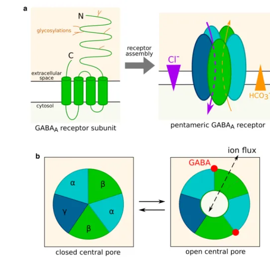
Mature subunits are integral plasma membrane proteins. Human subunits are 420–632 amino acid long and weigh between 52 and 59 kDa [34, 36]. They contain an extracel-lular N-terminal segment of 220–250 residues, which can be glycosylated; 4 transmembrane domains termed M1, M2, M3 and M4; and a short extracellular C-terminal segment (Fig. 1a).
A functional receptor contains five subunits (Fig. 1a), assembled in a circle around a chloride-permeable pore delineated by the M2 transmembrane domain of all five subunits [8]. Each monomer is thus in contact with two other subunits, respectively at its “principle”, or + , and “comple-mentary”, or −, faces.
The study of structure–function relationships of GABAA
receptors has long been hindered by the substantial diversity of conformations, making it difficult to stabilize a receptor population in a single state [43], and cryo-EM structures of GABAA receptors were only recently resolved (reviewed in
[44]). GABA-binding sites are located at interfaces between subunits on the surface of the extracellular part of the recep-tor. GABA binding induces a conformation change, typically when all binding sites are occupied, through a concerted rotation of the extracellular domains of the five mono-mers [45]. This rotation is translated to the transmembrane domains and results in variations of the opening of the cen-tral pore (Fig. 1b). Based on models of GABAA receptor
structure, Rossokhin predicted that the diameter of the chan-nel pore is mainly controlled by residues—2′, 9′ and 20′ of the M2 transmembrane domain [46]. The ring formed by the 9′ M2 residues of the five subunits is termed activation gate. In the main closed conformation, the diameter of the ring is only 2.5–3.4 Å. GABA-induced conformation change prompts the side chains of the activation ring residues to shift away from the central pore, whose diameter increases up to 3.4–7.6 Å, allowing ions to flow through the channel.
The most frequent subunit composition of GABAA recep-tors is 2 α, 2 β and 1 γ subunit [47, 48]. In this case, the 2 α and 2 β subunits in the receptor are most often, but not always, of the same isoform [48–53] and the receptor carries two GABA binding sites [54] located at the β+/α− subu-nit interfaces [44]. However, other stoichiometries can be observed [55]: ε, π and δ subunits can replace a γ subunit, and a θ subunit can replace a β subunit [33]. In addition, binary receptors (receptors containing two different classes of subunits instead of three, such as the αβ subtypes) are able to assemble in vitro. They were also described in vivo, but the evidence of their existence often comes from indirect
methods [56] or study of γ2 or δ deficient mice [57, 58]: it is therefore possible that they are artefactual or accidental.
ρ subunits usually assemble in homopentamers or het-eropentamers [59, 60], with five GABA binding sites per receptor [54], i.e. one at each subunit interface. However, in rare cases, ρ subunits assemble with α1 and/or γ2 subunits [61, 62].
GABAA receptors are classified into hundreds of subtypes
Due to the substantial subunit diversity, there are numerous subtypes of the GABAA receptor, characterized by the
com-bination of subunits they contain (e.g. α1β2γ3) [6].
GABAA receptors are expressed in numerous cell types, most notably in neurons. The expression patterns of the dif-ferent subunits determine which subtypes are assembled, as well as their cellular and subcellular location and their abun-dance. Numerous cells coexpress multiple GABAA subunits isoforms [48–53]. Consequently, a single cell can express several GABAA receptor subtypes [56, 63, 64].
The most abundant subtype in the mammalian nerv-ous system is α1β2γ2, possibly accounting for 60% of all GABAA receptors [65]. The other major subtypes are
α2β3γ2 and α3β3γ2 [66], while α4–6βγ2, α6β2–3δ, α4β2–3δ and ρ1–3 are less abundant. Minor subtypes whose physi-ological existence in vivo is deemed likely include α1βδ, α2β1γ1, αβε, αβπ, αβθ, α1α6βγ2, α1α6βδ and αβγ3 [10,
66]. Though it is unlikely that all possible subunit combina-tions exist and assemble into functional receptors, the large number of possible combinations of subunits expressed by a given cell explains that the number of existing functional subtypes is estimated to be as high as 500 [9]. Most of these subtypes are expected be very rare compared to the most abundant ones [66] and to be limited to a specific tissue, brain area or developmental phase, but they may still exert non-neglectable effects on brain function because of the high abundance of GABAA receptors compared to most neuronal
receptors [9] and because rare subtypes can be selectively enriched in certain neurons or synapses [12].
GABA
Areceptor ion conductance selectivity
GABAA receptors are ion channels. Different ions can flow
down their electrochemical gradient through the pore from one side of the membrane to the other, generating currents. The intensity, i.e. the net charge transfer per time unit, of a
Fig. 1 GABAA receptor
struc-ture and gating. a Strucstruc-ture of an isolated GABAA receptor
subunit and of a mature recep-tor. Five subunits, potentially belonging to different classes or isoforms, assemble into a channel permeable to chloride and bicarbonate ions. Ions flow through GABAA receptors down
their electrochemical gradient.
b Control of GABAA receptor
conductance by the receptor conformation. The central pore can be either closed or open as a result of agonist fixation on the receptor. In this example, the receptor is viewed from the extracellular space and the sub-units are ordered as in the most abundant subtypes, namely αβγ. The presence of two GABA binding sites corresponds to ternary receptors (composed of subunits from three different classes)
transmembrane ion current is the product of the ionic elec-trochemical gradient by the number and conductance of open channels.
Nopen is the number of open channels at the considered membrane; it depends on numerous parameters, notably GABA concentration. For each ion, ΔEion designates the cognate transmembrane electrochemical gradient and Gion
the conductance of a single open GABAA channel.
The conductance of GABAA receptors varies widely for
different ions: we will study in this paragraph how this selec-tivity shapes the currents mediated by GABAA receptors.
GABAA receptor chloride conductance
In most circumstances in the adult brain, K+ coupled
sec-ondary active Cl− transporters [67] generate an outwardly
directed chloride electrochemical gradient at the neuron plasma membrane [68]. Consequently, GABA-induced opening of GABAA receptors induces a chloride influx (Fig. 1a), thus a hyperpolarization of the plasma membrane and an inhibition of action potential generation. This is the most frequent mechanism of inhibitory neurotransmission, which occurs in a large proportion of brain synapses.
However, during development [69], GABAA receptors
mediate chloride efflux due to an inverted electrochemical gradient [70], and thus often activate the neuron [70–73]. Such a mechanism has also been observed in slices prepared from adult brains [74], but these findings have been ques-tioned because neurons in slices seem to have a higher intra-cellular chloride concentration than in vivo. This artifact may produce a non-physiological inversion of the chloride electrochemical gradient [75].
Membrane resistivity (ρ) is equal to the transmembrane potential (ΔE) divided by the intensity (I) of the current passing through a unit of membrane surface: ρ = ΔE × S/I (where S is the area of the membrane domain). The lower the resistivity is, the more intense is the transmembrane current generated by a given voltage gradient. The open-ing of channels, regardless if they mediate depolarizopen-ing or hyperpolarizing currents at the resting membrane potential, decreases membrane resistivity and promotes the dissipa-tion of acdissipa-tion potentials, a phenomenon termed shunting inhibition. Indeed, it enables ions to respond to the electro-chemical gradient created by the action potential with local transmembrane flows instead of the expected longitudinal flows that are necessary for propagating the action potential to other membrane locations. Depolarizing currents medi-ated by GABAA receptors can interfere with action
poten-tials propagation through shunting [76] or inactivation (1) dq dt =I = Nopen ∑ ions ΔEionGion.
of voltage-dependent Na+ channels [77], and can thus be
inhibitory instead of excitatory [78, 79]. Conversely, hyper-polarizations mediated by GABAA receptors may result in
neuronal activation through rebound spikes [80, 81]. The GABAA receptor-mediated depolarizations observed during development can induce an influx of calcium through voltage-dependent channels [80, 82], favoring neurite out-growth [83], synaptogenesis [84], neuron migration [85], and neuron survival [86].
Permeability of GABAA receptors to ions other than chloride
Although chloride ions account for most of GABAA receptor
conductance in vivo [3], GABAA receptors are also
perme-able to bicarbonate, formate, propionate, acetate, cyanide and halides [3, 4, 87]. Differences in GABAA receptor con-ductance and permeability to these anions are caused by a selectivity filter relying on recognition sites [3] and the limited pore diameter.
Because of CO2 production and conversion in HCO3− inside the cell, bicarbonate flows mediated by
GABAA receptors are outwardly directed (Fig. 1a), and thus
depolarize the cell membrane. In standard physiological con-ditions, the ratio of HCO3− vs Cl− permeability of GABA
A
receptors is comprised between 0.2 and 0.3 [88], i.e. chloride currents have a greater amplitude than bicarbonate currents and the net effect is hyperpolarization. However, prolonged GABA exposure can occasionally turn hyperpolarizing cur-rents into depolarizing ones [89], notably during epileptic seizures or in hypothalamic hamartoma neurons [90]. This phenomenon probably indicates a dissipation of the chloride gradient [91, 92] when the channel remains open for long periods. Under these conditions, the bicarbonate efflux may become larger than the chloride influx and GABAA receptors
may transiently become excitatory [72].
Intrinsic electrophysiological parameters
of the different GABA
Areceptor subtypes
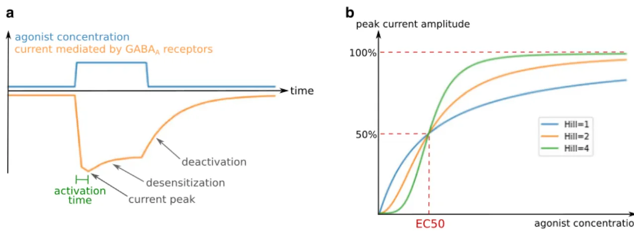
The impact of GABAA receptors on cellular
electrophysiol-ogy mostly depends on the time-course and amplitude of the chloride currents evoked by exposure to an agonist such as GABA. Typically, the current time-course following the introduction of an agonist in the receptor’s environment con-sists of an initial current peak, followed by a decrease in amplitude (Fig. 2a). This decrease ends with a stabilization at lower amplitude or a complete closure of the receptors, depending on agonist concentration, duration of exposure to the agonist, and on the receptor subtype. The effects of GABAA receptors on neuronal activity are determined pri-marily by the charge transfer (the product of the current
intensity with its duration) [93], but also by its synchroniza-tion with other electrical inputs, hence the importance of the shape of the current time-course [94, 95]. The current inten-sity at a certain time point is the product of the conductance of a single channel by the chloride electrochemical gradient and the number of open receptors (Eq. 1), which depends on kinetic parameters governing channel gating as well as receptor and agonist concentrations. Indeed, the effects of an agonist are dose-dependent: for example, the proportion of open receptors as a function of the GABA concentra-tion follows an allosteric pattern (Fig. 2b), characterized by a concentration threshold at which the proportion of open receptors sharply increases.
In most physiological situations, the cell response to a GABA input is driven by the joint action of dozens or hun-dreds or receptors that perceive the signal. The macroscopic scale (receptor populations) therefore appears more suited than the microscopic scale (single channels) to understand the biological consequences of GABAA receptor electro-physiology and is easier to study, which explains that most available information on GABAA receptor
electrophysiol-ogy covers macroscopic features. Nevertheless, macroscopic properties always emerge from the integration of single channel activity characteristics.
The electric activity of GABAA receptors can be
under-stood with a series of parameters that describe and explain their responses to the various patterns of agonist exposure. In this section, we will describe the macroscopic electro-physiological parameters that determine the activity of pop-ulations of GABAA receptors in response to GABA, and
we will highlight the differences between subtypes in that regard.
Electrophysiological parameters can be subdivided into kinetic parameters (activation time, desensitization time, deactivation time…) and so-called functional parameters (conductance, EC50, Hill coefficient…), which describe
respectively the current time-course and its dependence on agonist concentration. As will be exemplified below, some of these parameters are not independent.
Tools for the study of GABAA receptor electrophysiology
Before detailing the electrophysiological parameters of GABAA receptors, we will discuss two conceptual tools
that were instrumental in the discovery and understanding of these parameters: modeling and the use of channel modu-lating drugs.
Modeling
Numerous models have been used to interpret the growing amount of data on GABAA receptor electrophysiology, pre-dict receptor activity, and understand the microscopic origin of macroscopic parameters and large-scale behavior of chan-nel populations.
Most of the models used are Markovian. Markovian mod-els are discrete (time is discontinuous) and memory-free: the next step of evolution of a model depends only on its present state, and not on past states. Markovian models describe the activity of a channel with a series of states, each of them having its own conductance and being supposed to corre-spond to a specific receptor conformation. Transition rates can be a function of environmental parameters such as trans-membrane potential or agonist concentrations. Transition rates indicate the kinetics of channel activity as well as states occupancy at equilibrium. For example, in the model showed in Fig. 3a, if p is the proportion of receptors in the open state at a given time point, a proportion pkc of the receptor population transitions to the closed state before the next time point, while a proportion (1 − p)ko of receptor transitions to
a b
Fig. 2 General characteristics of GABAA receptor-mediated currents. a Typical GABAA receptor current time-course and kinetic electrophysi-ological parameters. b Dependence of GABAA receptor-mediated current intensity on agonist concentration for different Hill coefficients
the open state. At equilibrium, the net flux is null, meaning that pkc = (1 − p)ko, and therefore p = ko/(kc + ko).
Certain models specifically describe systems at equilib-rium. This simplification allows the replacement of transi-tion rates by equilibrium constants. In our example model, the equilibrium constant K would follow the equation
K = peq/(1 − peq) = ko/kc (Fig. 3b).
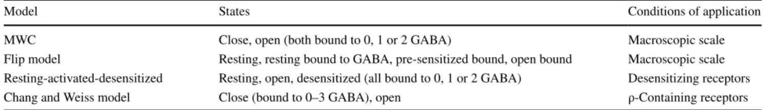
The most classic model used to describe GABAA
recep-tor activity is the Monod–Wyman–Changeux two-state concerted transition model (MWC) [96]. This equilibrium model takes into account one open and one closed confor-mations, both of which can be bound to zero, one or two GABA molecules (Fig. 3c). The MWC model requires only four parameters to entirely describe the receptor response to an agonist, which explains its attractiveness. It accurately describes GABAA activity at the macroscopic scale, and its
activation by numerous agonists and combination of agonists [47, 96–98]. This model has confirmed the presence of two GABA binding sites on ternary receptors [97].
Other models (Table 1) complexify the MWC model to fit more accurately to certain experimental data, such as the flip model, which includes a transient pre-sensitized closed
conformation with high agonist affinity [96, 99]. More com-plex models [100–104] consider a greater number of states. Although generally more accurate than simpler models, complex models often lack predictive value since it is dif-ficult to accurately estimate all the parameters of the model [96].
Complex models usually stem from the study of single channels, whose complex behavior requires several states to be accurately described, whereas fewer states are necessary to describe the properties of GABAA receptors activity at
the synaptic or cellular scale. Despite the likely existence of several conformations, the two-conformations MWC models provide accurate predictions at a macroscopic scale. This indicates that the receptor conformations can be separated into two groups with minor electrophysiological differences within each group [96, 105], or that some of them are occu-pied only transiently and are not involved in the steady-state circumstances that the MWC model is used to describe.
Electrophysiological parameters can be calculated as a function of transition rates and state occupancies in certain models. Modeling also helps to understand the interde-pendence between parameters, or to calculate parameters inaccessible to direct experimentation. For example, Chang and Weiss [106] estimated that ρ-containing receptors open when they bind GABA in at least 3 of their 5 binding sites.
Channel modulating drugs
The activity of GABAA receptors is modulated by subtype-selective agonists and antagonists belonging to different chemical families such as benzodiazepines or barbiturates and including anesthetic, anxiolytic or antiepileptic drugs.
Since primary and transfected cells can often express sev-eral subtypes simultaneously, it is necessary to distinguish from which subtype come the currents measured during electrical recordings. Subtype-selective channel modulating drugs facilitate in situ subtype identification [10].
For example, Lindquist and Birnir performed a patch-clamp single-channel study of GABAA extrasynaptic
recep-tors on dentate gyrus granule neurones and identified three types of electrophysiological signatures [107]. Receptors of the first, second and third type were potentiated respectively by zolpidem, flumazenil and THDOC, indicating that they belong respectively to α1βγ2, α4βγ2 and αβδ subtypes.
a
c
Fig. 3 Markovian models of receptor channel activity. a Simplest Markovian model of a receptor channel, with two unitary states: open (O) and closed (C). The transition rates describing channel opening and closure are respectively ko and kc. Both transition rates are a
func-tion of the agonist concentrafunc-tion [A]. b Applicafunc-tion of the two-states model to steady-state situations. K is the equilibrium constant, and depends on agonist concentration. c MWC model. L, r and K are con-stants depending on receptor structure and [G] is the GABA concen-tration. The open states are O (no GABA bound), OG (one GABA) and OG2 (two GABA); likewise, C, CG and CG2 are the close states
Table 1 Examples of Markovian steady-state models used for GABAA receptor study
Model States Conditions of application
MWC Close, open (both bound to 0, 1 or 2 GABA) Macroscopic scale
Flip model Resting, resting bound to GABA, pre-sensitized bound, open bound Macroscopic scale Resting-activated-desensitized Resting, open, desensitized (all bound to 0, 1 or 2 GABA) Desensitizing receptors Chang and Weiss model Close (bound to 0–3 GABA), open ρ-Containing receptors
Channel modulating drugs have also been used to study GABAA receptors structure and to map subtypes expression
in the brain. The benzodiazepine molecules [3H]Ro15-4513 and diazepam bind respectively all γ-containing subtypes and α1,2,3,5βγ receptors. Hence, under competition from diazepam, [3H]Ro15-4513 binds only α4,6βγ subtypes. Using this property, Korpi et al. mapped α4,6βγ receptors by autoradiography of [3H]Ro15-4513 in mouse brains and showed that they were overexpressed in δ−/− mice [108]. This proved that γ subunits can partly substitute δ.
Etomidate is a general anesthetic binding GABAA
recep-tors on their GABA binding sites and potentiating their response to GABA. This potentiation involves comparable allosteric shifts between αβδ and αβγ subtypes, showing that αβδ subtypes carry two GABA binding sites as had been previously described for αβγ subtypes [109].
Kinetic parameters Activation time
The delay between a change in GABA concentration and channel response varies between receptor subtypes [110]. The channel opening occurs in two phases: first, agonists bind to the receptor; second, the receptor conformation changes and the central pore opens. Thus, the delay in GABAA receptor opening upon GABA introduction in its
environment depends both on GABA binding kinetics [111] and on the kinetics of receptor conformational change upon GABA fixation [111]. GABA fixation kinetics is determined by GABA concentration [75, 100, 105], following an allos-teric pattern [112], and by the receptor affinity for GABA. At a given GABA concentration, the characteristic time of the GABA binding kinetics is theoretically inversely pro-portional to the receptor affinity for GABA: this relation has been observed in β2γ2S-containing subtypes [75]. However, at saturating GABA concentrations (in the order of 10 mM [112]), the conformational change kinetics becomes the lim-iting factor. This may explain why the δ subunit lowers the reactivity of the receptor compared to γ2 [113] despite a higher affinity. The activation time, i.e. the time to go from 10 to 90% of open channels after introduction of GABA, is 0.46 ms for α1β3γ2L at a GABA concentration of 1 mM, versus 2.4 ms for α1β3δ [114].
The activation times provided in Tables 2, 3 and 4 are calculated at saturating GABA concentration, thus should reflect the kinetics of conformation transition. In association with β3 and γ2L subunits, the rank order of α subunits in terms of activation time is α1 < α2 < α4 < α6 < α5 < α3, with α3 being three times slower than α1 [110–112, 114]. How-ever, when associated with β1 and γ2, α2 confers an activa-tion twice faster than α1 [100]. αβ subtypes [115], and even more ρ-containing subtypes [116], display an extremely slow
activation time lasting several tens of milliseconds. Recep-tors containing the ε subunit display slow activation in vivo [117]. However, this property has never been studied with recombinant ε-containing receptors, thus it may be conferred by another subunit.
The measured activation time depends on the size of the experimental settings. For example, macro-patches, in which several receptors gathered on a micrometric membrane domain are studied, display faster activation than whole-cell recordings [112]. It may be due to the time required for GABA to diffuse to all GABAA receptors, which is longer
for larger experimental models, or to cytoplasmic proteins that may modulate GABAA receptor kinetics in whole-cell studies [112].
Desensitization
Upon long exposure to GABA, GABAA receptors lose
their ability to open: this process is called desensitization (Fig. 2a). This mechanism is also observed when receptors are exposed to GABA transients at a high frequency and is then termed rundown (Fig. 4a) [118]. Desensitization pre-vents the transmission of abnormally strong inhibitory sig-nals during synaptic communication: if GABA is chronically present in the synapse rather than by interspaced bursts, as it is under normal conditions, postsynaptic GABAA receptors
will close [119].
In desensitized conformations, the M2 transmembrane domains are tilted and their -2′ residues obstruct the intra-cellular end of the central pore of GABAA channels [46].
The five monomers constituting the channel can transition between open and desensitized conformations independently from one another [120].
During prolonged GABA exposure, the proportion of open channels sometimes follows a decreasing exponential curve over time [75], thereby a characteristic time of desen-sitization (td) can be calculated. In other cases, the observed desensitization curve can be described as the addition of several exponential components [112, 121, 122] (Fig. 4b). In this situation, a single td can be computed as the average of the characteristic times of each component weighted on the amplitude of the related current (this is the procedure fol-lowed to calculate the td shown in Tables 2, 3 and 4).
Conse-quently, the calculated value of td depends on the duration of GABA exposure [111]. In long exposures, slow components gain in relative importance and the calculated td increases. In
addition, td depends strongly on GABA concentration [75]:
the curve of desensitization rate as a function of GABA con-centration follows approximately an allosteric pattern. When enough data are available for calculations, the td presented in
Tables 2, 3 and 4 are the asymptote of this curve at infinite GABA concentration.
The characteristic time of desensitization is usually in the order of 1 s, despite important variations. αβγ receptors con-taining the α4 subunit desensitize two to three times faster than those containing the α3 or α5 subunit, with α1 and α2 desensitizing almost as fast as α4, and α6 conferring an intermediary desensitization time [75, 111, 112]. However, when the receptor includes a δ subunit, α6 appears to confer faster desensitization than α1 [123]. β2 confers a slightly faster desensitization than β3, with β1 as an intermediate [124] or giving results similar to β2 [100]. Desensitization of γ2-containing receptors is two or three times faster than that of δ-containing receptors [113, 123, 125], but substantially slower than that of ε-containing receptors [126].
Certain subtypes do not desensitize or desensitize only partially. For example, homomeric ρ-containing receptors are almost insensitive to desensitization [5, 116, 127, 128]. Unlike γ-containing subtypes, receptors containing the δ subunit desensitize only partially [101]: α1β3δ currents decrease by only 11.6% upon 6 s exposure to saturating GABA concentration [123]. The α subunit plays a lesser but still notable role in determining the extent of desensiti-zation (Table 5): γ-containing subtypes desensitize incom-pletely only if they contain the α3 or α5 subunit [111], with a steady-state current in prolonged GABA exposure of up to 30% of the peak amplitude. α1β3 receptors containing two α and three β subunits desensitize more extensively than recep-tors containing three α and two β subunits [129].
Desensitization can be followed by a refractory period [114], during which receptors are unable to reopen. The length of this period is modeled as a function of several decreasing exponentials, indicating that GABAA receptors can adopt several closed conformations [114, 130], whose relative proportions depend on GABA concentration [130]. In vivo, the fast components of the refractory period disap-pear at low GABA concentration. In these conditions, desen-sitization is less extensive but its effects last longer [130,
131]. The length of the refractory period varies between sub-types, with receptors containing α4 or α5 being the fastest to recover (with a characteristic time of 25 ms following a 5 ms exposure to GABA), followed by α2 and α3 (about 100 ms), then α1 (about 200 ms) and finally α6 (about 400 ms) [111]. Nonetheless, characteristic times heavily depend on experimental procedures and method of calculation [112].
The study of desensitization prompted the development of complex, non-steady-state models of GABAA receptor
activity since the MWC model does not describe recep-tor desensitization and therefore applies well only to non-desensitizing subtypes or to short time-frames of activation. These limitations are tackled by three-state models such as the resting-activated-desensitized model (Table 1) [132]. Other models describe the different components of desensi-tization or refractory periods with several desensitized states [114, 120, 133]; the desensitization times and durations of refractory period components are represented by the transi-tion rates respectively to and from these states. Using one of these models, Gielen et al. [120] proposed that the fast component of desensitization corresponds to receptors with two of their five monomers adopting a desensitized confor-mation, while the slow component corresponds to receptors with at least three desensitized monomers.
Speed, extent and relative proportions of exponential components of desensitization are critical in shaping the time-course of the inhibitory post-synaptic current (IPSC) mediated by GABAA receptors and strongly differ between
subtypes [102, 106, 114, 134, 135]. For example, α6β3δ has no fast component, unlike α6β3γ2L [101, 123]. Con-sequently, despite the similarity of the characteristic time of their slow component of desensitization, α6β3δ appears to desensitize less extensively than α6β3γ2L and is less sensitive to the duration of GABA exposure. As a result, α6β3δ transmits inhibitory signals whose intensity is roughly proportional to the duration of GABA exposure,
a b
Fig. 4 Characteristics of GABAA receptor desensitization. a
Run-down. When GABAA receptors are exposed to frequent GABA
sients, a portion of the receptor population desensitizes at each tran-sient and does not recover before the next exposure, resulting in a
decrease in peak amplitude. b Biphasic desensitization. The current decay upon desensitization can often be modeled as the sum of two decreasing exponential components. A global desensitization rate can be computed from the characteristic time (τ) of each component.
while α6β3γ2L-mediated signals do not depend on exposure duration. This exemplifies that minor differences between subtypes at the microscopic scale can be amplified at larger scales. Furthermore, the time-course of IPSCs determines the strength and timing of neuronal inhibition, so limited differences in desensitization properties can have dramatic effects in the response of neuronal circuits to high-frequency inhibitory signals [101, 136].
Deactivation
GABAA receptors spontaneously release their ligands and close several milliseconds or tens of milliseconds after a drop in GABA concentration. The event of deactivation puts an end to the transmission of a hyperpolarizing signal dur-ing synaptic communication, after a brief GABA exposure (synaptic GABA bursts usually last no longer than a few mil-liseconds). Contrary to desensitized channels, deactivated channels can reopen without a refractory period.
Deactivation has been observed to be biphasic in vitro [100, 103, 111, 114, 122, 133] and in vivo [105, 137]; the relative proportions of the two components can vary depending on the receptor subtype. These two components may correspond to two conformations, one bound to two GABA molecules (the slow component) and the other to a
single one (the fast component) [102], but this hypothesis is debated [114]. Deactivation can sometimes be triphasic [56], once again depending on the subtype and probably on recording procedures, because certain protocols do not enable the recording of very fast components [112].
Homomeric ρ-containing receptors deactivate quickly [116]. Among α-containing subtypes, those containing α4 are the fastest to close, followed by α5 (1.5 times slower), α1 (2.5 times slower than α4), then α2 (5 times slower than α4), and finally α6 (6 times slower than α4) and α3 (about 10 times slower than α4). The γ2 subunit confers a slower deactivation than δ [138, 139]. The β2 subunit confers a substantially faster deactivation than β3 [140].
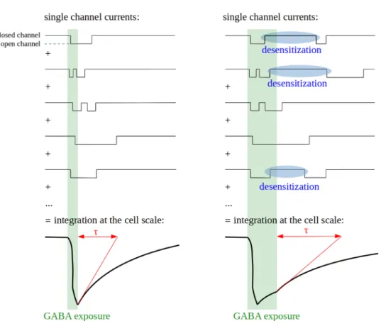
Most studies on deactivation have been conducted on whole-cell or multichannel patches, which should give results close to what is observed in vivo at the synaptic or cell scale. However, deactivation is much faster in single-channel (0.2–25 ms) than in single-channel cluster or whole cell (5–680 ms) recordings [141, 142]. This discrepancy likely stems from sequential closing and opening events of a single channel (Fig. 5), which are integrated in a single current time-course by whole-cell or multichannel patches record-ings [105, 143]. It exemplifies the apparent gap between the complex macroscopic properties of GABAA receptors and the single-channel activity they emerge from.
Fig. 5 Relationships between microscopic and macroscopic kinetics of channel opening. Deactivation is a macroscopic property resulting from the integration of single-channel currents time-courses. Its characteristic time (τ) is longer than mean channel open time due to possible channel reopen-ing events and is increased by desensitization due to the possi-ble reactivation of desensitized channels
The deactivation time increases in proportion with the extent of desensitization [102, 123, 135], maybe due to a high GABA affinity of desensitized states [135]. GABA is thus retained on desensitized or pre-desensitized receptors for tens of milliseconds, even when all GABA has been cleared from the extracellular space [144]. If the receptor is still bound to GABA when the refractory period ends, it is able to reopen immediately. When numerous receptors are considered together, this process results in a prolongation of the phase in which the current decreases, which is described as a slower deactivation (Fig. 5). Consequently, the fast components of desensitization prolong GABAA receptor responses to GABA and therefore synaptic communication while decreasing the current amplitude [102, 131].
Functional parameters Conductance
A single GABAA channel can display several conductance states, remarkably conserved between species and cell types, ranging around respectively 12, 20, 30 and 45 pS for the most frequent subtypes. The main conductance state is 30 pS, meaning that this is the state adopted by the channel in most circumstances when it is open [4]. The conductance states may correspond to different channel conformations.
Most GABAA receptor subtypes display very similar
con-ductance values (Tables 2, 3, 4) [145, 146]. However, the main conductance state of γ-containing receptors (~ 30 pS) [125] is higher than that of δ-containing receptors (~ 22 pS) [125], ρ-containing receptors (7 or 8 pS) [147, 148] and αβ binary subtypes (11–20 pS) [56].
GABA EC50
The curve of GABAA receptor peak current intensity as a function of GABA concentration follows an allosteric pat-tern (Fig. 2b), described by the equation of Hill:
where I is the current intensity, Imax its asymptote at infinite
agonist concentration, [A] the agonist concentration, and h the Hill coefficient (see “Hill coefficient”).
EC50 is the GABA concentration at which the current peak reaches 50% of its maximal amplitude. The EC50 par-tially reflects the receptor affinity for GABA: in general, the lower the EC50, the higher the affinity. However, the EC50 also depends on gating parameters such as the speed or max-imal probability of channel opening. This can be exemplified by a simple stochastic model with a single binding site and a single opening state.
(2)
I Imax =
[A]h
EC50h+ [A]h,
where A is an agonist that binds to a receptor site R, A + R is the vacant state of the receptor site, AR the occupied state,
AR* the open state, Kon and Koff, respectively the
associa-tion and dissociaassocia-tion constants of the agonist, and β and α are respectively the opening rate and closing rate constants of the channel. In this model, the true affinity of the recep-tor for its ligand is the microscopic dissociation constant
Kd = Koff/Kon. The receptor efficacy is defined as the
equi-librium constant of the closed-open isomerization E = β/α. In this model, the apparent affinity EC50 verifies EC50 = Kd/
(1 + E).
The receptor efficacy is related with Pomax, the maxi-mum fraction of receptors in the active state, by the rela-tion Pomax = E/(1 + E) = 1 − (EC50/Kd). Pomax can only
be estimated at saturating GABA concentration using non-stationary noise analysis [149]. However, the presence of several open states can render the estimation of receptor efficacy very challenging. Hence, the true affinity and effi-cacy of GABAA receptors are often inaccessible to accurate
measurements and the EC50 is used as a measure of the apparent affinity.
When certain receptors of a cluster open for the first time upon GABA exposure, others may be already closed, due to fast desensitization. It results in a smoothened shape of the current time-course through decreased peak amplitude and spreading of charge transfer over time. This phenomenon is stronger at high GABA concentrations, which explains why Pomax is often substantially inferior to 1 and EC50 is close to the true affinity Kd.
GABA EC50 is up to 100-fold lower than the concentra-tion (termed BC50) at which the probability of occupancy of the GABA binding sites is equal to 0.5 [150], and than the concentration at which the half-maximal activation rate is reached [100, 112]. This discrepancy is explained by dif-ferences in GABA affinity between receptor conformations [106] and by the influence of desensitization on EC50, as confirmed by the small difference between EC50 and BC50 in the non-desensitizing ρ1 subtype [106]. It indicates that the EC50 is not a purely thermodynamic parameter but partially depends on receptor kinetics. Macroscopic param-eters of GABAA receptor electrophysiology are complex and emerge from the interconnection of the numerous properties of single channels.
Significant differences in GABA EC50 can be observed between GABAA receptor subtypes, covering 2 orders of magnitude from around 0.5 µM to around 50 µM. Discrepan-cies are also observed for the same subtype in different stud-ies, due to differences in the cell type where the receptor was expressed, in the species of origin of the receptor subunits (3) A + R Kon ⇌ Koff AR 𝛽 ⇌ 𝛼 AR ∗,
or in experimental design (Tables 2, 3, 4). It nonetheless appears that the α subunit plays a crucial role in determining the receptor’s affinity for GABA (Table 5). The rank order of increasing GABA EC50 is α6 < α1 ~ α2 < α4 < α5 < α3 when these subunits are associated with β3 and γ2S [151], but α6 < α4 ~ α5 < α1 < α2 < α3 in association with β3 and γ2L [111]. The β isoform affects GABA affinity to a lim-ited degree, with the β3 isoform conferring a one to four-fold lower GABA EC50 than β1 and β2 (Table 6). The γ1 subunit confers a higher EC50 than γ2 when these subunits are associated with α5, but in association with α3, the rela-tionship is inverted. Several studies indicate a low EC50 in receptors containing the γ2S or γ3 subunits compared with the γ2L splice variant (Table 7). Receptors containing the δ subunit consistently display an EC50 around 5 times lower than γ2-containing receptors. ρ-containing subtypes are around 7 times more sensitive to GABA than the most abundant ternary receptors [147]. Subtypes containing two different isoforms of the same class generally show inter-mediate properties between the two corresponding subtypes containing only one of these two isoforms [125, 152–157]. Binary receptors have a very low GABA EC50 [146, 158], around 1 µM, which is as low as the most GABA sensitive ternary receptors. Binary α1β3 receptors containing two α and three β subunits have a lower GABA EC50 than α1β3 receptors with three α and two β subunits [129].
Hill coefficient
The Hill coefficient of a GABAA receptor depends on
its number of GABA binding sites and on cooperativity between GABA binding sites, the process by which the bind-ing of GABA to one site increases the affinity or accessibil-ity of the other site(s) for GABA. The higher the number of GABA binding sites or the extent of cooperativity are, the higher the Hill coefficient is. For Hill coefficients higher than 1, there is a threshold in GABA concentration where the opening probability of the receptor dramatically increases over a limited concentration span. The higher the Hill coef-ficient, the steeper the threshold (Fig. 2b).
In experimental systems with low temporal resolution, fast desensitization can decrease the resolution of the cur-rent peak, which might lead to errors in calculations of Hill coefficients. To the best of our knowledge, this phenom-enon has not been tested with GABAA receptors, but it was described in the glycine receptor [159] which belongs to the same receptor superfamily as GABAA receptors.
Most GABAA receptor subtypes have very similar Hill coefficients of around 1.5. However, binary αβ receptors and ε-containing receptors display low cooperativity [126,
146, 160, 161] with Hill coefficients below 1. On the oppo-site, ρ-containing subtypes display higher Hill coefficients than the most abundant ternary receptors [106, 128, 162],
probably due to the presence of 5 GABA-binding sites instead of 2 [54].
Subunits interplay in the integration
of subtype‑specific electrophysiologycal properties
Due to the remarkable evolutionary conservation of the sequence of GABAA receptor ortholog subunits, the electro-physiological properties of a certain subtype are expected to be similar in different species. To our knowledge, receptors of the same subtype but different species have never been compared in the same study. Tables 2, 3 and 4 highlight significant differences in the main parameters of certain sub-types between species, but important intraspecies differences are also observed, sometimes even when the subunits are expressed in the same cell line. Indeed, experimental pro-cedures and measurement techniques can have a substantial influence on the results [112]: it can therefore be difficult to compare different subtypes. A further complication is that many authors did not distinguish between the splice variants γ2S and γ2L. Still, conclusions can be drawn by comparing subtypes studied in the same article with the same protocols or when several articles give very similar results on the same subtype.
The electrophysiological properties of a ternary subtype are mainly determined by the α subunit isoform and the class of its “third subunit class” (Table 5, 6, 7). Few differences are observed between β or γ isoforms. Some of the differ-ences observed between β isoforms are thought to stem from their differential distribution and association with different isoforms of the α class. For example, preferential association of β3 with α2 and α3 instead of α1 is likely to explain the slower kinetics of the β3-containing subtypes as compared with the other β-containing subtypes [193]. Moreover, the electrophysiology of a subtype does not result in a straight-forward manner from the addition of the properties con-ferred by its subunits because of the complex interactions between these subunits: the properties of a subunit depend on its environment within the receptor.
Binary αβ receptors display low GABA EC50, con-ductance and Hill coefficient, and activate slowly. In most cases, they desensitize slowly and not extensively, except the α1β3 subtype which desensitizes as fast and extensively as α1β3γ2L [114]. Despite substantial differences with ter-nary receptors, biter-nary receptors helped to understand the molecular basis of ternary receptors electrophysiology. For example, using αβ2 subtypes, Olander et al. [115] showed that differences in activation and desensitization kinetics between α isoforms are determined by their transmembrane and intracellular domains. However, this study didn’t iden-tify the cause of variations in deactivation kinetics, since the different αβ2 subtypes investigated didn’t differ significantly in that parameter. Similarly, the extent of desensitization
Table 2 Main electrophysiological parameters of the different GABAA receptor subtypes in standard ionic conditions (subunits expressed in Xenopus oocytes) GABAA receptor subtype Single-channel
conductance (pS) GABA EC50 (µM) Hill coefficient Characteristic time of desensitization td (s)
Characteristic time
of deactivation (ms) Activation time (ms)
α1β1γ1 1.2 [167] α1β1γ2 30 [100] 4.5 [167] 6 [100] 1.2 [100] 0.492 [([GABA] = 1 mM)100] 0.6 [103] ([GABA] = 100 µM) 20.5 [100] 17.4 [103] 1.1 [1 [103100] (γ2S splice variant)] α1β1γ3 0.6 [167] α1β2γ1 0.67 [167] (human γ1, rat α1 and β2, expressed in 293 cells) α1β2γ2 32 [152] 27.9 [56] (γ2S splice variant) 5 [172] 7 [110] (γ2S splice variant) 1.71 [177] 1.6 [110] (γ2S splice variant) 2.12 [152] 1.4 [110] (γ2S splice
variant) 220 [splice variant)110] (γ2S α1β2γ3 30.2 [178] 1.3 [167] α1β2δ 6.71 [179] α1β3γ1 2.1 [167] α1β3γ2 28 [56] (γ2S splice variant) 27.5 [180] 1.7 [167] 2.9 [151] (γ2S splice variant) 7.6 [181] (γ2L splice variant) 14 [112] (γ2L splice variant) 36.2 [158] (γ2L splice variant) 14.1 [122] (γ2L splice variant) 1.7 [158] (γ2L splice
variant) 0.424 [of 1 and 3 mM of 111] (average GABA, γ2L splice variant) 0.95 [112] (γ2L splice variant, 1 mM of GABA) 68.6 [111] (γ2L splice variant) 143 [112] (γ2L splice variant) 82.5 [122] (γ2L splice variant) 0.55 [101] (γ2L splice variant) 0.603 [111] (γ2L splice variant) α1β3δ 27.5 [180] 7.4 [181] 63.4 [181] 86.8 [123] α2β1γ2 30 [100] 17 [184] 37 [100] 1.8 [100] 0.449 [([GABA] = 1 mM)100] 198.7 [82 (type 1) or 51 100] (type 2) [103] 0.5 [100] 0.5 [103] (γ2S splice variant) α2β1ε 11.2 [185] α2β2γ2 1.47 [177] α2β3γ2 5.2 [151] (γ2S splice
variant) 0.391 [of 1 and 3 mM of 111] (average GABA, γ2L splice variant)
110.6 [111] (γ2L
splice variant) 0.735 [111] (γ2L splice variant)
α3β1γ2 15.1 [167] (human α3, rat γ2 and β1, expressed in 293 cells) α3β2γ2 130 [187] 15.1 [167] (human α3, rat γ2 and β2, expressed in 293 cells) 75 [110] (γ2S splice variant) 2.21 [177] 1.5 [110] (γ2S splice variant) 1.1 [187] 3.2 [110] (γ2S splice
variant) 680 [splice variant)110] (γ2S
α3β3γ2 48 [151] (γ2S splice variant) 0.701 [111] (average of 1 and 3 mM of GABA, γ2S splice variant)
188.8 [111] (γ2L
splice variant) 1.788 [111] (γ2L splice variant)
α4β1γ2 1.40 [177] α4β1δ 0.30 [188] 0.17 [177] 1.57 [188] 174 [177] α4β2γ2 3.9 [189] 1.4 [167] α4β2δ 0.25 [179] α4β3γ2 7.6 [151] (γ2S splice variant) 15 [112] (γ2L splice variant) 0.408 [111] (average of 1 and 3 mM of GABA, γ2L splice variant) 0.711 [112] (γ2L splice variant, 1 mM of GABA) 24.8 [111] (γ2L splice variant) 109 [112] (γ2L splice variant) 0.951 [111] (γ2L splice variant)
of αβγ receptors was not matched by the corresponding αβ subtypes, with α1β2 desensitizing incompletely, contrary to α1β2γ. This confirms that novel properties emerge from the new subunits interactions introduced by γ or δ subunits in ternary receptors.
Receptors made of ρ subunits react very slowly to changes of GABA concentration, do not desensitize and deactivate quickly; they have low conductance and GABA EC50 but a high Hill coefficient.
The response of GABAA receptors to particular GABA
inputs can be predicted from the parameters that have been described so far. For example, due to similar conductance, faster activation and slower deactivation, α2β1γ2 medi-ates tenfold higher charge transfers than α1β1γ2 when it is activated [100], but the higher GABA EC50 of the former means that stronger inputs are necessary to activate it.
Several studies found a great consistency between the electrophysiological activity of in vitro recombinant and in vivo endogenous GABAA receptors [60, 102, 103, 137,
152, 154, 194] (reviewed in [195]). The parameter values
given above should therefore be relevant enough in physi-ological contexts to explain the activity and function of GABAA receptors in the nervous system.
GABA
Areceptors mediate different types
of currents at the neuronal scale
As receptors of the most abundant inhibitory neurotransmitter, GABAA receptors are expressed in most neurons and
influ-ence their electric activity. Depending on the neuronal type, GABAA receptors can be located in the dendrites, the cell body
and/or the axons. They are highly enriched at post-synaptic sites [194, 196], where they usually mediate inhibitory neu-rotransmission, but can also be found extrasynaptically and perisynaptically. Despite the preferential synaptic localization of α1, α2, α3, and especially γ2 [197], no GABAA receptor
subunit has yet been found exclusively in synapses [196, 198]. Receptors containing the α4, α5, α6, γ1, γ3 or δ subunits, as well as binary αβ subtypes, are mainly extrasynaptic [56, 196,
Table 2 (continued) GABAA
receptor subtype
Single-channel
conductance (pS) GABA EC50 (µM) Hill coefficient Characteristic time of desensitization td (s)
Characteristic time
of deactivation (ms) Activation time (ms)
α4β3δ 0.35 [188] 1.8 [188] α5β1γ2 5.6 [167] α5β2γ2 5.8 [187] 4.2 [167] 0.47 [177] 1.5 [187] α5β2γ3 4.9 [190] 1.9 [190] α5β3γ2 24.9 [180] 11.6 [151] (γ2S splice
variant) 1.315 [of 1 and 3 mM of 111] (average GABA, γ2L splice variant)
41.5 [111] (γ2L
splice variant) 1.247 [111] (γ2L splice variant)
α6β1γ2 0.5 [167] α6β2γ1 0.4 [167] α6β2γ2 5.2 [156] (γ2S splice variant) 1.4 [189] 0.34 [172] 0.50 [179] (γ2S splice variant) α6β2δ 0.52 [156] (γ2S splice variant) 1.2 [167] 0.21 [179]
α6β3γ2 1 [151] (γ2S splice variant) 0.596 [111] (average of 1 and 3 mM of GABA, γ2L splice variant)
163.8 [111] (γ2L
splice variant) 1.052 [111] (γ2L splice variant)
α6β3δ 340.4 [123]
ρ1 1.7 [116] 3.5 [116] 14.5 [116] (30 µM of
GABA) 154 [116]
ρ2 2.6 [116] 3.3 [116] 8.2 [116] (30 µM of
GABA) 180 [116]
Roman: rat subunits; bold: human subunits; bold italics: mouse subunits. The Hill coefficient, GABA EC50, main conductance state and charac-teristic times of activation, deactivation and desensitization of the most abundant subtypes in vivo, i.e. ternary subtypes and ρ homopentamers, are presented. Information on other subtypes can be found in [13, 103, 112, 114, 116, 123, 125, 130, 146, 152–157, 176, 180, 182, 191, 192]
Table 3 Main electrophysiological parameters of the different GABAA receptor subtypes in standard ionic conditions (subunits expressed in
mouse L929 cells)
Roman: rat subunits; bold: human subunits; italics: bovine subunits. The Hill coefficient, GABA EC50, main conductance state and character-istic times of activation, deactivation and desensitization of the most abundant subtypes in vivo, i.e. ternary subtypes and ρ homopentamers, are presented. Information on other subtypes can be found in [13, 103, 112, 114, 116, 123, 125, 130, 146, 152–157, 176, 180, 182, 191, 192] GABAA
receptor subtype
Single-channel
con-ductance (pS) GABA EC50 (µM) Hill coefficient Characteristic time of desensitization td (s)
Characteristic time of
deactivation (ms) Activation time (ms) α1β1γ1 5.2 [165] α1β1γ2 30 [125] (γ2L splice variant) 29.3 [145] (γ2S splice variant) 7.1 [146] 5.2 [168] (γ2L splice variant) 5.1 [169] (γ2L splice variant) 6.2 [125] (γ2L splice variant) 1.7 [146] 1.9 [168] (γ2L splice variant) 1.9 [169] (γ2L splice variant) 1.4 [125] (γ2L splice variant) 5 [125] (γ2L splice variant) 6.0 [145] (γ2S splice variant) α1β1δ 22 [125] No desensitization observed [125] 400 [125] α1β2γ2 29 [145] (γ2S splice variant) 11 [171] α1β3γ2 25.9 [114] 26.8 [130] 14 [11.6 [171130]] (γ2L splice variant) 15.5 [111] (γ2L splice variant) 15.5 [182] (γ2L splice variant) 1.48 [111] (γ2L splice variant) 1.5 [182] (γ2L splice variant) 0.462 [114] ([GABA] = 1
mM, γ2L splice variant) 76.1 [splice variant)114] (γ2L 0.46 [variant)114] (γ2L splice
α1β3δ 23.8 [114] 26.7 [130] 2.8 [4.4 [130182]] 1.4 [182] 1.26 [mM)114] ([GABA] = 1 42.8 [114] 2.4 [114] α1β3ε 24 [161] (rat α1 and β3, and human ε, in L929 cells) 0.8 [161] (rat α1 and β3, and human ε, in L929 cells) 0.9 [161] (rat α1 and β3, and human ε, in L929 cells) α2β3γ2 25 [111] (γ2L splice variant) α3β3γ2 35.8 [111] (γ2L splice variant) α4β3γ2 10.7 [111] (γ2L splice variant) 2.57 [113] 1.3 [113] 2.5 [113] ([GABA] = 100 µM) α4β3δ 0.5 [113] 1.3 [113] 4.8 [113] ([GABA] = 100 µM) α5β1γ2 32.8 [157] (γ2L splice variant) 26 [124] (γ2L splice variant) 1.69 [157] (γ2L splice variant) α5β3γ2 22 [133] (γ2L splice
variant) 5.7 [variant)157] (γ2L splice 6 [124] (γ2L splice
variant)
9.4 [111] (γ2L splice variant)
1.54 [157] (γ2L
splice variant) 51.8 [splice variant)133] (γ2L 0.9 [variant)133] (γ2L splice
α5β3γ3 26.9 [191] 1.5 [191] 1.6 [191] α5β3π 23.8 [191] 1.3 [191] 1.4 [191] α6β2γ2 2 [171] α6β2δ 0.2 [171] α6β3γ2 2 [171] (γ2L splice variant) 2.25 [111] (γ2L splice variant) 1.04 [111] (γ2L splice variant) α6β3δ 0.3 [171]
197, 199], although the α4, α5 and α6 subunits can also be found in synapses at medium concentrations.
Due in part to the diversity in electrophysiology of the different subtypes, GABAA receptors mediate several types of currents in
neurons. These currents, and by extension the subtypes involved in their generation, have different biological functions.
Phasic activity
Post-synaptic GABAA receptors primarily have a phasic activity: they mediate intense currents of regulated duration
upon binding GABA molecules released in the synaptic cleft by the pre-synaptic neuron. Phasic receptors are involved in synaptic communication and are the main mediators of inhibitory signals [200]. The most abundant synaptic recep-tors of the mature brain belong to subtypes containing the γ2 subunit associated with β subunits and α1 or α2 [114,
201]. Their electrophysiological properties (see Tables 5, 6,
7) ensure the speed and specificity of inhibitory post-syn-aptic currents (IPSCs) generated in response to pre-synpost-syn-aptic GABA release, ensuring efficient neuronal communication. Indeed, the GABA concentration reaches 2-5 mM in the
Table 4 Main electrophysiological parameters of the different GABAA receptor subtypes in standard ionic conditions (subunits expressed in Xenopus oocytes)
Roman: rat subunits; bold: human subunits; italics: bovine subunits; bold italics: mouse subunits. The Hill coefficient, GABA EC50, main con-ductance state and characteristic times of activation, deactivation and desensitization of the most abundant subtypes in vivo, i.e. ternary subtypes and ρ homopentamers, are presented. Information on other subtypes can be found in [13, 103, 112, 114, 116, 123, 125, 130, 146, 152–157, 176,
180, 182, 191, 192]
GABAA receptor subtype GABA EC50 (µM) Hill coefficient
α1β1γ1 25 [163]
41 [164] 75 [166]
α1β1γ2 9.8 [170] (γ2S splice variant)
19.9 [126] (γ2S splice variant) 1.36 [126] (γ2S splice variant)
α1β1δ 4.9 [170] α1β1ε 4.0 [126] 0.85 [126] α1β2γ2 41 [153] 16 [132] (γ2L splice variant) 45.8 [47, 173, 174] 34 [97] (γ2L splice variant) 55 [175] 20 [155] 20 [163] 4.61 [176] (γ2L splice variant) 1.39 [153] 1.59 [174] 1.57 [47, 173] 1.38 [97] (γ2L splice variant) 1.4 [175] 1.38 [176] (γ2L splice variant) α1β3γ2 8 [163] α2β1γ1 39.8 [183] α2β1γ2 30.6 [183] α3β1γ1 114 [163] α3β1γ2 240 [154] 208 [163] 200 [186] (γ2S splice variant) 98 [164] α3β1γ3 32 [163] α3β1ε 2.3 [186] α3β1θ 81 [186] α3β2γ2 487 [154] 11 [163] α3β3γ2 28 [163] α5β1γ2 17 [154] 15 [163] α5β2γ2 14 [154] α5β3γ1 24 [163] α5β3γ2 3 [163] (γ2L splice variant) α5β3γ3 2 [163] α6β2γ2 6.7 [153] 1.6 [155] 0.82 [153] ρ1 0.81 [106] 2.83 [106] ρ2 1.19 [162] 2.17 [162]
synaptic cleft during a GABA burst [134, 202], a concen-tration much higher than the EC50 of any GABAA
recep-tor subtype (Tables 2, 3, 4). Low affinity receptors are thus activated by GABA bursts without being activated by back-ground GABA concentration (Fig. 7a), which ensures that receptor activation is dependent on pre-synaptic inputs. The quick activation of most phasic receptors increases the speed at which neuronal networks communicate, and allows the receptors to respond to GABA bursts despite the extremely fast clearance of GABA in the synapse (whose time constant is only 0.3–0.6 ms) [134].
The transmission of a strong inhibitory signal requires maximization of charge transfer. This is ensured by the high receptor concentration (there are usually tens to hundreds of receptors per inhibitory synapse) [56, 73] and the mainte-nance of the IPSC for relatively long times [102]. Indeed, the slow or intermediate deactivation of phasic receptors enables IPSCs to last several tens of milliseconds. In addition, slow deactivation is associated with fast and complete desensi-tization, which explains the common observation of IPSC
depression upon high frequency stimulation, a phenomenon termed rundown (Fig. 4a) [203, 204]. This process protects neurons against pathological excessive inhibition in case of defective clearance of GABA, or excessive GABA emission in the synaptic cleft.
Different GABAA receptor subtypes can be located at
dif-ferent synapses of the same neuron. This phenomenon ena-bles precise modulation of neurotransmission because iden-tical signals received at different synapses will not induce the same phasic current in the postsynaptic neuron [196,
205, 206]. Several subtypes can even be expressed in the same synapse [207–210]. In such a case, the post-synaptic current triggered by GABA is a composite of the currents mediated by the individual subtypes.
Ternary subtypes (αβγ or αβδ) and ρ-containing subtypes are coexpressed at the axon terminals of retinal bipolar cells [208, 210] and mediate different types of phasic currents: ternary receptors mediate fast and brief currents, whereas ρ-containing receptors mediate long and delayed currents that can be explained by their slow activation and lack of desensitization [211]. Since ρ-containing subtypes have the lowest GABA EC50 (Tables 2, 3, 4), it is possible that at low GABA concentration, only the slow and long currents take place. Therefore, the diversity of phasic receptors facili-tates complex and diverse responses to GABA inputs in a dose-dependent manner [5, 212]. In retinal bipolar cells, the contribution of different GABAA receptor subtypes to the
generation of inhibitory currents varies between cell sub-populations [213, 214], probably because of differences in
Table 6 Properties conferred by the “second subunit class” of the receptor
Isoform β1 β2 β3
GABA EC50 Medium Medium Moderate Desensitization Medium Medium Moderately slow Deactivation Medium Moderately slow
Table 7 Properties conferred by the “third subunit class” of the receptor
Isoform γ1 γ2L γ3 δ ε π
Conductance Medium Medium Medium Low Low Medium
GABA EC50 Medium Medium (γ2L variant) or moderate
(γ2S variant) Moderate Low Medium Moderate
Hill coefficient Medium Medium Medium Medium Low Medium
Desensitization Complete and moderately fast Slow and incomplete Fast
Deactivation Medium Moderately fast
Activation Fast Slow
Table 5 Properties conferred by the “first subunit class” of the receptor
isoform α1 α2 α3 α4 α5 α6
GABA EC50 Medium Medium Very high Medium Medium Very low
Desensitization Complete and
moderately fast Complete and moderately fast Slow and incomplete Fast and complete Slow and incomplete Medium speed and complete
Refractory period Long Medium Medium Short Short Very long
Deactivation Moderately fast Moderately slow Very slow Very fast Fast Slow Activation Fast Moderately fast Slow Medium Moderately slow Medium