Advances in Non-Heme Diiron Modeling Chemistry: Developing Functional Protein Mimics Through Ligand Design
and Understanding Dioxygen Activation
by
Loi Hung Do
B.S., Chemistry/Biochemistry University of California, San Diego, 2006
Submitted to the Department of Chemistry in Partial Fulfillment of the Requirements for the degree of
DOCTOR OF PHILOSOPHY IN CHEMISTRY at the
Massachusetts Institute of Technology May 2011
© Massachusetts Institute of Technology, 2011 All rights reserved
Signature of Author: ______________________________________________________________________________ Department of Chemistry May 02, 2011 Certified by: ______________________________________________________________________________ Stephen J. Lippard Arthur Amos Noyes Professor of Chemistry
Thesis Supervisor Accepted by:
______________________________________________________________________________ Robert W. Field Professor of Chemistry Chairman, Departmental Committee on Graduate Students
This doctoral thesis has been examined by a committee of the Department of Chemistry as follows:
______________________________________________________________________________ Richard R. Schrock Frederick G. Keyes Professor of Chemistry Committee Chairman
______________________________________________________________________________ Stephen J. Lippard Arthur Amos Noyes Professor of Chemistry
Thesis Supervisor
______________________________________________________________________________ Daniel G. Nocera Henry Dreyfus Professor of Energy and Professor of Chemistry
Advances in Non-Heme Diiron Modeling Chemistry: Developing Functional Protein Mimics Through Ligand Design
and Understanding Dioxygen Activation
by
Loi Hung Do
Submitted to the Department of Chemistry in Partial Fulfillment of the Requirements for the Degree of Doctor of Philosophy in Chemistry
ABSTRACT Chapter 1
A comprehensive review of diiron modeling in the Lippard group over the past thirty years is presented. This account describes the different strategies employed to prepare biomimetic complexes of non-heme diiron protein active sites, highlighting the accomplishments of the past as well as the challenges for the future. Studies of various model systems have led to a more profound understanding of the fundamental properties of carboxylate-bridged diiron units and their reactivity toward molecular oxygen and organic substrates. The key principles and lessons that have emerged from these studies have been an inspiration for the original work presented in this thesis.
Chapter 2
A series of phenoxylpyridyl and phenoxylimine ligands, H2LR,R’ (compounds derived from
bis(phenoxylpyridyl)diethynylbenzene, where R = H, Me, or t-Bu, and R’ = H, or Ph) and H2BIPSMe,Ph (bis((phenylphenoxyl)iminephenyl)sulfone) were synthesized as platforms for
non-heme diiron(II) protein model complexes. UV-vis spectrophotometric studies and preparative-scale reactions of [LR,R’]2– or [BIPSMe,Ph]2–, where [LR,R’]2– and [BIPSMe,Ph]2–are the deprotonated forms of H2LR,R’ and H2BIPSMe,Ph, respectively, with Fe(II) revealed that the presence of
sterically protective ortho phenol substituents is necessary to obtain discrete dinuclear species. Reaction of [LMe,Ph]2– with Fe(II) in THF afforded the doubly-bridged compound [Fe2(LMe,Ph)2(THF)3] (1), which was characterized in the solid state by X-ray crystallography. A
large internal cavity in this complex facilitates its rapid reaction with dioxygen, even at –50 ºC, to produce the thermodynamically stable [Fe2(µ-O)(LMe,Ph)2] (2) species. Reaction of 18O2 instead
of 16O2 with 1 led to a shift in the Fe–O–Fe vibrational frequency from 833 cm-1 to 798 cm-1,
confirming the presence of the µ-oxodiiron(III) core and molecular oxygen as the source of the bridging oxo group. The [LMe,Ph]2– ligand is robust toward oxidative decomposition and does not
display any reversible redox activity. Chapter 3
A dinucleating macrocycle, H2PIM, containing phenoxylimine metal-binding units has been prepared. Reaction of H2PIM with [Fe2(Mes)4] (Mes = 2,4,6-trimethylphenyl) and sterically hindered carboxylic acids, Ph3CCO2H or ArTol
CO2H (2,6-bis(p-tolyl)benzoic acid), afforded complexes [Fe2(PIM)(Ph3CCO2)2] (1) and [Fe2(PIM)(Ar
Tol
CO2)2] (2), respectively. X-ray diffraction studies revealed that these diiron(II) complexes closely mimic the active site structures of the hydroxylase components of bacterial multi-component monooxygenases (BMMs), particularly the syn disposition of the nitrogen donor atoms and the bridging μ-η1
η2 and μ-η1
η1
modes of the carboxylate ligands at the diiron(II) centers. Cyclic voltammograms of 1 and 2 displayed quasi-reversible redox couples at +16 and +108 mV vs. ferrocene/ferrocenium,
afforded a silver(I)/diiron(III) heterotrimetallic complex, [Fe2(μ-OH)2(ClO4)2(PIM)(Ar Tol
CO2)Ag] (3), which was structurally and spectroscopically characterized. Complexes 1 and 2 both react rapidly with dioxygen. Oxygenation of 1 afforded a (μ-hydroxo)diiron(III) complex [Fe2(μ-OH)(PIM)(Ph3CCO2)3] (4), a hexa(μ-hydroxo)tetrairon(III) complex [Fe4(μ-OH)6(PIM)2(Ph3CCO2)2] (5), and an unidentified iron(III) species. Oxygenation of 2 exclusively formed di(carboxylato)diiron(III) products. X-ray crystallographic and 57
Fe Mössbauer spectroscopic investigations indicated that 2 reacts with dioxygen to give a mixture of (μ-oxo)diiron(III) [Fe2(μ-O)(PIM)(ArTol
CO2)2] (6) and di(μ-hydroxo)diiron(III) [Fe2(μ-OH)2(PIM)(ArTol
CO2)2] (7) complexes in the same crystal lattice. Compounds 6 and 7 spontaneously convert to a tetrairon(III) complex, [Fe4(μ-OH)6(PIM)2(Ar
Tol
CO2)2] (8), when treated with excess H2O. The possible biological implications of these findings are discussed. Chapter 4
To investigate how protons may be involved in the dioxygen activation pathway of non-heme diiron enzymes, the reaction of H+ with a synthetic (µ-1,2-peroxo)(carboxylato)diiron(III) complex was explored. Addition of an H+ donor to [Fe2(O2)(N-EtHPTB)(PhCO2)]2+ (1·O2, where N-EtHPTB = anion of N,N,Nʹ ,Nʹ
-tetrakis(2-benzimidazolylmethyl)-2-hydroxy-1,3-diamino-propane) resulted in protonation of the carboxylate rather than the peroxo ligand. Mössbauer and resonance Raman spectroscopic measurements indicate that the Fe2(O2) core of the protonated
complex [1·O2]H+ is identical to that of 1·O2. In contrast, the benzoate ligand of [1·O2]H+
displays significantly different IR and NMR spectral features relative to those of the starting complex. The [1·O2]H+ species can be converted back to 1·O2 upon treatment with base,
indicating that protonation of the carboxylate is reversible. These findings suggest that in the reaction cycle of soluble methane monooxygenases and related diiron proteins, protons may
induce a carboxylate shift to enable substrate access to the diiron core and/or increase the electrophilicity of the oxygenated complex.
Chapter 5
To explore additional methods to interrogate the properties of diiron protein intermediates, studies of the vibrational profiles of (µ-1,2-peroxo)diiron(III) species were pursued using nuclear resonance vibrational spectroscopy (NRVS). Comparison of the NRVS of [Fe2(O2
)(N-EtHPTB)(PhCO2)]2+ (1·O2) to that of the diiron(II) starting material [Fe2(N-EtHPTB)(PhCO2)]2+
(1) revealed that the oxygenated complex displays new frequencies above 350 cm-1, which are attributed to the Fe–O–O–Fe core vibrations based on 18O2/16O2 isotopic labeling studies. The
peak at 338 cm–1has not been previously observed by resonance Raman spectroscopy. Empirical normal mode analysis provides a qualitative description of these isotopic sensitive modes. The NRVS of [Fe2(µ-O2)(HB(iPrpz)3)2(PhCH2CO2)2] (4·O2, where HB(iPrpz)3 =
tris(3,5-diisopropyl-pyrazoyl)hydroborate) was also measured and shows several Fe2(O2) modes between 350–500
cm–1.
Appendix A
Attempts to prepare a diiron(IV) complex described in the literature led to several unexpected discoveries. Reaction of tris((3,5-dimethyl-4-methoxy)pyridyl-2-methyl)amine (R3TPA) with
iron(III) perchlorate decahydrate and sodium hydroxide afforded a (µ-oxo)(µ-hydroxo)diiron(III) [Fe2(µ-O)(µ-OH)(R3TPA)2](ClO4)3 complex (1), rather than [Fe2(µ-O)(OH)(H2
O)-(R3TPA)2](ClO4)3 (B) as previously reported. The putative diiron(III) starting material B is
formed only at low temperature when excess water is present. Compound 1 hydrolyzes acetonitrile to acetate under ambient conditions. The acetate-bridged diiron compound, [Fe2
(µ-various spectroscopic methods and elemental analysis. The identity of the acetate bridged complex was confirmed by comparing the structural and spectroscopic characteristics of 4A to those of an independently prepared sample of [Fe2(µ-O)(µ-CH3CO2)(R3TPA)2](ClO4)3.
Thesis Supervisor: Stephen J. Lippard
Preface
The goal of synthetic modeling is to illuminate the underlying inorganic chemistry adopted by nature to achieve specific and complex transformations. A family of metalloenzymes that catalyzes the regio- and stereoselective oxidation of hydrocarbons using the earth abundant molecule O2 is the bacterial multi-component monooxygenases (BMMs). The BMMs are
sophisticated biological machineries that require several protein components to function, including a hydroxylase that contains a diiron active site, a reductase that shuttles electrons from nicotinamide adenine dinucleotide (NADH) to the hydroxylase, and a regulatory protein that controls electron transfer and substrate oxidation. In addition to studying the biomolecules directly, our laboratory has employed a complementary approach using synthetic modeling to interrogate the structure and reactivity of the carboxylate-bridged diiron unit of the BMM active sites.
To place the work in this thesis into context, a chronological review of diiron modeling in the Lippard laboratory is presented in Chapter 1. This account highlights the various strategies utilized to prepare mimics of non-heme diiron protein active sites, with particular emphasis on rational ligand design. Despite the advances that have been made since the early 1980s, developing a functional protein mimic is still a significant challenge. To obtain a scaffold that can support a diiron core having the same coordination environment/geometry as that in the protein, a series of syn N-donor ligands were prepared. The diiron complexes assembled using these ligands, however, were too substitutionally labile to be useful as model compounds. Chapter 2 describes our attempt to prepare a more kinetically stabilizing ligand framework; the bis(phenoxylpyridine) and bis(phenoxylimine) ligands are anionic when deprotonated and form six-membered chelate rings upon complexation with iron. These ligands, however, spontaneously form bis(syn N-donor)diiron units when treated with iron(II) salts. To prevent undesired association of two syn N-donor ligands in a diiron complex, the two phenyl groups of the bis(phenoxylimine) compound were linked to afford a macrocycle, PIM2–. Chapter 3 reports the synthesis and characterization of di(µ-carboxylato)(PIM)diiron(II) complexes that are excellent structural models of the BMM active sites in their reduced state. The [Fe2PIM] unit is
stable under oxidizing conditions and accommodates changes in the binding mode of the carboxylate moieties upon reaction with O2. Although further modifications to the aryl groups of
PIM2– are necessary to prevent formation of tetranuclear species, the macrocyclic framework is an important breakthrough in our goal to access more advanced diiron protein models.
To gain insight into the O2 reaction pathway, another research objective was to examine
the characteristics of well-defined diiron complexes that mimic the oxygenated intermediates formed during the BMM catalytic cycle. Extensive biochemical studies revealed that the generation and activation of [Fe2(O2)] units in the BMMs occur through proton dependent
processes. To interrogate the possible role of protons in the biological O2 reduction mechanism,
the study presented in Chapter 4 explores the effect of H+ on the stability of a synthetic
(µ-1,2-peroxo)(carboxylato)diiron(III) model compound. Resonance Raman (RR), infrared (IR), nuclear magnetic resonance (NMR), and Mössbauer spectroscopic studies indicate that the carboxylate is protonated rather than the dioxygen moiety upon treating the synthetic [Fe2(O2)] complex with
H+. These results suggest that during O2 activation in the catalytic cycle of the BMMs, protons
might induce a carboxylate shift in the diiron core, possibly increasing the electrophilicity of the [Fe2(O2)] unit or facilitating substrate access to the active site.
Inspired by nuclear resonance vibrational spectroscopic (NRVS) studies of mononuclear iron complexes, we initiated a project to evaluate the feasibility of applying this technique to probe the vibrational profile of diiron protein intermediates. As described in Chapter 5, to obtain spectroscopic benchmarks for possible [Fe2(O2)] species that occur during the BMM reaction
cycle, the vibrational spectra of two (peroxo)diiron(III) model compounds were recorded using the synchrotron facility at SPring8, Japan. For one of the compounds, 16O2/18O2 studies revealed
three isotopic sensitive peaks in the NRVS, one of which was not previously detected by RR spectroscopy. Normal coordinate analyses provide a qualitative description of the modes that involve vibrations of the [Fe2(O2)] core. These results suggest that NRVS may be a useful tool
for studying diiron protein samples if an appropriate set of conditions can be met.
As a spectroscopic standard of an [FeIV2(O)2] unit for the NRVS studies described above,
attempts were made to prepare a di(µ-oxo)bis(tris(pyridyl-2-methyl)amine)diiron(IV) complex that was reported in the literature. As summarized in Appendix A, our efforts led to several unexpected observations that prompted a re-evaluation of the identity of the diiron(III) precursor. Our data suggest that the (µ-oxo)(hydroxo)(aquo)diiron(III) compound reported is only present in sufficient amounts in wet solvent at low temperature. Furthermore, the diron(III) complex mediates hydrolysis of acetonitrile to acetate when H2O is present. These findings raise some
concerns regarding the purported generation of high-valent diiron(IV) species derived from the mischaracterized diiron(III) starting material.
Diiron modeling has been at the heart of our research program for many years and will continue to be a vital research area for many more. The sophistication of today’s chemical toolbox should enable synthetic chemists to overcome the obstacles that have previously limited significant advances in biomimetic chemistry.
TABLE OF CONTENTS Page Title Page 1 Signature Page 2 Abstract 3 Preface 9 Table of Contents 11 List of Figures 14 List of Charts 16 List of Schemes 17 List of Tables 18 List of Abbreviations 19
List of Compound Identifiers 23
Chapter 1. Rationally Designed Synthetic Mimics of Diiron Protein Active Sites: A
Chronological Persepctive 27
1.1. Introduction 28
1.2. Non-Heme Diiron Proteins 29
1.3. Mononucleating N-Heterocyclic Ligands 31
1.4. Dicarboxylate Ligands 34
1.5. Terphenylcarboxylate Ligands 38
1.6. Dinucleating Polynitrogen Ligands 43
1.7. Syn N-Donor Ligands 45
1.8. Macrocyclic Ligands 48
1.9. Concluding Remarks 49
1.10. References 52
Chapter 2. Kinetic Stabilization of Diiron Complexes Using Phenoxylpyridine and
Phenoxylimine Syn N-Donor Ligands 61
2.1. Introduction 62
2.2. Experimental 64
Synthesis 66
UV-vis Spectrophotometric Studies 81
2.3. Results and Discussion 82
Ligand Design and Synthesis 82
Reaction of Complex 1 with Dioxgyen 99
2.4. Conclusion 104
2.5. References 105
Chapter 3. Redox Behavior and Dioxygen Reactivity of a Macrocyclic Carboxylate-Bridged Diiron(II) Mimic of Bacterial Monooxygenase Active Sites 111
3.1. Introduction 112
3.2. Experimental 114
Synthesis 117
3.3. Results 126
Ligand Design and Synthesis 126
Assembly of Diiron(II) Complexes 127
Redox Chemistry 136
Reactivity with O2 141
3.4. Discussion 154
Macrocyclic Ligands for Constructing Biomimetic Carboxylate-Bridged Diiron
Models 154
Reactivity of Diiron(II) Mimics and Their Biological Implications 156
3.5. Conclusion 159
3.6. References 159
Chapter 4. Carboxylate as the Protonation Site in (Peroxo)diiron(III) Model
Complexes of Soluble Methane Monooxygenase and Related Diiron Proteins 165
4.1. Introduction 166
4.2. Experimental 169
Synthesis 170
Spectroscopic Studies 173
4.3. Results and Discussion 175
Mössbauer and Resonance Raman Spectroscopy 176
Infrared Spectroscopy 180
NMR Spectroscopy 183
4.4. Conclusion 186
4.5. References 186
Chapter 5. Characterization of Synthetic (µ-1,2-Peroxo)diiron(III) Complexes by
Nuclear Resonance Vibrational Spectroscopy 191
Synthesis 195
NRVS Sample Preparation 196
Nuclear Resonance Vibrational Spectroscopy (NRVS) 197
Empirical Force Field Normal Mode Analysis 197
5.3. Results and Discussion 198
NRVS of Diiron N-EtHPTB Complexes 198
NRVS of Iron HB(iPrpz3)– Complexes 200
Empirical Normal Coordinate Analysis 202
5.4. Conclusion 205
5.5. References 205
Appendix A. Evaluating the Identity of an Electron Rich Bis(tris(pyridyl-2-methyl)amine)diiron(III) Complex and Its Involvement in the Hydrolysis of Acetonitrile
211
A.1. Introduction 212
A.2. Experimental 214
Synthesis 217
A.3. Results and Discussion 218
Characterization of Diiron(III) Complexes 218
Effect of Solvent, Water, and Temperature on the Stability of 1 230
Hydrolysis of Acetonitrile to Acetate 232
Validity of the Reported Results 234
A.4. Conclusion 235
A.5. References 235
Biographical Sketch 239
Curriculum Vitae 240
LIST OF FIGURES
Page Chapter 2
Figure 2.1. Absorption spectra of FeII/Na2LH,H, FeII/Na2LH,Ph, and FeII/Na2LH,Ph+
sodium triphenylacetate. 88
Figure 2.2. Single wavelength plots of the optical changes associated with addition of
Fe(II) to Na2LH,H, Na2LH,Ph, Na2LtBu,Ph, and Na2BIPSMe,Ph. 90
Figure 2.3. Absorption spectra of FeII/Na2LtBu,Ph and FeII/Na2BIPSMe,Ph. 90
Figure 2.4. Thermal ellipsoid (50%) diagram of the X-ray structure of 1. 94
Figure 2.5. Electronic spectra of 1 and 2 in THF. 95
Figure 2.6. 1H NMR spectra of 1 and 2 in THF-d8. 96
Figure 2.7. Zero-field 57Fe Mössbauer spectra of polycrystalline 1 and 2 at 90 K. 96 Figure 2.8. Cyclic and differential pulse voltammograms of 1 and 2. 98 Figure 2.9. Absorption spectra of 1 and ferrocenium in THF. 99 Figure 2.10. Stick figure diagram of the X-ray structure of 2. 100
Figure 2.11. Cyclic voltammograms of 2 in THF. 102
Figure 2.12. Stopped-flow absorption spectra of the reaction of 1 with O2. 103
Chapter 3
Figure 3.1. Absorption spectra of FeII/PIM2– and FeII/PIM2–/Ph3CCO2Na. 128
Figure 3.2. Thermal ellipsoid (50%) diagram of the X-ray structure of 1. 129 Figure 3.3. Thermal ellipsoid (50%) diagram of the X-ray structure of 2. 130 Figure 3.4. Absorption spectra of 1 and 2 in CH2Cl2. 133
Figure 3.5. Zero-field 57Fe Mössbauer spectrum of polycrystalline 1 at 80 K. 133
Figure 3.6. EPR spectrum of 1 in 2-MeTHF at 5K. 134
Figure 3.7. 1H NMR spectra of 1 and 2 at RT. 135
Figure 3.8. Cyclic voltammograms of 1 and 2 in CH2Cl2 at RT. 136
Figure 3.9. Absorption spectra of 3 and 2/AgSbF6 in CH2Cl2. 138
Figure 3.10. Thermal ellipsoid (50%) diagram of the X-ray structure of 3. 140 Figure 3.11. Absorption spectra of 1/O2, 4, and 5 in CH2Cl2. 141
Figure 3.12. 1H NMR spectra of 1/O2, 4, and 5 at RT. 142
Figure 3.13. Thermal ellipsoid (50%) diagram of the X-ray structure of 4. 143 Figure 3.14. Thermal ellipsoid (50%) diagram of the X-ray structure of 5. 144 Figure 3.15. Zero-field 57Fe Mössbauer spectrum of polycrystalline 4 and 5 at 80 K. 146 Figure 3.16. Zero-field 57Fe Mössbauer spectrum of 1/O2 in benzene-d6 at 80 K. 148
Figure 3.20. Hybrid stick and space-filling diagram of the X-ray structures of 4 and 6. 150 Figure 3.21. Zero-field 57Fe Mössbauer spectra of 2, 2/O2, and 2/O2 + H2O. 153
Figure 3.22. Comparison of the diiron cores of sMMOH and the synthetic models. 155 Chapter 4
Figure 4.1. Absorption spectra of 1a·O2 with protons and base in CH3CN. 176
Figure 4.2. Absorption spectra of 2·O2 with protons in CH3CN. 177
Figure 4.3. Zero-field 57Fe Mössbauer spectra of 1a·O2, [1a⋅O2]H+, 2⋅O2, and [2⋅O2]H+
in CH3CN at 80 K. 178
Figure 4.4. Resonance Raman spectra of 1a·O2, [1a⋅O2]H+, 1b·O2, and [1b⋅O2]H+ in
CH3CN. 179
Figure 4.5. Resonance Raman spectra of 2·O2 and [2⋅O2]H+ in CH3CN. 179
Figure 4.6. IR spectra of 1a and 1b in the solid-state and in solution. 181 Figure 4.7. Solution IR spectra of 1a⋅O2, 1b⋅O2, [1a⋅O2]H+, and [1b⋅O2]H+. 182
Figure 4.8. Solution IR spectra of 2·O2 and [2⋅O2]H+. 183
Figure 4.9. 1H NMR spectra of 1a·O
2, [1a⋅O2]H+, 2·O2 and [2⋅O2]H+ in CH3CN. 184
Figure 4.10. 19F NMR spectra of 2·O
2 and [2⋅O2]H+ in CH3CN and CH2Cl2. 185
Chapter 5
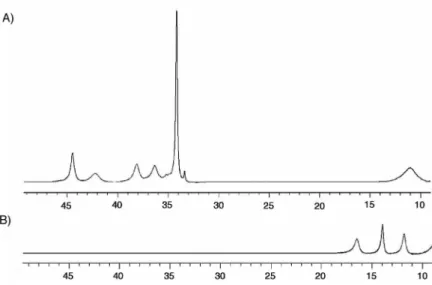
Figure 5.1. NRVS of polycrystalline 1 at ~10 K. 199
Figure 5.2. NRVS of frozen THF solutions of 1·16O
2 and 1·18O2 at ~10 K. 200
Figure 5.3. Resonance Raman spectra of 1·16O
2 and 1·18O2 in CH3CN. 201
Figure 5.4. NRVS of 4, 4/16O
2, and 4/18O2 at ~10 K. 202
Figure 5.5. Normal coordinate calculations for 1·O2 of modes involving vibrations of the
Fe2(O2) core. 204
Appendix A
Figure A.1. Zero-field Mössbauer spectra of the crude reaction material, 1, 4A, and 4B
at 80 K. 219
Figure A.2. IR spectra of FeIII/R
3TPA/OH–, 1, 4A, and 4B and their difference plots. 221
Figure A.3. Thermal ellipsoid (50%) diagram of the X-ray structure of 1. 222 Figure A.4. Thermal ellipsoid (50%) diagram of the X-ray structure of 4A. 225 Figure A.5. Absorption spectra of 1, 4A, and 4B in CH2Cl2. 227
Figure A.6. 1H NMR spectra of 1, 4A, and 4B in CD2Cl2. 228
Figure A.7. Thermal ellipsoid (50%) diagram of the X-ray structure of 4B. 229 Figure A.8. Absorption spectra of 1 in CH2Cl2, CH3CN, and CH2Cl2/MeOH. 231
Figure A.9. Optical changes of 1 in CH3CN at various temperatures and in the presence
of water. 232
LIST OF CHARTS
Page Chapter 1
Chart 1.1. The tethered substrates that were successfully oxidized by reaction with O2
when integrated into a diiron(II) complex. 41
Chart 1.2. Examples of crystallographically characterized [Fe2(syn N-donor)2]
complexes. 47
Chapter 2
Chart 2.1. The active site structure of sMMOHred and a proposed synthetic mimic. 63
Chart 2.2. The series of phenoxylpyridyl and phenoxylimine dinucleating ligands
synthesized. 84
Chapter 3
Chart 3.1. Syn N-donor ligands containing mixed N,O metal binding units. 113 Chapter 4
Chart 4.1. Depiction of the (peroxo)diiron(III) complexes that have been structurally
determined by X-ray crystallography. 167
Chapter 5
Chart 5.1. Possible geometries of the (µ-peroxo)diiron(III) unit. 193 Chart 5.2. Structural depiction of compounds 1·O2, 2·O2, 3·O2, and 4·O2. 194
Appendix A
LIST OF SCHEMES
Page Chapter 1
Scheme 1.1. Dioxyen reactivity of Hr, RNR, and sMMOH. 30 Scheme 1.2. Proposed mechanism for the reaction of [Fe2(µ-ArTolCO2)4(4-tBupy)2] with
dioxygen. 40
Scheme 1.3. Proposed reaction of O2 with a sterically-hindered PIM2– variant. 49
Chapter 2
Scheme 2.1. Synthesis of H2LVH,H and H2LH,H. 85
Scheme 2.2. Synthesis of H2LH,Ph, H2LMe,Ph, and H2LtBu,Ph. 86
Scheme 2.3. Synthesis of H2BIPSMe,Ph. 86
Scheme 2.4. Reaction of H2LH,H and H2LMe,Ph with iron(II). 92
Chapter 3
Scheme 3.1. Reaction of sMMOH with dioxygen. 112
Scheme 3.2. Synthesis of H2PIM. 126
Scheme 3.3. Reaction of 1 and 2 with dioxygen and silver perchlorate. 157 Chapter 4
Scheme 4.1. Proposed catalytic cycle of sMMOH. 167
Scheme 4.2. Reaction of 1a with dioxygen and protons. 169 Appendix A
Scheme A.1. Reaction of B leading to formation of high-valent diiron species. 213 Scheme A.2. Propose reaction pathway for the formation of 4A from 1. 232
LIST OF TABLES
Page Chapter 1
Table 1.1. Various ligands employed to prepare diiron protein model complexes. 32 Chapter 2
Table 2.1. X-ray crystallographic data and refinement for 1. 93 Chapter 3
Table 3.1. X-ray crystallographic data and refinement for 1, 2, and 3. 131
Table 3.2. Spectroscopic data for 1-8. 132
Table 3.3. Bond valence sum analysis for 3, 6, and 7. 139 Table 3.4. X-ray crystallographic data and refinement for 4, 5, and 6/7. 145 Chapter 4
Table 4.1. Spectroscopic data for 1a·O2, [1a⋅O2]H+, 2⋅O2, and [2⋅O2]H+. 178
Appendix A
Table A.1. Summary of spectroscopic data for FeIII/R3TPA/OH–, 1, 4A, and 4B. 220
Table A.2. X-ray crystallographic data and refinement for 1, 4A, and 4B. 223 Table A.3. Select structural parameters of 1 and two structurally related diiron TPA
complexes. 224
LIST OF ABBREVIATIONS 2-Ph2P(O)py 2-pyridyldiphenylphosphine oxide
2-Ph2Py 2-pyridyldiphenylphosphine
3-Fpy 3-fluoropyridine 4-tBupy 4-tert-butylpyridine 4-CNpy 4-cyanopyridine
5-Et3-TPA tris((5-ethylpyridyl)-2-methyl)amine
6Me2-BPP bis((6-methylpyridyl)-2-methyl)propionate amine
6-Me3TPA tris((6-methylpyridyl)-2-methyl)amine
[BIPSMe,Ph]2– anion of bis((phenylphenoxyl)iminephenyl)sulfone
[G3]CO2– third generation dendrimer appended terphenylcarboxylate
[LH,H]2– anion of bis(phenoxylpyridyl)diethynylbenzene
[LH,Ph]2– anion of bis((phenylphenoxyl)pyridyl)diethynylbenzene [LMe,Ph]2– anion of bis(phenyl(p-cresol)pyridyl)diethynylbenzene
[LtBu,Ph]2– anion of bis((o-phenyl-p-tert-butylphenoxyl)pyridyl)diethynylbenzene [LVH,H]2– anion of bis(phenoxylpyridyl)diethynylveratrole
Abs absorption
AMO alkene monooxygenase
APD avalanche photodiode detector Ar4-FPhCO2– 2,6-bis(p-fluorophenyl)benzoate ArtBuCO2– 2,6-bis(p-(tert-butyl)phenyl)benzoate ArTolCO2– 2,6-bis(p-tolyl)benzoate Asp aspartate bdptz 1,4-bis(2,2ʹ -dipyridylmethyl)phthalazine BEPEAN 2,7-bis(bis(2-(2-(5-ethyl)pyridyl)ethyl)aminomethyl)-1,8-napthyridine BIPhMe bis(1-methylimidazol-2-yl)phenylmethoxymethane
BMMs bacterial multi-component monooxygenases
BPEAN 2,7-bis(bis(2-(2-(5-methyl)pyridyl)ethyl)aminomethyl)-1,8-napthyridine BPMAN 2,7-bis(bis(2-pyridylmethyl)aminomethyl)-1,8-napthyridine
bpy 2,2ʹ -bipyridine
BVS bond valence sum
BXDK2– benzyl substituted variant of XDK2–
Cp cyclopentadienyl
CV cyclic voltammetry
DAFA2– 1,8-bis(dimethylaminomethylethynyl)-3,6-di(tert-butyl)fluorine-9-yl-acetate DFT density functional theory
DMF dimethylformamide
DMSO dimethyl sulfoxide DNA deoxyribonucleic acid
DPV differential pulse voltammetry EPR electron paramagnetic resonance
E1/2 average of anodic and cathodic redox potential
ET electron transfer
Et2BCQEBEt 1,2-bis(3-ethynyl-8-carboxylatequinoline)-4,5-diethynylbenzene methyl ester
EXAFS extended X-ray absorption fine structure
Fc ferrocene
Fc+ ferrocenium
FTIR fourier transform infrared
GC-MS gas chromatography-mass spectrometry
Glu glutamate
H[BArF4] bis(diethyl ether)hydronium tetrakis((3,5-trifluoromethyl)phenyl)borate
His histidine
HMDS hexamethyldisilazide
Hmv mixed-valent diiron(II,III) state of soluble methane monooxygenase
Hr hemerythrin
Im2DET bis(N-methylimidazole)diethynyltriptycene
iPrCO2– isobutyrate
IR infrared
k kinetic rate constant
Kcom comproportionation constant
Lut 2,6-lutidine
MArTolCO
22– 1,3-bis(aminomethyl)-4,6-diisopropylbenzene linked bis(terphenylcarboxylate)
Mc Methylococcus capsulatus
Mes 2,4,6-trimethylphenyl
metHr the oxidized inactive form of hemerythrin
1,3-diaminopropane
N-MeIm N-methylimidazole
NMR nuclear magnetic resonance
N,N-Bn2en N,N-dibenzylethylenediamine
NRVS nuclear resonance vibrational spectroscopy
N-tBuIm N-tert-butylimidazole
oxyHr dioxygen-bound form of hemerythrin
PDK4– anion of α,α-5,15-bis(α-N-(Kemp’s triacid imido)-o-tolyl)-2,8,12,18-tetraethyl-3,7,13,17-tetramethylporphyrin PIC2DET bis(picolinic methyl ester)diethynyltriptycene
PIM2– dibenzylether linked bis(3-(methylphenoxylimine)phenyl)sulfone
PH phenol hydroxylase
Ph4DBA2– dibenzofuran-4,6-bis(diphenylacetate)
PhCO2– benzoate
PhCyCO2– 1-phenylcyclohexylcarboxylate
Ph-bimp 2,6-bis(bis(2-(1-methyl-4,5-diphenylimidazolyl)methyl)aminomethyl)-4-methylphenolate
pKa acid dissociation constant, log scale
PXDK2– propyl substituted varient of XDK2–
py pyridine
R2 ribonucleotide reductase subunit containing the diiron active site R3TPA tris((3,5-dimethyl-4-methoxypyridyl)-2-methyl)amine
RNR ribonucleotide reductase
RR resonance Raman
RT room temperature
sMMO soluble methane monooxygenase
sMMOH soluble methane monooxygenase hydroxylase
sMMOHred the active form of soluble methane monooxygenase hydroxylase
sMMOHox the resting state of soluble methane monooxygenase hydroxylase
sMMOR soluble methane monooxygenase reductase
t-BuCH2CO2– 1-tert-butylacetate
t-BuCO2– pivalate
T4MoH toluene 4-monooxygenase hydroxylase
THF tetrahydrofuran
TMEDA N,N,Nʹ ,Nʹ -tetramethylethylenediamine
TP– tris(pyrazolyl)borate
TPA tris(pyridyl-2-methyl)amine TrpCO2– triptycenecarboxlate
UV-vis ultraviolet-visible
XDK2– anion of m-xylenediamine bis(Kemp’s triacid)imide
δ isomer shift
ΔEQ quadrupole splitting
LIST OF COMPOUND IDENTIFIERS Chapter 1
1 [FeIII2(µ-O)(µ-CH3CO2)2(TP)2]
2 [FeIII2(µ-OH)(µ-CH3CO2)2(TP)2]+
3 [FeII2(µ-HCO2)3(HCO2)(BIPhMe)2]
4 [FeIII2(µ-O)(µ-HCO2)2(HCO2)2(BIPhMe)2]
5 [FeIII2(µ-O)(MPDP)(bpy)2Cl2]
6 [FeIII2(µ-O)(MPDP)(BIPhMe)2Cl2]
7 [FeIII2(µ-O)(MPDP)(TP)2]
8 [FeIII2(µ-O)(XDK)(CH3OH)5(H2O)]2+
9 [FeII2(XDK)(µ-PhCyCO2)(PhCyCO2)(py)2]
10 [FeII2(µ-OH)(Ph4DBA)(TMEDA)2(CH3CN)]+
11 oxygenated species of [FeII2(µ-OH)(Ph4DBA)(TMEDA)2(CH3CN)]+
12 [FeII3(PDK)(Lut)(Br)2(HBr)]
13 [FeII2(µ-ArTolCO2)2(ArTolCO2)2(THF)2]
14A quadruply-bridged form of [FeII2(ArTolCO2)4(4-tBupy)2]
14B triply-bridged form of [FeII2(ArTolCO2)4(4-tBupy)2]
14C doubly-bridged form of [FeII2(ArTolCO2)4(4-tBupy)2]
15 [FeIIFeIII(ArTolCO2)4(4-tBupy)2]+
16 putative [FeIIIFeIV(µ-O)2] species
17 [FeIII2(µ-OH)2(µ-ArTolCO2)2(ArTolCO2)2(4-tBupy)2]
18 [FeII2(µ-ArTolCO2)2(ArTolCO2)2(N,N-Bn2en)2]
19 [FeIII2(µ-OH)2(µ-ArTolCO2)(ArTolCO2)3(N-Bnen)(N,N-Bn2en)]
20 [FeII2([G3]-CO2)4(4-CNpy)2]
21 [FeII2(µ-H2O)2(µ-ArTolCO2)2(ArTolCO2)2(4-tBupy)2]
22 [FeII2(µ-H2O)2(µ-Ar4-FPhCO2)2(Ar4-FPhCO2)2(THF)2(H2O)]
23 [FeII2(µ-ArTolCO2)4(4-CNpy)2]
24 [FeII2(µ-OH)(BPEAN)(SO3CF3)]2+
25 [FeII2(µ-OH)(BEPEAN)(SO3CF3)]2+
26 putative [FeIII2(OOH)] species
27 [FeII2(BPMAN)(µ-PhCyCO2)2]2+
28 [FeII2(µ-OH)(µ-ArTolCO2)(bdptz)(CH3CN)(SO3CF3)]+
29 [FeIII2(µ-O)(µ-ArTolCO2)(bdptz)(acetone)(SO3CF3)]2+
30 [FeII2(Et2BCQEBEt)(µ-ArTolCO2)3]+
32 [FeII2(LMe,Ph)2(THF)2]
33 [FeIII2(µ-O)(LMe,Ph)2]
34 [FeII2(µ-ArTolCO2)2(PIM)]
35 [FeII2(µ-Ph3CCO2)2(PIM)]
36 [FeIII2(µ-O)(ArTolCO2)2(PIM)]
37 [FeIII2(µ-OH)2(ArTolCO2)2(PIM)]
38 [FeIII4(µ-OH)6(PIM)2(ArTolCO2)2]
39 [FeII2(µ-RCO2)2(bulky PIM)]
40 [FeIII2(µ-OH)2(RCO2)2(L)(bulky PIM)]
41 [FeIII4(µ-OH)6(bulky PIM)2(RCO2)2]
Chapter 2 A 1-Benzyloxy-2-iodobenzene B 2-(2-Benzyloxyphenyl)-5-bromopyridine C 5-Bromo-2-(2-methoxyphenyl)pyridine D 2-Phenylanisole E 2-Bromo-6-phenylanisole F 5-Bromo-2-(2-methoxybiphenyl-3-yl)pyridine G 1-Benzyloxy-2,6-dibromo-4-methylbenzene H 1-Benzyloxy-2,6-dibromo-4-tert-butylbenzene I 1-Benzyloxy-2-bromo-4-methyl-6-phenylbenzene J 1-Benzyloxy-2-bromo-6-phenyl-4-tert-butylbenzene K 5-Bromo-2-(2-benzyloxy-5-methylbiphenyl-3-yl)pyridine L 5-Bromo-2-(2-benzyloxy-5-tert-butylbiphenyl-3-yl)pyridine M 2-(5-Methylbiphenyl-2-yloxy)tetrahydro-2H-pyran N 3-Bromo-5-methylbiphenyl-2-ol O 1-Benzyloxy-2,6-dibromo-4-methyl-benzene P 2-Benzyloxy-3-bromo-5-methylbenzaldehyde Q 2-Benzyloxy-5-methyl-3-phenylbenzaldehyde R 2-Hydroxy-5-methyl-3-phenylbenzaldehyde 1 [FeII2(LMe,Ph)2(THF)2]
2 [FeIII2(µ-O)(LMe,Ph)2]
B 2,2'-[(Oxybis(methylene))bis(5-methyl-[1,1'-biphenyl]-3',2-diyl)] bis(oxy)bis(tetrahydro-2H-pyran) C Bis(3-(2-hydroxy-5-methylphenyl)benzyl)ether
D Bis(3-(2-hydroxy-5-methylphenyl-3-carbaldehyde)benzyl)ether 1 [FeII2(PIM)(Ph3CCO2)2]
2 [FeII2(PIM)(ArTolCO2)2]
3 [FeIII2(µ-OH)2(ClO4)2(PIM)(ArTolCO2)Ag]
4 [FeIII2(µ-OH)(PIM)(Ph3CCO2)3]
5 [FeIII4(µ-OH)6(PIM)2(Ph3CCO2)2]
6 [FeIII2(µ-O)(PIM)(ArTolCO2)2]
7 [FeIII2(µ-OH)2(PIM)(ArTolCO2)2]
8 [FeIII4(µ-OH)6(PIM)2(ArTolCO2)2]
Chapter 4
1a [FeII2(N-EtHPTB)(Ph12CO2)]2+
1b [FeII2(N-EtHPTB)(Ph13CO2)]2+
1a·16O2 [FeIII2(µ-16O2)(N-EtHPTB)(Ph12CO2)]2+
1a·18O2 [FeIII2(µ-18O2)(N-EtHPTB)(Ph12CO2)]2+
1b·16O2 [FeIII2(µ-16O2)(N-EtHPTB)(Ph13CO2)]2+
1b·18O2 [FeIII2(µ-18O2)(N-EtHPTB)(Ph13CO2)]2+
[1a·16O2]H+ [FeIII2(µ-16O2)(N-EtHPTB)(Ph12CO2H)]3+
[1a·18O2]H+ [FeIII2(µ-18O2)(N-EtHPTB)(Ph12CO2H)]3+
[1b·16O2]H+ [FeIII2(µ-16O2)(N-EtHPTB)(Ph13CO2H)]3+
[1b·18O2]H+ [FeIII2(µ-18O2)(N-EtHPTB)(Ph13CO2H)]3+
2 [FeII2(N-EtHPTB)(C6F5CO2)]2+
2·16O2 [FeIII2(µ-16O2)(N-EtHPTB)(C6F5CO2)]2+
2·18O2 [FeIII2(µ-18O2)(N-EtHPTB)(C6F5CO2)]2+
[2·16O2]H+ [FeIII2(µ-16O2)(N-EtHPTB)(C6F5CO2H)]3+
[2·18O2]H+ [FeIII2(µ-18O2)(N-EtHPTB)(C6F5CO2H)]3+
Chapter 5
1 [FeII2(N-EtHPTB)(PhCO2)]2+
1·16O2 [FeIII2(µ-16O2)(N-EtHPTB)(PhCO2)]2+
1·18O2 [FeIII2(µ-18O2)(N-EtHPTB)(PhCO2)]2+
2·O2 [FeIII2(µ-O2)(N-EtHPTB)(OPh3)2]3+
4-Br [FeII(HB(iPrpz)3)Br]
4 [FeII(HB(iPrpz)3)(PhCH2CO2)]
4·16O2 [FeIII2(µ-16O2)(HB(iPrpz)3)2(PhCH2CO2)2]
4·18O2 [FeIII2(µ-18O2)(HB(iPrpz)3)2(PhCH2CO2)2]
Appendix A
A [FeIIIFeIV(µ-O)2(5-Et-TPA)2]3+
B [FeIII2(µ-O)(OH)(H2O)(R3TPA)2]3+
C [FeIII2(µ-O)(OOH)(R3TPA)2]3+
D [FeIV2(µ-O)(OH)(O)(R3TPA)2]3+
E [FeIIIFeIV(µ-O)(OH)(O)(R3TPA)2]2+
F [FeIIIFeIV(O)2(R3TPA)2]3+
G [FeIV2(O)2(R3TPA)2]4+
1 [FeIII2(µ-O)(µ-OH)(R3TPA)2]3+
2 [FeIII2(µ-O)(OH)(CH3CN)(R3TPA)2]3+
3 [FeIII2(µ-O)(µ-CH3CONH)(R3TPA)2]3+
4A [FeIII2(µ-O)(µ-CH3CO2)(R3TPA)2](ClO4)3·(CH3CN)3
Chapter 1
Rationally Designed Synthetic Mimics of Diiron Protein Active Sites:
A Chronological Perspective
1.1. Introduction
Understanding the role of metal ions in dictating protein structure and function is a central theme in bioinorganic research.1-4 In addition to the arsenal of modern physical techniques and instrumentation available for studies of biomolecules,5 synthetic modeling has been pursued as a complementary tool to interrogate complex biological systems.6 It provides a convenient simplification of elaborate macromolecules in a real, rather than a virtual (e.g. computational),7 platform. By reducing a metalloprotein to its functional core, it is possible to gain insight into whether the chemistry that occurs at the metal center is predominantly a result of the inorganic component alone or a consequence of the protein complex as a whole. Synthetic modeling also provides an opportunity to access chemistry that has not yet been achieved artificially using simple biological building blocks under ambient conditions. Some of the most remarkable transformations in nature, such as nitrogen fixation,8-11 water splitting,12 and hydrogen production,13 are performed by metalloenzymes. Reproducing such chemical reactivity in an efficient manner would not only revolutionize the chemical industry but also provide new sustainable sources of energy.14 Finally, the synthetic challenges of modeling chemistry will push the limits of current synthetic methodologies. Although natural product synthesis has been largely aided by an understanding of organic chemistry,15 inorganic synthesis is not governed by the same set of well-defined rules.16 Controlling the nuclearity and kinetic stability of metal clusters or components using organic ligands is still a significant challenge.
Small-molecule metalloprotein mimics are prepared using one of two design strategies.4 The biomimetic approach seeks to duplicate the active site structure as faithfully as possible, particularly in matching the identity and geometric arrangement of ligands in the primary
synthetic model shares some common features with those of the protein core, unrestricted by the type or position of ligands coordinated to the metal center. For modeling studies, the former is preferred over the latter because biomimetic complexes more accurately reproduce the characteristics of the biological center under investigation.
The following is a chronological account of the strategies and tactics employed in the Lippard group to prepare structural and functional mimics of diiron protein active sites. This work will describe the rational basis for ligand design and selection as well as the notable findings that resulted from studies of the various model systems. The reader is referred elsewhere for more general reviews of synthetic diiron modeling.17-22
1.2. Non-Heme Diiron Proteins
Non-heme carboxylate-bridged diiron proteins are involved in essential physiological processes.23,24 Although their roles vary, the first step in their respective reaction mechanisms involves binding and activation of dioxygen. In hemerythrin (Hr),25 the O2 molecule coordinates
to a single iron site and acquires two electrons and a proton to generate a (hydroperoxo)diiron(III) unit (Scheme 1.1, top). This process is reversible and is the basis for O2
transport in some marine invertebrates. Unlike hemerythrin, ribonucleotide reductase (RNR)26 and bacterial multi-component monooxygenases (BMMs)27 consume dioxygen to perform catalytic reactions (Scheme 1.1, middle and bottom, respectively). Activation of O2 by these
enzymes leads to formation of (peroxo)diiron(III) or di(µ-oxo)diiron(IV) complexes that are capable of oxidizing organic moieties.28-32 The high-valent diiron intermediate in the R2 subunit of RNR generates a tyrosyl radical that initiates the first step in DNA biosynthesis, whereas that in the hydroxylase component of the BMMs is responsible for inserting an oxygen atom into the
C–H bond of hydrocarbon substrates. The most well-studied member of the BMM family is soluble methane monooxygenase (sMMO),33 which is unique for its ability to hydroxylate methane to methanol. Recent studies have revealed that other classes of enzymes containing carboxylate-bridged diiron motifs also exist in biology.34-36
Scheme 1.1. Dioxygen reactivity of hemerythrin (Hr, top), ribonucleotide reductase (RNR, middle), and soluble methane monooxygenase hydroxylase (sMMOH, bottom). The active site structures of the proteins in various redox states are depicted.
These diiron proteins contain two iron atoms that are coordinated by imidazole and carboxylate residues, where at least one of the carboxylate groups bridges the two metal centers.23 The structural similarities between the active sites of this protein family indicate that
their functional differences are derived from variations in their Asp/Glu/His amino acid combinations and/or careful tuning of their tertiary and quaternary protein structures. Discerning
the factors that contribute to their mechanism of action is an important goal in synthetic model studies.19,20
1.3. Mononucleating N-Heterocyclic Ligands
The first successful attempt to prepare a mimic of the Hr core was achieved using tris(pyrazolyl)borate (TP–) (Table 1.1),37,38 a tripodal ligand commonly employed in inorganic coordination chemistry. Reaction of TP–, iron(III), and acetate led to the spontaneous self-assembly of [FeIII2(µ-O)(µ-CH3CO2)2(TP)2] (1), the structure of which closely matches that of
metHr, the inactive form of Hr.25 Reaction of 1 with an H+ donor led to formation of [FeIII2
(µ-OH)(µ-CH3CO2)2(TP)2]+ (2), a stable protonated analogue of 1.39 Spectroscopic and magnetic
measurements of the (µ-oxo)di(µ-carboxylato)diiron(III) and (µ-hydroxo)di(µ-carboxylato)diiron(III) compounds provided, for the first time, benchmarks for identifying such units in biology.40,41 When 1 was exposed to H218O, the 16O bridge was readily exchanged for 18O, indicating that a lack of exchange with water in the protein is due to inaccessibility of H
2O
to the diiron core rather than an intrinsic property of the [FeIII2O] unit. An undesirable feature of
1 and 2 is that the facial capping nature of the TP– ligands blocks having open sites for binding of O2. Furthermore, reduction of the diiron(III) species resulted in irreversible dissociation to
monoiron complexes.38
To access the asymmetric iron centers in Hr, the denticity of the capping ligand was reduced from three to two, using bis(1-methylimidazol-2-y1)phenylmethoxymethane (BIPhMe) (Table 1.1).42,43 Stirring iron(II) formate with BIPhMe in a methanol solution provided [FeII2
(µ-HCO2)3(HCO2)(BIPhMe)2] (3). X-ray crystallography revealed a dinuclear structure with both
five- and six-coordinate iron sites, in which each metal ion is bound by a BIPhMe ligand and bridged by three formate groups. A terminal acetate group completes the coordination sphere of
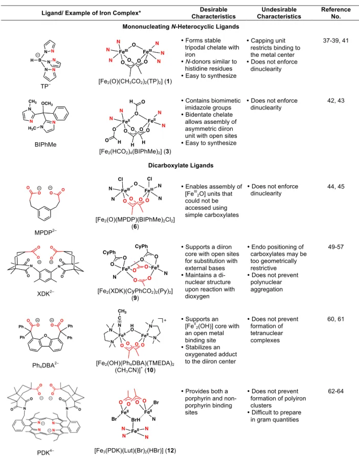
Table 1.1. Various Ligands Employed to Prepare Diiron Protein Model Complexes.
Ligand/ Example of Iron Complex* Characteristics Desirable Characteristics Undesirable Reference No. Mononucleating N-Heterocyclic Ligands
B N N H N N N N TP– FeIII FeIII O O O O N N N N N N O [Fe2(O)(CH3CO2)2(TP)2] (1) Forms stable tripodal chelate with iron N-donors similar to histidine residues Easy to synthesize Capping unit restricts binding to the metal center Does not enforce
dinuclearity 37-39, 41 OCH3 N N N N H3C CH3 BIPhMe FeII FeII O O O O H H N N O N N O O H O H
[Fe2(HCO2)4(BIPhMe)2] (3)
Contains biomimetic imidazole groups Bidentate chelate
allows assembly of asymmetric diiron unit with open sites Easy to synthesize
Does not enforce
dinuclearity 42, 43 Dicarboxylate Ligands O O O O MPDP2– FeIII FeIII O O O O Cl N N N N O Cl [Fe2(O)(MPDP)(BIPhMe)2Cl2] (6) Enables assembly of [FeIII 2O] units that could not be accessed using simple carboxylates
Does not enforce
dinuclearity 44, 45 N O O O O N O O O O XDK2– FeII FeII O O O O O O N O N O CyPh CyPh
[Fe2(XDK)(CyPhCO2)2(Py)2] (9)
Supports a diiron core with open sites for substitution with external bases Maintains a di-nuclear structure upon reaction with dioxygen Endo positioning of carboxylates may be too geometrically restrictive Does not prevent
polynuclear aggregation 49-57 O Ph Ph O O Ph Ph O O Ph4DBA2– FeII FeII H O O O O N N N N N O C CH3 +
[Fe2(OH)(Ph4DBA)(TMEDA)2 (CH3CN)]+ (10)
Supports an [FeII
2(OH)] core with an open metal binding site Stabilizes an
oxygenated adduct to the diiron center
Does not prevent formation of tetranuclear complexes 60, 61 FeII FeII BrH O O O O FeII N N N N Br Br N Provides both a porphyrin and non-porphyrin binding sites
Does not prevent formation of polyiron clusters
Difficult to prepare in gram quantities
Table 1.1. (Continued)
Ligand/ Example of Iron Complex* Characteristics Desirable Characteristics Undesirable Reference No. Terphenylcarboxylate Ligands O O ArTolCO 2– ArTol FeII FeII O O O ArTol ArTol O O O O O ArTol N N
[Fe2(ArTolCO2)4(4-tBuPy)2] (14A) Forms diiron complexes in the presence of Fe(II) salts and an appropriate base Stabilizes
high-valent iron species Easy to synthesize The steric encumbrance of the 2,6-aryl groups restricts access to the diiron core. Does not prevent formation of polyiron species 69-72, 74-90
Dinucleating Polynitrogen Ligands
N N N N N N N N BPEAN FeII FeII H O F3CO2SO N N N N N N N N 2+ [Fe2(OH)(BPEAN)(SO3CF3)]2+ (24) Contains a “masked carboxylate” to bridge two metal centers Stabilizes diiron species in multiple oxidation states Nitrogen-rich, rather than carboxylate-rich 95-98 N N N N N N bdptz FeII FeII H O F3CO2SO N N N N N N + O O ArTol N C CH3
[Fe2(OH)(bdptz)(ArTolCO2) (SO3CF3)(CH3CN)]+ (28)
Forms very stable bimetallic compounds Nitrogen-rich, rather than carboxylate-rich 99, 100
syn N-Donor Ligands
N N EtO O OEt O Et2BCQEBEt O FeII FeII O O O ArTol ArTol O O ArTol N N O O +
[Fe2(ArTolCO2)3(Et2BCQEBEt)]+ (30)
Enforces the syn stereochemistry of nitrogen donors relative to the Fe–Fe vector Accommodates binding of external carboxylates to the diiron core Neutral oxygen donors, rather than anionic Quinoline ester groups do not sufficiently stabilize the iron centers 101, 102-108 N N O O DAFA– N.A. Contains a bridging carboxylate unit in addition to two adjacent amine groups Not sufficiently pre-organized, could not metallate with iron No clear advantage over Et2BCQEBEt design 109
* The donor atoms highlighted in red in the iron complexes (second column) are derived from the donor groups, also shown in red, of the featured ligand (first column).
Table 1.1. (Continued)
Ligand/ Example of Iron Complex* Characteristics Desirable Characteristics Undesirable Reference No. Macrocyclic Ligands O O O O H N N H N H H N MArTolCO 22– N.A.
May help control the nuclearity of the resulting iron complex
Metallation of this ligand was not successful 113 O H3C O O CH3 N N S O O PIM2– O FeII FeII O O ArTol O ArTol N N O O
[Fe2(ArTolCO2)2(PIM)] (34)
Supports a carboxylate-bridged diiron(II) unit Maintains a
dinuclear core upon reaction with O2 Can be sterically
tuned without obstructing access to the metal binding pocket. Does not prevent formation of tetrairon species Phenolate and
imine donors are not biomimetic
114
* The donor atoms highlighted in red in the iron complexes (second column) are derived from the donor groups, also shown in red, of the featured ligand (first column).
the octahedral iron atom. Complex 3 reacts instantaneously with dioxygen to give [FeIII
2
(µ-O)(µ-HCO2)2(HCO2)2(BIPhMe)2] (4), with no detectable intermediates. Manometric measurements
indicated that 0.5 equiv of O2 was consumed per diiron(II) complex, suggesting that 4 is formed
through a tetranuclear dioxygen species. Unfortunately, the behavior of 3 toward dioxygen is markedly different than that of deoxyHr, which binds O2 reversibly (Scheme 1.1, top).25
1.4. Dicarboxylate Ligands
To increase the structural integrity of the model compounds, several dicarboxylate ligands were explored as dinucleating platforms. The first of these is m-phenylenedipropionate (MPDP2-, Table 1.1),44,45 which was selected because the distance between its β-methylene
(5) (bpy = 2,2’-bipyridine) and [FeIII2(µ-O)(MPDP)(BIPhMe)2Cl2] (6, Table 1.1), complexes that
could not be obtained from self-assembly with simple monocarboxylate anions. The cyclic voltammogram of [FeIII2(µ-O)(MPDP)(TP)2] (7), an analogue of 1, showed that mononuclear
[FeII(TP)2] and [FeIII(TP)2]+ species were generated upon electrochemical reduction and
oxidation, respectively. These results demonstrated that linking the carboxylate groups using MPDP2– does not impart additional stability to the diiron models.
To obtain a more stable bridging framework, m-xylenediamine bis(Kemp’s triacid)imide (H2XDK, Table 1.1), a compound developed from studies of molecular recognition in organic
chemistry,46,47 was employed. The rigid conformation of the carboxylate units in XDK2– provides a well-defined cleft for assembly of homo- and heterodimetallic complexes.48-51 A diiron(III) compound, [FeIII2(µ-O)(XDK)(CH3OH)5(H2O)](NO3)2 (8), was readily prepared from iron(III)
nitrate and XDK2–.52 Kinetic studies of the substitution of 2,2´-bipyridine or N-methylimidazole with the coordinated solvents in 8 suggested that anion binding and exchange at the active site of hemerythrin proceed with rates similar to those exhibited by small-molecules; thus, the protein scaffold of Hr does not alter the intrinsic rate of terminal ligand exchange at the diiron center. Preparation of a series of pseudohalide bound diiron(III) compounds, with the formula [FeIII2
(µ-O)(XDK)(bpy)2(X)2] (where X = NCS–, NCSe–, or N3–), allowed detailed studies of the
spectroscopic signatures of molecules with terminal ligation to carboxylate-bridged diiron units.53 A notable discovery is that the appearance of only one asymmetric 15NNN– stretch is not sufficient to discount the possibility of a terminally coordinated azide group, for isotopically shifted peaks may occur too close to one another in energy to be resolved.
The XDK2– ligand also supports carboxylate-bridged diiron(II) units with the general composition [FeII2(XDK)(µ-RCO2)(RCO2)(N-donor)2], where R = t-Bu- (pivalate), PhCy-
(1-phenylcyclohexylcarboxylate), Ph- (benzoate), iPr- (isobutyrate), or tBuCH2-
(1-tert-butylacetate); N-donor = py (pyridine), 3-Fpy (3-fluoropyridine), N-MeIm (N-methylimidazole), or N-tBuIm (N-tert-butylimidazole).54-56 More sterically-protecting XDK2– variants, containing either propyl (PXDK2–) or benzyl (BXDK2–) substituents in place of the methyl groups on the Kemp’s triacid moiety, were also successfully employed to assemble similar diiron(II) compounds. The asymmetric bridging mode of the ancillary carboxylate to the diiron(II) core is determined by both steric and electronic factors.56 X-ray structural studies suggested that greater steric repulsion, between XDK2– and the external carboxylate, and more basic N-donors favor a
syn,syn-bidentate bridging mode of the ancillary carboxylate rather than a syn,anti-monodentate
one. For complexes that have sufficiently bulky groups, such as [FeII2
(XDK)(µ-PhCyCO2)(PhCyCO2)(py)2] (9, Table 1.1), exposure to O2 led to formation of stable peroxo
adducts at low temperature.56 Although these (peroxo)diiron(III) species could not be crystallized for X-ray diffraction analysis, resonance Raman measurements indicate that the dioxygen molecule is bound in a µ-1,2 fashion. Reactivity studies of the [FeIII2(O2)] units revealed that they
are nucleophilic,57 rather than electrophilic like the oxygenated intermediates in the BMMs.58,59 When warmed above -65°C, the [FeIII2(O2)] species rapidly decayed, initiating a radical chain
pathway that oxidized solvents with weak to intermediate C–H bond strengths. Despite having the same ligand stoichiometry as that of the (peroxo)diiron(III) species in sMMOH (Hperoxo) and
related enzymes,33 the synthetic analogues do not reproduce the substrate scope, product selectivity, or reaction mechanism of the diiron monooxygenases. It is possible that the doubly-bridging XDK2– ligand is too rigid in the synthetic models to achieve an Hperoxo-like structure, a
The search for a ligand that is sufficiently pre-organized yet structurally flexible led to examination of other dicarboxylate motifs. One potential candidate was the compound dibenzofuran-4,6-bis(diphenylacetate) (Ph4DBA2-, Table 1.1).60,61 Similar to XDK2–, Ph4DBA2–
has two orthogonal carboxylate groups that can support an {FeIII2O}4+ core. The Ph4DBA2–
ligand, however, has more conformational freedom because the C–C bond between its carboxylate groups and the dibenzofuran linker can rotate freely. The compound [FeII2
(µ-OH)(Ph4DBA)(TMEDA)2(CH3CN)]+ (10, Table 1.1), where TMEDA =
N,N,N´,N´-tetramethylethylenediamine, was prepared from Ph4DBA2–, triethylamine, TMEDA, and iron(II)
triflate. The structure of 10 is unique because it is the first synthetic complex to reproduce the (µ-hydroxo)di(µ-carboxylato)diiron(II) core of deoxyHr25 with an open coordination site for binding of a terminal ligand. When 10 was treated with dioxygen at -78 °C in the presence of 3 equiv of
N-MeIm in CH2Cl2 or in neat EtCN, a red-orange species (11) appeared that decayed after ~10
min. The UV-vis, Mössbauer, resonance Raman, and EXAFS spectra of the transient intermediate closely match those of oxyHr (Scheme 1.1, top), strongly suggesting that 11 contains a (hydroperoxo)(µ-oxo)diiron(III) unit. Unfortunately, oxygenation of 10 is irreversible and led to decomposition to a tetrairon(III) cluster.
A fourth dicarboxylate ligand, α,α-5,15-bis(α-N-(Kemp’s triacid imido)-o-tolyl)-2,8,12,18-tetraethyl-3,7,13,17-tetramethylporphyrin (H4PDK, Table 1.1), was prepared by
replacing the m-xylenediamine linker of XDK2– with a porphyrin unit.62 The construct was designed so that activation of O2 within a trimetallic cavity would offer the possibility of
supplying additional electrons to the carboxylate-bridged diiron centers, much like the reductase component of sMMO.33 A triiron(II) compound, [FeII3(PDK)(Lut)(Br)2(HBr)] (12, where Lut =
When iron(II) chloride was used instead of iron(II) bromide in the preparation, a mixed-valent heptairon chloride cluster was isolated.64 Due to the complicated nature of the iron complexes of PDK4–, no further studies were pursued using this ligand.
The more pre-organized dicarboxylate ligands XDK2– and Ph4DBA2– impart enhanced
stability to their respective diiron complexes and allow detection of O2 adducts at low
temperature. A common problem with the dicarboxylate ligands, however, is that they do not prevent aggregation of the diiron compounds into oligo- and polynuclear clusters,65 an undesired reaction that is detrimental to the synthesis of accurate models. Furthermore, the conformationally rigid ligand framework may be a liability in terms of accessing oxygenated diiron species that could further react with external substrates.
1.5. Terphenylcarboxylate Ligands
To reduce the geometric constraints of the ligand platform, bulky terphenylcarboxylates were employed. Unlike simple benzoates that form polynuclear clusters with iron, 66-68 the sterically-hindered 2,6-bis(p-tolyl)benzoate (ArTolCO2–, Table 1.1) and
2,6-bis(p-fluorophenyl)benzoate (Ar4-FPhCO2–) self-assemble into discrete diiron compounds in the
presence of iron salts and an appropriate base.69,70 The first iron complex synthesized in this series is [FeII2(µ-ArTolCO2)2(ArTolCO2)2(THF)2] (13), which adopts a windmill structure with two syn,syn-bridging carboxylates and two bidentate terminal carboxylates in the solid-state. The
coordinated THF molecules in 13 can be readily substituted with N-donors to afford the corresponding [FeII2(ArTolCO2)4(N-donor)2] complex. Use of 4-tert-butylpyridine (4-tBuPy) as
solution was demonstrated by variable temperature NMR spectroscopy.70 Oxygenation of 14A at -78°C resulted in irreversible formation of a deep green intermediate that decayed to a [FeIII2
(µ-OH)2(µ-ArTolCO2)2(ArTolCO2)2(4-tBupy)2] (17) product.71,72 The di(µ-hydroxo)diiron(III) unit of
17 closely resembles that of the oxidized core of sMMOH.73 Characterization of the green intermediate revealed two mixed-valent complexes, a diiron(II,III) (15) and a diiron(III,IV) (16) species, that are present in equal amounts. EPR and magnetic Mössbauer measurements indicated that 15 and 16 have spin states of 9/2 and 1/2, respectively. The assignment of 15 as a diiron(II,III) compound was confirmed by comparing its spectroscopic properties to those of a crystallographically characterized [FeIIFeIII(ArTolCO2)4(4-tBupy)2] complex.74,75 As shown in the
proposed mechanism in Scheme 1.2, reaction of 14A with dioxygen proceeds through a carboxylate shift, in which either one or two of the bridging ArTolCO2– groups rearrange to adopt
terminal positions, providing an open site for O2. Binding of dioxygen to 14B or 14C affords a
(µ-peroxo)diiron(III) or a di(µ-oxo)diiron(IV) intermediate that further reacts with 14A, giving an equal mixture of 15 and 16. This reaction is noteworthy because it is the first synthetic example in which treatment of a diiron(II) precursor with O2 resulted in formation of an iron(IV)
species, a process that parallels the chemistry of several non-heme diiron enzymes.23
Although the steric hindrance provided by the terphenycarboxylates could suppress undesired reactions involving bond-making processes, it could not eliminate deleterious intermolecular electron transfer (ET) reactions.71,72 To prevent the paddlewheel diiron(II) complex from quenching the oxygenated intermediates, bidentate ancillary ligands were used to favor the assembly of windmill rather than paddlewheel species. The doubly-bridged [FeII2
(µ-ArTolCO2)2(ArTolCO2)2(N,N-Bn2en)2] (18) complex, where N,N-Bn2en =
Scheme 1.2. A proposed mechanism for the reaction of [FeII2(µ-ArTolCO2)4(4-tBupy)2] (14A)
with O2.71,72
reacted with dioxygen, but instead of producing a green intermediate, N-dealkylation occurred affording [FeIII
2(µ-OH)2(µ-ArTolCO2)(ArTolCO2)3(N-Bnen)(N,N-Bn2en)] (19) (where Bnen =
N-benzylethylenediamine) and benzaldehyde. Isotopic labeling with 18O2 demonstrated that the
oxygen atom in benzaldehyde was derived from dioxygen. When a non-coordinating N,N-Bn2en
analogue, such as N,N-dibenzylpropylamine, was treated with O2 in the presence of either
mononuclear or dinuclear iron(II) complexes, the yield of benzaldehyde was significantly reduced. Detailed mechanistic studies suggested that oxidative N-dealkylation of 18 involved single electron transfer, proton abstraction, and rearrangement.78,79 The fortuitous discovery that high-valent diiron terphenylcarboxylate complexes could be intercepted by tethered substrates enabled studies of the reactivity of oxygenated intermediates toward organic moieties. Attachment of benzyl,80,81 ethyl,81 ethynyl,82 phenoxyl,83 phosphido,80,84 or sulfido84,85 units to an amine or pyridine ligand afforded a series of tethered substrates that could be easily incorporated
complex to O2, the reaction products were analyzed by gas chromatography-mass spectrometry
(GC-MS). Chart 1.1 depicts the substrates that were successfully oxidized by this method. When 2-pyridyldiphenylphosphine (2-Ph2Ppy) was employed in excess, catalytic oxidation to
2-pyridyldiphenylphosphine oxide (2-Ph2P(O)py) was observed.80,84 Because external
non-coordinating substrates cannot be oxidized by [Fe2(ArTolCO2)4(THF)2]/O2, these results suggest
that intramolecular reactions are preferred over intermolecular ones in the terphenylcarboxylate diiron complexes. This behavior is most likely due to reaction of the active diiron oxidant with solvent or adventitious reductants before it can be intercepted by the desired external substrates.
Chart 1.1. The tethered substrates that were successfully oxidized with O2 when integrated into
a diiron(II) terphenylcarboxylate complex. The starting substrate is depicted on top and the product isolated after oxygenation is shown directly below.
One strategy to prevent premature deactivation of the oxygenated diiron intermediate was to attach dendritic groups to the terphenylcarboxylate ligand that would shield the diiron core from participating in intermolecular ET reactions.86 The complex [FeII2([G-3]CO2)4(4-CNpy)2]
(20) (where [G-3]CO2– = third-generation dendrimer-appended terphenylcarboxylate and
4-CNpy = 4-cyanopyridine) was synthesized in a manner analogous to that for the simpler diiron(II) compounds. Treatment of 20 with dioxygen resulted in formation of a diiron(II,III)
N NH2 Ph Ph H O H2N H O OMe OMe N P Ph Ph N P Ph Ph O N S N S O N S O N S O O N Ph N Ph OH N N O N S N S O NH2 S Ph H O
intermediate that was postulated to have a superoxo moiety. This colored intermediate is capable of oxidizing dihydroanthracene, albeit in only modest yields.
The influence of water on the structure and reactivity of the diiron(II) terphenylcarboxylate compounds was also examined. Treatment of 14A with excess water afforded a diaqua-bridged diiron(II) species, [FeII2(µ-H2O)2(µ-ArTolCO2)2(ArTolCO2)2(4-tBupy)2]
(21), in which the terminal carboxylates exhibit strong hydrogen-bonding interactions with the bridging waters.87 In addition to having a diaqua bridge, a third H2O molecule occupies a
terminal position in [FeII2(µ-H2O)2(µ-Ar4-FPhCO2)(Ar4-FPhCO2)3(THF)2(H2O)] (22).88 When a
large excess of water was added to 22 complete dissolution occurred, giving the fully aquated [FeII(H2O)2]2+ cation. These observations suggest that the accessibility of water within diiron
enzyme active sites may be a control element for achieving different functions using a common structural motif. Furthermore, the presence of excess amounts of water may be destructive to the integrity of the carboxylate-bridged diiron core. Stopped-flow kinetic studies using [FeII2
(µ-ArTolCO2)4(4-CNpy)2] (23) showed that it reacts with H2O ~1000 times faster than with O2 and
that aquated 23 reacts with O2 ~10 times faster than anhydrous 23.89,90 Coordination of water to
23 most likely induces rearrangement from a paddlewheel to a windmill complex, facilitating more rapid binding of dioxygen to the diiron centers.
The iron chemistry of a variety of other monocarboxylate ligands was evaluated. When the steric bulk of the terphenylcarboxylate was increased using 2,6-bis(p-(tert-butyl)phenyl)benzoate (ArtBuCO2–), reaction with iron(II) salts afforded a tetrairon cluster.91
When the steric demand of the carboxylate was reduced using 2-biphenylcarboxylate, an assortment of di-, tri-, and tetranuclear species was obtained.92 Replacing the


![Figure 2.4. Ortep thermal ellipsoid (50%) diagram of the X-ray crystal structure of [Fe 2 (L Me,Ph ) 2 (THF) 3 ] (1) with a partial numbering scheme](https://thumb-eu.123doks.com/thumbv2/123doknet/13886901.447142/94.918.251.667.131.539/figure-thermal-ellipsoid-diagram-crystal-structure-partial-numbering.webp)


![Figure 2.10. A stick figure representation of the low-resolution X-ray crystal structure of [Fe 2 (µ- (µ-O)(L Me,Ph ) 2 ] (2) in two different views](https://thumb-eu.123doks.com/thumbv2/123doknet/13886901.447142/100.918.227.693.350.729/figure-stick-figure-representation-resolution-crystal-structure-different.webp)


