HAL Id: hal-00527276
https://hal.archives-ouvertes.fr/hal-00527276
Submitted on 18 Oct 2010
HAL is a multi-disciplinary open access
archive for the deposit and dissemination of
sci-entific research documents, whether they are
pub-lished or not. The documents may come from
teaching and research institutions in France or
abroad, or from public or private research centers.
L’archive ouverte pluridisciplinaire HAL, est
destinée au dépôt et à la diffusion de documents
scientifiques de niveau recherche, publiés ou non,
émanant des établissements d’enseignement et de
recherche français ou étrangers, des laboratoires
publics ou privés.
Specific Inhibition of Chemiluminescent Activity by
Pathogenic Vibrios in Hemocytes of Two Marine
Bivalves: Pecten maximus and Crassostrea gigas
Christophe Lambert, Jean-Louis Nicolas
To cite this version:
Christophe Lambert, Jean-Louis Nicolas.
Specific Inhibition of Chemiluminescent Activity by
Pathogenic Vibrios in Hemocytes of Two Marine Bivalves: Pecten maximus and Crassostrea gigas.
Journal of Invertebrate Pathology, Elsevier, 1998, �10.1006/jipa.1997.4707�. �hal-00527276�
Specific Inhibition of Chemiluminescent Activity by
Pathogenic Vibrios in Hemocytes of Two Marine Bivalves:
Pecten maximus
and
Crassostrea gigas
C. Lambert and J. L. Nicolas
Laboratoire de Physiologie des Invertébrés, Direction des ressources vivantes, Aquaculture, Centre de Brest, IFREMER, B.P.70, 29280 Plouzané, France
Hemocytes from two adult bivalves, Pecten maximus and Crassostrea gigas, were exposed to 12 different bacterial strains including 2 Alteromonas spp. (U1 and T413), 2 type strains of vibrios (V. anguillarum ATCC 19264 and V. alginolyticus ATCC 17749), 1 vibrio (S322) pathogenic to C. gigas larvae, 2 vibrios (V110 and V117) virulent to Ostrea edulis larvae, and 5 different strains of a same Vibrio sp. (group A496) isolated from moribund P. maximus larvae. After 1.5 h contact with bacteria, zymosan particles were added to the hemocytes and the chemiluminescent (CL) activity of the cells was measured over 6 h. Analysis of CL activity after bacterial inoculation indicated that most of the strains could initiate the respiratory burst. However, the intensity of CL was not related to the virulence of the bacteria. In contrast the CL activity after stimulation by zymosan was modulated by the previous exposure of bacteria. This second CL response may depend on the virulent characteristics of the bacteria. As evidence, the strain S322 completely inhibited the CL activity of oyster hemocytes, whereas in the scallop hemocytes the CL activity was only moderately repressed. Inversely, the strainA496 was very effective in disrupting the CL activity in scallop hemocytes and reduced the CL activity to 30% in oyster hemocytes. V. anguillarum completely inhibited scallop and oyster hemocytes, whereas the strain U1 decreased the CL activity only by 20%. Finally, the measurement of CL activity allowed to partially elucidate the mechanism of infection as well as to determine some characteristics of bacterial virulence.
Key Words: Pecten maximus; Crassostrea gigas; bacteria; chemiluminescence; hemocytes; respiratory burst; Vibrio sp.
INTRODUCTION
Hemocytes can be considered as a primary line of defence in molluscs (Volety and Chu, 1995), especially by their ability to phagocytose. When membranes are stimulated by foreign particles (parasites, bacteria. . .), hemocytes initiate a metabolic pathway (‘‘respiratory burst,’’ RB) together with the production of numerous cytotoxic reactive oxygen intermediates (ROIs), hydroxyl radical (·OH), hydrogen peroxide (H2O2), singlet oxygen (1O2 ), superoxide anion (O2 2). These excited molecules produce a single photon during the
relaxation to ground state. The reactive oxygen intermediates are also able to oxidize present and nontoxic molecules, such as luminol and luciferin (chemiluminescent process). This process amplifies the photon emissions up to a level that can be detected by a liquid scintillation counter (Wishkovsky, 1988).
Increased interest in the chemiluminescence (CL) technique or ROIs production has encouraged further study in hemocyte activity. This has been supported by recent works on marine bivalve molluscs including
Mytilus edulis (Pipe, 1992), Patinopecten yessoensis (Nakamura et al., 1985), Crassostrea virginica (Larson et al., 1989), and specially in Pecten maximus (Le Gall et al., 1991), Ostrea edulis, and Crassostrea gigas (Bachère et al., 1991). However, this test is not always adapted to investigate immune response in all bivalves. Thus the
clam, Ruditapes decussatus, and the cockle, Cerastoderma edule (Lopez et al., 1994) did not generate any detectable CL activity during phagocytosis of inert particles, such as zymosan.
The CL activity of bivalve hemocytes have been measured for different purposes such as the effect of contaminants (Pipe and Coles, 1995) and host–pathogen interaction. For the last point, only the parasites including Rickettsia (Le Gall et al., 1991), Perkinsus marinus (Anderson et al., 1992; Volety and Chu, 1995), and Bonamia ostrea (Mourton et al., 1992, Chagot et al., 1992) have been investigated up to now. If different experiments were reported about the interaction between bacteria and bivalve hemocytes (Rodrick and Ulrich, 1984; Alvarez et al., 1995) it was not quantified by the measurement of CL activity.
The measurement of CL response to study the pathogenic activity of different bacterial strains is more relevant than observation by light microscopy because of its swiftness and provision of quantitative data. In fish
for instance, authors showed that some pathogenic bacteria were able to stimulate CL activity and generate reactive oxygen intermediates: Aeromonas hydrophila with tilapia hemocytes (Oreochromis aureus; Leung et al., 1995), V. anguillarum with sea bass hemocytes (Morone saxatilis; Stave et al., 1985) and with trout hemocytes (Oncorhynchus mykiss; Sakai et al., 1991).
The present work had a double objective: to examine the ability of bacterial strains from C. gigas, O.
edulis, and P. maximus larval culture, to trigger the CL activity of hemocytes, and to investigate their detrimental
effect on hemocytes by measuring the CL response at a second stimulation with zymosan.
MATERIALS AND METHODS
Bivalves
Two species of bivalve were used, the scallop P. maximus and the Pacific oyster C. gigas. Twenty adult scallops, 3 or 4 years old, and 20 adult oysters were collected in the bay of Brest (Brittany). Scallops were stored at a temperature of 15°C in a 450-liter sea-water tank equipped with sand filter and air lift system. They were fed just close to satiation on a drip issue of four microalgae mixed including Isochrysis affinis galbana (1 to 2.6 3
109 cells/day/animal), Pavlova lutheri (1 to 2.6 x 10⁹ cells/day/animal), Chaetoceros calcitrans (0.5 to 1.3 x 10⁹ cells/day/animal), Skeletonema costatum (0.3 to 0.5 x 10⁹ cells/day/animal).
They were maintained, for at least 2 weeks, in constant conditions of temperature and feeding before sampling of hemocytes. For C. gigas, the same conditions were applied, except the temperature (18°C) and the diet (Isochrysis affinis galbana: 0.7 to 1.1 x 10⁹ cells/day/animal; Pa. lutheri: 0.7 to 1.1 x 10⁹ cells/day/ animal;
Ch. calcitrans: 0.4 to 0.5 x 10⁹ cells/day/ animal; S. costatum: 0.2 to 0.3 x 10⁹ cells/day/animal).
Hemolymph Sampling
Either two, three, or four individuals were first placed on ice and all the solutions for dilution were previously sterilised and cooled. Hemolymph was withdrawn from the pericardial cavity, through the adductor muscle, using a sterile needle, fitted to a 1-ml syringe. Volumes ranging from 1 to 3 ml (P. maximus) and 0.5 to 1 ml (C. gigas) per individual were usually obtained, after repeated withdrawals if necessary, and pooled. Hemolymph was rapidly twofold diluted in modified anti-aggregant alsever solution (MAS; Bachère et al., 1988). Hemocytes, in a 50-µl sample of the hemolymph–MAS mixture, were counted using a Malassez cell. Concentrations in the mixture generally ranged from 2 to 4 x 10⁶ hemocytes per milliliter for individual scallops and 0.35 to 1.5 x 10⁶ for individual oysters.
Only the samples with 1 x 10⁶ hemocytes/ml for Pecten and 0.5 x 10⁶ for Crassostrea or more (in the hemolymph/MAS 1/1 mixture) were kept. Hemocyte numbers below these were not sufficient after dilution of MAS (final concentration of MAS: 2.5%; Bachère et al., 1991).
Inoculated Bacteria
Twelve strains were used (cf. Table 1). Five strains (A365, A496, A060, A601, A700) of the same vibrio species, assessed by DNA/DNA hybridization (unpublished results) were pathogenic for P. maximus larvae. They were isolated over 5 years during episodic mortalities in a hatchery and previously described by Nicolas et al. (1996). Two Vibrio sp. (V117, V110) were isolated from moribund O. edulis larvae and one Vibrio sp. (S322) from moribund C. gigas larvae. Two Alteromonas (U1, T413) were found in dominant bacterial flora of healthy P. maximus larval culture. All these strains were affiliated to these different genera after sequencing of 16S rDNA and phylogenetic analysis. They are available from our laboratory. Two type pathogenic strains of vibrios were used as reference bacteria: V. anguillarum ATCC 19264 and V. alginolyticus ATCC 17749. Bacterial strains were cultured in Zobell 2216E liquid medium (Oppenheimer and Zobell, 1952) at 18°C without shaking for 48 hr (end of the exponential phase). The bacterial cells were collected by centrifugation (5000g, 10 min), washed twice in sterile sea water (SSW), and resuspended in SSW. Concentration was checked by outside diameter measurement at 540 nm. Correlation between direct counts and optical density had previously been established.
Hemocytes were inoculated with appropriate bacterial dilutions in 50 µl, in a ratio determined by preliminary experiments with a series of bacterial concentrations including 5, 20, 50, 100, 200 cells/hemocyte, with the strain A496. This strain was chosen because it exhibited the most regular virulence of the strains specifically pathogenic to scallop larvae. For oyster hemocytes the same ratio was used and gave similar results as to those obtained with scallop hemocytes, including complete inhibition and gradation of response with different bacteria.
Chemiluminescence chemicals
Luminol solution was freshly prepared by 100-fold dilution inSSWof a stock solution: 5-amino-2,3-dihydro- 1,4-phthalazinedione, 1021 M; dimethyl-sulfoxide 1 M; stored at -20°C. For the zymosan particles solution, 40 mg of zymosan A (Sigma) was suspended in 10 ml SSW, boiled for 30 min, washed twice, and then resuspended in SSW, counted, and stored in aliquots at -20°C.
CL Assays
This protocol was adapted from Bachère et al. (1991). The hemolymph/MAS solution was carefully vortexed and distributed into plastic scintillation vials (type pico prias vial, 6000192, Packard), at 105 hemocytes for scallop and 5 x10⁴ for oyster per vial. This solution was made up to 2 ml with luminol solution (200 µl) and SSW. MAS was adjusted to a final concentration of 2.5% if necessary.
Generation of chemiluminescence was then measured with a liquid scintillation counter (Tri-Carb 2000 CA, Packard) in ‘‘single photon count’’mode. The counter was set on repeated sequential counts. Each vial was counted for 30 sec. For the 60 vials, the counting lasted 43 min and 12 countings (cycles) were performed during 8 hr. The results were expressed in counts per minute (cpm). In a previous experiment with scallop hemocytes, the measure, performed at 12-min interval, indicated that the peak of activity was achieved 45 min after addition of zymosan in agreement with the data of Le Gall et al. (1991). According to Lopez et al. (1996), and Larson et
al. (1989), the oyster (C. gigas and C. virginica) hemocytes reacted faster and the maximum of activity occurred
between 20 and 30 min after addition of zymosan. However, the ratio between control sample and any sample with bacteria remained similar over two cycles at least after the peak. Finally, the measurement at every 43 min allowed to conciliate technical constraints and to accurately follow the ratios of CL activity.
The CL response of hemocytes stimulated by zymosan particles was tested at two different temperatures, 17 and 26°C, in order to determine the effect of temperature.
The measurement started with a delay of 30 min after the distribution of hemocytes into the vials, in order to allow hemocyte fixation to the vial surfaces.
Bacterial Stimulation
The CL base activity was recorded during a first cycle (cycle 1) and then the 12 bacterial strains were inoculated in four replicates, using a single volume (50 µl) and gentle mixing (pipeting). Every vial was inoculated just after its CL base measure. Three sets of four vials were not inoculated with bacteria and received the same volume of SSW and luminol. One set without hemocytes was used as the ‘‘zero level’’ sample; one set with hemocytes nonstimulated by bacteria was used as the ‘‘base level’’ sample and a second set with nonstimulated hemocytes was used as the ‘‘control’’ in the second part of the experimentation (zymosan
stimulation). After inoculation of bacteria, the CL activity was recorded over two cycles (cycles 2 and 3) (cf. Table 2).
The CL activity of the hemocytes stimulated by bacteria was compared with that of the base level hemocytes, 43 min after inoculation. Results were expressed in a ‘‘CL activity multiplying coefficient’’ (Mc) calculated by the ratio
Mc = CLba/CLb,
with CLba as activity of hemocytes stimulated by bacteria and CLb as activity of the base level hemocytes. This coefficient expressed the stimulation of RB by bacteria.
In order to determine if bacteria alone could generate CL activity, the 12 bacteria were added separately to the vials containing luminol, SSW, and MAS but without hemocytes.
Zymosan Stimulation
After 1.5 hr of contact with bacteria, the solution of zymosan particles was added at the concentration of 80 particles/hemocyte, optimal concentration previously determined by Bachère et al. (1991), in all sets except the zero level and the base level samples. The CL activity was then recorded for nine consecutive cycles (cf. table 2).
The CL activity of hemocytes, previously in contact with bacteria and stimulated by zymosan, was compared to that of the control sample, 43 min after stimulation. Results were expressed in a ‘‘residual CL activity coefficient’’ (Ra) calculated by the ratio
Ra = CLba - CLb/CLc - CLb,
with CLba as activity of hemocytes stimulated by zymosan after 1.5 hr contact with bacteria; CLb as activity of the base level hemocytes; and CLc as activity of the control hemocytes. This coefficient expressed the inhibitory effect of bacteria on CL activity of hemocytes stimulated by zymosan particles. Significant differences between CL responses of treated groups were determined using a multifactor analysis of variance procedure with
alpha = 0.01 or 0.05. All experiments were performed four times with oyster hemocytes and five times with
scallop hemocytes.
RESULTS
Preliminary Experiments
The increase of the bacterial concentration from 5 to 200 bacterial cells per hemocyte showed progressive inhibition of the hemocytes CL activity (Fig. 1) compared with the noninoculated hemocytes. Significant, but not complete inhibition occurred at 100 cells/ hemocyte. All the following CL experiments were made with this concentration. At temperatures of 17 and 26°C, CL activity of C. gigas hemocytes was not strongly modified (Fig. 2a).
FIG. 1. Effect of bacteria/hemocyte ratio (strain A496) on CL activity (cpm) of P. maximus hemocytes after activation by zymosan particles
(1, bacterial inoculation; 2, zymosan addition).
Conversely, the CL activity of scallop hemocytes decreased dramatically by 75% when temperatures were increased from 17 to 26°C (Fig. 2b). For this study, temperatures during CL activity recording were stabilised at 18/20°C for scallop hemocytes and 22/24°C for oyster hemocytes. These temperatures were more compatible with technical constraints than 17 and 26°C and allowed to obtain high levels of CL response.
Activation of Chemiluminescence by Bacterial Inoculation (Figs. 3 and 4)
The CL activity generated by bacteria (without hemocytes) was weak or nonexistent, except for T 413 and U1. High CL activity observed after 43 min with T413 and U1 alone (5 x 10⁴ to 8 x 10⁴ cpm) was similar to the activity detected with oyster (9 to 20 x 104 cpm) or scallop (3 to 9 x 10⁴ cpm) hemocytes.
For P. maximus hemocytes, most of strains induced a significant (n = 5, alpha = 0.05) or highly significant (n = 5, alpha = 0.01) increase of CL activity in comparison with the ‘‘base level’’ sample, except the
V. anguillarum and, tacking into account the self activity of bacteria, T 413 and U1. The ‘‘multiplying
coefficient’’ (Mc) values were ranged between 1.56 and 5.26. However, the CL response may vary within the test samples. During five observations one of the most variable responses occurred with A 601 and ranged from 2.02 to 6.09. According to the CL response, bacteria could be clustered into three groups (Fig. 3): poorly active strains (U1 to T413), fairly active (V117 to A060), and highly active (S322), but the statistical tests did not validate these three groups.
For oyster hemocytes as for scallop hemocytes the analysis of results does not concern the strains U1 and T413. The strains A365, A060, A601, A700, V110 induced a significant (n = 4, alpha = 0.05) or highly significant (n = 4, alpha = 0.01) increase of CLactivity in comparison with the control samples. Mc values were in the range of 1.30 to 2.64. For strains A496, V117, S322, and V. alginolyticus, global statistical analysis on the four tests rejected a significant difference with the control sample. However, their capacity to induce CL activity was real as they all triggered significant RB at least once. It was quite difficult to separate the different strains according to their CL responses (Fig. 4) because there was no significant difference between the nine strains. V.
anguillarum is quite remarkable because it did not trigger the RB in either of the hemocyte types. On the
FIG. 2. (a) Variation of CL activity (cpm) of C. gigas hemocytes after activation by zymosan particles, according on temperature (1,
zymosan addition). (b) Variation of CL activity (cpm) of P. maximus hemocytes after activation by zymosan particles, according on temperature (1, zymosan addition).
FIG. 3. Mean (n = 5, 1SD) multiplying coefficient (Mc) of P. maximus hemocytes after exposure to different bacterial strains. (Significance versus base: not significant (NS); *alpha = 0.05; **alpha = 0.01.)
Bacterial Inhibition of Zymosan-Stimulated CL Activity (Figs. 5 and 6)
For P. maximus hemocytes, the contact between hemocytes and most of the strains tended to disturb hemocyte responses to the zymosan stimulant. The responses were not significant between the control and strains U1 and T413 over the five tests. Another group composed of strains A365, V110, and V117 moderately but significantly inhibited the CL activity (about 40% of a decrease). A third group including S322, A060, V.
alginolyticus, A601, and A700, reduced the hemocyte response to 60%. V. anguillarum and A496 almost
completely disrupted the CL activity (Fig. 5). For C. gigas hemocytes, similar inhibition of the CL activity occurred. The strain U1 appeared again as a neutral bacterium without significant difference in comparison to the control. In contrast the ‘‘neutrality’’ of T413 was not confirmed in this case.
Finally bacteria could be clustered into three groups, the first with U1, the second with partially inhibiting bacteria (from 53 to 70% decrease in the response) and the third composed of V. anguillarum, V.
alginolyticus, and S322 which led to complete inhibition (Fig. 6).
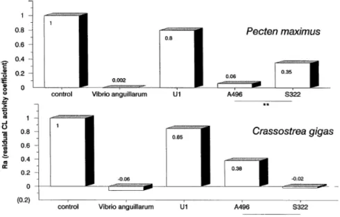
The strains A496 and S322 acted differently according to the bivalve hemocyte species (Fig. 7). In scallop hemocytes, the Ra with A496 was very weak and significantly different (n = 5, alpha = 0.01) to the one with S322. Inversely in oyster hemocytes, S322 provoked complete inhibition of CL activity whereas A496 left almost 40% CL activity (n = 4, alpha = 0.05). The strains A365, A060, A601, and A700, belonging to the same pathogenic Vibrio sp. as A496, were much less active than the latter strain in scallop hemocytes, especially the strain A365. Besides they produced an equivalent inhibitory effect in oyster hemocytes. The strain V117 isolated from the flat oyster (O. edulis) larvae exhibited a greater inhibitory effect in oyster hemocytes than in scallop hemocytes. This effect was less evident with isolate V110. V. alginolyticus behaved exactly like S322 with both hemocyte types.
FIG. 4. Mean (n = 4, 1SD) multiplying coefficient (Mc) of C. gigas hemocytes after exposure to different bacterial strains. (Significance versus base: not significant (NS); *alpha = 0.05, **alpha = 0.01.)
FIG. 5. Mean (n = 5, 1SD) residual CL activity (expressed by the coefficient Ra) of P. maximus hemocytes stimulated by zymosan particles, 2 hr after inoculation of bacteria. (Significance versus control: not significant (NS); *alpha = 0.05; **alpha = 0.01; ***alpha = 0.001.)
DISCUSSION
Although Wishkovsky (1988) reported that temperature control was one of the two main problems of liquid scintillation counter utilization for CL activity measure, many authors eluded the problem, specifying that experiments were performed at room temperature. Preliminary experiments have shown that the CL activity of C.
gigas hemocytes was not strongly modified by temperatures of 17 and 26°C (also observed by Alvarez et al.,
1989) whereas the CL activity of scallop hemocytes decreased by 75% when the temperature was increased from 17 to 26°C. It is therefore extremely important to limit temperature variation during the recording of CL activity.
In addition, the physiological state of the animals is an important source of variability in hemocyte CL response. For example, it has been observed that hemocyte concentration can double between fasted animals and well fed ones. Pipe and Coles (1995) testing M. edulis observed a considerable interanimal variability in total hemocyte counts from the same site and Bachère et al. (1991) observed with O. edulis and C. gigas that the CL response varied considerably in intensity and also in swiftness, in 10 oysters. These facts confirm the importance of working with adult bivalves of similar origins, stored in identical and stable conditions.
Validity of this method could be limited by the fact that the bacterial strains studied are pathogenic to bivalve larvae and not to adults from which hemocytes were taken, for obvious technical reasons. In addition, it was well known that infection of adult bivalves by bacteria was rare (Nottage and Birkbeck, 1990; Getchell, 1991). If the adult hemocytes were susceptible to bacteria it may be because the experimental conditions, especially bacterial rate (100 cells per hemocyte) was high and probably not encountered in natural conditions.
FIG. 6. Mean (n = 4, 1SD) residual CL activity of C. gigas (expressed by the coefficient Ra) hemocytes stimulated by zymosan particles, 2 hr after inoculation of bacteria. (Significance versus control: not significant (NS); *alpha = 0.05; **alpha = 0.01; ***alpha = 0.001.)
On the other hand, means of defence in bivalve larvae are little known. Nevertheless, phagocytosis capacity of C. virginica larvae hemocytes was shown by electron microscopy observations (Elston and Leibovitz, 1980) and for M. edulis larvae hemocytes by observation of fluorescent bacteria internalization (Dyrynda et al., 1995). Finally, since the larval hemocytes are functional, they may have some similarity with adult hemocytes, especially their susceptibility to bacteria.
The measure of CL activity, as already underlined by several authors, represented an excellent tool to investigate the pathogen–hemocyte interaction. The experiments demonstrated the capacity of current bacteria to elicit RB, except V. anguillarum, T413, and U1 when taking their self CL activity into account. During the experiment to determine the optimal concentration of A496 (Fig. 1), this strain did not trigger the RB. This was probably due to the high basal level of hemocytes, about three times superior to the usual level (3 to 9 x 10⁴ cpm), which may have masked the effect of bacteria.
This capacity would then confirm the antimicrobial action of this mechanism. The level of generated CL did not permit differentiation of pathogenic from nonpathogenic bacteria. Some authors reported that virulent strains induced a higher CL activity than non or less virulent strains (Leung et al., 1995; Stave et al., 1985), but Leung et al. (1995) also observed the contrary. As for the parasite Bonamia (Mourton et al., 1992; Chagot et al., 1992) and Rickettsia (Le Gall et al., 1991) the ability of V. anguillarum to penetrate into hemocytes without triggering RB may permit it to survive in phagocyte cells. In this way S322, which generated the strongest response in both hemocyte types, may be quickly destroyed and may be considered as less virulent than V.
anguillarum.
According to Mauel (1984) two mechanisms are involved in parasite survival in hemocytes: resistance to hydrolytic activity of lysosomes and capacity to inhibit the RB or inactive toxic compounds generated by macrophage oxidative metabolism. So the measure of CL activity alone, cannot account for bacterial inhibition of other destructive mechanisms. Nevertheless the primary inhibition of CL activity may confer a certain
advantage to V. anguillarum. Further experiments would be necessary to determine if enzymes, such as catalase, glutathione peroxidase, dismutase superoxide, and alkaline phosphatase are involved in the primary inhibition of CL activity.
The inhibitory effect of bacteria on the CL response on the addition of zymosan gave more convergent results. They were reproducible and allowed the validation of this method for the examination of some aspects of bacterial virulence. The inhibition of zymosan CL response by bacteria may correspond to two events. After some bacteria were internalized by phagocytosis, enzyme(s) and toxin(s), actively released by intact bacterial cells or passively by lysed bacteria, may block the RB. Second, these toxins or others may lead to hemocyte death. The saturation of nonself receptor by bacteria which should prevent the secondary phagocytosis of zymosan seems least probable. Indeed, that supposes noninternalization of bacteria without previous damage to the hemocyte. Such a phenomenon to our knowledge has not yet been reported.
FIG. 7. Comparison of mean residual CL activity (expressed by the coefficient Ra) of P. maximus and C. gigas hemocytes, with four
bacterial strains: Vibrio anguillarum, U1, A496, S322. (Significance A496 versus S322: n = 4 (oyster), n = 5 (scallop); *alpha = 0.05; **alpha = 0.01.)
CL test assisted the differentiation of two interesting Vibrio strains, S322 and A496. Higher inhibition of CL activity by these two strains was observed with hemocytes of their habitual host. Thus this method may be useful to determine the virulence of bacteria toward different organisms. Nevertheless previous experimental exposures of larvae to bacteria (Nicolas et al., 1996) rejected for some cases a direct relation between neutralization of hemocytes and bacterial virulence.
C. gigas larvae exposed to the strain V110, which was as active as T413, A365, or A700 toward oyster
hemocytes, died some days later but not with neutral strain T413 or the strains pathogenic to scallop larvae. The strains A700 and A601, which were moderately active against scallop hemocytes, also provoked an outbreak of disease in scallop larvae, 2 or 3 days after exposure, in the same manner as A496. Inversely, isolates A060 and A365 failed to infect scallop larvae under the same conditions despite moderate activity against P. maximus hemocytes. In reports not yet published, the same strain of V. anguillarum which presented high activity in hemocytes was unable to kill scallop larvae whereas virulence of strains A700, A601, and A496 was confirmed again.
he absence of a direct relationship between cytotoxicity to hemocyte and ability to kill larvae for these bacteria confirm that different means of defence other than hemocytes alone may exist. Inversely bacteria may invade larval tissues without being destroyed by hemocytes which constitute only an ultimate barrier against pathogens.
To conclude, this work places further emphasises on the specific action of some vibrios toward their host hemocytes. In other words, some strains studied were able to ‘‘improve their offensive capacity’’ against target species. These Vibrio capable of neutralizing the hemocytes quickly may possess efficient cytotoxin(s). The CL activity would be an interesting biological test to facilitate their isolation.
In an other way, the use of CL test may possibly be better applied to the determination of the acquisition of immunity during larval development. If phagocytosis capacity was already shown by microscopy observations, the moment when hemocytes start to synthesize cytotoxic reactive oxygen intermediates, with RB pathway, is unknown. The CL test, used with constituent cells from disaggregated larvae (technical method given by Odintsova and Khomento (1991), with Japanese scallop larvae) would be an interesting approach.
ACKNOWLEDGMENTS
We thank E. Bachère, E. Lane, and D. Prieur for critical reading of the manuscript and for useful discussions.
REFERENCES
Alvarez, M. R., Friedl, F. E., Johnson, J. S., and Hinsch, G. W. 1989. Factors affecting in vitro phagocytosis by oyster hemocytes. J.
Invertebr. Pathol. 54, 233–241.
Anderson, R. S., Paynter, K. T., and Burreson, E. M. 1992. Increased reactive oxygen intermediate production by hemocytes withdrawn from
Crassostrea virginica infected with Perkinsus marinus. Biol. Bull. 183, 463–481.
Bachère, E., Chagot, D., and Grizel, H. 1988. Separation of Crassostrea gigas hemocytes by density gradient centrifugation and counterflow centrifugal elutriation. Dev. Comp. Immunol. 12, 549–559.
Bachère, E., Hervio, D., and Mialhe, E. 1991. Luminol-dependent chemiluminescence by hemocytes of two marine bivalves, Ostrea edulis and Crassostrea gigas. Dis. Aquat. Org. 11, 173–180.
Chagot, D., Boulo, V., Hervio, D., Miahle, E., Mourton, C., and Grizel, H. 1992. Interactions between Bonamia ostreae (Protozoa: Ascetospora) and hemocytes of Ostrea edulis and Crassostrea gigas (Mollusca: Bivalvia): Entry mechanisms. J. Invertebr. Pathol.
59, 241– 249.
Dyrynda, E. A., Pipe, R. K., and Ratcliffe, N. A. 1995. Host defence mechanisms in marine invertebrate larvae. Fish Shellfish Immunol. 5, 569–580.
Elston, R., and Leibovitz, L. 1980. Pathogenesis of experimental vibriosis in larval American oysters, Crassostrea virginica. Can. J. Fish.
Aquat. Sci. 37, 964–978.
Getchell, R. G. 1991. Diseases and parasites of scallops. In ‘‘Scallops: Biology, Ecology and Aquaculture. Developments in Aquaculture and Fisheries Science’’ (S. E. Shumway, Ed.), Vol. 21, pp. 471–494. Elsevier, New York.
Larson, K. G., Robertson, B. S., and Hetrick, F. M. 1989. Effect of environmental pollutants on the chemiluminescence of hemocytes of the American oyster Crassostrea virginica. Dis. Aquat. Org., 131–136.
Le Gall, G., Bachère, E., and Mialhe, E. 1991. Chemiluminescence analysis of the activity of Pecten maximus hemocytes stimulated with zymosan and host-specific rickettsiales-like organisms. Dis. Aquat. Org. 11, 181–186.
Leung, K.Y., Low, K.W., Lam, T. J., and Sin,Y. M. 1995. Interaction of the fish pathogen Aeromonas hydrophyla with tilapia, Oreochromis
aureus (Steindachner) phagocytes. J. Fish Dis. 18, 435–447.
Lopez, C., Villalba, A., and Bache`re, E. 1994. Absence of generation of oxygen radicals coupled with phagocytosis by the hemocytes of the Clam, Ruditapes decussatus (Mollusca: bivalvia). J. Invertebr. Pathol. 64, 188–192.
Mauel, J. 1984. Mechanisms of survival of protozoan parasites in mononuclear phagocytes. Parasitol. 88, 579–592.
Mourton, C., Boulo, V., Chagot, D., Hervio, D., Miahle, E., and Grizel, H. 1992. Interactions between Bonamia ostreae (Protozoa: Ascetospora) and hemocytes of Ostrea edulis and Crassostrea gigas (Mollusca: Bivalvia): In vitro system establishment. J.
Invertebr. Pathol. 59, 235–240.
Nakamura, M., Mori, K. M., Inooka, S., and Nomura, T. 1985. In vitro production of hydrogen peroxide by the amoebocytes of the scallop,
Patinopecten yessoensis (Jay). Dev. Comp. Immunol. 407–417.
Nicolas, J. L., Corre, S., Gauthier, G., Robert, R., and Ansquer, D. 1996. Bacterial problems associated with scallop (Pecten maximus) larval culture. Dis. Aquat. Org. 27(1), 67–76.
Nottage, A. S., and Birkbeck, T. H. 1990. Interactions between different strains of Vibrio alginolyticus and hemolymph fractions from adult
Mytilus edulis. J. Invertebr. Pathol. 56, 15–19.
Odintsova, N. A., and Khomenko, A. V. 1991. Primary cell culture from embryos of the Japanese scallop Mizuchopecten yessoensis.
Cytotechnology 6, 49–54.
Oppenheimer, C. H., and Zobell, C. E. 1952. The growth and viability of sixty three species of marine bacteria as influenced by hydrostatic pressure. J. Mar. Res. 11, 10–18.
Pipe, R. K. 1992. Generation of reactive oxygen metabolites by the hemocytes of the mussel Mytilus edulis. Dev. Comp. Immunol. 16, 11– 122.
Pipe, R. K., and Coles, J. A. 1995. Environmental contaminants influencing immune function in marine bivalve molluscs. Fish Shellfish
Immunology 5, 581–595.
Rodrick, G. E., and Ulrich, S. A. 1984. Microscopical studies on the hemocytes of bivalves and their phagocytic interaction with selected bacteria. Helgoländer Meeresuntersuchungen, Helgoländer Meeresunters. 37, 167–176.
Sakai, M., Konishi, M., Atsuta, S., and Kobayashi, M. 1991. The Chemiluminescent response of leukocytes from the anterior kidney of rainbow trout Oncorhynchus mykiss vaccinated with Vibrio anguillarum, Streptococcus sp. or Renibacterium salmoninarum.
Nippon Suisan Gakkaishi 57(2), 237–241.
Stave, J.W., Roberson, B. S., and Hetrick, F. M. 1985. Chemiluminescent responses of striped bass, Morone saxatilis (Walbaum), phagocytes to Vibrio spp. J. Fish Dis. 8, 479–483.
Volety, A. K., and Chu, E. 1995. Suppression of chemiluminescence of eastern oyster (Crassostrea virginica) hemocytes by the protozoan parasite Perkinsus marinus. Dev. Comp. Immunol. 19(2), 135–142.
Wishkovsky, A. 1988. Chemiluminescence: an advanced tool for measuring phagocytosis. In ‘‘Disease Processes in Marine Bivalve Molluscs.’’ (W. S. Fisher, Ed.). American Fisheries Society Special Publication 18, pp. 292–298.