HAL Id: hal-02354341
https://hal.archives-ouvertes.fr/hal-02354341
Submitted on 7 Nov 2019
HAL is a multi-disciplinary open access
archive for the deposit and dissemination of sci-entific research documents, whether they are pub-lished or not. The documents may come from teaching and research institutions in France or abroad, or from public or private research centers.
L’archive ouverte pluridisciplinaire HAL, est destinée au dépôt et à la diffusion de documents scientifiques de niveau recherche, publiés ou non, émanant des établissements d’enseignement et de recherche français ou étrangers, des laboratoires publics ou privés.
(Porifera)
Alexander Ereskovsky, Daria B. Tokina, Stephen Baghdiguian, Emilie Le Goff,
Andrey Lavrov
To cite this version:
Alexander Ereskovsky, Daria B. Tokina, Stephen Baghdiguian, Emilie Le Goff, Andrey Lavrov. Transdifferentiation and mesenchymal-to-epithelial transition during regeneration in Demospongiae (Porifera). Journal of Experimental Zoology Part B: Molecular and Developmental Evolution, Wiley, 2020, 334 (1), pp.37-58. �10.1002/jez.b.22919�. �hal-02354341�
For Peer Review
Transdifferentiation and mesenchymal-to-epithelial transition during regeneration in Demospongiae (Porifera)
Journal: JEZ Part B: Molecular and Developmental Evolution Manuscript ID JEZ-B-2019-06-0045.R1
Wiley - Manuscript type: Research Article Date Submitted by the
Author: n/a
Complete List of Authors: Ereskovsky, Alexander; CNRS, Aix-Marseille University, Institut Méditerranéen de Biodiversité et d’Ecologie marine et continentale (IMBE); Saint-Petersburg State University, Biological Faculty,
depertment of Embryology; Koltzov Institute of Developmental Biology of Russian Academy of Sciences
Tokina, Daria; CNRS, Aix-Marseille University, Institut Méditerranéen de Biodiversité et d’Ecologie marine et continentale (IMBE)
Saidov , Danial; Dept. of Invertebrate Zoology, Biological Faculty, Lomonosov Moscow State University, 119234, Leninskie gory 1-12, Moscow
Baghdiguian, Stephen ; ISEM, Univ Montpellier, CNRS, EPHE, IRD Le Goff , Emilie; ISEM, Univ Montpellier, CNRS, EPHE, IRD
Lavrov, Andrey; Lomonosov Moscow State University, Faculty of Biology, Invertebrate Zoology
For Peer Review
1 Title1 Transdifferentiation and mesenchymal-to-epithelial transition during regeneration
2 in Demospongiae (Porifera) 3
4 Alexander V. Ereskovsky1,2,3*, Daria B. Tokina1, Danial M. Saidov4, Stephen
5 Baghdiguian5, Emilie Le Goff5, Andrey I. Lavrov2,6
6
7 1 Institut Méditerranéen de Biodiversité et d’Ecologie marine et continentale (IMBE),
8 Aix Marseille University, CNRS, IRD, Avignon University, Station Marine d’Endoume, Rue 9 de la Batterie des Lions, 13007, Marseille, France
10 2 Dept. Embryology, Faculty of Biology, Saint-Petersburg State University,
11 Universitetskaya emb. 7/9, 199034, Saint-Petersburg, Russia
12 3 Koltzov Institute of Developmental Biology of Russian Academy of Sciences, Russia,
13 Moscow
14 4 Dept. of Invertebrate Zoology, Biological Faculty, Lomonosov Moscow State
15 University, 119234, Leninskie gory 1-12, Moscow, Russia
16 5 ISEM, Univ Montpellier, CNRS, EPHE, IRD, Montpellier, France.
17 6 Pertsov White Sea Biological Station, Biological Faculty, Lomonosov Moscow State
18 University, 119234, Leninskie gory 1-12, Moscow, Russia 19
20 Text figures – 16
21 Abbreviated title: Demosponges regeneration 22
23 *Correspondence to: Alexander V. Ereskovsky, Institut Méditerranéen de Biodiversité 24 et d’Ecologie marine et continentale (IMBE), Aix Marseille University, CNRS, IRD, Avignon 25 University, Station Marine d’Endoume, Rue de la Batterie des Lions, 13007, Marseille, 26 France, Tel. +33 04 91 04 16 21 ; Fax : e-mail alexander.ereskovsky@imbe.fr,
27 This work was supported by grants of Russian Foundation for Basic Research n° 16-04-28 00084, and the Russian Science Foundation n° 17-14-01089 (histological and ultrastructural 29 studies).
1Funding information. This work was supported by grants of Russian Foundation for Basic Research n° 16-04-00084, the Russian Science Foundation n° 17-14-01089 (histological and ultrastructural studies). This work also is a contribution to Labex OT-Med (n° ANR-11-LABX-0061) and has received funding from Excellence Initiative of Aix-Marseille University_A*MIDEX, a French ''Investissements d'Avenir'' program for travel expenses.
3 4 5 6 7 8 9 10 11 12 13 14 15 16 17 18 19 20 21 22 23 24 25 26 27 28 29 30 31 32 33 34 35 36 37 38 39 40 41 42 43 44 45 46 47 48 49 50 51 52 53 54 55 56 57 58 59 60
For Peer Review
3031 Abstract
32 Origin and early evolution of regeneration mechanisms remain among the most pressing 33 questions in animal regeneration biology. Porifera have exceptional regenerative capacities 34 and, as early Metazoan lineage, are a promising model for studing evolutionary aspects of 35 regeneration. Here, we focus on reparative regeneration of the body wall in the Mediterranean 36 demosponge Aplysina cavernicola. The epithelialization of the wound surface is completed 37 within two days, and the wound is completely healed within two weeks. The regeneration is 38 accompanied with the formation of a mass of undifferentiated cells (blastema), which consists 39 of archaeocytes, dedifferentiated choanocytes, anucleated amoebocytes, and differentiated 40 spherulous cells. The main mechanisms of A. cavernicola regeneration are cell 41 dedifferentiation with active migration and subsequent redifferentiation or transdifferentiation 42 of polypotent cells through the mesenchymal-to-epithelial transformation. The main cell 43 sources of the regeneration are archaeocytes and choanocytes. At early stages of the 44 regeneration, the blastema almost devoid of cell proliferation, but after 24 hpo and up to 72 45 hpo numerous DNA-synthesizing cells appear there. In contrast to intact tissues, where vast 46 majority of DNA-synthesizing cells are choanocytes, all EdU-labeled cells in the blastema are 47 mesohyl cells. Intact tissues, distant from the wound, retains intact level of cell proliferation 48 during whole regeneration process. For the first time, the apoptosis was studied during the 49 regeneration of sponges. Two waves of apoptosis were detected during A. cavernicola 50 regeneration: the first wave at 6-12 hpo and the second wave at 48-72 hpo.
51
52 Keywords: demosponges, regeneration, mesenchymal-to-epithelial transformation, blastema, 53 apoptosis, transdifferentiation.
54
55 Highlights
56 1) Regeneration in the demosponge Aplysina cavernicola is accompanied with the formation 57 of a mass of undifferentiated cells (blastema).
58 2) The main mechanisms of A. cavernicola regeneration are cell dedifferentiation with active 59 migration and subsequent redifferentiation or transdifferentiation of polypotent cells - 60 archaeocytes and choanocytes - through the mesenchymal-to-epithelial transformation.
61 3) Apoptosis during regeneration of A. cavernicola participate in damaged cells elimination 62 and associated with the extensive ejection of spherulous cells from wound area.
63 3 4 5 6 7 8 9 10 11 12 13 14 15 16 17 18 19 20 21 22 23 24 25 26 27 28 29 30 31 32 33 34 35 36 37 38 39 40 41 42 43 44 45 46 47 48 49 50 51 52 53 54 55 56 57 58 59 60
For Peer Review
64 Introduction65 In spite of big interest in various problems concerning origin and early steps of 66 evolution in animal regeneration, morphogenesis, cell turnover etc., up to now there are 67 surprisingly small number of the studies, dealing with ultrastructural, morphogenetic, cell and 68 genetic aspects of sponge reparative regeneration.
69 Phylum Porifera consists of four classes: syncytial Hexactinellida, and cellular 70 Calcarea, Homoscleromorpha and Demospongiae. The last class is the largest and includes 71 about 80% of living sponges. Studies of regeneration in sponges have begun on demosponges 72 (Cavolini, 1785; Vaillant, 1869; Weltner, 1893). However, there are only few papers, 73 concerning ultrastructural description of the morphogenesis and cell behavior in reparative 74 regeneration of sponges. Moreover, three of them dealing with the regeneration of specific 75 “organs” after amputation (oscular diaphragm in Hippospongia communis (Thiney, 1972), 76 oscular tube in Ephydatia fluviatilis (Sukhodolskaya, 1973), and papillae in Polymastia 77 (Boury-Esnault, 1976)). Reparative regeneration of the body wall is described only in seven 78 species. Regeneration in Spongilla lacustris (Brondsted, 1953), Halichondria panicea 79 (Korotkova & Nikitin, 1969), Geodia barretti (Hofmann et al., 2003) and Halisarca caerulea 80 (Alexander et al., 2015) was studied only with light microscopy. In the case of H. caerulea, 81 the light microscopy studies were supplemented with the investigations of the cell 82 proliferation during the regenerative processes (Alexander et al., 2015). Reparative 83 regeneration in Chondrosia reniformis was investigated with light and scanning electron 84 microscopy (SEM) (Pozzolini et al., 2019). Finally, SEM studies supplemented with time-laps 85 recordings were done for regeneration in Hymeniacidon heliophila (Coutinho et al., 2017). 86 At the same time, our complex detailed investigations of reparative regeneration, done 87 with TEM, SEM, epifluorescent and light microscopy, immunocytochemistry and time-laps 88 recordings, in homoscleromorphs (Ereskovsky et al., 2015), calcareous sponges (Ereskovsky 89 et al., 2017; Lavrov et al., 2018) and demosponges (Borisenko et al., 2015) show a high 90 diversity of morphogenesis, cell mechanisms, and cell turnover, accompanying these 91 processes.
92 Thus, having comprehensive data about mechanisms of the regeneration for only one 93 species from the huge and very diverse class Demospongiae, we cannot make any 94 generalizations regarding the mechanisms of regeneration of this class of Porifera.
95 Representatives of the genus Aplysina are widely distributed in subtropical and tropical 96 coastal waters (Bergquist & Cook, 2002). They are considered proper models in chemical 97 ecology and microbiology (Azevedo et al., 2008; Betancourt-Lozano et al., 1998; Thoms et
3 4 5 6 7 8 9 10 11 12 13 14 15 16 17 18 19 20 21 22 23 24 25 26 27 28 29 30 31 32 33 34 35 36 37 38 39 40 41 42 43 44 45 46 47 48 49 50 51 52 53 54 55 56 57 58 59 60
For Peer Review
98 al., 2004, 2006). Aplysina cavernicola is a popular and promising model for various 99 researches, dealing with the sponge cell composition (Vacelet, 1966, 1967, 1970, 1971, 1975; 100 Vacelet & Gallissian, 1978), bacterial symbionts (Vacelet, 1975; Fiedrich et al., 1999; 101 Hentschel et al., 2001; Thoms et al., 2003), three-dimensional skeletal scaffolds (Vacelet, 102 1971a,b; Garrobe et al. 1973; Ehrlich et al. 2010a, b), biochemistry and secondary metabolites 103 (D'Ambrosio et al., 1982, 1983; Ciminiello et al., 1997; Reverter et al., 2016), and temporal 104 variability of secondary metabolism (Reverter et al., 2016). Life cycle researches showed that 105 A. cavernicola is an oviparous sponge whose reproductive period lasts barely one month 106 (Gallissian & Vacelet, 1976; Reverter et al., 2016).
107 The present study was aimed at investigation of the reparative regeneration in 108 Mediterranean demosponge Aplysina cavernicola (Vacelet, 1959). The wide range of methods 109 allow us to make a comprehensive analysis of mechanisms, which contribute to the 110 regeneration in this species, including cell behavior and migrations, morphogenetic process, 111 cell proliferation and apoptosis.
112
113 Materials and methods 114
115 Sampling
116 Aplysina cavernicola (Vacelet, 1959) (Demospongiae, Verongida) is a perennial 117 sciaphilous species inhabiting coralligenous formations or the entrance of submarine caves 118 generally between 8 to 60 m in the Mediterranean Sea (Figure 1). It presents a typical 119 yellowish color. For regeneration experiments A. cavernicola specimens were collected by 120 SCUBA diving in September-November 2017, July-August and October 2018 and March 121 2019 near Maire Island, Marseille (43.2096° N; 5.3353° E) at a depth of 12 - 15 m. Collected 122 sponges were maintained in a 100 l laboratory aquarium with running natural seawater at a 123 temperature of 15-16°C for 36 hours for sponge adaptation.
124
125 Surgical operations
126 Two type of surgical operations have been conducted: i) in situ, and ii) in laboratory. 127 For in situ operations six individuals were used. The experiment was performed in June 2018 128 at the site of sponge sampling. In each sponge wounds were made in a wall of a cylindrical 129 outgrowth using a sharp stainless dissecting scalpel. The wounds had a uniform size of 3 cm2
130 in area and 1 cm deep. Each wound was measured and photographed at t = 0, 2, 7, 12, and 32 131 days post operation, using digital camera Nikon D300 equipped with waterproof camera
3 4 5 6 7 8 9 10 11 12 13 14 15 16 17 18 19 20 21 22 23 24 25 26 27 28 29 30 31 32 33 34 35 36 37 38 39 40 41 42 43 44 45 46 47 48 49 50 51 52 53 54 55 56 57 58 59 60
For Peer Review
132 housing SUBAL ND300 and flash INON Z-240.133 Surgical operations in laboratory were performed as an excision of a small part 134 (approximately 0.3-0.5x0.3-0.5 cm) of the body wall at the base of a cylindrical outgrowth 135 (Figure 2). A total of 24 individuals were used in the body wall regeneration experiments 136 (Supporting Table 1).
137 The surgical operations were done manually under a stereomicroscope using scalpel. 138 After the operations the sponges with excised body wall were maintained in a 100 l laboratory 139 aquarium with running natural seawater at a temperature of 15-16°C. The sponges were 140 inspected and photographed using a stereomicroscope Leica M165FC (Leica) equipped with a 141 digital camera Leica DFC 320 (Leica) and LAS Store and Recall v.4.1 software (Leica). The 142 observations were done at 3, 6, 12, 18, 24, 36, 48, 72, 96, and 120 hours post operation (hpo). 143
144 Light and electron microscopy
145 Specimens were fixed overnight at 4°C by 2.5% glutaraldehyde (Ted Pella) on 0.2M 146 cacodylate buffer (pH 7.4) and post-fixed for 2 h with 1% OsO4 (Spi Supplies) on the same
147 buffer at room temperature (RT). Between fixation and post-fixation specimens were twice 148 rinsed with cacodylate buffer for 30 min. Finally, specimens were dehydrated in an ethanol 149 series at RT and stored in 70% ethanol at 4°C.
150 For semi-thin sections and transmission electron microscopy (TEM) specimens were 151 embedded in Araldite (Sigma-Aldrich) epoxy embedding media according to the 152 manufacturer instructions. Semi-thin sections (1 µm) were cut on a Reichert Jung 153 ultramicrotome (Reichert) and Ultramicrotome PowerTome XL (RMC Boeckeler) and then 154 stained with 1% toluidine blue – 0.2% methylene blue mixture. The semi-thin sections were 155 studied under a WILD M20 microscope (Wild). Digital photos were taken with a Leica 156 DMLB microscope (Leica) using Evolution LC color photo capture system 157 (MediaCybernetics).
158 Ultrathin sections (60–80 nm) were cut with a Leica UCT6 and an Ultramicrotome 159 PowerTome XL, equipped with a Drukkert 45° diamond knife, and contrasted with 4% 160 aqueous uranyl acetate. Ultrathin sections were studied under Zeiss-1000 (Carl Zeiss) 161 transmission electron microscope.
162 For scanning electron microscopy (SEM), fixed specimens were critical-point-dried, 163 sputter-coated with gold-palladium, and observed under Hitachi S 570 (Hitachi) microscope. 164
165 Spherulous cell counting
3 4 5 6 7 8 9 10 11 12 13 14 15 16 17 18 19 20 21 22 23 24 25 26 27 28 29 30 31 32 33 34 35 36 37 38 39 40 41 42 43 44 45 46 47 48 49 50 51 52 53 54 55 56 57 58 59 60
For Peer Review
166 Spherulous cells were counted on images of semi-thin sections of intact sponge tissues 167 and regenerating specimens at 6, 12, 24, 48, and 96 hpo. The images were obtained with a 168 Leica DMLB microscope (Leica) at 40x magnification using Evolution LC color photo 169 capture system (MediaCybernetics). At each stage three images, arising from three 170 independent individuals, were used for counting. Spherulous cells were counted in the 171 approximate 50-µm thick lane beneath the exopinacoderm in intact sponges or beneath wound 172 surface in regenerating individuals. The area of the studied lanes was measured for each 173 image, and number of spherulous cells were extrapolated for an area of 1 mm2. Cell counting
174 and area measuring were done with ImageJ v.1.48 software (National Institute of Health). For 175 each stage mean value and standard deviation were calculated (Supporting Table 4).
176 Statistical analysis was performed in R (R Core Team, 2019) with basic package “stats” 177 ver. 3.6.0 (R Core Team, 2019) and additional packages “agricolae” ver. 4.2-0 (de 178 Mendiburu, 2019), “car” 3.0-3 (Fox & Weisberg, 2019) and graphic package “ggplot2” ver. 179 3.3.1 (Wickham, 2019). To analyze the results, analysis of variance (ANOVA) was performed 180 to evaluate differences for spherulous cell count in intact tissues and at different stages of 181 regeneration. For ANOVA we prerequisitely performed box-cox transformation with λ=0 to 182 normalize our data, and Leven’s test for homogeneity of variances (p = 0.3744). For pairwise 183 comparisons, we performed Tukey’s honestly significant test and Duncan’s multiple range 184 test from “agricolae” package with 0.95 confidence level (Supporting Table 4).
185
186 Cell proliferation investigations
187 A total of 27 individuals with the excised body wall were used in cell proliferation 188 studies (Supporting Table 2). The 5-Ethynyl-2′-deoxyuridine (EdU) (Thermo Fisher 189 Scientific), which incorporates in nuclear DNA during its synthesis in S-phase and marks 190 DNA-synthesizing cells, was used as a label for cell proliferation. The EdU stock solution 191 was prepared in DMSO (MP Biomedicals). The optimal EdU concentration and incubation 192 time were elucidated during the preliminary studies with the intact tissues of Aplysina 193 cavernicola.
194 Labeling of DNA-synthesizing cells in the regenerating sponges were conducted during 195 the following time periods: 0-6, 6-12, 0-24, 24-48, 48-72, 120-144 hpo. The EdU 196 concentration in the experiments with 6-hour incubation period was 600 µM and in the 197 experiments with 24-hour incubation period – 200 µM. Three individuals were used at each 198 time period. Three additional sponges were cultured in FSW without the EdU and served as 199 negative technical controls (Supporting Table 2).
3 4 5 6 7 8 9 10 11 12 13 14 15 16 17 18 19 20 21 22 23 24 25 26 27 28 29 30 31 32 33 34 35 36 37 38 39 40 41 42 43 44 45 46 47 48 49 50 51 52 53 54 55 56 57 58 59 60
For Peer Review
200 The cell proliferation was also studied in the intact tissues of A. cavernicola. Three 201 individuals were incubated 6 hours in FSW with 600 µM EdU and three individual – 24 hours 202 in FSW with 200 µM EdU. One individual was cultured in FSW without the EdU and served 203 as a negative technical control (Supporting Table 2). The mode of EdU incubation did not 204 significantly influence the pattern of the staining of DNA-synthesizing cells: after both types 205 of incubation sponge tissues show essentially the same amount and localization of the DNA-206 synthesizing cells.
207 During the EdU incubation, intact and regenerating sponges were cultivated in glass 208 vessels with 200 ml FSW supplemented with required amount EdU at 13°C.
209 After the incubation period, all individuals were rinsed twice with FSW and fixed with 210 4% PFA (Sigma-Aldrich) in PBS (Amresco, Inc.) for 12-15 hours at 4°C. Fixed specimens 211 were rinsed with PBS and the Click-iT reaction were performed in the following mixture: 4 212 мМ CuSO4 (ChimMed), 20 mg/ml Sodium L-ascorbate (Sigma-Aldrich) and 10 µM
Sulfo-213 Cyanine3 Azide (Lumiprobe) in PBS. Finally, the specimens were rinsed several times with 214 PBS and stained with DAPI (Sigma-Aldrich).
215 Stained specimens were mounted in 90% glycerol-DABCO (Sigma-Aldrich) and 216 studied with a CLSM Nikon A1 (Nikon) using lasers with 405 nm, 488 nm and 546 nm 217 wavelength. The tissues beneath the wound surface and tissues no less than 1 cm away from 218 the wound were studied in each regenerating specimen.
219 The obtained Z-stacks and images were processed with ImageJ v.1.48 software 220 (National Institute of Health). Nuclei measuring were done on separate optical slices with NIS 221 Elements Viewer v. 4.5 (Nikon) and JR Screen Ruler v. 1.5 (Spadix Software). For all 222 measurements mean value and standard deviation were calculated.
223
224 Apoptosis investigation
225 Four individuals were used for studies of apoptosis during regeneration and in intact 226 sponge tissues (Supporting Table 3). The studies were performed using the In Situ Cell Death 227 Detection Kit (Roche) or Click-iT Plus TUNEL Kit (Thermo Fischer Scientific). Both kits 228 detect apoptotic cells using the TUNEL assay, i.e. by attaching labeled nucleotides to double-229 stranded DNA breaks that occur at the later stages of apoptosis.
230 Intact tissue and wounded areas at 6, 12, 24 and 48 hpo were fixed at 4°C by 4% PFA 231 (Sigma-Aldrich) on PBS (Amresco, Inc.). Fixed specimens were rinsed with PBS and treated 232 according to the manufacturer instructions for apoptotic cell visualization. Finally, the 233 specimens were rinsed several times with PBS and stained with DAPI (Sigma-Aldrich).
3 4 5 6 7 8 9 10 11 12 13 14 15 16 17 18 19 20 21 22 23 24 25 26 27 28 29 30 31 32 33 34 35 36 37 38 39 40 41 42 43 44 45 46 47 48 49 50 51 52 53 54 55 56 57 58 59 60
For Peer Review
234 Samples, incubated with DNase I recombinant purified from bovine pancreas (Thermo 235 Fisher Scientific) prior to the TUNEL reaction, were used as positive technical controls. 236 Samples, incubated without the TdT enzyme during the TUNEL reaction, were used as 237 negative technical controls.
238 Stained specimens were mounted in Mowiol (12%) or 90% glycerol-DABCO (Sigma-239 Aldrich) and studied with a confocal microscope TCS-SPE (Leica) or CLSM Nikon A1 240 (Nikon) using lasers with 405 nm, 488 nm and 546 nm wavelength. The tissues beneath the 241 wound surface and tissues no less than 1 cm away from the wound were studied in each 242 regenerating specimen.
243
244 Field Study Permissions
245 No specific permissions were required for these locations because the study was done 246 outside national parks, private lands or protected areas. We declare that the field studies did 247 not involve endangered or protected species.
248
249 Results 250
251 Intact sponge morphology and histology
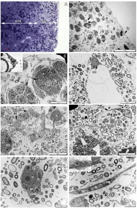
252 The body of Aplysina cavernicola has a branchy shape, with each cylindrical branch 253 having 1-2 cm in diameter (Figure 1). Sponge tissues are dense and elastic. The sponge has 254 leuconoid organization of aquiferous system (numerous small choanocyte chambers, scattered 255 in the mesohyl). The skeleton represented exclusively by organic (spongin) fibers, covered 256 with chitin (Ehrlich et al., 2010a), thus the surgical operations are easily conducted.
257 The body is composed of the peripheral ectosome and the internal endosome, bearing 258 numerous choanocyte chambers (Figure 3A). The ectosomal region is up to 30 μm thick and 259 consists of three layers: (1) external parts of the T-shaped exopinacocytes, connected by non-260 specialized cell junctions (Figure 3B,C) and covered by an acellular cuticle; (2) layer 261 containing collagen fibrils, cell bodies of exopinacocytes and scattered spherulous cells; and 262 (3) the inner layer, consisting of condensed collagen fibrils and spherulous cells. The 263 endosome (Figure 3A) composes the major part of the sponge body. It includes choanocyte 264 chambers, consisted of flagellated choanocytes, aquiferous canals, lined by endopinacocytes, 265 (Figure 3D,E) and the mesohyl with the skeleton, abundant symbiotic bacteria and scattered 266 specialized sponge cells.
3 4 5 6 7 8 9 10 11 12 13 14 15 16 17 18 19 20 21 22 23 24 25 26 27 28 29 30 31 32 33 34 35 36 37 38 39 40 41 42 43 44 45 46 47 48 49 50 51 52 53 54 55 56 57 58 59 60
For Peer Review
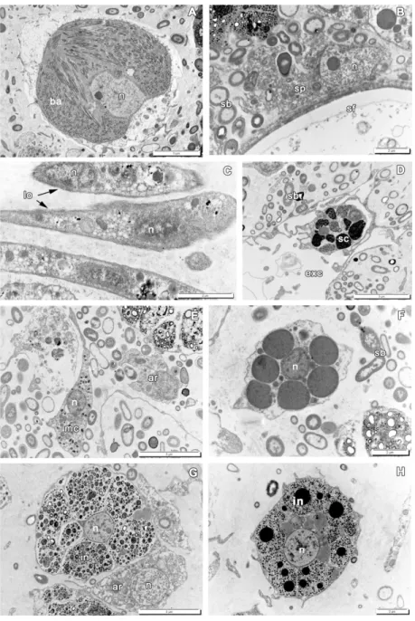
267 Populations of free cells in the mesohyl of A. cavernicola include: lophocytes, 268 archaeocytes, pocket cells, contractile cells (myocytes) (Figure 3F,H), spherulous cells at 269 different stages of their maturation with two principal morphotypes: larger cells with clear 270 inclusions (Figures 3C, 4G) and smaller ones with dense inclusions (Figure 4D,H), 271 bacteriocytes, microgranular cells, spongocytes (Figure 4A-F) (Vacelet, 1966, 1967, 1970, 272 1971, 1975; Vacelet & Gallissian, 1978).
273
274 Regeneration
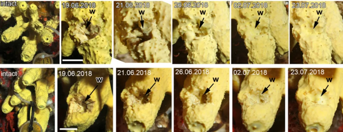
275 In spite of minor individual differences wound healing in Aplysina cavernicola, which 276 is expressed in the epithelialization of the wound surface, is completed within two – six days 277 (Figure 1). During our observations we did not reveal any significant differences in the onset 278 of the stages and course of the regeneration, as well as in the morphogenesis accompanying it, 279 across studied individuals. The regeneration ends within two weeks, when the wound is 280 completely healed: only a small depression on the surface remains in its place.
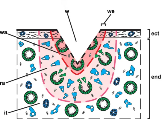
281 At the histological level the observed regeneration processes can be subdivided into 282 three stages: 1) internal milieu isolation – formation of a clot (3 – 12 hpo), 2) wound healing – 283 epithelization (12 – 24 hpo), and 3) restoration of ectosome and endosome (36 – 96 hpo). 284 For detailed description of morphogenesis and cell behavior, accompanying the 285 regeneration, we propose to subdivide a wound and tissue around the wound on several areas 286 (Figure 5):
287 Wound – a break in the continuity of any bodily tissue due to injury.
288 Wound area – tissues directly adjacent to an excised part of the sponge body; their structure is 289 severely disrupted during the surgery.
290 Edge of the wound – peripheral parts of the wound, which is in direct contact with intact 291 tissues.
292 Regeneration area – an area of the sponge body (tissue), which is not directly affected by 293 surgery, but in which anatomical structures (choanocyte chambers, canals of aquiferous 294 system and skeleton) are reorganized, and the normal composition and distribution of cells 295 disrupted due to their participation (dedifferentiation, transdifferentiation and migration) in 296 the regenerative processes. The dimension of this area could vary, depending on size and type 297 of the injury and on individual characteristics of a sponge.
298 Intact issues – ectosome and endosome areas that are not affected by surgery and are not 299 directly involved in regeneration and retaining the normal organization.
3 4 5 6 7 8 9 10 11 12 13 14 15 16 17 18 19 20 21 22 23 24 25 26 27 28 29 30 31 32 33 34 35 36 37 38 39 40 41 42 43 44 45 46 47 48 49 50 51 52 53 54 55 56 57 58 59 60
For Peer Review
300 Stage I – Internal milieu isolation301 Immediately after the surgical excision of ectosome with the directly adjacent endosome, 302 the wound surface retracts, leaving the surface of the intact ectosome protruding around the 303 edges of the wound. The ectosome and the upper areas of endosome are destroyed in the 304 wound area.
305 During the first 3 hours post operation (hpo), the wound surface is covered with 306 exudate, cell debris and numerous symbiotic bacteria. The extracellular matrix (ECM) does 307 not show any signs of a condensation (Supporting Figure 1A-C). All cells in the wound area 308 undergo morphological modifications. The epithelial cells - endopinacocytes and choanocytes 309 of choanocyte chambers, begin losing contacts with adjacent cells in their epithelial layers and 310 change their shape from trapeziform (choanocytes) and flat (endopinacocytes) to spherical or 311 amoeboid (Figure 6). These dedifferentiated cells mix with the mesohyl cell population. 312 During transformation of the choanocytes, their collar of microvilli and flagellum are 313 resorbed (Figure 6D). However, the elements of the flagellar apparatus (the basal body and 314 accessory centriole located near the nucleus) persist in the transformed cells for 315 approximately two days and serve as the natural marker of dedifferentiated choanocytes. The 316 dedifferentiated endopinacocytes have no specific morphological characteristics and therefore 317 these cells cannot be distinguished from other mesohyl cells. The non-secreting cells of the 318 mesohyl (archaeocytes, lophocytes, dedifferentiated choanocytes and endopinacocytes) 319 actively phagocyted cell debris, symbiotic and invasive microbes, including diatoms in the 320 wound area (Figure 6). Thus, all cells in the wound area, except the spherulous cells, are filled 321 with large phagosomes. The ectosome, surrounding the wound slightly contracts and bends 322 inward. After 3 hpo an active migration of the mesohyl cells and dedifferentiated choanocytes 323 towards the wound surface begins from wound area, as these cells assume an elongated shape 324 with the long axis, perpendicular to the wound surface (Figure 6). There are no changes in the 325 regeneration area in this period.
326 At 6 hpo, the wound surface is aligned and becomes flat and smooth. It is covered by a 327 thick layer of ECM, containing fragments of cells, dispersed symbiotic bacteria and few 328 spherulous cells (some of which are beginning their dedifferentiation) (Figure 7A; Supporting 329 Figure 1D). The amount of the spherulous cells in the wound area at this stage of the 330 regeneration significantly decreases and is approximately 2,8-fold lower than in the ectosome 331 of intact sponges (Figure 8; Supporting Table 4). This structure can be referred as a 332 regenerative clot, by analogy with other animals (Carlson, 2007). The thickness of the clot is 333 from 12 to 20 µm. 3 4 5 6 7 8 9 10 11 12 13 14 15 16 17 18 19 20 21 22 23 24 25 26 27 28 29 30 31 32 33 34 35 36 37 38 39 40 41 42 43 44 45 46 47 48 49 50 51 52 53 54 55 56 57 58 59 60
For Peer Review
334 The cells of wound area have numerous small or few large phagosomes, which 335 contained fragments of the spherulous and granular cells (Figure 7B-F). Such cells could be 336 found not only in the upper part of the wound, but also in the deeper zone up to 100 µm 337 beneath the wound surface. There still no visible changes in the regeneration area at this 338 stage.
339 At 12 hpo a regenerative clot at the wound surface is getting thinner, and wound surface 340 become aligned, flat and smooth (Figure 9A). The peripheral parts of the wound are clearly 341 limited by the flat outgrowths of intact exopinacocytes (Figure 9F).
342 In the wound area, сell distribution begins ordering, and ECM condensation occurs 343 (Supporting Figure 1E). The number of dedifferentiated choanocytes and endopinacocytes, 344 archaeocytes, and lophocytes increases in this area in comparison with the previous period of 345 regeneration (Figure 9B-E). Majority of these cells migrated from regenerated area. In 346 contrast, the number of the spherulous cells shows insignificant variations and remains 347 approximately the same as at 6 hpo (Figure 8; Supporting Table 4). Some of these spherulous 348 cells undergo the dedifferentiation, which is accompanied by the release of the spherules from 349 the cells. The amount of free mesohyl and concentration of the symbiotic bacteria decrease in 350 comparison with previous periods of regeneration (Figure 9C, D).
351 Simultaneously, the migration of the amoeboid cells (archaeocytes, dedifferentiated 352 choanocytes) from the regeneration area to the wound area and wound surface proceeds 353 (Figure 9C,E), and some of the migrating cells reach the wound surface, where they become 354 oriented with the long axis parallel to the wound surface (Figure 9B). Also first elongated 355 contractile cells (myocytes) appear in the regeneration area.
356
357 Stage II. Wound healing - epithelization (12 – 24 hpo)
358 At 24 hpo a cell mass, consisting of heterogenous dedifferentiated and undifferentiated 359 (archaeocytes) cells, is formed under the upper part of the wound area. We considered it as a 360 blastema, as structurally it resembles blastemas formed during regeneration of other animals. 361 Blastema occupies the wound area and the upper part of the regeneration area.
362 Simultaneously, the upper part of the wound area becomes similar to intact sponge 363 ectosome, showing structured ECM with collagen fibrils (Figure 10A,B; Supporting Figure 364 1F). The wound surface is mosaically covered with very thin superficial outgrowths of 365 developing T-shaped exopinacocytes, arising from blastema cells of the heterogeneous origin 366 (Figure 10B,C,E). New exopinacocytes do not yet form close contacts with each other. In 367 some places submerged nucleated bodies of these cells are found. New exopinacocytes, in
3 4 5 6 7 8 9 10 11 12 13 14 15 16 17 18 19 20 21 22 23 24 25 26 27 28 29 30 31 32 33 34 35 36 37 38 39 40 41 42 43 44 45 46 47 48 49 50 51 52 53 54 55 56 57 58 59 60
For Peer Review
368 contrast to the cells located deeper under the wound surface, have only few phagosomes. 369 There is no directed movement (contraction or creeping) of the intact exopinacoderm 370 surrounding the wound.
371 The number of spherulous cells in the wound area significantly increases in 372 approximately 2,3-folds in comparison with the previous stage of regeneration (Figure 8; 373 Supporting Table 4). These cells show typical appearance (Figure 10B). Their shape is oval, 374 not amoeboid, as in the earlier stages of regeneration. In contrast, near the wound surface, the 375 number of dedifferentiated choanocytes, archaeocytes and spherulous cells decreases (Figure 376 10C-F). The lophocytes, granular cells, and myocytes are absent from this zone. The 377 choanocyte chambers or their fragments completely disappear from the regeneration area at 378 this stage of regeneration. They occur at a depth of about 200 µm, as in intact sponge.
379
380 Stage III. Restoration of ectosome and endosome (36 – 96 h)
381 At 48 hpo the ectosome of the regenerate is completely restored, but superficial cuticle, 382 characteristic for intact sponge, is still absent (Figure 11A,B; Supporting Figure 1G). The 383 wound epithelization is finished by new exopinacocytes, arose from the heterogeneous 384 dedifferentiated cells population and archaeocytes. Some of new exopinacocytes clearly show 385 their origin from the dedifferentiated choanocytes, as they still bear flagellar basal apparatus 386 (Figure 11C). According to the orientation of this flagellated complex, it can be assumed that 387 the basal part of the former choanocyte is flattened. Archaeocytes, reaching the wound, flatten 388 and assume position parallel to the surface (Figures 10E,F, 11D). New exopinacocytes have 389 normal T-shape and cell contacts.
390 During formation of the exopinacoderm active elimination of small apoptotic 391 spherulous cells with compact inclusions and cells, filled with big heterophagosomes, begins 392 (Figure 11E, F). However, the number of the spherulous cells shows insignificant variations 393 and remains approximately the same as at 24 hpo (Figure 8; Supporting Table 4).
394 During this stage, the choanocyte chambers and aquiferous canals of the endosome are 395 restored by association of previously disaggregated choanocytes and endopinacocytes, 396 respectively (Figure 12). Archaeocytes also participate in the development of new choanocyte 397 chambers (Figure 12D). Cells that form new structure of aquiferous system contact each other 398 and connect by interdigitations. Individual choanocytes form groups of cells, forming 399 structures less compact than in the intact choanocyte chamber (Figure 12D-F). These cells 400 gradually transform from mesenchymal morphology to epithelial.
3 4 5 6 7 8 9 10 11 12 13 14 15 16 17 18 19 20 21 22 23 24 25 26 27 28 29 30 31 32 33 34 35 36 37 38 39 40 41 42 43 44 45 46 47 48 49 50 51 52 53 54 55 56 57 58 59 60
For Peer Review
401 At 96 hpo, the regeneration is almost complete: the ectosome obtains the cuticle, while 402 the endosome of the regeneration area begins to recover: choanocyte chambers and aquiferous 403 canals are gradually developing (Figure 13; Supporting Figure 1H). The number of 404 spherulous cells in the regenerated ectosome significantly increases, reaching the intact level 405 (Figure 8; Supporting Table 4). The free cells of the mesohyl, with the exception of 406 specialized secretory cells, still contain phagosomes.
407
408 Cell proliferation
409 The DNA-synthesizing (EdU-labelled) cells are irregularly distributed in the intact 410 tissues of A. cavernicola (Figure 14). Numerous DNA-synthesizing cells are located in the 411 sponge endosome (Figure 14B). The majority of these cells are choanocytes with small 412 (3.87±0.46 µm; n=45) round to oval nuclei (Figure 14D). Nevertheless, some mesohyl cells 413 with large (4.84±0.44 µm; n=50) round nuclei are also labelled by EdU (Figure 14E). These 414 mesohyl DNA-synthesizing cells occurs all over sponge mesohyl: in the ectosome, endosome 415 and tissues, adjacent to the large exhalant canals of the aquiferous system, which are 416 structurally similar to the ectosome. The ectosome and tissues, adjacent to the large exhalant 417 canals contain only few such mesohyl DNA-synthesizing cells (Figure 14A, C).
418 During the regeneration, the pattern of the cell proliferation dramatically changes 419 (Figure 15). Immediately after the surgical operation, the level of the cell proliferation in the 420 tissues, adjacent to the wound, decreases sharply: at 0-6 hpo, DNA-synthesizing cells are 421 completely absent in a 100-µm zone below the wound surface (Figure 15A); at 6–12 hpo and 422 0-24 hpo, rare DNA-synthesizing cells appear in the tissues, located 30–40 µm below the 423 wound surface. The majority of the labelled cells are choanocytes from disintegrating 424 choanocyte chambers, however some mesohyl cells are also labelled. Similarly, to the intact 425 tissues, the labelled choanocytes have small (3.85±0.4 µm; n=13) round to oval nuclei, while 426 labelled mesohyl cells are characterized by large (4.79±0.37 µm; n=16) round nuclei.
427 After 24 hpo, further changes in the cell proliferation pattern occurs in the regeneration 428 area. The samples at 24-48 hpo and 48-72 hpo show a similar pattern (Figure 15B, D): in a 429 100-µm zone below the wound surface, numerous DNA-synthesizing cells occur. Some of 430 these cells are located just beneath the wound surface. Virtually all labeled cells are located in 431 the mesohyl, since the choanocyte chambers disappear in the tissues, adjacent to the wound, 432 by this time. These DNA-synthesizing cells have large (4.99±0.39 µm; n=16) round nuclei, 433 similar to EdU-labelled mesohyl cells from intact tissues. Occasionally, single
DNA-3 4 5 6 7 8 9 10 11 12 13 14 15 16 17 18 19 20 21 22 23 24 25 26 27 28 29 30 31 32 33 34 35 36 37 38 39 40 41 42 43 44 45 46 47 48 49 50 51 52 53 54 55 56 57 58 59 60
For Peer Review
434 synthesizing choanocytes occur in the tissues, located 60-70 µm below the wound surface, 435 where intact choanocyte chambers could be retained.
436 At 120-144 hpo, after the recovery of ectosome and endosome structure in the 437 regeneration area, the cell proliferation returns to the normal state, showing the intact pattern 438 with majority of DNA-synthesizing cells, occurring in choanocyte chambers. However, the 439 level of cell proliferation is still lower than in intact tissues (Figure 15E).
440 In the tissues, distant from the wound, the intact cell proliferation pattern persists during 441 the whole regeneration process (Figure 15F).
442
443 Apoptosis
444 We found no apoptotic cell in the intact tissue of A. cavernicola.
445 Two waves of apoptosis occur during the regeneration process. The level of apoptosis is 446 low during both waves with few apoptotic cells, located only at the upper part of the wound. 447 In the endosome under the wound and in tissues distant from the zone of regeneration 448 apoptotic cells are completely absent.
449 The first wave is associated with the early stages of regeneration. Apoptotic cells appear 450 at 6 hpo, and their number gradually decline during further regeneration (Figure 16A). At 12 451 hpo only single apoptotic cells occur at the upper parts of the wound area (Figure 16B), while 452 at 24 hpo they are virtually absent. Apparently, this wave of apoptosis participates in the 453 elimination of damaged cells from the wound area.
454 The second wave of apoptosis occurs at 48 hpo, when relatively numerous apoptotic 455 cells appears at the upper parts of the wound area (Figure 16C). This wave is probably 456 associated with the active elimination of cells through the emerging exopinacoderm during 457 later stage of regeneration (Figure 11E,F).
458
459 Discussion 460
461 1 General
462 In vivo observations
463 Sponges are well known for their capacity to regenerate not only small body parts, but 464 also after substantial partial mortality and damage. In situ monitoring of naturally and 465 experimentally generated wounds confirms high recovery capacity of sponges from different 466 taxa and of different growth forms (Connes, 1966, 1968; Ayling, 1983; Hoppe, 1988; 467 Duckworth, 2003; Henry & Hart, 2005; Wulff, 2010, 2013). These studies also discovered
3 4 5 6 7 8 9 10 11 12 13 14 15 16 17 18 19 20 21 22 23 24 25 26 27 28 29 30 31 32 33 34 35 36 37 38 39 40 41 42 43 44 45 46 47 48 49 50 51 52 53 54 55 56 57 58 59 60
For Peer Review
468 that regeneration can be influenced both by characteristics of the wound and by inherent 469 characteristics of particular species of sponges.
470 Our observations in situ showed that wound epithelialization under natural conditions 471 occurs in Aplysina cavernicola during two days. Other investigations provided with different 472 demosponges showed the same results (Maas, 1910; Brondsted, 1953; Connes, 1966, 1968; 473 Korotkova & Nikitin, 1969; Korotkova et al., 1983; Diaz, 1979; Hoppe, 1988; Hofmann et al., 474 2003; Wulff, 2010, 2013; Alexander et al., 2014; Borisenko et al., 2015; Pozzolini et al., 475 2019).
476 Previous investigations of regeneration demonstrated that in many individuals of the 477 same species the amount of damage, type of damage, size of the sponge, and location on the 478 individual sponge can influence recovery, and even susceptibility to further damage by other 479 agents (Henry & Hart, 2005; Wulff, 2010, 2013). However, we find small individual 480 variability during regeneration of A. cavernicola, probably because the wounds were applied 481 in the same areas of the sponges and had the same size.
482
483 Histological observations
484 In this work we showed that regeneration processes in Aplysina cavernicola in general 485 follow the stages similar to those described in other massive sponges with leuconoid 486 aquiferous system - Demospongiae and Homoscleromorpha (Brondsted, 1953; Korotkova & 487 Nikitin, 1969; Diaz, 1979; Hofmann et al., 2003; Borisenko et al., 2015; Ereskovsky et al., 488 2015; Pozzolini et al., 2019), and in various eumetazoans (Korotkova, 1997; Carlson, 2003). 489 Therefore, A. cavernicola regeneration includes three main stages: 1) internal milieu isolation 490 - formation of “regenerative clot”, 2) wound healing - epithelization, and 3) restoration of 491 damaged structures - ectosome and endosome. The wound epithelization in A. cavernicola 492 occurs without formation of the regenerative membrane, a structure, characteristic for 493 regeneration of asconoid and syconoid calcareous sponges (Jones, 1957; Korotkova, 1961, 494 1962; Ereskovsky et al., 2017; Lavrov et al., 2018).
495 The main mechanisms of Aplysina cavernicola reparative regeneration of the body wall 496 are (1) cell dedifferentiation with their subsequent redifferentiation, (2) transdifferentiation, 497 and (3) active migration of polypotent cells (archaeocytes and choanocytes) to the wound. 498 The same basic mechanisms are also characteristic for regeneration in other studied 499 demosponges: Halichondria panicea (Korotkova & Nikitin, 1969), Suberites massa (Diaz, 500 1979), Halisarca dujardinii (Borisenko et al., 2015), and Suberites domuncula (our 501 unpublished results). However, these mechanisms strongly differ from those, participating in
3 4 5 6 7 8 9 10 11 12 13 14 15 16 17 18 19 20 21 22 23 24 25 26 27 28 29 30 31 32 33 34 35 36 37 38 39 40 41 42 43 44 45 46 47 48 49 50 51 52 53 54 55 56 57 58 59 60
For Peer Review
502 regeneration process in calcareous sponges and homoscleromorphs, which show clear 503 epithelial organization with specialized cell junctions in the epithelia (Ereskovsky et al., 2015; 504 Lavrov et al., 2018).
505
506 2. Morphogenetic mechanisms of A. cavernicola regeneration
507 Main morphogenesis during body wall regeneration in A. cavernicola is mesenchymal-508 to-epithelial transformation (MET). Indeed, at 12 hpo archaeocytes and dedifferentiated 509 choanocytes as well as the spherulous cells begin to move toward wound area, where they 510 form a blastema-like structure. Part of external blastema cells begins to flat and form cover 511 sponge epithelium (exopinacoderm) through the MET.
512 However, experimentally induced (by surgical operation) epithelial-to-mesenchymal 513 transition (EMT) proceeds MET in A. cavernicola. During this process choanocytes and 514 endopinacocytes leave the choanocyte chambers and the canals of aquiferous system, 515 correspondingly, and move into the mesohyl, dedifferentiating and assuming an amoeboid 516 shape. This morphogenesis is one of central for the creation of numerous organs and complex 517 tissues during embryonic development, asexual reproduction, and regeneration and has been 518 well described in Eumetazoa (Keller et al., 2003; Lim & Thiery, 2012). EMT is also well 519 known during the normal ontogeny of sponges and occurs in the course of embryonic 520 development, and larval metamorphosis (reviewed in Ereskovsky et al. 2013). This 521 morphogenesis was described during the first stages of regeneration of other investigated 522 demosponges and homoscleromorphs (Korotkova & Nikitin, 1969; Borisenko et al., 2015; 523 Ereskovsky et al., 2015).
524 After wound epithelization, the restoration of elements of the aquiferous system begins. 525 Both choanocyte chambers and aquiferous system canals are formed through MET: separate 526 amoeboid cells coalescence into groups and gradually transform from mesenchymal 527 morphology to epithelial-like cells (choanocytes and endopinacocytes, respectively).
528 The same MET mechanism is characteristic for exopinacoderm and choanoderm 529 formation during regeneration of other investigated demosponges: Halichondria panicea 530 (Korotkova & Nikitin, 1969), Halisarca dujardini (Borisenko et al., 2015), and 531 basopinacoderm formation in Hymeniacidon heliophila regeneration (Coutinho et al., 2017). 532 It is fundamentally different from the regeneration in Calcarea and Homoscleromorpha, 533 in which epithelial morphogenesis, e.g. flattening and spreading of epithelial layers, plays a 534 leading role in the restoration of main structures (exopinacoderm and elements of aquiferous 535 system) (Ereskovsky et al., 2015; Lavrov et al., 2018).
3 4 5 6 7 8 9 10 11 12 13 14 15 16 17 18 19 20 21 22 23 24 25 26 27 28 29 30 31 32 33 34 35 36 37 38 39 40 41 42 43 44 45 46 47 48 49 50 51 52 53 54 55 56 57 58 59 60
For Peer Review
536537 3. Blastema
538 In regenerative biology blastema is a mass of undifferentiated cells, located beneath the 539 wound and produced by the dedifferentiation of many cell types and/or migration of 540 polypotent cells. In some cases, differentiation of blastema cells, and further reconstruction 541 the lost part of the body could be accompanied with cell proliferation. In this case, the mitotic 542 rate of the blastema slows down as the structure grows, and it ceases completely when the 543 new structure reaches the original size (Santos-Ruiz et al., 2002; Carlson, 2007; Tsonis, 2008; 544 Vervoort, 2011).
545 The formation of a concentration of cells under the wound in the middle stages of a 546 body wall regeneration has been shown for several demosponges (Thiney, 1972; Boury-547 Esnault, 1976; Borisenko et al., 2015; Coutinho et al., 2017). This cellular concentration 548 consists of archaeocytes and choanocytes, which are principal stem cells of demosponges 549 (Funayama, 2018), dedifferentiated cells (e.g., pinacocytes) and specialized differentiated 550 cells (e.g., gray cells) (Thiney, 1972; Boury-Esnault, 1976; Borisenko et al., 2015; Coutinho 551 et al., 2017). This structure could be referred as a blastema, characteristic for a regeneration in 552 many animals.
553 Similarly, in the current study, we refer to the undifferentiated cell mass beneath a 554 wound during the end of second and beginning of third stages of regeneration in A. 555 cavernicola as a blastema. In A. cavernicola, it consists of archaeocytes, dedifferentiated 556 choanocytes, mixed population of anucleated amoebocytes (dedifferentiated pinacocytes, 557 myocytes, lophocytes), and differentiated spherulous cells.
558 At early stages of regeneration, the blastema almost devoid of DNA-synthesizing cell, 559 but after 24 hpo and up to 72 hpo numerous DNA-synthesizing cells appears there. However, 560 even during this period the level of cell proliferation in the blastema is much lower in 561 comparison with intact sponge tissues. In contrast, intact tissues distant form the wound 562 retains intact level of cell proliferation during whole regeneration process. The similar pattern 563 of cell proliferation has been described during body wall regeneration of Halisarca caerulea 564 (Alexander et al., 2014) and Halisarca dujardinii (Borisenko et al., 2015), for which 565 decreased proliferation in the wound area is characteristic.
566 Moreover, in contrast to intact tissues of A. cavernicola, where vast majority of DNA-567 synthesizing cells are choanocytes, all EdU-labeled cells in the blastema are mesohyl cells, as 568 blastema lacks choanocyte chambers. These EdU-labeled cells could arise directly in blastema 569 through cell divisions or be EdU-labeled migrating descendants of proliferating cells in intact
3 4 5 6 7 8 9 10 11 12 13 14 15 16 17 18 19 20 21 22 23 24 25 26 27 28 29 30 31 32 33 34 35 36 37 38 39 40 41 42 43 44 45 46 47 48 49 50 51 52 53 54 55 56 57 58 59 60
For Peer Review
570 tissues, distant from the wound. This issue requires further investigations, using additional 571 markers, like antibodies against phospho-histone H3 for reveling cells in M-phase and cell 572 tracers for the visualization of cell migrations.
573 Importantly, studied sponges from classes Calcarea (Leucosolenia cf. variabilis) 574 (Lavrov et al., 2018) and Homoscleromorpha (Oscarella lobularis) (Ereskovsky et al., 2015) 575 demonstrate a distinct mode of regeneration without blastema formation. In these sponges 576 regeneration occurs due to the local remodeling of intact tissues, adjacent to the wound. 577 Moreover, the cell proliferation is neither affected nor contributes to the regeneration at any 578 stage.
579
580 4. Dedifferentiation, transdifferentiation and cell sources in A. cavernicola 581 regeneration
582 Transdifferentiation is the transformation of one type of already differentiated cell into 583 another type of differentiated cell. In some cases, transdifferentiation is accompanied by cell 584 division, while in other cases it is not (Shen et al., 2004).
585 Sponge cell transdifferentiation is likely a driving force accompanying their restoration 586 processes (Korotkova, 1997; Lavrov & Kosevich, 2014; Adamska, 2018). However, 587 transdifferentiating cell types and mechanisms of this process vary in different sponge species 588 (Diaz, 1979; Gaino et al., 1995; Korotkova, 1997; Ereskovsky et al., 2015; Borisenko et al., 589 2015; Lavrov & Kosevich, 2016; Lavrov et al., 2018). For example, direct transdifferentiation 590 of cells in intact epithelia (pinacocytes and choanocytes) has been demonstrated during the 591 reparative regeneration of the homoscleromorph Oscarella lobularis (Ereskovsky et al., 592 2015), the calcareous sponges Sycon lingua (Korotkova, Efremova, & Kadantseva, 1965; 593 Korotkova, 1972b), and Leucosolenia ssp. (Lavrov et al., 2018). In these cases, the layer of 594 choanocytes transdifferentiates into layer of pinacocytes without loss of epithelial structure, 595 without mesenchymal-to-epithelial transformation, and without contribution from cell 596 proliferation.
597 Cell transdifferentiation have also been described during reparative regeneration in all 598 investigated demosponges (for review see: Korotkova, 1997; Borisenko et al., 2015). While 599 archaeocytes directly differentiate into new cells, choanocytes and pinacocytes undergo 600 transdifferentiation to give rise for other cell types. It is characteristic, mainly, for 601 choanocytes from disintegrated choanocyte chambers, which can transdifferentiate, for 602 example, into exopinacocytes (Korotkova & Nikitin, 1969a,b; Borisenko et al., 2015). 603 Endopinacocytes in a less extent could also transdifferentiate into exopinacocytes (Thiney,
3 4 5 6 7 8 9 10 11 12 13 14 15 16 17 18 19 20 21 22 23 24 25 26 27 28 29 30 31 32 33 34 35 36 37 38 39 40 41 42 43 44 45 46 47 48 49 50 51 52 53 54 55 56 57 58 59 60
For Peer Review
604 1972; Borisenko et al., 2015). The principal difference of the transdifferentiation in 605 demosponges in comparison with Homoscleromorpha + Calcarea lineage is an occurrence of 606 the disruption of epithelial layers and cell dedifferentiation prior to this process in the 607 demosponges.
608 The similar processes were observed during A. cavernicola regeneration. The 609 archaeocytes and dedifferentiated cells participate in the restoration of the lost structures in 610 this sponge. We identify two cells types, which undergo dedifferentiation during A. 611 cavernicola regeneration: choanocytes, spherulous cells, and population of anucleated cells of 612 mesohyl, including dedifferentiated pinacocytes, myocytes, and lophocytes. The choanocytes, 613 despite their dedifferentiation and loss of the flagella and collars of microvilles, are well 614 distinguished on ultrathin sections due to the persistence of two centrioles in their apical part. 615 Anucleated amoebocytes differ from archaeocytes by a smaller nucleus without nucleolus and 616 the presence of small phagosomes. The dedifferentiation of the spherulous cells through a 617 complete or almost complete loss of specific inclusions (spherules) was completely 618 unexpected process. However, the dedifferentiation of spherulous cells is not so rapid, as in 619 other dedifferentiating cells, because of high specialization of these secretory cells.
620 We identified two main cellular sources for the restoration of the lost tissue and 621 structures in A. cavernicola: archaeocytes and choanocytes. Both cell types contribute to the 622 restoration of the exopinacoderm and choanocyte chambers. The restoration of the choanocyte 623 chambers is carried out mainly due to the redifferentiation of the choanocytes, and to lesser 624 extent due to the differentiation of archaeocytes.
625 However, the future of dedifferentiated anucleated amoebocytes and spherulous cells 626 could not be traced. This issue requires the search and use of a special marker of these cells. It 627 is possible that dedifferentiated pinacocytes and spherulous cells could also participate in the 628 formation of the new exopinacoderm.
629 According to last investigations in Porifera there are not only two types of adult stem 630 cells (ASC): archaeocytes and choanocytes (Funayama, 2018), but at least four, including 631 pinacocytes and particular amoeboid vacuolar cells (Ereskovsky et al., 2015; Fierro-Constain 632 et al., 2017; Lavrov et al., 2018).
633 There are the differences in these ASC distributions among different sponge classes. In 634 Demospongiae the principal (pluripotent) ASC are archaeocytes and choanocytes, however 635 the experiments with their regeneration (including present data on A. cavernicola 636 regeneration) and dissociated cells aggregation demonstrated, that pinacocytes could also be 637 ASC (Korotkova & Nikitin, 1969a,b; Borisenko et al., 2015; Lavrov & Kosevich, 2016;
3 4 5 6 7 8 9 10 11 12 13 14 15 16 17 18 19 20 21 22 23 24 25 26 27 28 29 30 31 32 33 34 35 36 37 38 39 40 41 42 43 44 45 46 47 48 49 50 51 52 53 54 55 56 57 58 59 60
For Peer Review
638 Ereskovsky et al., 2016). In Calcarea archaeocytes absent and pluripotent ASC in these 639 sponges are choanocytes and pinacocytes (Korotkova, 1961a, 1962b; Korotkova & 640 Gelihovskaia, 1963; Lavrov et al., 2018). In Homoscleromorpha the pluripotent ASC are as 641 in Calcarea the choanocytes and pinacocytes, but also mesohyl amoeboid vacuolar cells 642 (Gaino et al., 1986; Ereskovsky, 2010; Ereskovsky et al., 2015; Fierro-Constain et al., 2017). 643
644 5. Spherulous cells and their role in regeneration
645 Similarly, to intact tissues of A. cavernicola (Vacelet, 1967), two principal morphotypes 646 of spherulous cells occurs in the wound area of this sponge: larger cells with clear inclusions 647 and smaller ones with dense inclusions. In fact, these morphotypes represent different stages 648 of the ontogenesis of spherulous cells (Vacelet, 1967). According to Vacelet (1967) in intact 649 sponge “dense” small spherulous cells concentrate mostly in the ectosome and around big 650 exhalant canals, while “clear” larger ones are distributed in the mesohyl of endosome.
651 We have detected that in A. cavernicola the wound surface was free from invasive 652 microbes from the first hours after injury up to the end of the regeneration. All microbes, that 653 we detected at the wound surface and in the wound area during regeneration, shows 654 ultrastructure identical to the microbes, located in the deeper sponge tissues and described as 655 symbiotic in the previous papers (Vacelet, 1975; Fiedrich et al., 1999; Hentschel et al., 2001). 656 Such defense against invasive microbes is probably provided by the particular chemical 657 substances in the spherulous cells.
658 X-ray microanalysis revealed that these specialized cells of Aplysina produce and store 659 brominated metabolites (Turon et al., 2000). Turon et al. (2000) demonstrated that the 660 concentration of bromine peaks is slightly higher in the spherules from “dense” spherulous 661 cells than in “clear” ones. Thompson et al. (1983) have detected bromine in the spherulous 662 cells of Caribbean Aplysina fistularis. Spherulous cells have been shown to produce defense 663 metabolites in other species (Thompson et al., 1983; Bretting et al., 1983; Uriz et al., 1996a). 664 In addition, it was showed that tissue damage in Aplysina induces a bioconversion of 665 isoxazoline alkaloids into aeroplysinin-1 and secondary metabolite dienone (Thoms et al., 666 2006). This reaction is likely catalyzed by enzymes, and it may be ecologically important as 667 the dienone has the strong antibacterial power and toxicity (Reverter et al., 2016). Moreover, 668 this injury-induced reaction takes place within less than 1 min after wounding (Thoms et al., 669 2006).
670
671 6. Apoptosis during A. cavernicola regeneration
3 4 5 6 7 8 9 10 11 12 13 14 15 16 17 18 19 20 21 22 23 24 25 26 27 28 29 30 31 32 33 34 35 36 37 38 39 40 41 42 43 44 45 46 47 48 49 50 51 52 53 54 55 56 57 58 59 60
For Peer Review
672 We have directly visualized and investigated apoptosis in the regeneration of sponges 673 for the first time. The only previous data on the involvement of apoptosis in sponge 674 regeneration emerge from the comparative transcriptomic analysis of the early stages of body 675 wall regeneration in Halisarca caerulea (Kenny et al., 2017).
676 We have observed two waves of apoptosis during Aplysina cavernicola regeneration. 677 The first wave, occurring around 6 hpo, is probably involved in the elimination of damaged 678 cells and is characteristic for the early stages of regeneration of various animals, from 679 cnidarians to vertebrates (Vlaskalin et al., 2004; Tseng et al., 2007; Chera et al., 2009; 680 Pellettiere et al., 2010; DuBuc et al., 2014; Brandshaw et al., 2015; Kenny et al., 2017; Cebrià 681 et al., 2018). For Hydra head regeneration after mid-gastric bisection (Chera et al., 2009) and 682 tail regeneration in tadpole larva of Xenopus laevis (Tseng et al., 2007) it was shown that 683 early wave of apoptosis not only clean a wound from damaged cells, but also generate signals, 684 essential for initiation of subsequent regenerative processes. Inhibition of this apoptotic wave 685 completely abolishes the regeneration. In particular, in both cases, apoptotic cells initiate a 686 synchronous burst of proliferative activity in neighboring cells (Tseng et al., 2007; Chera et 687 al., 2009), which is similar to the well-known apoptosis-induced compensatory proliferation 688 in the imaginal disks of Drosophila (Fan & Bergmann, 2008; Bergmann & Steller, 2010). 689 Apoptotic cells may be involved in the proliferation induction also during A. cavernicola 690 body wall regeneration, as numerous DNA-synthesizing cells appears in the blastema only 691 after the first apoptotic wave, at 24 hpo. These observations illustrate a novel active 692 instructing role of apoptosis in morphogenetic processes, in contrast to its canonical passive 693 role as a destructive agent (Duffy, 2012).
694 The second wave of apoptosis during A. cavernicola regeneration is likely associated 695 with the extensive thrown out of spherulous cells though the forming exopinacoderm. 696 Spherulous cells of A. cavernicola demonstrate the similar behavior in the intact sponges. 697 Being one of the few terminally differentiated sponge cell lines, spherulous cells leave the 698 mesohyl through aquiferous system canals or external surfaces (Vacelet, 1967). Their release 699 outside from sponge is part of their normal physiology and possibly involved in the release of 700 various chemicals (Uriz et al., 1996a; Ternon et al., 2016) and/or discharge processes 701 (Maldonado, 2016) to the environment. The ultrastructure of spherulous cells eliminated 702 through the exopinacoderm during the third phase of A. cavernicola regeneration is identical 703 to that of the spherulous cells at the last stage of their evolution (Vacelet, 1967). Similarly, in 704 the intact tissues of H. caerulea, many caspase-3 positive cells have a spherulous cell 705 morphology (De Goeij et al., 2009), which indicates that apoptosis involved in the waste
3 4 5 6 7 8 9 10 11 12 13 14 15 16 17 18 19 20 21 22 23 24 25 26 27 28 29 30 31 32 33 34 35 36 37 38 39 40 41 42 43 44 45 46 47 48 49 50 51 52 53 54 55 56 57 58 59 60
For Peer Review
706 control system in sponges in addition to maintaining tissue homeostasis (De Goeij et al., 707 2009).
708 Thus, the described increase in the level of apoptosis at the late stages of the A. 709 cavernicola regeneration could be referred as a general tissue response, involved in the 710 restoration of the normal sponge physiology in the area of injury, rather than in regeneration 711 process itself. In planarians, the similar late general apoptotic response is also described, but it 712 participates in the intensive remodeling of intact tissues, adjacent to a wound, to restore 713 proper scale and proportions (Pellettiere et al., 2010; Cebrià et al., 2018).
714
715 Conclusion
716 Finally, we can made some principal conclusions:
717 1. In Porifera there are two principal mode of reparative regeneration: blastemal 718 regeneration and tissue remodeling. From the literature data and results, obtained in this work 719 we can conclude that first mode is characteristic for Demospongiae regeneration and the 720 second – for Calcarea + Homoscleromorpha clade.
721 2. The results of our research also showed that sponges have more than two lines of 722 adult stem cells, the potencies of which are clearly manifested during reparative regeneration. 723 Our data support previous investigations, showed that archaeocytes and choanocytes are main 724 players, or principal adult stem cells during demosponges regeneration (Korotkova & Nikitin, 725 1969; Thiney, 1972; Boury-Esnault, 1976; Diaz, 1979; Borisenko et al. 2015). However, the 726 pinacocytes also could be considered as multipotent stem cells. Moreover, for the first time 727 we showed, that spherulous cells of Aplysina cavernicola have a capacity to dedifferentiation 728 during regeneration. It is first evidence that highly specialized cells of demosponges have 729 capacity to dedifferentiation and possibly transdifferentiation.
730 3. Cell transdifferentiation plays an extremely significant role in sponge regeneration, as 731 we have shown that regardless of the phylogenetic position, type of aquiferous system, and 732 structure of epithelia, all studied sponges intensively utilize it for restoration of lost structures 733 (Borisenko et al., 2015; Ereskovsky et al., 2015, 2017; Lavrov et al., 2018).
734 4. For the first time was investigated apoptosis during sponge regeneration. In 735 Aplysina cavernicola regeneration this processes participate in damaged cells elimination and 736 associated with the extensive ejection of spherulous cells from wound area.
737 738 Author contributions 3 4 5 6 7 8 9 10 11 12 13 14 15 16 17 18 19 20 21 22 23 24 25 26 27 28 29 30 31 32 33 34 35 36 37 38 39 40 41 42 43 44 45 46 47 48 49 50 51 52 53 54 55 56 57 58 59 60
For Peer Review
739 A.V.E. and A.I.L. designed the study, conducted the experimental procedures with the living 740 animals; A.I.L. performed the cell proliferation studies; A.V.E. and D.B.T. performed 741 histological and ultrastructural studies. A.V.E.: Project administration and funding 742 acquisition; S.B., E.L.G., and A.I.L.: performed the apoptosis studies; A.V.E. and A.I.L. 743 prepared the manuscript with contributions from all authors. All authors reviewed and 744 approved the final manuscript.
745
746 Acknowledgements
747 Authors gratefully thank Alexandre Altié of Plateforme C2VN de Microscopie Électronique 748 TIMONE, Aix-Marseille Université, France, the Electron Microscopy Laboratory of the 749 Shared Facilities Center of Lomonosov Moscow State University sponsored by the RF 750 Ministry of Education and Science and Research Resource Center for Molecular and Cell 751 Technologies at St. Petersburg State University, the staff of the Common Service of 752 morphology in IMBE, and Dr. Jean Philippe Mévy (IMBE) - for help and assistance with 753 electron and confocal microscopy studies. Data used in this study were partly produced using 754 the Montpellier RIO imaging platform (confocal microscopy) (Montpellier, France). This 755 work was supported by grants of Russian Foundation for Basic Research no. 16-04-00084, the 756 Russian Science Foundation no. 17-14-01089 (histological and ultrastructural studies), and 757 Metchnikov fellowship 2019 by French Embassy in Russia. This work also is a contribution 758 to Labex OT‐Med (n° ANR-11-LABX-0061) and has received funding from Excellence 759 Initiative of Aix‐Marseille University-A*MIDEX, a French ’’Investissements d’Avenir’’ pro-760 gram for travel expenses.
761 3 4 5 6 7 8 9 10 11 12 13 14 15 16 17 18 19 20 21 22 23 24 25 26 27 28 29 30 31 32 33 34 35 36 37 38 39 40 41 42 43 44 45 46 47 48 49 50 51 52 53 54 55 56 57 58 59 60