Pseudomonas diversity in western Algeria: role
in the stimulation of bean germination and common bean
blight biocontrol
Slimane Mokrani&Abdelwahab Rai&
Lakhder Belabid&Ameur Cherif&Hanane Cherif& Mouna Mahjoubi&Elhafid Nabti
Accepted: 8 August 2018
# Koninklijke Nederlandse Planteziektenkundige Vereniging 2018 Abstract The aim of this work was to determine the functional diversity of soil bacteria belonging to the Pseudomonas genus, to study their effects on bean (Phaseollus vulgaris L) seed germination and their biocontrol potential of common bean blight. Bacte-ria were isolated and identified based on physiolog-ical and biochemphysiolog-ical characters and BOX-PCR. Followed by qualitative and/or quantitative analysis of their secondary metabolites. 50 soil bacteria were affected to the two groups of fluorescent (72%) and non-fluorescent Pseudomonads (28%). The UPGMA (Unweighted Pair Group Method with Arithmetic Mean) showed five phenons of carbon sources as-similation; at the time that BOX-PCR profiling re-sulted in five clusters characterized by 29 different
haplotypes. (66%) isolates induced phosphate solu-bilization; (24%) were HCN producers, (21%) showed IAA production and all isolates had pro-duced siderophores. In vitro antibacterial activity against Xapf showed 26.67 and 24 mm of inhibition zone using the two isolates P. grimontii P25 and P. cepatia P7, respectively. Similarly, the same iso-lates significantly reduced Xapf-bean common blight intensity, while their co-inoculation was less effective. The isolate P. cepatia P7 was highly ef-fective on seed germination and root growth prop-erties, then P. grimontii P25. Thus, the selected isolates could play a crucial bean growth promotion and bean common blight biocontrol as alternative to chemicals for crop yield enhancement.
https://doi.org/10.1007/s10658-018-1566-9
S. Mokrani
:
E. Nabti (*)Laboratoire de Maitrise des Energies Renouvelables, Faculté des Sciences de la Nature et de la Vie, Université de Bejaia, 06000 Bejaia, Algeria
e-mail: elhnabti1977@yahoo.fr
S. Mokrani
e-mail: distillateur@yahoo.fr
S. Mokrani
:
L. BelabidDepartment Agronomy, Laboratory of Research on Biological Systems and Geomantic (L.R.S.B.G), University of Mustapha Stumbouli, P.O. Box 305, 29000 Mascara, Algeria
L. Belabid
e-mail: belabidl@yahoo.fr
A. Rai
Faculté des Sciences de la Nature et de la Vie et des Sciences de la Terre, Université Akli Mohand Oulhadj, 10000 Bouira, Algeria e-mail: abdelwahabrai@yahoo.fr
A. Cherif
:
H. Cherif:
M. MahjoubiHigher Institute for Biotechnology, Laboratory of Biotechnology and Bio-Geo Resources Valorization (L.B.B.G.R.V),
University of Manouba, Sidi Thabet Biotechpole, 2020 Sidi Thabet, Ariana, Tunisia
A. Cherif e-mail: cherif.ameur@gmail.com H. Cherif e-mail: cherifhanene@gmail.com M. Mahjoubi e-mail: mounamahjoubi@gmail.com
Keywords Pseudomonas . Plant growth promoting bacteria and Xanthomonas axonopodis pv. phaseoli var. fuscans
Introduction
Nutritional problems touch about 2 billion people worldwide (Hawkes and Ruel 2008). In addition, the world’s population, estimated around 7 billion people at this time, is predicted to jump to around 10 billion in the next 50 years, which requires significant increase in food productivity human life continuity on the planet (Glick2014). In this context, agricultural practices are the major source of food and feed around the world. Without enough agricultural production, catastrophic scenarios are objectively expected in the next few years (Giovannucci et al.2012).
Modern agriculture is facing major challenges be-cause of climate changes, increasing world population, natural resources scarcity and food transitions to more meat products (Urruty2017). Thus, the produced crop need to be equipped with disease eradication systems, salt-, drought-, heavy metal-tolerance characters, and better nutritional values (Armada et al.2014). In some regions of the globe, up to 30% agricultural yield is lost every year due to both abiotic and biotic stresses. These last are the result of damages done to plants by other living organisms such as bacteria, viruses, fungi, para-sites, insects, weeds, and cultivated or native plants (Touraev and Jones2015).
Soil physical and chemical properties control (tem-perature, soil salinity, moisture, etc.) and management practices (crop rotation, irrigation, chemical control etc.) may prevent diseases and reduce losses in agriculture. However, the most appropriate strategy for plant dis-eases controlling consists of developing resistant varie-ties and selecting for high seed quality (Maloy 2005). One of the typical features of modern agriculture is to increase productivity through external chemical inputs, including fertilizers, pesticides, fungicides and herbi-cides (Abd-Alla et al. 2014). However, the excessive chemical application may cause environmental disor-ders that could affect both soil quality and plants’ health. In addition, chemical application promotes resistant pathogen emergence and decreases beneficial organism populations in soil (Silva et al.2004).
Common bean (Phaseolus vulgaris L.) is one of the most important legumes worldwide because of its
extensive production, higher consumption and nutri-ent values, and its commercial impact (Popovic et al.
2012). It provides (15%) of protein, (30%) of the caloric requirement of the world’s population (McConnell et al. 2010). The annual common bean production is about 17 million tons, constituting an important agricultural crop and a protein source for human diets (Karakaya and Özcan 2001). However, bean production is confronted with numerous attacks of devastating diseases. Among them, common blight is one of the five major bacterial diseases of bean crops, leading to important yield losses (Broughton et al. 2003; Silué et al. 2010). The dis-ease can attack leaves, stems, pods, seeds and is difficult to deal, where chemical control by copper compound formulations seems inefficient (Fourie
2002; Zanatta et al.2007).
The necessity to reduce chemical products for more sustainable agriculture obliged researchers and farmers to seek for new solutions. Among these, using beneficial bacteria for soil health restoration and plant growth, improvement seems to be promising (Vale et al.2010). Thus, bacteria like those belonging to the genera Pseu-domonas, Azospirillum, Bacillus, Azotobacter, Burkholderia, and Enterobacter are able to colonize plant rhizosphere, root and shoot interior/surface, and can promote plant growth. Such bacteria are referred to as Plant Growth Promoting Rhizobacteria (PGPR) (Glick and Bashan1997; Beneduzi et al.2012; Gupta and Kaushal2017). The genus Pseudomonas belongs to Gamma-Proteobacteria. They are Gram negative, motile and mostly oxidase positive. The presence of a charac-teristic green fluorescent pigment subdivides the genus Pseudomonas to fluorescent and non-fluorescent Pseu-domonads. Many studies had shown that Pseudomo-nads are potential biological control agents (Wei et al.
1996). They are largely used to protect a wide range of plants from several biotic attacks, enhancing plant growth and effectively reducing many plants diseases’ severity (Beneduzi et al.2012).
The overall objective of this work was to isolate soil bacteria belonging to the genus Pseudomonas from different regions in western Algeria. After that, the isolated bacteria were biochemically identified, screened for their BOX-PCR profiles, investigated for their ability to produce PGP traits such as HCN, Indole Acetic Acid, siderophores, phosphate solubilization and their antibacterial activity against the isolated common blight-causing Xanthomonas axonopodispv. Phaseoli
var. fuscans (Xapf). Finally, the effectiveness of two selected Pseudomonas isolates in protecting bean seed-lings against the aforementioned pathogen and their role as promoters of bean germination was evaluated.
Material and methods
Soil sampling and bacterial isolation
Rhizospheric and non-rhizospheric soil samples were collected between 2010 and 2014 from the provinces of Mascara, Saida, Sidi Bel Abbes, Relizane and Tlemcen, all located in western Algeria (Table 1). Samples were collected in sterile bags and immediate-ly transported to the laboratory for bacterial isolation using the method described by Pepper and Gerba (2004). Thus, 10 g of each sample were mixed in 90 mL PBS (Phosphate Buffered Saline) and placed under agitation (200 rpm/30 min). One mL of the obtained solution was inoculated in nutrient broth then incubated for 48 h (28 °C). 100 μL of the resulting cultures were spreaded on Petri dishes con-taining either Cetrimide (Harmonized, Hemedia®) or 10% egg white-supplemented King B agar (BD Difco™) (Garibaldi1967) and incubated at 28 °C or 37 °C/48 h. Characteristic colonies of the genus Pseu-domonas were purified and conserved for further stud-ies. Phytopathogenic isolate named Xapf and 50 iso-lates were selected from 10 different sites and named from P1 to P50 as shown in Table1.
Identification and characterization of isolates
Biochemical and physiological characterization
The production of characteristic pigments of the ge-nus Pseudomonas (pyoverdine and pyocianine) was realized following the protocol of Cho and Tiedjen (2000). Determination of lecithinase production, gel-atin liquefaction, KOH solubility, oxidase tests, and utilization of L-arabinose, D-xylose, sodium tartrate, glucose, D-alanine, tryptophan, sorbitol and L-arginine as sole carbon source was tested as de-scribed by Goszczynska et al. (2000). Strain abilities to grow at 4 °C and 41 °C were determined according to Rhodes (1959). Nitrate reduction and denitrifica-tion activities were tested as described by Delif et al. (2005). Pectinase production, Polyhydroxybutirate (PHB) accumulation, and isolates’ pathogenicity on bean leaves were tested according to Reetha et al. (2014), Panigrahi and Badveli (2013) and Saettler et al. (1989), respectively. Following the obtained results, species identification was established accord-ing to the dichotomous keys and LOPAT test described by Holt et al. (1994) and Lelliott et al. (1966), respectively. Differences between the obtain-ed Biovars were determinobtain-ed as describobtain-ed by Palleroni (1984). Species and Biovars identification was achieved using the Advanced Bacterial Identification Software (ABIS) available on http://www.tgw1916. net/bacteria_Pseud.html.
The phytopathogenic isolate was identified using specific phenotypic characters of colonies obtained on
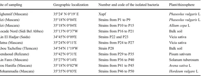
Table 1 Soil sampling, geographic localization and bacterial isolation
Site of sampling Geographic localization Number and code of the isolated bacteria Plant/rhizosphere Tighennif (Mascara) 35°24′ N 0°19′ E Xapf Phaseolus vulgaris L Tizi (Mascara) 35°18′N 0°04′E Strains from P1 to P9 Phaseolus vulgaris L Tizi (Mascara) 35°18′N 0°04′E Strains from P10 to P15 Allium cepa L Rocade Nord (Sidi Bel Abbes) 35°13′N 0°37′W Strains from P16 to P21 Balk soil Ain El Hadjar (Saida) 34°45′N 0°09′E Strains P22 and P23 Vicia sativa Slatna (Mascara) 35°24′N 0°11′E Strains from P24 to P27 Vicia sativa Abou Tachefine (Tlemcen) 34°54′N 1°19′W Strain P28 Balk soil Bendaoud (Relizane) 35°42′N 0°31′E Strains from P29 to P35 Pisum sativum Ain Fares (Mascara) 35°27′N 0°14′E Strains from P36 to P40 Solanum tuberosum Bou Hanifia (Mascara) 35°18′N 0°02′W Strains from P41 to P45 Avena sativa L Mohammadia (Mascara) 35°35′N 0°03′E Strains from P46 to P50 Hordeum vulgare L
the semi-selective medium PTSCA. The isolate’s identity was then confirmed by pathogenicity test on bean leaves.
BOX-PCR fingerprinting
DNA extraction was performed following William et al. (2012). Pure colonies of the isolated bacteria were treated by standard protocol for PCR manipu-lation. After DNA extraction, BOX-PCR were per-formed using mixtures of 1 × PCR buffer, 2 mM MgCl2,0.1 Mm dNTPs, 0.8 μM BOX-A1R primer (5′-CTA CGG CAA GGC GAC GCT GAC G-3′), 5% dimethylsulfoxyde, 1.3 U TaqDNA polymerase and standardized 15 ng of genomic DNA in a final volume of 30μL. The PCR mixtures were denatured (94 °C/1 min), subjected to 35 cycles (94 °C/1 min, 45 °C/1 min and 72 °C/2 min) and to a final exten-sion (72 °C/10 min). PCR products were checked on 1.5% agarose gel by electrophoresis (100 V/30 min) and revealed using Gel DOC (Biorad).
PGP traits characterization
Phosphate solubilization
Bacteria were streaked on Pikovskaya’s agar me-dium containing (per liter of distilled water): (0.5 g yeast extract, 10 g dextrose, 5 g Ca3(PO4)2,
0.5 g (NH4)2SO4, 0.2 g KCl, 0.1 g MgSO47H2O,
0.0001 g MnSO4H2O, 0.0001 g FeSO47H2O and
15 g agar, pH 7.4). After incubation at 28 °C/ 6 day (s), isolates inducing a clear zone around their colonies were considered as Phosphate Solu-bilizing Bacteria (PSB). Results were expressed as the clear halo diameter: PSI = A-B, where A is the total diameter (Colony + halo zone) and B is the colony diameter (Nautiyal 1999).
Siderophores production
Siderophores were detected on Chrome Azurol-S (CAS) agar medium. Isolates were streaked on Petri plates containing CAS medium and incubated at 30 °C for 48 h. Formation of orange halos around colonies indicated siderophore production by bacte-ria (Yeole et al. 2001). In addition, siderophores were quantified as described by Christina et al. (2015). Accordingly, Isolates were grown in
modified King’s B broth and incubated under con-tinuous agitation (28 °C/24–30 h/150 rpm). After incubation, cell free supernatant, obtained by centri-fugation (14.000×g/10 min at 4 °C), was mixed with 0.5 mL CAS solution. The obtained color was spec-trophotometrically (630 nm) recorded after 20 min incubation in darkness. A control, prepared with 0.5 mL CAS solution and 0.5 mL uninoculated King’s B broth, was used. Siderophore percentage was estimated through color variation as compared to the control using the formula: [(Ar - As) /Ar] × 100, where (Ar) is the control absorbance and (As) is the sample absorbance (CAS assay solution + Cell-free supernatant).
Hydrogen cyanide secretion
Hydrogen cyanide (HCN) production was screened as described by Lorck (1948). Bacterial isolates were streaked on nutrient agar supplemented with 0.44% L-glycine. Whatman paper discs were soaked in an alkaline picrate solution (0.5% picric acid, 2% Na2CO3) and deposited in the lids of Petri dishes.
The system was sealed with parafilm and incubated at 28 °C for 4 day(s).
To quantify HCN amount, the method described by Bakker and Schippers (1987) was performed. Thus, King’s B broth-containing tubes, amended with 0.44% filter sterilized L-glycine, were inoculat-ed with bacterial isolate. Whatman paper strips (10 cm × 1 cm) were soaked in an alkaline picrate solution and introduced at the tubes tops. Tubes were then sealed with parafilm and incubated at 28 °C/ 72 h. Color changes from yellow to brown or reddish brown indicates moderate or high HCN production, respectively. The total cyanide content (TCC) in Whatman paper was estimated by using the following equation: TCC (ppm) = 396 × OD (510 nm), where OD is the broth absorbance after incubation.
Indole acetic acid synthesis
Isolates were evaluated for their ability to synthesize IAA, largely known to stimulate plant growth. Therefore, Qualitative IAA estimation was deter-mined as described by Bric et al. (1991). Luria Bertani (LB) medium supplemented with 5 mM L-tryptophan was used. IAA and/or its analogs pro-duction in the medium lead to a red-pink color
formation. For other indoles, color becomes yellow or brown yellow.
Quantitative IAA evaluation was realized following the protocol of Jeyanthi and Ganesh (2013). 10 mL of medium were inoculated with fresh bacterial cultures then incubated under shaking for 72 h (28 °C/120 rpm). Then, 1 mL of supernatant, obtained by centrifugation (2.253×g/20 min at 4 °C), was mixed with 3 mL Salkowski’s reagent (50 mL of 35% of Perchloric acid, 1 mL of 0.5 M FeCl3). After incubation (room temper-ature in darkness), IAA amount was spectrophotometri-cally determined (540 nm). Finally, IAA concentration was estimated by the standard curve prepared using pure IAA solution (25–200 μg/mL).
Isolation of Xanthomonas axonopodis pv. phaseoli var. fuscans
To isolate the common blight-causing Xanthomonas axonopodis pv. phaseoli var. fuscans (Xapf), a Modified method of Saettler et al. (1989) was used. Bacteria was isolated from4 to 5 months old of infected Phaseolus vulgaris L plant showing common blight characteristic symptoms from Tighennif (Mascara) (Table1). There-fore, infected leaves were macerated in sterile demineralized water and serially diluted (up to 10−2) in sterile saline water. 100μL of each dilution were spread on Petri plates containing PTSCA medium and incubat-ed at 28 °C for 3 to 6 days. Typical colonies of Xapf were then purified and tested to confirm their pathoge-nicity on bean plants of trifoliate leaf stage (2 to 3 weeks after sowing). Infection was performed by soaking a sterile gauze in the pathogenic bacterial suspension (standardized to 108 CFU/mL using distilled water). The first two trifoliate leaves were weakly rubbed by the gauze on both sides and plants were maintained at 25 ± 5 °C under high humidity for 5 to 7 day (s).
In vitro antibacterial activity of the isolated bacteria against Xapf
Isolate antibacterial activity was tested following a mod-ified protocol of Skathivel and Gnanamanickam (1987). A standardized Xapf culture suspension was prepared by scraping 48–72 h-old culture. The suspension was then molted with a previously cooled (42 °C) King’s B agar and versed in Petri plates. From 7 day (s) old cultures, the antagonistic isolates were inoculated by deposing agar disks of (6 mm) on the plates already
inoculated with the pathogen. Controls without antago-nist (sterile agar disks) and without pathogen (sterile king’s B agar) were used. The plates were then incubat-ed at 28 °C and periodically checkincubat-ed to detect inhibition zones appearance around the antagonist agar disks dur-ing 2 to 3 days. The results of the antibacterial activity were expressed as measuring three times the clear halo diameter: Aa = D-d, where D is the total diameter (Col-ony + halo zone) and d is col(Col-ony diameter. Based on this test, two strains (P7 and P25) were selected and tested for their ability to inhibit the common blight and to enhance been germination.
Biocontrol of bean common blight caused by the isolated Xapf
The isolated bacteria that revealed in vitro antibac-terial activities were in vivo screened for their ability to biocontrol the common bean blight caused by our isolated pathogen Xapf (Al-Saleh 2014). Therefore, 48 h-old cultures of two selected bacteria (P7 and P25) were centrifuged (14.000×g for 10 min) and bacterial pellets were washed twice with sterilized distilled water. The strains’ optical densitywas ad-justed to (OD610 nm= 0.45) to obtain bacterial
sus-pensions of about 107CFU/mL. The phytopathogen Xapf was grown on King’s B agar medium and incubated at 28 °C for 48–72 h. Pure Xapf colonies were scraped from the cultures, re-suspended in sterile distilled water, and their optical density was spectrophotometrically adjusted to (OD610 nm= 0.6),
corresponding to 108CFU/mL.
Four treatments were realized to study the possi-ble role of the two selected in vitro antagonistic isolates and their combination [P (7) + P (25)] pro-portion (1:1) on Xapf propagation on bean leaves. Each treatment was composed of 10 plants and prepared by spraying bean leaves with the prepared bacterial suspensions. Three days later, plants were infected by spraying the Xapf suspension on their leaves until complete saturation of the leaves sur-face. Controls without treatment or with only infect-ed with the isolate Xapf were usinfect-ed. Inoculatinfect-ed plants were immediately enclosed in plastic bags at high humidity for 48 h.
At the end of the experiment, disease severity was estimated by measuring the percentage of infected leaves. In addition, symptoms intensity was deter-mined using a predefined scale (from F0 to F3): F0
(without task), F1 (one unbroken task with oily as-pect), F2 (1 to 3 brown tasks surrounded with a halo or leaf side-necrosis), F3 (50% of the leaf with pale or yellow necrosis with diffuse color) (Bernier2011).
Effect of the bacterial isolates on bean seed germination
The two bacterial isolates, previously assayed as biocontrol agents of common bean blight, were test-ed, individually and in combination, for their effect on bean seed germination using a modified protocol of Shweta et al. (2008). Seeds were disinfected with 2% sodium hypochlorite solution for 3 min then washed three times with sterile distilled water. Seeds of each treatment were soaked in a standardized bacterial suspension (107 CFU/mL) for 3 h, dried and deposited in agar water-containing Petri plates (0.8% agar). Controls with seeds soaked in sterile distilled water were similarly prepared. Three Petri dishes by treatment and 10 seeds in each of them were used. After incubation at 25 °C for 5 days in darkness, germinated seeds (G) and the number of lateral roots (NLR) were counted. A numerical meth-od was used to measure primary root lengths (PRL) and root area (RA) using the Image J software.
The stimulation percentage of each parameter (S %) as compared to the control was expressed using the formula: S % = [(testmv-controlmv)/controlmv)] × 100; where (controlmv) is the mean value of the control for each parameter, and testmv is the mean value of the treated seeds for the same parameter.
Statistical analysis
In vivo study of seed germination assays was analyzed by one-way ANOVA. Seed growth properties and bio-logical control of common blight were analyzed by two-way ANOVA. All results were statistically compared to their corresponding controls using Graph Pad Prism V. 6.0.The DendroUPGMA (available on:http://genomes. urv.cat/UPGMA/) was used to realize dendrograms of carbon sources assimilation and BOX-PCR profiling of the isolated bacteria. For this, the pattern was score specified with binary characters (1 = positive growth and 0 = absence of growth) for carbon sources assimila-tion and (1 = presence and 0 = absence of band) for BOX-PCR profiling (Garcia-Vallve et al.1999).
Results
Identification and characterization of the isolated bacteria
Biochemical and physiological identification of 50 bacterial isolates (supplementary material) had shown high metabolic diversity. Thirty-four isolates developed pale green pigmentation in King’s B agar medium and liberated a characteristic odor of soft mist. Exposing of Petri plates to UV light under (365 nm) revealed a fluorescent green pigment. Two isolates produced pyorubin (red pigment) which is a specific pigment of the species Pseudo-monas aeruginosa. That indicated that bacteria forming a total of 36 (72%) isolates belong to the fluorescent Pseudomonas group; whereas, 14 (28%) bacterial isolates do not produce this specific char-acteristic pigment, indicating that these isolates be-long to the non-fluorescents Pseudomonas group. Within 15 days incubation, 49 (98%) isolates had shown positive reaction for oxidase, 37 (74%) iso-lates for reduction of nitrates and 50 (100%) isoiso-lates solubilize KOH. They were able to assimilate most of carbon sources tested: 45 (90%) isolates assimi-lated glucose, 50 (100%) isolates assimiassimi-lated ala-nine, 20 (40%) isolates assimilated arginine and 27 (54%) isolates assimilated xylose. 15 (30%) isolates assimilated L-tryptophan, 31 (62%) isolates assimi-lated sodium tartrate and 29 (58%) isolates liquefied gelatin. 2 (4%) isolates only accumulated PHB. A total of 19 (38%) isolates grew at 4 °C and 11 (22%) isolates grew at 41 °C.
Biochemical and physiological characterization
Identification of bacterial isolates by ABIS software revealed that 36 (72%) isolated bacteria belonged to fluorescent Pseudomonads, whereas the non-fluorescent Pseudomonads were represented by 14 (28%) isolates. The predominance of Pseudomonas fluorescens Biovars (1, 2, 3, 4 and 5) 23 (44%) was clearly observed. Among them, Pseudomonas fluorescens Biovars 5 was represented with 10 isolates (20%). Moreover, Pseudomonas putida Biovars (A) and Pseudomonas cichorii were represented by four isolates for each one. The less represented species principally belong the non-fluorescent Pseudomonads: P. stutzeri 3 (6%), P. pseudomallei 2 (4%), P. plantarii 2 (4%),
P. cepatia 2 (4%) isolates, P. corrugota 1 (2%), P. lini 1 (2%) and P. maltophila 1 (2%) isolate.
The distribution of the dominant isolated bacteria P. fluorescens Biovar 5 according to their samples showed their abundance in Pisum sativum (Bendaoud) and Hordium vulgare L (Mohamadia) (Table2).
Numerical analysis of phenotypic characteristics revealed high polymorphism among Pseudomonas isolates. The 50 isolates tested are grouped into five major phenons (Fig. 1). Phenons 1, 2, 3, 4, and 5 that consist a total of 2 (4%), 9 (18%), 11 (22%), 3 (6%) and 25 (50%) isolates, respectively. Phenon 5 was the main and diversifies, assembling fluores-cent and non-fluoresfluores-cent Pseudomonads species, including isolates belonging to P. putida Biovar A. The second major phenons 2 and 3 regrouping
heterogeneous Pseudomonas species. P. fluorescens Biovar 5 isolates were distributed between these two clusters. Phenons 1 and 4 were the less repre-sented phenons, comprising only isolates belonging to Pseudomonas alcaligenes and Pseudomonas stutzeri, respectively.
BOX-PCR fingerprinting
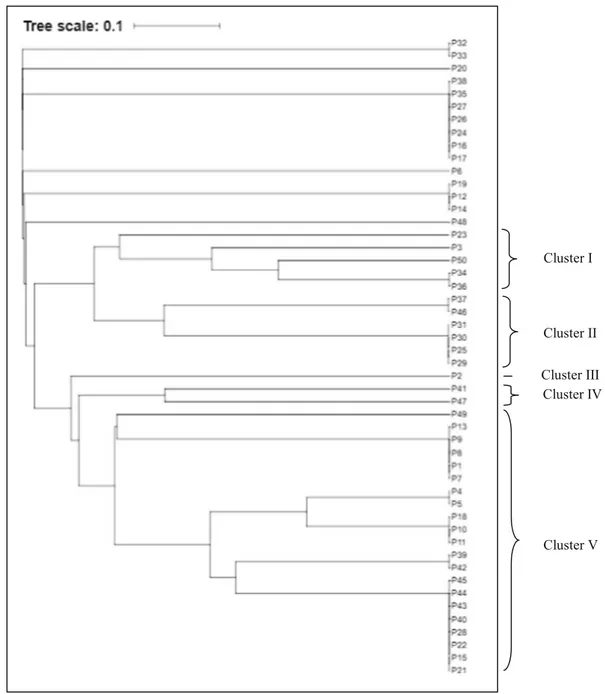
Molecular diversity of Pseudomonas isolates was also confirmed by the molecular level. BOX-PCR showed 29 haplotypes characterized by 1 to 7 bands, ranging from 100 bp to 1600 bp size. Nu-merical BOX-PCR analysis differentiated the spe-cies belonging to Pseudomonas. DNA fingerprinting pattern result in a dendrogram showing five differ-ent clusters (Fig. 2). Cluster V formed the major cluster, comprising 21 (42%) isolates belonging to fluorescent and non-fluorescent Pseudomonads groups, including most isolates belonging to P. putida Biovar A. Clusters I and II were the second major cluster, comprising 5 (10%) and 6 (12%) isolates respectively. Those two clusters grouped the largest part of Pseudomonas fluorescens Biovar 5 isolates. Clusters III and IV were the less represented clusters including 1 (2%) and 2 (4%) isolates, respectively. 14 (28%) isolates did not fall into the dendrogram and revealed together their distinct profile and genotypic identities.
BOX-PCR profiling results analysis indicate better classification of the isolates taxonomically and simulta-neously with the carbon source assimilation. Compari-son between BOX-PCR profiling and metabolic groups suggested this observation is valid. Clear resemblances among phenons and clusters were observed.
PGP trait characterization
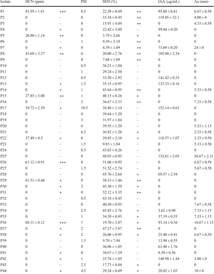
Functional properties of the isolated bacteria showing qualitative and/or quantitative production of secondary metabolites were regrouped in (Table3).
Phosphate solubilization
33 (66%) Pseudomonas isolates induced solubiliza-tion of tri-calcium phosphate on Pikovskaya’s medi-um by forming clear zones around the colonies. Whereas, 22 (34%) isolates did not show any halo around their colonies, indicating that they do not Table 2 Results of biochemical identification, obtained using the
Bacterial Identification Software (ABIS) Isolate Species Isolate Species P1 P. pseudomallei P26 P. grimontii P2 P. plantarii P27 P. grimontii P3 P. corrugota P28 P. cichorii
P4 P. alcaligenes P29 P. fluorescens Biovar 5 P5 P. stutzeri P30 P. fluorescens Biovar 5 P6 P. plantarii P31 P. fluorescens Biovar 5 P7 P. cepatia P32 P. alcaligenes P8 P. pseudomallei P33 P. fluorescens Biovar 1 P9 P. cepatia P34 P. fluorescens Biovar 2 P10 P. fluorescens Biovar 2 P35 P. fluorescens Biovar 5 P11 P. fluorescens Biovar 2 P36 P. fluorescens Biovar 5 P12 P. stutzeri P37 P. fluorescens Biovar 5 P13 P. fluorescens Biovar 2 P38 P. fluorescens Biovar 3 P14 P. fluorescens Biovar 3 P39 P. fluorescens Biovar 3 P15 P. aeruginosa P40 P. putida Biovar A P16 P. fluorescens Biovar 2 P41 P. fluorescens Biovar 2 P17 P. stutzeri P42 P. aeruginosa P18 P. cichorii P43 P. putida Biovar A P19 P. lini P44 P. putida Biovar A P20 P. fluorescens Biovar 2 P45 P. cichorii
P21 P. cichorii P46 P. fluorescens Biovar 5 P22 P. maltophila P47 P. fluorescens Biovar 3 P23 P. fluorescens Biovar 4 P48 P. fluorescens Biovar 5 P24 P. putida Biovar A P49 P. fluorescens Biovar 5 P25 P. grimontii P50 P. fluorescens Biovar 5
solubilize tricalcium phosphates. Phosphate solubili-zation index (PSI) ranged from 0.5 to 10.5 after 6 days inoculation. Isolate P17 induced the highest PSI (10.5); moderate PSI was registered for P44 and P45 (4.5), while P20 and P21 gave a PSI of about 6 and 6.5, respectively. The weakest PSI was recorded for isolates P1, P10, P12, P24 and P32 (0.5). Other isolates induced PSI index varying from 1 to 3 or did not solubilize phosphate.
Siderophores production
After 72 h of incubation, siderophore production by 28 (56%) Pseudomonas isolates on CAS agar
medium showed the appearance of clear zones with orange color, representing iron chelation. 22 (34%) isolates do not show any change surround growth. Quantitative analysis using CAS solution revealed that all isolates produced siderophores. Percentages of siderophores production varied from 3.79 ± 2.66% to 52.12 ± 3.35% for isolates P5 and P31, respectively.
Hydrogen cyanide secretion
Evaluation of the capacity of isolates to produce hydro-gen cyanide revealed a qualitative change of Whatman paper N°1 color from yellow to reddish brown that
Phenon 1
Phenon 5 Phenon 4 Phenon 3 Phenon 2
confirmed production of HCN. Of all isolates tested, 24 (48%) had shown HCN production on nutrient agar and 26 (52%) shown negative production. Quantitative HCN production in King’s B broth revealed only 12 (24%) of isolates producers and 38 (76%) shown a negative result. Isolate P45 had exhibited the lowest production of 20.59 ± 2.86 ppm, whereas the highest production was unregistered for the isolate P1 of 83.95 ± 1.81 ppm. Other isolates had shown moderate or none HCN production.
Indole acetic acid synthesis
Qualitative IAA production revealed that 28 (56%) iso-lates produced IAA. 22 (43%) isoiso-lates shown negative results. Quantitative IAA production on TSB broth sup-plemented with (0.5%) of L-tryptophan revealed 21 (42%) isolates IAA producers; whereas, 29 (48%) iso-lates did not produce IAA. A varying level of IAA production was recorded for the low production of 3.62 ± 0.90μg/ml for isolate P34, to the high production
Cluster I
Cluster V Cluster IV Cluster III Cluster II
Table 3 PGP traits: qualitative and quantitative estimation
Isolate HCN (ppm) PSI SED (%) IAA (μg/mL) Aa (mm)
P1 83.95 ± 1.81 +++ 0.5 22.58 ± 0.69 ++ 95.88 ± 0.41 6.67 ± 0.58 P2 0 – 0 33.18 ± 0.45 ++ 119.85 ± 32.1 4.00 ± 0 P3 0 – 0 15.91 ± 4.04 ++ 0 4.33 ± 0.58 P4 0 + 0 22.42 ± 3.09 – 99.04 ± 0.20 0 P5 26.80 ± 1.14 ++ 0 3.79 ± 2.66 + 0 0 P6 0 – 1 9.39 ± 3.19 ++ 0 0 P7 0 + 0 4.39 ± 1.89 ++ 73.69 ± 0.20 24 ± 0 P8 43.69 ± 3.37 ++ 0 20.00 ± 2.76 ++ 105.00 ± 2.34 0 P9 0 – 0 7.88 ± 1.89 ++ 0 0 P10 0 – 0.5 36.21 ± 1.84 – 0 0 P11 0 – 1 29.24 ± 2.50 – 0 0 P12 0 – 0.5 35.30 ± 2.92 – 141.42 ± 0.35 0 P13 0 + 1.5 35.15 ± 0.95 – 133.33 ± 8.16 0 P14 0 + 1 43.64 ± 0.95 ++ 0 5.33 ± 0.58 P15 27.85 ± 3.08 ++ 1 40.15 ± 0.26 + 0 0 P16 0 – 2 36.67 ± 2.33 ++ 0 7.33 ± 0.58 P17 39.73 ± 2.39 + 10.5 38.48 ± 1.14 – 152.14 ± 0.61 0 P18 0 – 0 38.64 ± 1.20 – 0 0 P19 0 – 3 31.97 ± 1.84 – 0 0 P20 0 – 6 39.55 ± 1.20 – 0 5.33 ± 1.15 P21 0 – 6.5 36.82 ± 1.20 + 0 2.33 ± 0.58 P22 37.49 ± 8.5 + 0 39.85 ± 2.10 + 118.57 ± 1.07 3.33 ± 0.58 P23 0 – 1.5 9.85 ± 1.84 – 0 5.33 ± 0.58 P24 0 – 0.5 43.03 ± 0.26 – 0 0 P25 0 – 0 48.03 ± 0.95 – 132.61 ± 2.03 26.67 ± 2.31 P26 61.12 ± 0.91 +++ 0 51.06 ± 0.95 – 0 6.67 ± 0.58 P27 0 – 1.5 51.52 ± 2.74 – 0 5.67 ± 0.58 P28 0 – 0 45.76 ± 2.66 + 69.57 ± 2.58 0 P29 61.51 ± 0.46 + 0 38.33 ± 1.46 ++ 0 0 P30 0 + 2 45.30 ± 1.39 + 0 0 P31 0 + 0 52.12 ± 3.35 ++ 0 0 P32 0 – 0.5 43.18 ± 0.45 – 0 0 P33 0 – 0 40.30 ± 0.95 – 0 7.67 ± 0.58 P34 0 – 2.5 45.45 ± 2.76 + 3.62 ± 0.90 7.33 ± 1.15 P35 0 – 1 34.39 ± 0.95 + 37.19 ± 0.55 7.33 ± 1.15 P36 68.11 ± 4.12 +++ 1 19.70 ± 3.87 + 93.14 ± 0.36 10.67 ± 1.15 P37 0 + 2 47.27 ± 3.87 ++ 0 0 P38 0 + 2 26.06 ± 0.95 + 25.40 ± 0.41 6.67 ± 0.58 P39 0 – 1.5 9.70 ± 7.94 – 13.98 ± 0.55 0 P40 0 – 0 36.06 ± 1.05 – 61.48 ± 1.76 0 P41 0 + 0 36.67 ± 1.39 – 8.50 ± 0.36 0 P42 0 + 1 25.76 ± 1.05 – 148.98 ± 1.44 2.00 ± 0 P43 0 – 2.5 17.73 ± 4.04 + 0 0 P44 0 + 4.5 29.24 ± 0.69 + 28.02 ± 1.03 10 ± 0
of 152.14 ± 0.61 μg/ml for isolate P17 and 148 ± 1.44μg/ml for isolate P42.
Isolation of Xanthomonas axonopodis pv.phaseoli var. fuscans
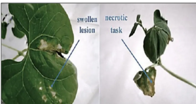
Phytopathogenic isolate Xapf had shown characteristic colonies of Xanthomonas axonopodis pv. Phaseoli on the semi-selective medium PTSCA (Fig. 3). Colonies were yellow, round, mucoid with whole margins. In addition, the fuscans type was distinguished by diffusion of brown pigment, after incubation at 28 ± 2 °C for 3 to 6 days. Confirmation of Xapf identification was obtained by verification of pathogenicity on bean leaves; in the initial stage of the disease (Fig. 4), symptoms infection on leaves appears as a small lesion soaked with water. When the disease progresses (Fig.4), lesions become larger and were developed to brown, dry tasks with yellow halos. These lesions consist of irregular regions of dry brown tissue, frequently produced at the margins of leaves.
In vitro antibacterial activity of the isolated bacteria against Xapf
Results obtained in the present study by the dual culture technique revealed that 23 (46%) of Pseudomonas iso-lates exert potent antibacterial activity against Xanthomonas axonopdis pv. phaseoli var. fuscans (Table 3). The highest antibacterial activities recorded were 26.67 ± 2.31 mm for P. grimontii P25, followed by 24.00 ± 0 mm for P. cepatia P7.The lowest antagonistic activity was registered for P42 Pseudomonas aeruginosa of 2.00 ± 0 mm.
Biocontrol of bean common blight caused by the isolated Xapf
The two antagonistic isolates P. grimontii P25 and P. cepatia P7 were tested for their biological control potential against common blight been caused by the pathogenic isolate Xapf (Fig. 5). Application of isolates P7 and P25 alone or in combination reduced the number of common blight tasks on bean leaves as compared to the control leaves only infected by the pathogenic isolate.
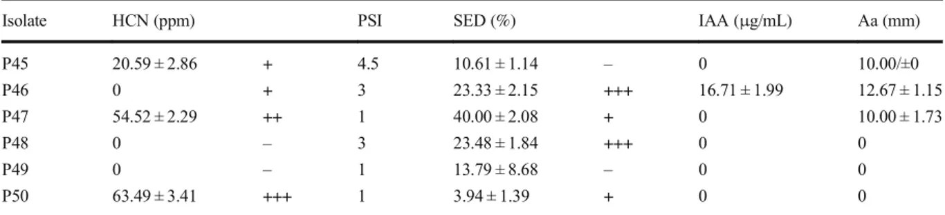
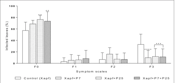
Biological control results analyzed by ANOVA indicated that application of the two isolates P7 or P25 alone or in combination decreased the disease intensity (Fig. 6). Significant decrease of symptom scales F0 (leaf without task) and F3 (50% of the leaf with pale or yellow necrosis with diffuse color) were observed, whereas symptom scales F1 (one unbroken task with oily aspect) and F2 (1 to 3 brown tasks surrounded with a halo or leaf side-necrosis) were not highly affected. The highest increase of non-infected leaves, represented by scale F0, was obtained through inoculation by the Table 3 (continued)
Isolate HCN (ppm) PSI SED (%) IAA (μg/mL) Aa (mm)
P45 20.59 ± 2.86 + 4.5 10.61 ± 1.14 – 0 10.00/±0 P46 0 + 3 23.33 ± 2.15 +++ 16.71 ± 1.99 12.67 ± 1.15 P47 54.52 ± 2.29 ++ 1 40.00 ± 2.08 + 0 10.00 ± 1.73 P48 0 – 3 23.48 ± 1.84 +++ 0 0 P49 0 – 1 13.79 ± 8.68 – 0 0 P50 63.49 ± 3.41 +++ 1 3.94 ± 1.39 + 0 0
Qualitative and/or quantitative determination of PGP traits PSI: phosphate solubilization index. SED: Siderophores. Aa: Antibacterial activity. (+++) Highly positive reaction, (++) Moderate positive reaction, (+) Low positive reaction, (−) Negative reaction
Fig. 3 Characteristic brown diffusible pigment of Xanthomonas axonopodis pv. phaseoli var. fuscans isolates on PTSCA medium
isolate P25 (71.82%) followed by co-inoculation P7 + P25 (73.6%) as compared to the control (57.2%). The three inoculations decreased diseased intensity scale F3. Highest decrease was recorded through inoculation by isolate P7 up to (9.8%) compared to the control (33%); followed by the isolate P25 and co-inoculation with P7+ P25 (11.3 and 11.94%, respectively).
Effect of the isolated bacteria on bean seed’s germination
Inoculation of Phaseolus vulgaris seed with Pseu-domonas isolates P. cepatia P7, P. grimontii P25, and co-inoculation with P7 + P25 increased seed germination as compared to uninoculated controls (Fig. 7). Application of isolate P7 alone was highly Fig. 4 Pathogenicity test of
Xanthomonas axonopodis pv. phaseoli var. fuscans (Xapf) showing swollen lesion as initial stage of common blight bean (on the left) and necrotic task as advanced stage of common blight bean (on the right)
Fig. 5 High contrast-uncolored leaves pictured from the experi-mentBbiological control of common blight caused by Xapf^. a: leaves only infected by the pathogen Xapf (control). b: leaves infected by the pathogen Xapf and treated by the combination
P7 + P25. c: leaves infected by the pathogen Xapf and treated by P25. d: leaves infected by the pathogen Xapf and treated by P7. Black regions of leaves indicate unaffected zones
effective on seed germination. A significant increase of seed germination when treated with isolate P7 was recorded (66.6%). However, P25, alone and in combination with P7, did not show any significant increase in seeds germination rate.
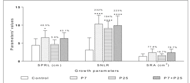
Results obtained from seedling growth properties treated by Pseudomonas isolates, applied alone or in combination, shown a significant increase of SNLR (Stimulation of Number of Lateral Root) and SPRL (Stimulation of Primary Root Length). Root areas SRA (Stimulation of Root Area) was not highly enhanced (Fig.8). Application of isolate P7 increased (SNLR) up to 232% and co-inoculation P7 + P25 up to 223%.
Inoculation of seed by P7 increase (SPRL) up to 48.5%. Thus, P7 was highly effective on stimulation of seed growth properties. Followed by the co-inoculation P7 + P25 that revealed considerable stimulation of (SNLR).
Discussion
In the last 20 years, bacteria of the genus Pseudomonas have occupied an important place among the most stud-ied soil bacteria. Their remarkable capacity to inhibit several phytopathogenic agents in soil and to enhance
0 2 0 4 0 6 0 8 0 1 0 0 X a p f+ P 7 X a p f+ P 2 5 X a p f+ P 7 + P 2 5 C o n t r o l ( X a p f ) * * * * * * * * * * * F 0 F 1 F 2 F 3 In fe cte d le a ve s (% ) S y m p to m s c a le s
Fig. 6 Biological control of common blight intensity caused by Xapf by Pseudomonas. Values represent the percentages of infect-ed leaves. F0, F1, F2 and F3 represent symptom scales as shown in
material and methods (**: significant difference at p < 0.01; ***; significant difference at p < 0.005; ****: significant difference at p < 0.001) Ge rmi n at e ds e e d s( 1 0s e e d s /p la te ) 0 2 4 6 8 1 0 6 6 . 6 % * 1 3 . 2 % 2 0 % C o n tr o l P 7 P 2 5 P 7 + P 2 5 T r e a t m e n t s
Fig. 7 Effect of Pseudomonas isolates on seed germination of Phaseolus vulgaris L. Values represent the average number of germinated seeds. Percentages represent comparisons to control. (*: significative difference at p < 0.05)
plant growth is mainly due to their high metabolic diversity (Wei et al.1996).
In this work Pseudomonas fluorescens Biovar (1, 2, 3, 4 and 5) was the most isolated species, representing 22 (44%). Biovar 5 represented the most dominant Biovar with 20% isolates, followed by P. putida Biovar A and P. cichorii 4 with 8% isolates for each. This was probably due to the medium selectivity (cetrimide) and the incubation temperature (from 30 °C to 37 °C). We also revealed the abundance of P. fluorescens Biovar 5 in Pisum sativum and Hordeum vulgare rhizospheric soil samples. Similar results were reported by Meliani (2012), mentioning that among 70 strains, the dominat-ing Pseudomonas bacteria from divers rhizospheric plant were P. fluorescens and P. putida, representing 38.57% of the total. Sutra et al. (2000) also reported that among 58 fluorescent Pseudomonas isolates, 41 were identified as P. fluorescens Biovar 5 and 17 as P. putida Biovar A in banana rhizospheric soil.
The capacity of Pseudomonas to use different carbon sources is an important characteristic in microorganism natural selection. The characterization results of our iso-lates showed their ability to use most of the tested carbon sources, which is consistent with the results reported by Palleroni (1993). The same author revealed the high metabolic and ecological diversity of the Pseudomonas genus. Otherwise, Bossis et al. (2000) confirmed that Levan production, denitrification, gelatin liquefaction and L-tryptophan assimilation are characteristic of differ-ent Biovars of P. putida and P. fluorescens.
Pseudomonas capacity to assimilate several carbon sources can affect and contribute to their diversity in a specific environment. According to Zak et al. (1994), the rhizobacterial functional diversity is defined as the degradation rate that determines the whole comport-ment of a microbial community against several sub-strates assemblage. The Pseudomonas diversity ob-served in our results (five physicochemical phenons) may occur from their extracellular enzymes’ diversity and their high adaptability from one rhizospheric soil to another. As the phylogenic diversity that was inves-tigated by BOX-PCR profiling showing five distinct clusters forming from the lowest (2%) to the highest (47.62%) of the isolated species. BOX-PCR uses re-petitive extragenic palindromic-PCR (BOX A1R). This single primer binds to repetitive sequences in bacterial genomes that are conserved and specific for each species (Fitriyah et al. 2013). In this study, 14 (28%) isolates did not fall into any of the clusters, indicating the high Pseudomonas variability and di-versity in western Algeria.
Physicobiochemical and phylogenic diversity of Pseudomonas isolates is probably due to the charac-teristics of soil samples and the types of plant rhizo-spheres from which they were isolated. Plant root exudates could be largely implicated in determining the soil microbial diversity and metabolic/functional diversity of rhizospheric bacteria (Lakshmi et al.
2015). In addition, the environmental characteristics, differing from one sampling site to another, are also
0 5 1 0 1 5 C o n t r o l P 7 P 2 5 P 7 + P 2 5 S P R L ( c m ) S N L R S R A ( c m2) 4 8 . 5 % * 2 3 2 % * * * * 2 2 3 % * * * * 1 9 4 % * * * * 5 . 8 % 4 3 . 1 % 7 7 . 8 % 1 8 . 7 % 7 8 . 7 % Pa ra m et er s' va lu es G r o w t h p a r a m e t e r s
Fig. 8 Effect of Pseudomonas isolates on seed growth properties of Phaseolus vulgaris L. Values represent the stimulation of the properties of germinated seeds. The percentages represent the stimulation of seed properties compared to the control (100%).
SPRL: Stimulation of primary root length. SNLR: Stimulation of number of lateral roots. SRA: stimulation of root area (*: signifi-cant difference at p < 0.05; ****: signifisignifi-cant difference at p < 0.0001)
determining factors of the obtained bacterial diversity (Fierer and Jackson 2006). Our results confirm that the genus Pseudomonas is able to colonize the rhizo-spheres of several plants, being highly competent in environments with different biological and physico-chemical characteristics (Wei et al.1996).
The main interest in investigations with different species of the genus Pseudomonas, other than their high diversity and rhizo-competence, lies in their ability to produce several metabolites of high agricultural interest such as IAA, siderophores, hydrolytic enzymes and antibiotic compounds (Dorjey et al.2017). In this work, 33 (66%) isolates showed high ability to solubilize insoluble phosphate with a PSI ranging from 0.5 to 10.5. Several reports indicated that different bacterial species, particularly rhizosphere colonizing bacteria, have the ability to liberate organic phosphates or to solubilize insoluble inorganic phosphate compounds. These bacteria make available the soluble phosphates to plants and, in return, gain root-borne carbon com-pounds, mainly sugars and organic acids, necessary for bacterial growth (Khan et al. 2010). In addition, all isolates tested in this study produced siderophores. Siderophores are low molecular weight metabolites that play an important role in supplying iron to plants (Radzki et al.2013). Bacteria of the genus Pseudomonas are known to produce high amounts of siderophores such as pyochelin, pseudobactin and pyoverdine (Neilands1989; Mezaache et al.2014). Such character-istics are largely used to select beneficial soil bacteria that help to supply iron to plant, but also to compete with other pathogenic organisms for the limited available-iron in soil. Siderophores production by bacteria is a key mechanism by which they eliminate pathogenic fungi by competition, and hence promoting plant growth and defenses (Peer et al.1990). Moreover, siderophores are important for their producers survival, colonization and iron metabolism control (Adams et al.1994).
Twenty-four percent of the isolated Pseudomonas showed ability to produce HCN. It is a secondary me-tabolite, commonly produced by rhizosphere Pseudo-monads. HCN may directly enhance plant growth or inhibit plant pathogens development (Schippers et al.
1990). 24% of the isolated bacteria produced HCN. HCN production depends the substrate amino acids composition. Thus, glycine is the direct precursor of microbial cyanide production (Askeland and Morrison
1983). The rate of microbial HCN production in the rhizosphere also depends on the amino acid composition
of root exudates. In addition, environmental factors such as light intensity and soil water potential may also affect root exudation rate, exudate composition, and hence HCN production (Schippers et al. 1990). HCN can indirectly interfere with phosphorus availability and, when synthesized by a PGPB, it appears to act syner-gistically with other biocontrol traits employed by the same bacterium (Hinsinger 2001; Olanrewaju et al.
2017). Moreover, HCN is known as an inhibitor of the cytochrome c oxidase as well as other important micro-bial metallo-enzymes, participating in pathogen biocon-trol and plant growth enhancement (Nandi et al.2017). IAA-producing PGPR are believed to increase root growth and root length (Vessey 2003). In this study, 21% isolates produced IAA. Isolates P7 and P25 produced 73.69 ± 0.20 and 132.61 ± 2.03μg/mL IAA, respectively. Resulting in a significant increase of bean SNLR and SPRL. IAA production controls a wide variety of processes in plant development and plant growth: low concentrations of IAA can stimu-late primary root elongation; whereas high IAA levels stimulate the formation of lateral roots, de-crease the primary root length, and inde-crease root hair formation (Vacheron et al.2013).
The high in vitro antibacterial activities of the two isolates P. grimontii P25 and P. cepatia P7 could be explained by their ability to secrete one or a combination of different PGP compounds (Dorjey et al.2017). Qual-itative and quantQual-itative characterization of secondary metabolites such as phosphate solubilization, siderophores, HCN and IAA are the main characteristics in PGPR selection. Such metabolites can contribute to Xapf biological control, but also to bean growth stimu-lation observed in this work. Chin-A-Woeng et al. (2003) reported that the metabolic diversity of PGPR makes of them interesting candidates as biofertilizers and biological control agents of plant diseases. In addi-tion, their high ability to colonize roots and to maintain strong density and diversity population is probably due to their rhizocompetence and their capacity to metabo-lize efficiently several root exudates compounds.
This study of biological control of Xapf by isolated bacteria revealed that isolate P. cepatia P7, reduced significantly the disease intensity, then isolate P. grimontii P25 or co-inoculation with P7 + P25, al-though isolate P25 applied in vitro gave the highest antibacterial activity. P. cepatia strain was used as a potentially effective biological control agent by Cart-wright and Benson (1995). Bacterial biocontrol agents
(BCA) effective in vitro are not necessarily effective in vivo. Giorgio et al. (2016) reported that out of 162 Rhizobacteria isolates, 60 inhibited the growth of com-mon bacterial blight (CBB) in in vitro condition. But only six of these, when applied to seeds, reduced disease symptoms in in vitro and greenhouse assays. Corrêa et al. (2017) suggest also a relationship between the region of origin of Xapf strains and the efficacy of BCA to control the commune blight bean.
In this study, P. cepatia P7 alone induced the highest significant seed germination rate. Also, seed growth analysis revealed that the isolate P7 was the most effec-tive on seed growth stimulation (SNLR and SPRL). This bacterium was isolated from rhizospheric soil of Phaseolus vulgaris, which presupposes that the isola-tion origin could play an important role in seed germi-nation, bacterial colonization and adaptation. The co-inoculation with P7 + P25 was less effective, probably due to competition that maintained a lower density population of isolate P7. Lemanceau (1992) reported that some bacterial strains, belonging in particular to the group of Pseudomonas fluorescent spp., seem to improve the germination of seeds of different plants (colza, corn and tomato). Kapilan and Thavaranjit (2015) reported also that the application of Pseudomo-nas strains showed high promoting effect on bean seed germination. Boruah et al. (2003) reported that bacteri-zation bean seed with fluorescent Pseudomonas strains significantly increased the root biomass and length of roots. Bacterial strains produced more than one-fold increase in root length that ranged from 13 to 17%. The inoculation of bean with bacterial strains P. extremorientalis TSAU20 and P. chlororaphis TSAU13 increased root length, even under saline stress conditions (Egamberdieva 2011). Kragelund and Nybroe (1996) mentioned that there was competition between Pseudomonas fluorescens Ag1 and Alcaligenes eutrophus JMP134 in co-inoculation (pJP4) during col-onization of barley roots.
Variation in the degree of promoting effect on seed germination depends on the variety of plant seeds, bacterial species used, the rate of colonization, amount of substance causing the effect, and the in-ternal physical factors of seeds (Carrillo-Castañeda et al. 2002; Krishnaswamy and Seshu 1990). The ability of a strain or bioinoculant combination to promote plant growth changes with the cultivated variety. This could be due to the variation in the rhizosphere dynamics of a given bacterial isolate
which in turn could be caused by the change in plant root exudates (Kumar et al. 2015). In terms of this in vivo study, we can conclude that application of isolate P. cepatia P7 was more effective on seed germination, seed growth properties, and biological control of Xapf compared to P. grimontii P25 or their concomitant application. Isolate P7 was isolated from rhizospheric soil of Phaseolus vulgaris, when inocu-lated on the same plant, it showed high effectiveness as biological agent and biofertilizer. It shows the crucial role of origin isolation in biological control of plant disease and growth stimulation, besides the traits properties of PGPR.
Conclusion
In conclusion, the selected Pseudomonas isolates could play a crucial role in common bean blight biocontrol and beans yield promotion through siderophore, HCN and IAA production, phosphate solubilization, and other metabolite secretions. The selected strains also showed high stimulation of bean seed germination. Further stud-ies are needed to investigate their security and their long-term effect on plants, soil and the microbial com-munity of the inoculated environment. Finally, these bacteria could present promising agricultural applica-tions in the future.
References
Abd-Alla, M. H., El-Enany, A. W. E., Nafady, N. A., Khalaf, D. M., & Morsy, F. M. (2014). Synergistic interaction of Rhizobium leguminosarum bv. viciae and arbuscular mycor-rhizal fungi as a plant growth promoting biofertilizers for faba bean (Viciafaba L.) in alkaline soil. Microbiological Research, 169(1), 49–58.
Adams, C., Dowling, D. N., O'Sullivan, D. J., & O'Gara, F. (1994). Isolation of a gene (pbsC) required for siderophore biosynthesis in fluorescent Pseudomonas sp. strain M 114. Molecular Genetics and Genomics.
https://doi.org/10.1007/bf00284199.
Al-Saleh, M. A. (2014). Evaluation of Saudi fluorescent pseudo-monads isolates as a biocontrol agent against citrus canker disease caused by Xanthomonas citri subsp citri. Egyptian Academic Journal of Biological Sciences, 6(2), 1–7. Armada, E., Roldán, A., & Azcon, R. (2014). Differential
activity of autochthonous bacteria in controlling drought stress in native Lavandula and Salvia plants species under drought conditions in the natural arid soil. Microbial Ecology, 67(2), 410–420.
Askeland, R. A., & Morrison, S. M. (1983). Cyanide produc-tion by Pseudomonas fluorescens and Pseudomonas aeruginosa. Applied and Environmental Microbiology, 45(6), 1802–1807.
Bakker, A. W., & Schippers, B. (1987). Microbial cyanide pro-duction in the rhizosphere in relation to potato yield repro-duction and Pseudomonas spp-mediated plant growth-stimulation. Soil Biology and Biochemistry, 19, 451–457.https://doi. org/10.1016/0038-0717(87)90037-x.
Beneduzi, A., Ambrosini, A., & Passaglia, L. M. (2012). Plant growth-promoting rhizobacteria (PGPR): Their potential as antagonists and biocontrol agents. Genetics and Molecular Biology, 35, 1044–1051. https://doi.org/10.1590/s1415-47572012000600020.
Bernier, M. (2011). Étude de la variabilité des symptômes pathologiques affectant le haricot commun. In Ecole Supérieure d'Agriculture. Institut Nationale de Recherche Agronomique, FRA. http://prodinra.inra.fr/ft?id=4F54 AFCF-2B62-483F-A833-D1CC5DADDADA. Accessed 22 July 2014.
Boruah, H. P. D., Rabha, B. K., Saikia, N., & Kumar, B. S. D. (2003). Fluorescent Pseudomonas influences palisade meso-phyll development and spatial root development in Phaseolus vulgaris. Plant and Soil, 256, 291–301.
https://doi.org/10.1023/A:1026197300684.
Bossis, E., Lemanceau, P., Latour, X., & Gardan, L. (2000). The taxonomy of Pseudomonas fluorescens and Pseudomonas putida: Current status and need for revision. Agronomie, 20, 51–63.https://doi.org/10.1051/agro:2000112.
Bric, J. M., Bostock, R. M., & Silverstone, S. E. (1991). Rapid in situ assay for indole acetic acid production by bacteria immobilized on nitrocellulose membrane. Applied and Environmental Microbiology, 57(2), 535–538.
Broughton, W. J., Hernandez, G., Blair, M., Beebe, S., Gepts, P., & Vanderleyden, J. (2003). Beans (Phaseolus spp.) -model food legumes. Plant and Soil. https://doi. org/10.1023/a:1024146710611.
Carrillo-Castañeda, G., Muños, J. J., Peralta-Videa, J. R., Gomez, E., Tiemannb, K. J., Duarte-Gardea, M., & Gardea-Torresdey, J. L. (2002). Alfalfa growth promotion by bacteria grown under iron limiting conditions. Advances in Environmental Research, 6, 391–399. https://doi. org/10.1016/s1093-0191(02)00054-0.
Cartwright, D. K., & Benson, D. M. (1995). Comparison of Pseudomonas species and application techniques for biocon-trol of Rhizoctonia stem rot of Poinsettia. Plant Disease.
https://doi.org/10.1094/pd-79-0309.
Chin-A-Woeng, T. F. C., Bloemberg, G. V., & Lugtenberg, B. J. J. (2003). Mechanisms of biological control of phytopathogen-ic fungi by Pseudomonas spp. In G. Stacey & N. T. Keen (Eds.), Plant-microbe interactions (pp. 173–225). The American Phytopathologial Society: Saint Paul.
Cho, J. C., & Tiedjen, J. M. (2000). Biogeography and degree of endemicity of fluorescent pseudomonads strains in soil. Applied and Environmental Microbiology, 66, 5448–5456.
https://doi.org/10.1128/aem.66.12.5448-5456.2000. Christi na, J. A., Aruna, S. S., Anbumalarmathi, J.,
Umamaheswari, K., & Shyamala, K. (2015). Studies on siderophores production by microbial isolates obtained from aquatic environment. European Journal of Experimental Biology, 5(10), 41–45.
Corrêa, B. O., Soares, V. N., Sangiogo, M., de Oliveira, J. E. R. E., & Moura, A. E. B. (2017). Interaction between bacterial biocontrol-agents and strains of Xanthomonas axonopodis pv. phaseoli effects on biocontrol efficacy of common blight in beans. African Journal of Microbiology Research.
https://doi.org/10.5897/ajmr2017.8565.
Delif, L. R., Tarnawski, S., Hamelin, J., Philippot, L., Aragno, M., & Fromin, N. (2005). Frequency and diversity of nitrate reductase genes among nitrate-dissimilating Pseudomonas in the rhizosphere of perennial grasses grown in field conditions. Microbial Ecology, 49, 63–72.
https://doi.org/10.1007/s00248-003-0228-3.
Dorjey, S., Dolkar, D., & Sharma, R. (2017). Plant growth pro-moting Rhizobacteria Pseudomonas: A Review. International Journal of Current Microbiology and Applied Sciences.https://doi.org/10.20546/ijcmas.2017.607.160. Egamberdieva, D. (2011). Survival of Pseudomonas
extremorientalis TSAU20 and P. chlororaphisTSAU13 in the rhizosphere of common bean (Phaseolus vulgaris) under saline conditions. Plant, Soil and Environment, 57(3), 122–127. Fierer, N., & Jackson, R. B. (2006). The diversity and
biogeogra-phy of soil bacterial communities. Proceedings of the National Academy of Sciences, 103, 626–631.https://doi. org/10.1073/pnas.0507535103.
Fitriyah, D., Arimurti, S., & Senjarini, K. (2013). Physiological and molecular characteristics of bacterial isolates from Bandealit Coastal Area Jember, East Java, Indonesia. HAYATI Journal of Biosciences, 20(2), 89–93.
Fourie, D. (2002). Distribution and severity of bacterial diseases on dry beans (Phaseolus vulgaris L.) in South Africa. Journal of Phytopathology. https://doi.org/10.1046/j.1439-0434.2002.00745.x.
Garcia-Vallve, S., Palau, J., & Romeu, A. (1999). Horizontal gene transfer in glycosyl hydrolases inferred from codon usage in Escherichia coli and Bacillus subtilis. Molecular Biology and Evolution, 16, 1125–1134. https://doi. org/10.1093/oxfordjournals.molbev.a026203.
Garibaldi, J. A. (1967). Media for the enhancement of fluorescent pigment production by Pseudomonas species. Journal of Bacteriology, 94(5), 1296–1299.
Giorgio, A., Lo Cantore, P., Shanmugaiah, V., Lamorte, D., & SanteIacobellis, N. (2016). Rhizobacteria isolated from common bean in southern Italy as potential biocontrol agents against common bacterial blight. European Journal of Plant Pathology, 144, 297–309. https://doi. org/10.1007/s10658-015-0767-8.
Giovannucci, D., Scherr, S. J., Nierenberg, D., Hebebrand, C., Shapiro, J., Milder, J., & Wheeler, K. (2012). Food and agriculture: The future of sustainability. A strategic input to the Sustainable Development in the 21st Century (SD21) project. New York: United Nations Department of Economic And Social Affairs, Division For Sustainable Development, https://sustainabledevelopment.un. org/content/documents/agriculture_and_food_the_future_ of_sustainability_web.pdf. Accessed 13 July 20018. Glick, B. R. (2014). Bacteria with ACC deaminase can promote
plant growth and help to feed the world. Microbiological Research, 169(1), 30–39.
Glick, B. R., & Bashan, Y. (1997). Genetic manipulation of plant growth-promoting bacteria to enhance biocontrol of phyto-pathogens. Biotechnology Advances, 15(2), 353–378.
Goszczynska, T., Serfontein, J. J., & Serfontein, S. (2000). Intoducion to practical microbioloy. Pretoria: Plant Protection Research Institute.
Gupta, S., & Kaushal, R. (2017). Plant growth promoting Rhizobacteria: Bioresouce for enhanced productivity of Solanaceous vegetable crops. Acta Scientific Agriculture, 1(3), 10–15.
Hawkes, C. & Ruel, M. T. (2008). From Agriculture to Nutrition: Pathways, Synergies and Outcomes. Agricultural and Rural Development Notes; No. 40.World Bank, Washington, DC. © World Bank. https://openknowledge.worldbank. org/handle/10986/9511License: CC BY 3.0 IGO. Accessed 22 April 2018.
Hinsinger, P. (2001). Bioavailability of soil inorganic P in the rhizosphere as affected by root-induced chemical changes: A review. Plant and Soil, 237, 173–195. https://doi. org/10.1023/A:1013351617532.
Holt, J. G., Krieg, N. R., Sneath, P. H. A., Staley, J. T., & Williams, S. T. (1994). Bergey’s manual of determinative bacteriology. Baltimore: Williams and Wilkins Co..
Jeyanthi, V., & Ganesh, P. (2013). Production, optimization and characterization of Phytohormone indole acetic acid by Pseudomonas fluorescence. International Journal of Pharmaceutical and Biological Archive, 4(3), 514–520. Kapilan, R., & Thavaranjit, A. C. (2015). Promotion of vegetable
seed germination by soil borne bacteria. Archives of Applied Science Research, 7(8), 17–20.
Karakaya, A., & Özcan, S. (2001). Susceptibility of different bean (Phaseolus vulgaris L.) cultivars to Agrobacterium tumefaciens. Turkish Journal of Biology, 25(4), 447–452. Khan, M. S., Zaidi, A., Ahemad, M., Oves, M., & Wani, P.
A. (2010). Plant growth promotion by phosphate solu-bili zing fungi-current perspective. Archives of Agronomy and Soil Science, 56, 73–98. https://doi. org/10.1080/03650340902806469.
Kragelund, L., & Nybroe, O. (1996). Competition between Pseudomonas fluorescens Ag1 and Alcaligenes eutrophus JMP134 (pJP4) during colonization of barley roots. FEMS Microbiology Ecology, 20(1), 41–51.
Krishnaswamy, V., & Seshu, D. V. (1990). Germination after accelerate aging and associated characters in rice varieties. Seed Science and Technology, 18(1), 147–156.
Kumar, G. P., Desai, S., Amalraj, E. L. D., & Pinisetty, S. (2015). Impact of seed bacterization with PGPR on growth and nutrient uptake in different cultivable varieties of green gram. Asian Journal of Agricultural Research. https://doi. org/10.3923/ajar.2015.113.122.
Lakshmi, V., Kumari, S., Singh, A., & Prabha, C. (2015). Isolation and characterization of deleterious Pseudomonas aeruginosa KC1 from rhizospheric soils and its interaction with weed seedlings. Journal of King Saud University Science, 27, 113– 119.https://doi.org/10.1016/j.jksus.2014.04.007.
Lelliott, R., Billing, E., & Hayward, A. C. (1966). A determinative scheme for the fluorescent plant pathogenic Pseudomonas. Journal of Applied Microbiology, 29, 470–489.https://doi. org/10.1111/j.1365-2672.1966.tb03499.x.
Lemanceau, P. (1992). Effets bénéfiques de rhizobactéries sur les plantes: exemple des Pseudomonas spp fluorescents. Agronomie, 12, 413–437. https://doi.org/10.1051 /agro:19920601.
Lorck, H. (1948). Production of hydrocyanic acid by bacteria. Physiologia Plantarum, 1, 142–146. https://doi. org/10.1111/j.1399-3054.1948.tb07118.x.
Maloy, O. C. (2005). Plant disease management. The Plant Health Instructor.https://doi.org/10.1094/PHI-I-2005-0202-01. McConnell, M., Mamidi, S., Chikara, R. L. S., Rossi, M., &
McClean, R. P. P. (2010). Syntenic relationships among legumes revealed using a gene-based genetic linkage map of common bean (Phaseolus vulgaris L.). Theoretical and Applied Genetics. https://doi.org/10.1007/s00122-010-1375-9.
Meliani, A. (2012). Contribution à l’étude de la diversité écologique et fonctionnelle des Pseudomonas fluorescens. PhD thesis, université d’Oran, Algérie.
Mezaache, A. S., Haichour, N., Guechi, A., & Zerroug, M. M. (2014). Telluric Pseudomonads metabolites involved in the antagonism to phytopathogenic fungi. Global Journal of Biology, Agriculture & Health Sciences, 3(1), 71–77. Nandi, M., Selin, C., Brawerman, G., Fernando, W. G. D., & de
Kievit, T. (2017). Hydrogen cyanide, which contributes to Pseudomonas chlororaphis strain PA23 biocontrol, is upreg-ulated in the presence of glycine. Biological Control, 108, 47–54.https://doi.org/10.1016/j.biocontrol.2017.02.008. Nautiyal, C. S. (1999). An efficient microbiological growth
medi-um for screening phosphate solubilizing microorganisms. FEMS Microbiology Letters, 170, 265–270.
Neilands, J. B. (1989). Siderophores systems of bacteria and fungi. In T. J. Beveridge & R. J. Doyle (Eds.), Metal ions and bacteria (pp. 141–164). New Jersey: Wiley.
Olanrewaju, O. S., Glick, B. R., & Babalola, O. O. (2017). Mechanisms of action of plant growth promoting bacteria. World Journal of Microbiology and Biotechnology, 33, 197.
https://doi.org/10.1007/s11274-017-2364-9.
Palleroni, N. J. (1984). Genus I Pseudomonas. In N. R. Krieg & J. G. Holt (Eds.), Bergey’s manual of determinative bacteriolo-gy (pp. 141–199). Baltimore: Williams and Wilkins Co.. Palleroni, N. J. (1993). Pseudomonas classification. A new
case history in the taxonomy of Gram-negative bacteria. Antonie Van Leeunhoek, 64, 231–251. https://doi. org/10.1007/bf00873084.
Panigrahi, S., & Badveli, U. (2013). Screening, isolation and quantification of PHB-producing soil bacteria. International Journal of Engineering and Science Invention, 2(9), 1–6. Peer, R., Kuik, A. J., Rattink, H., & Schippers, B. (1990).
Control of Fusarium wilt in carnation grown on rock wool by Pseudomonas sp. strain WCS417r and by Fe-Eddha Neth. Journal of Plant Pathology. https://doi. org/10.1007/bf01974251.
Pepper, I. L., & Gerba, C. P. (2004). Environmental microbiology: A laboratory manual. Elsevier: Academic Press.
Popovic, T., Starovic, M., Aleksic, G., Zivkovic, S., Josic, D., Ignjatov, M., & Milovanovic, P. (2012). Response of differ-ent beans against common bacterial blight disease caused by Xanthomonas axonopodis pv. phaseoli. Bulgarian Journal of Agricultural Science, 18(5), 701–707.
Radzki, W., Gutierrez Manero, F. J., Algar, E., Lucas Garcia, J. A., Garcia-Villaraco, A., & Ramos, S. B. (2013). Bacterial siderophores efficiently provide iron to iron-starved tomato plants in hydroponics culture. Antonie Van Leeuwenhoek, 104, 321–330.https://doi.org/10.1007/s10482-013-9954-9.
Reetha, S., Selvakumar, G., Bhuvaneswari, G., Thamizhiniyan, P., & Ravimycin, T. (2014). Screening of cellulase and pectinase by using Pseudomonas fluorescens and Bacillus subtilis. International Letters of Natural Sciences, 13, 75–80.
https://doi.org/10.18052/www.scipress.com/ilns.13.75. Rhodes, M. E. (1959). The characterization of Pseudomonas
fluorescens. Journal of General Microbiology, 21, 221–263.
https://doi.org/10.1099/00221287-21-1-221.
Saettler, A. W., Schaad, N. W., & Roth, D. A. (1989). Detection of bacteria in seeds and other planting material. Saint Paul: The American Phytopathological Society.
Schippers, B., Bakker, A., Bakker, P., & Van Peer, R. (1990). Beneficial and deleterious effects of HCN-producing pseu-domonads on rhizosphere interactions. Plant and Soil, 129, 75–83.https://doi.org/10.1007/bf00011693.
Shweta, B., Maheshwari, D. K., Dubey, R. C., Arora, D. S., Bajpai, V. K., & Kang, S. C. (2008). Beneficial effects of fluorescent pseudomonads on seed germination, growth pro-motion, and suppression of charcoal rot in groundnut (Arachis hypogea L.). Journal of Microbiology and Biotechnology, 18(9), 1578–1583.
Silué, S., Jacquemin, J., & Baudoin, J. (2010). Utilisation des mutations induites pour l'étude de l'embryogenèse chez le haricot Phaseolus vulgaris L. et deux plantes modèles, Arabidopsis thaliana (L .) He y nh. et Ze a may s. Biotechnology, Agronomy, Society and Environment, 15(1), 195–205.
Silva, H. S. A., da Silva Romeiro, R., Macagnan, D., de Almeida Halfeld-Vieira, B., Pereira, M. C. B., & Mounteer, A. (2004). Rhizobacterial induction of systemic resistance in tomato plants: Non-specific protection and increase in enzyme activ-ities. Biological Control, 29(2), 288–295.
Skathivel, N., & Gnanamanickam, S. S. (1987). Evaluation of P. fluorescens for suppression of sheath rot disease and for enhancement of grain yield in rice, Oriza sativa. L. Applied and Environmental Microbiology, 53(1987), 2036–2059. Sutra, L., Risède, J. M., & Gardan, L. (2000). Isolation of
fluores-cent pseudomonads from the rhizosphere of banana plants antagonistic towards root necrosing fungi. Letters in Applied Microbiology, 31, 289–293. https://doi.org/10.1046/j.1472-765x.2000.00816.x.
Touraev, A. & Jones, J. (2015). Plant biotic stresses and ResistanceMechanisms II. International Conference. Fitotron.
http://viscea.org/wp-content/uploads/2017/05/Abst-BOOK-PBSRM.pdf. Accessed 1 September 2017.
Urruty, N. (2017) Robustesse du rendement du blé tendre face aux perturbations abiotiques et biotiques: cadre méthodologique et leviers agronomiques. University of Poitiers, France.
http://www.theses.fr/2017POIT2253. Acceced 01/04/2018. Vacheron, J., Desbrosses, G., Bouffaud, M. L., Touraine, B.,
M o ë n n e - L o c c o z , Y. , M u l l e r, D . , L e g e n d r e , L. , Wisniewski-Dyé, F., & Prigent-Combaret, C. (2013). Plant growth-promoting rhizobacteria and root system functioning. Frontiers in Plant Science, 4, 1–19. Vale, M., Seldin, L., Araújo, F. F., Lima, R., & Maheshwari, D.
K. (2010). Plant growth and health promoting bacteria. In D. K. Maheshwari (Ed.), Plant growth promoting rhizobacteria: Fundamentals and applications (pp. 21– 43). Berlin: Springer.
Vessey, J. K. (2003). Plant growth promoting rhizobacteria as biofertilizers. Plant and Soil, 255, 571–586.https://doi. org/10.1023/A:1026037216893.
Wei, G., Kloepper, J. W., & Tuzun, S. (1996). Induced sys-temic resistance to cucumber diseases and increased plant growth by plant growth promoting rhizobacteria under field conditions. Phytopathology, 86, 221.
https://doi.org/10.1094/phyto-86-221.
William, S., Feil, H., & Copeland, A. (2012). Bacterial genomic DNA isolation using CTAB. Sigma, 50, 6876.
Yeole, R. D., Dave, B. P., & Dube, H. C. (2001). Siderophores production by fluorescent pseudomonads colonizing roots of certain crop plants. Indian Journal of Experimental Biology, 39(5), 464–468.
Zak, J. C., Willing, M. R., Moorhead, D. L., & Wildman, H. G. (1994). Functional diversity of microbial communities: A quantitative approach. Soil Biology and Biochemistry, 26, 1101–1108.https://doi.org/10.1016/0038-0717(94)90131-7. Zanatta, Z. G., Moura, A. B., Maia, L. C., & Santos, A. S. D. (2007). Bioassay for selection of biocontroller bacteria against bean common blight (Xanthomonas axonopodis pv. phaseoli). Brazilian Journal of Microbiology.https://doi. org/10.1590/s1517-83822007000300024.