HAL Id: hal-01854294
https://hal.archives-ouvertes.fr/hal-01854294
Submitted on 28 Jan 2020
HAL is a multi-disciplinary open access
archive for the deposit and dissemination of
sci-entific research documents, whether they are
pub-lished or not. The documents may come from
teaching and research institutions in France or
abroad, or from public or private research centers.
L’archive ouverte pluridisciplinaire HAL, est
destinée au dépôt et à la diffusion de documents
scientifiques de niveau recherche, publiés ou non,
émanant des établissements d’enseignement et de
recherche français ou étrangers, des laboratoires
publics ou privés.
Autumnalamide targeted proteins of the immunophilin
family
Jon Andoni Sánchez, Amparo Alfonso, Olivier P Thomas, Luis Botana
To cite this version:
Jon Andoni Sánchez, Amparo Alfonso, Olivier P Thomas, Luis Botana.
Autumnalamide
tar-geted proteins of the immunophilin family. Immunobiology, Elsevier, 2017, 222 (2), pp.241-250.
�10.1016/j.imbio.2016.09.016�. �hal-01854294�
Autumnalamide targeted proteins of the immunophilin family
Jon Andoni Sánchez
a, Amparo Alfonso
a, Olivier P. Thomas
b,c, Luís M. Botana
a,∗aDepartamento de Farmacología, Facultad de Veterinaria, Universidad de Santiago de Compostela, Lugo 27002, Spain bGeoazur, UMR Université Nice Sophia Antipolis-CNRS-IRD-OCA, 250 rue Albert Einstein, 06560, Valbonne, France cMarine Biodiscovery, School of Chemistry, National University of Ireland Galway, University Road, Galway, Ireland
Previous works with autumnalamide reported that Store Operated Calcium (SOC) channels were blocked through mitochondrial modulation. In the present paper we studied the effect of autumnalamide on ion-omycin Ca2+ fluxes. Thus, autumnalamide did not modify ionomycin-sensitive intracellular pools while the ionomycin-induced Ca2+ influx was blocked with similar potency whether the incubation was done before or after ionomycin-sensitive pools depletion. Nevertheless, autumnalamide was not able to inhibit ionomycin-induced Ca2+influx once the membrane channels were activated. Moreover, the compound efficiently inhibited flufenamic acid (FFA) Ca2+ release induced in this organelle but no the next influx. Since in previous work the effect of autumnalamide was inhibited by cyclosporine A (CsA), structures that target this drug were studied. Therefore, the affinity of autumnalamide for cyclophilin D (Cyp D) was examined. The KDobtained for Cyp D- autumnalamide was 1.51 ± 1.399. Moreover, the KDfor Cyp A- autumnalamide was calculated. The peptide had a similar order of Cyp A binding affinity than CsA (8.08 ± 1.23 and 6.85 ± 1.1 M respectively). After testing autumnalamide-binding capacity for Cyp A, the activity of this compound on Cyp A pathway was tested. Thus, the effect on interleukin (IL)-2 release on activated T-lymphocytes was checked. Autumnalamide was able to reduce IL-2 levels near to T cells in resting conditions. Next, the effect over calcineurin and NFATc1 was also evaluated. While CsA inhibits both calcineurin and NFATc1, autumnalamide did not produce any effect. From these results we can con-clude that, autumnalamide targeted mitochondrion and prevent T-cells from IL-2 production through the modulation of SOC Ca2+channels.
1. Introduction
Filamentous cyanobacteria of the genus Phormidium have a worldwide distribution and present a large chemical diversity with nearly 200 described species. The genus Phormidium forms mats on wet soil, mud, wetted rocks, macrophytes and in stand-ing and runnstand-ing waters. Up to now, several species from different genus have been cultivated for the production of large quantities of biomass with wide range of applications, including animal and human nutrition, as biofertilizers in agriculture, cosmetics or with energy purpose to produce biodiesel (Markou and Georgakakis, 2011). Nevertheless, the use of active metabolites in the pharma-ceutical sector is less studied. In the case of cyanobacteria, the production of toxic metabolites with therapeutic potential has
∗ Corresponding author.
E-mail addresses:jonandoni.sanchez@rai.usc.es(J.A. Sánchez),
amparo.alfonso@usc.es(A. Alfonso),olivier.thomas@nuigalway.ie(O.P. Thomas),
Luis.Botana@usc.es(L.M. Botana).
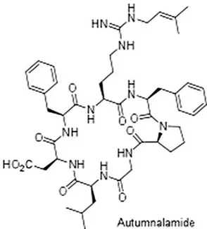
attracted the attention of pharmacy industry (Coates et al., 2013). Autumnalamide is a prenylated cyclic peptide obtained from the cyanobacteria Phormidium autumnale whose chemical structure has been recently elucidated (Fig. 1) (Audoin et al., 2014). This specie is one of the most common of the genus Phormidium, which colonizes stones in streams and rivers (Komárková, 2003). Initial studies on the biological activity of this natural compound has shown that autumnalamide is able to induce calcium influx block-ade through store operated calcium channels (SOC channels) in SH-SY5Y neuroblastoma cells due to an effect at the mitochondrial level, disrupting the mitochondrial membrane potential (MMP) and inducing the opening of mitochondrial permeability transi-tion pore (mPTP). Moreover, the effect of autumnalamide on SOC channels was abolished by pre-incubation with the immunosup-pressant drug cyclosporine A (CsA), (Audoin et al., 2014). The mPTP complex is a multiproteic structure composed by different proteins. Among them, cyclophilin D (Cyp D) is an essential component with a molecular weight of 22 kDa (Elrod and Molkentin, 2013; Javadov and Kuznetsov, 2013). CsA is a cyclic 11-amino-acid peptide iso-lated from the fungus Tolypocladium inflatum and was described as
a potent and selective mPTP inhibitor due to Cyp D binding in the matrix and on the inner surface of the mitochondrial membrane (Elrod and Molkentin, 2013; Hoppert et al., 2001). Besides Cyp D binding and mPTP opening inhibition, CsA has the ability to bind to all Cyclophilins (Cyps) present in the cell with different affinities due to a high degree of sequence conservation in their structure (Wang and Heitman, 2005; Fruman et al., 1994). The Cyps protein family structurally contain two main protein domains, a cyclophilin (Cyp)-like domain, which is the conserved domain across all family members and the specific domain depending on the family member (Lee and Kim, 2010). These proteins are present in all eukaryote and prokaryote cells like plants, fungi, bacteria, insects and human cells with a common peptidyl-prolyl isomerase activity. That is, Cyps cat-alyze the isomerisation of the peptide bonds from trans to cis form at the proline residues and facilitates protein folding among other functions (Wang and Heitman, 2005; Kofron et al., 1991; Davis et al., 2010). The location of these proteins is very broad and can be found in different organelles and also in the cytosol or in the extracellular medium (Lee and Kim, 2010; Kumari et al., 2013). Our initial stud-ies on the biological activity of autumnalamide have shown that the compound induced the formation of mPTP. The mPTP can be induced by different stimulus over the proteins that form this struc-ture like Cyp D (Audoin et al., 2014). Due to this and since the effect of autumnalamide on SOC channels was blocked by CsA, the main objective of this work was to go into detail about the knowledge of mechanism of action of autumnalamide.
2. Materials and methods
2.1. Chemicals and solutions
Carboxymethyl dextran (CM5) sensor chips, Hank’s Bal-ance Solution Surfactant P20 (HBS-EP) buffer (pH 7.4, 0.01 M 4-(2-hydroxyethyl)-1-piperazineethanesulfonic acid (HEPES), 0.15 M NaCl, 3 mM Ethylenediamine tetraacetic acid (EDTA), 0.005% polysorbate), amine coupling kit (1-ethyl-3- (3-dimetylaminopropyl) carbodiimide hydrochloride (EDC) and N-hydroxysuccinimide (NHS) and ethanolamine-HCl were sup-plied by BiacoreAB (Uppsala, Sweden). Percoll was obtained from Pharmacia (Uppsala, Sweden). Plastic tissue-culture dishes were purchased from Falcon (Madrid, Spain). RPMI and Foetal calf serum (FBS) were bought from Gibco (Glasgow, UK). The Pan T cell Isolation Kit (human) and the monoclonal antibody to human CD3, clone BW264/56, FITC ware purchased from Miltenyi Biotec (Germany). Human interleukin (IL)-2 ELISA kit was obtained from Invitrogen (Spain). Active human Cyclophilin D (Cyp D) and Active Human Cyclophilin A (Cyp A) full-length proteins were from Abcam (Cambridge, UK). Anti-nuclear factor of activated T cells (NFAT) c1 mouse monoclonal antibody [AT1C3] to NFATc1 and rabbit polyclonal anti-lamin B1 antibody were from Abcam (UK). FURA-2AM was obtained from Molecular Probes. Polyvinylidene fluoride (PVDF) membrane was from Millipore (Temecula, CA). Polyacrylamide gels and molecular weight marker Precision Plus Protein Standards Kaleidoscope was from BioRad (Barcelona, Spain). The Protease Inhibitor Complete Tablets and Phosphatase Inhibitor Cocktail Tablets were from Roche (Spain). Bovine serum albumin (BSA) and CsA (purity≥ 98.5%), Flufenamic acid (FFA), carbonyl cyanide 4-(trifluoromethoxy) phenylhydrazone (FCCP) and the rest of Chemicals and reagents were obtained from Sigma-Aldrich (Madrid, Spain). The strain of Phormidium autumnale was purchased from the Culture Collection of Algae and Protozoa (UK, CCAP1446/10). The strain was cultured and extracted as previously described (Audoin et al., 2014). Autumnalamide was isolated with purity higher than 95% as demonstrated by its1H
NMR spectrum. The composition of saline solution (PBS) used for
Fig. 1. Autumnalamide chemical structure.
human T lymphocytes purification was in (mM): 137 ClNa, 8.2 Na2HPO4, 1.5 KH2PO4, 3.2 KCl and 2 EDTA. The composition of
Umbreit saline solution was in (mM): 119 NaCl, 1.2 Mg (SO4), 1.2
NaH2PO4, 22.8 NaHCO3, 5.9 KCl, 1 CaCl2and 1 g/L Glucose. The pH
was equilibrated between 7.2-7.3. Stock solutions of drugs were done in dimethylsulphoxide (DMSO).
2.2. Human neuroblastoma culture cells and human T lymphocytes isolation
Neuroblastoma cell line SH-SY5Y was purchase from Ameri-can Type Culture Collection (ATCC) Number CRL-2266. The cells were plated in 25 cm2flasks at a cultivation ratio of 1:10. The cells
were maintained in Eagle’s Minimum Essential Medium (EMEM) from ATCC and F12 Medium (Invitrogen) 1:1 supplemented with 10% fetal bovine serum (FBS) (Gibco) plus 100 UI/mL penicillin and 100g/mL streptomycin. The neuroblastoma cells were expanded weekly using 0,05% trypsin/EDTA (1x) (Invitrogen).
Peripheral lymphocytes were isolated from human fresh hep-arinised blood from healthy volunteers as previously described (Alfonso et al., 2001). The blood was diluted in the same proportion with PBS plus EDTA 2 mM previously equilibrated at room tem-perature. 4 mL of diluted blood were placed over 3 mL of isotonic percoll (57,5%) carefully avoiding the mixture of the two phases. Once the tubes were prepared they were centrifuged at 3000 rpm, 25 min at room temperature. After centrifugation, different phases were obtained and only the fraction that containing the population of white blood cells was collected and washed three times by cen-trifugation with PBS-EDTA at 1500 rpm, 10 min, room temperature to remove remaining percoll. Lymphocyte purity was always higher than 80%. T lymphocytes were purified from this population with a human Pan T cell Isolation Kit. This is an indirect magnetic label-ing system for the isolation of untouched T cells. T cell purity was always higher than 95%. Assessment of cell purity was performed by flow cytometry by using a monoclonal antibody to human CD3 labeled with FITC. Viability (>95%) was determined by trypan blue exclusion. Pure T cells were maintained in RPMI 1640 plus 10% FBS and plated in 24 plastic tissue-culture dishes in humidified 5% CO2
2.3. Measurements of cytosolic free calcium
Cells were seeded onto 18 mm glass cover slips and used between 48 and 72 h after plating at a density of 120.000 cells/glass cover slip. For cytosolic Ca2+measurements, cells were washed
twice with saline solution (Umbreit) supplemented with 0.1% BSA. Umbreit composition was (mM): NaCl 119, Mg (SO4) 1.2, NaH2PO4
1.2, NaHCO3 22.85, KCl 5.94, Glucose 0.1% and CaCl2 1. In all
assays the solutions were equilibrated with CO2before being used,
adjusting the final pH between 7.2–7.4. The cells were loaded with the Ca2+-sensitive fluorescent dye FURA-2 AM (0,5M) for
6,5 min at 37◦C and 300 rpm. Loaded cells were washed twice with saline solution and the cover slips were placed in a thermostatic chamber (Life Sciences Resources, UK). Cells were viewed using a Nikon Diphot 200 microscope equipped with epifluorescence optics (Nikon 40X- immersion UV- Fluor objective). Addition of drugs was made by aspiration and addition of fresh bathing solution with the drug to the incubation chamber. Cytosolic Ca2+levels as
FURA-2 ratio was obtained from the images collected by fluorescent equipment, Ultra-high-speed wavelength switching illumination system (Lambda-DG4) for excitation and Lambda 10-2 for emis-sion from Sutter Instruments Co., USA. The Light source was a xenon arc bulb and the different wavelengths used were chosen with fil-ters. Cells were excited at 340 and 380 nm lights alternately and emission was collected at 510 nm.
2.4. Viability assay
Cells were incubated with different concentrations of autum-nalamide ranging from 0.001 to 10M for 24 and 48 h. After incubation, cells were washed twice with saline solution. MTT (500g mL−1) dissolved in saline solution was added to the
96-well microplate for 1 h at 37◦C (300 rpm) in the dark. Finally the MTT incubation media was removed and cells were washed once before the addition of SDS at 5%. Colored formazan salt aggregates were measured at 595 nm in a spectrophotometer plate reader. 2.5. Binding experiments: surface activation, ligand
immobilization and binding
A Biacore X SPR biosensor with Control Software and BIAeval-uation software version 3.0 from Biacore (GE Healthcare, Uppsala, Sweden) was used to check the binding between molecules. Sen-sor surface activation and ligand immobilization were performed by using HBS-EP as running buffer at a flow rate of 5L min−1
and 25◦C. CM5 sensor chip were used as surface where Cyp D or Cyp A was immobilized as ligand. The CM5 chip is a glass slide coated with a thin layer of gold with a matrix of carboxymethylated dextran covalently attached. The CM5 chip was activated using an amine coupling kit. Following manufacture instructions, a mixture (1:1, v/v) of EDC and NHS was applied for 2 min over the sensor chip. After activation, the ligand, 100g/mL−1 of Active human
Cyp A or Cyp D full-length protein dissolved in sodium acetate 10 mM at pH 4.5 was added. In these conditions, a typical cova-lent binding curve was obtained. Then, the CM5 chip surface was washed with HBS buffer and no fall in the signal was observed, indicating the protein immobilization onto the sensor chip surface. Finally, ethanolamine-HCl was injected to deactivate the remain-ing active esters, remove non-covalently bound protein and avoid non-specific binding (Sanchez et al., 2015; Alfonso et al., 2014). Next, analytes were added to check the binding between them and Cyp D or Cyp A. Individual binding curves for each analytes were analyzed by determining the kinetic constant of analytes-Cyp A/D binding, namely, the observed rate constant (Kobs), the association rate constant (Kass), the dissociation rate constant (Kdiss), and the
kinetic equilibrium dissociation constant (KD). At equilibrium, by
definition, Kdiss/Kass= KD. The pseudo-first-order association rate
constants Kobs(s−1) were determined by using the 1:1 Langmuir
association model of BiaEvaluation software (BiaCore, Uppsala, Sweden). Then a representation of Kobsagainst the corresponding
concentration of compound was done. These plots follow a linear correlation coefficient. From the equation of this representation, Kass, M−1s−1, gradient of the Plot, and Kdiss, s−1, intercept of the
Plot was obtained. Within these two values, the kinetic equilib-rium dissociation constant KDfor the analyte-Cyp A/D binding was
obtained.
The duration of the sample injection was 2 min at 10L min−1
flow rate. Next, dissociation of bounded molecules in HBS-EP buffer flow was studied. The bounded drugs were removed from the chip surface before the next injection by adding 1 M Glycine-HCl at pH 2.5 for 1 min. The association phase was used to quantify the compound-Cyp A/D interactions.
2.6. Interleukin 2 release
Human T lymphocytes at the concentration of 1× 10⁶cells mL−1
were plated in 24 well plates and pre-treated for 2 h with autum-nalamide (10M). Then, cells were stimulated with Concanavalin A (Con A) at 50g/mL for 48 h to induce IL-2 release. The amount of IL-2 released to the culture medium was evaluated using a Human IL-2 ELISA kit.
2.7. Calcineurin phosphatase activity assay
Calcineurin phosphatase activity was used as a measure of Cyp A activity by means of a calcineurin Phosphatase Assay Kit (Enzo Life Sciences, Inc., Farmingdale, NY). This is a complete colorimetric assay kit to measure calcineurin phosphatase activity. The Cyp-A-drug complex was allowed to form for 1 h at room temperature. 1 nM of Cyp A and different concentrations of drugs were employed dissolved in deionised water. After 1 h, recombinant calmodulin (0.25M) and recombinant calcineurin (40 units per well) were added to the complex formed between Cyp A and Autumnalamide or CsA. The complex was incubated for 30 min. Next; RII phospho-peptide substrate was added at a final concentration of 0.15 mM allowing the development of the reaction for 1 h at room tempera-ture. Then 100L of development reagent was added and incubated for 20 min at room temperature. Finally, the A620of the 96-well
plate samples was measured using Multi-Mode Microplate Reader SynergyTM4 equipment from Biotek (Winooski, VT, USA).
2.8. Western blotting
Cells were incubated first with autumnalamide or CsA for 2 h after which Con A (50g/mL) was added for 48 h. After incu-bation, T cells were centrifuged and washed twice with ice-cold saline solution (Umbreit) at 1500 rpm, 5 min and 4◦C. Then, the pellet was resuspended in 50L of hypotonic lysis buffer solu-tion to extract cytosolic proteins for 15 min with the following composition mM: 20 Tris-HCl, 10 NaCl and 3 MgCl2, 10X
phos-phatase inhibitors and 7X protease inhibitor. Cells were pelleted at 3000 rpm, 10 min at 4◦C to remove the cytosolic protein frac-tion. The supernatant was collected and the pellet obtained was resuspended in 30L of nuclear lysis buffer composed by: 100 mM Tris, 2 mM NaVO4, 10 mM NaCl, 1 mM EDTA, 1 mM EGTA, 1 mM NaF,
20 mM Na4P2O7, 1% Triton-X-100, 10% Glycerol, 0,1% SDS and 0,5%
Deoxychocolate Sodium containing 1 mM PMSF and 10X protease inhibitor cocktail for 30 min vortexing every 10 min. Samples were then centrifuged at 14000g at 4◦C for 30 min. The supernatants of interest were collected as protein nuclear fraction. The deter-mination of protein concentration was done using Direct Detect (Millipore) using bovine serum albumin (BSA) as standard protein.
Fig. 2. Effects of autumnalamide and ionomycin cytosolic Ca2+profile in SH-SY5Y
neuroblastoma cells. A. First arrow indicates the addition of 2M ionomycin. Second arrow shows addition of 10M of autumnalamide. The third arrow indicates the addition of Ca2+1 mM to the bath solution B. Cytosolic Ca2+profile
in cells first pre-incubated with 10M autumnalamide (first arrow) followed
Samples of cell lysates containing 10g of nuclear fraction of total protein were used for electrophoresis. Samples for electrophore-sis were dissolved in a 15–20% polyacrylmide gel and blotted to PVDF membrane by reduced SDS-PAGE. To determine the protein size and also to monitor the progress of the electrophoresis run, Precision Plus Protein Standards Kaleidoscope molecular weight marker was used. Then, membranes were blocked with 0,5% BSA in washing buffer (PBS + 0.1% Tween) and incubated 10 min with primary anti-NFATc1 (1:1000). After three washes with washing buffer (PBS + 0.1% Tween 20), membranes were incubated with sec-ondary anti-Mouse IgG conjugated with horseradish peroxidase. The immunoreactive bands were detected using the Supersignal West Pico or Supersignal West Femto Maximum Sensitivity Sub-strate (Pierce) and the Diversity GeneSnap software (Syngene). NFATc1 signal was normalized using Lamin B1 (1:1000).
2.9. Statistical analysis
All experiments were carried out by duplicate a minimum of three times. Results were analyzed by using one-way analysis of variance ANOVA with Dunnett’s post hoc analysis. p values < 0.05 were considered statistically significant. All results were expressed as the mean± SEM of 3 or more experiments.
3. Results
Considering the results obtained in a previous work, autum-nalamide was able to completely inhibit SOC channels by its effect over mitochondrion (Audoin et al., 2014). Therefore, several addi-tional calcium (Ca2+) experiments were carried out in order to
clarify the effect of autumnalamide on Ca2+fluxes different from
SOC. To do this, the effect of autumnalamide on ionomycin Ca2+
fluxes was studied. Ionomycin is a Ca2+ionophore that induces
intracellular pools depletion and allows Ca2+entrance from the
extracellular medium (Morgan and Jacob, 1994). Thus, autum-nalamide was added in a Ca2+-free medium after ionomycin pools
depletion, and a significant cytosolic Ca2+influx reduction was
observed after ion addition (Fig. 2A). In the same way, when the compound was incubated in a Ca2+-free medium before ionomycin
addition, no modification on intracellular stores was observed nei-ther on ionomycin-sensitive pools depletion. Nevertheless, once Ca2+ ion was restored, a significant ion-influx inhibition was
obtained comparing to ionomycin (Fig. 2B). Moreover, autum-nalamide did not inhibit the ion entrance once the influx is activated (Fig. 2C). Thus, autumnalamide did not affect intracellular pools modulated by ionomycin; nevertheless cytosolic Ca2+entrance was
reduced a 30% when the incubation with autumnalamide was done after or before ionomycin-sensitive pools depletion. Therefore, as it was reported, the compound inhibits SOC entrance.
In order to confirm if autumnalamide inhibits other Ca2+influx
different from SOC, flufenamic acid (FFA) was employed. FFA is a non-steroidal drug that acts inducing Ca2+release from the
mito-chondrion in a Ca2+free medium (Jiang et al., 2012; Damsker et al.,
2009; Zeng et al., 2012). Thus, when FFA was incubated in the absence of Ca2+, a significant Ca2+release from internal stores was
induced. In addition, when Ca2+was added to the medium an ion
influx was observed (Fig. 3A). Nevertheless, when autumnalamide was incubated before the FFA addition, the Ca2+release from
mito-chondrion was significantly inhibited, besides after addition of Ca2+
the ion reached similar levels than FFA (Fig. 3A). Therefore, the
by ionomycin 2M (second arrow). The third arrow indicates 1 mM of Ca2+addition
to the bath solution. C. The first arrow shows the ionomycin addition. The second arrow shows the Ca2+addition to the medium and the third the addition of 10M of
autumnalamide. The experiments were performed in duplicate. The results of each replica are mean of 20 cells. Mean± SEM of three independent experiments.
Fig. 3. Effect of FCCP or autumnalamide and FFA on cytosolic Ca2+profile in
SH-SY5Y neuroblastoma cells. A. First arrow indicates autumnalamide addition and the second arrow the FFA addition. The third arrow indicates the addition of Ca2+. B. The
FCCP addition to the medium (first arrow) prior to FFA addition (second arrow). The third arrow indicates the addition of the ion to the Ca2+-free bathed solution. The
experiments were performed in duplicate. The results of each replica are mean of 20 cells. Mean± SEM of three independent experiments.
compound does not affect mitochondrial Ca2+influx, however the
release of the ion from this organelle was largely reduced. In order to know the effect of other mitochondrial uncouplers on FFA, FCCP was employed (Ma et al., 2012). As shown inFig. 3B, FCCP produced a small increase in Ca2+levels; and the following addition of FFA
did not induce a Ca2+release. Thus, after the ion addition to the
bath solution, a similar Ca2+influx as FFA control was observed.
Therefore, as in the case of autumnalamide, the prior incuba-tion with FCCP inhibited the Ca2+release induced by FFA without
affecting the next Ca2+influx. Summarizing the results described
above, both, FCCP and autumnalamide were able to decrease the release of Ca2+from mitochondrion induced by FFA; nevertheless
the cytosolic Ca2+entrance was not blocked. In addition,
autum-nalamide inhibited cytosolic Ca2+influx induced by the ionophore
while ionomycin-sensitive intracellular pools were not modified. Therefore, autumnalamide targeted mitochondrion without induc-ing Ca2+release from this organelle but inhibiting SOC influx.
In order to explain the effect of autumnalamide on Ca2+handling
and since in a previous work it was observed the effect of autum-nalamide inhibiting Ca2+influx, which was reverted by CsA (Audoin
et al., 2014), the structures that this drug targets were studied. With
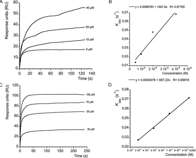
this purpose, the highly sensitive biosensor Biacore X was used to determine the binding between mitochondrial Cyp D, used as lig-and, attached to the sensor surface and using autumnalamide as ligate. First, CsA was used as control of binding for the immobilized Cyp D. AsFig. 4A shows, when different concentrations of CsA (5, 10, 20 and 40M) were added over the immobilized Cyp D, typical association curves profiles were obtained. In the presence of 5M CsA the signal was 10.05 RU, while in the presence of 40M of CsA the response reached 31.8 RU. Individual binding curves from
Fig. 4A were analyzed as described in the material and methods sec-tion. Kobsfor each concentration of CsA were obtained. When Kobs
were represented against each CsA concentration, (Fig. 4B), a lin-ear regression with a correlation coefficient of r = 0.97985 for Cyp D was obtained. From this representation, Kass, M−1s−1, slope, Kdiss,
s−1, and Y-intercept, were obtained. Within these two values, the kinetic equilibrium dissociation constant KD(Y-intercept/slope) for
the CsA-Cyp D association was obtained. The value of this constant was 4.5× 10⁻⁶± 3.05 × 10⁻⁶M for the CsA-Cyp D binding assay
(Fig. 4B). After setting up these conditions the binding of Cyp D and autumnalamide was next studied. In this case, different concentra-tions of autumnalamide (30, 50, 70 and 100M) were dissolved in HBS-EP buffer and added onto the previously immobilized Cyp D. Thus, asFig. 4C shows, association curves for the binding of autumnalamide with Cyp D were obtained with responses rang-ing from 10 RU at the lowest autumnalamide concentration to 220 RU in the presence of 100M of the compound. As in the case of CsA, the interaction between autumnalamide and Cyp D follows a pseudo-first order kinetic where Kobswas calculated. When each
Kobswas represented against the corresponding concentration of
autumnalamide fromFig. 4C, linear regression plots with a cor-relation coefficient of r = 0.98, for the binding of autumnalamide to Cyp D were obtained. From these representations the kinetic equilibrium constant for the binding of autumnalamide to Cyp D was calculated. In the case of the Cyp D-autumnalamide binding, a KD= 1.51± 1.399 M (Fig. 4D) was obtained. Due to Cyps share
a common region domain and since autumnalamide showed good binding affinity for Cyp D, the effect of this compound over Cyp A, another important immunophilin, was also evaluated. Straight-away, the same procedure was followed for Cyp A immobilization over a CM5 chip as described in Material and Methods. Thus, when different concentrations of CsA were added over the immobilized Cyp A, typical association curves were obtained. In the presence of 5M CsA the signal was 17.54 RU, while in the presence of 40M of CsA the response reached 55.45 RU (Fig. 5A). In this case, a KDfor CsA-Cyp A was 6.83× 10⁻⁶± 1.1 × 10⁻⁶M (Fig. 5B). In the
same way, different concentrations of autumnalamide were dis-solved in HBS-EP buffer and added onto the immobilized Cyp A. Thus, asFig. 5C shows, association curves for the binding of autum-nalamide with Cyp A were obtained with responses ranging from 34.11RU at the lowest concentration to 102.5 RU at the highest con-centration of autumnalamide. From the association curves a KDof
8.08± 1.23 M was obtained (Fig. 5D). So far the results obtained here indicated that autumnalamide showed higher affinity for Cyp D than CsA and also a good affinity for Cyp A, although slightly smaller than that of CsA.
Hence, autumnalamide showed Cyp A binding, and also acts by inhibiting SOC influx; both targets are related with the mod-ulation of interleukins (ILs) (Sweeney et al., 2009; Djuric et al., 2000), we then tested the effect of autumnalamide on IL-2 pro-duction in human T lymphocytes. In previous work the effect of autumnalamide in SH-SY5Y cell viability was checked for 1 h, and the compound produced a 42% of decrease in cell viability at the concentration of 25M (the highest concentration tested) (Audoin et al., 2014). In order to develop experiments with prolonged expo-sure times and in different cellular model, it was necessary to check cell toxicity of autumnalamide. To do this, different concentrations
Fig. 4. CsA/autumnalamide association curves and ligand binding analysis. A. Cyp D-CsA association: association curves after addition of different amounts of CsA to
immobilized Cyp D. Different CsA concentrations were injected using HBS-EP as running buffer and a flow rate of 10g mL−1. The association curves were obtained after
subtraction of their respective solvent control. B. Analysis of ligand binding: kinetic plot of apparent association rate constant Kobs(s−1) obtained from plot A (calculated
by BiaEvaluation software) versus CsA concentration. C. Cyp D-autumnalamide association: association curves after addition of different amounts of autumnalamide to immobilized Cyp D. Different autumnalamide concentrations were injected using HBS-EP as running buffer and a flow rate of 10g mL−1. The association curves were
obtained after subtraction of their respective solvent control. D. Analysis of ligand binding: kinetic plot of apparent association rate constant Kobs(s−1) obtained from plot C
(calculated by BiaEvaluation software) versus autumnalamide concentration. Representative experiment of n = 3.
ranges of autumnalamide were tested for 24 and 48 h in human T lymphocytes. AsFig. 6shows, the concentrations tested for the two exposure times did not produced any changes on cell viability. Once again, the concentration of 10M was selected to develop the current experiments at 48 h.
To develop IL-2 experiments, a control of human T lymphocytes was treated with Con A alone, to induce IL-2 release, or in combi-nation with autumnalamide. After 48 h of incubation, IL-2 release in the culture medium was measured. AsFig. 7shows, the stim-ulation of human T lymphocytes with Con A for 48 h efficiently induces IL-2 production comparing with non-stimulated control cells from 47.72± 1.58 pg mL−1to 742,77± 152.23 pg mL−1
respec-tively (p≤ 0.001). In this conditions, when T lymphocytes were pre-incubated for 2 h with 10M autumnalamide, before stimula-tion with ConA for 48 h, a reducstimula-tion from 742,77± 152.23 pg mL−1
to 111.61± 3.4 pg mL−1 (p≤ 0.01). The well-known IL-2 inhibitor
CsA was used as positive control of IL-2 inhibition. Thus, when cells were pre-incubated with CsA 0.2M for 2 h before Con A stimu-lation, IL-2 production was significantly reduced to control values 146.25± 111.34 pg mL−1(p≤ 0.01). From these results we conclude
that both compounds suppressed IL-2 release from human acti-vated T lymphocytes with a similar efficacy.
In view of these results two additional experiments were per-formed to evaluate the mechanisms underlying the effects of autumnalamide in IL-2 blockade. Some compounds, as in the case
of CsA, act docking calcineurin phosphatase activity over NFATc1 and the next IL-2 induction due to the binary complex formed with Cyp A (Randak et al., 1990). Therefore, in view of the binding of autumnalamide to Cyp A, the ability of autumnalamide to inhibit calcineurin phosphatase activity was next compared. In this case, a compound concentration close to the calculated KD was used.
AsFig. 8shows, when Cyp A was incubated in the presence of 6M CsA a significant reduction in calcineurin phosphatase activity of 28.33± 3.07% (p ≤ 0.01) was observed. In the same way, in the presence of 10M of autumnalamide a significant 18.87 ± 2.21% (p≤ 0.001) reduction was observed over calcineurin phosphatase activity.
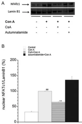
It is known that T lymphocyte stimulation with Con A induce a cytosolic Ca2+ increase (Smith-Garvin et al., 2009). As a
con-sequence, Cyp A forms a complex with calcineurin that binds to and dephosphorylates cytosolic NFATc1. The dephosphorylation of the nuclear factor leads to the translocation to the nucleus. The increase in the nuclear expression of NFATc1 in a transcription-ally active form leads to the increase of IL-2 production (Liu, 2009; Xiong et al., 2013). Therefore, the nuclear expression of NFATc1 in activated human peripheral T lymphocytes in the presence of autumnalamide was next checked and compared with the effect of CsA in the same condition. Representative western blot bands for these results are shown inFig. 9A. As shown inFig. 9B, the quantifi-cation of western blot band intensities in each condition showed
Fig. 5. CsA/atumnalamide association to immobilized Cyp A and ligand binding analysis. A. Cyp A-CsA association: association curves after addition of different amounts of
CsA to immobilized Cyp A. Different CsA concentrations were injected using HBS-EP as running buffer and a flow rate of 10g mL−1. The association curves were obtained
after subtraction of their respective solvent control. B. Analysis of ligand binding: kinetic plot of apparent association rate constant Kobs(s−1) obtained from plot A (calculated
by BiaEvaluation software) versus CsA concentration. C. Cyp A-autumnalamide association: association curves after addition of different amounts of autumnalamide to immobilized Cyp A. Different autumnalamide concentrations were injected using HBS-EP as running buffer and a flow rate of 10g mL−1. The association curves were
obtained after subtraction of their respective solvent control. D. Analysis of ligand binding: kinetic plot of apparent association rate constant Kobs(s−1) obtained from plot C
(calculated by BiaEvaluation software) versus autumnalamide concentration. Representative experiment of n = 3.
that when cells were stimulated with Con A for 48 h nuclear band intensity of NFATc1 exhibited a 67.21± 0.12% (p ≤ 0.01) increase comparing with T cells in resting conditions. Pre-incubation of the cells with CsA before activation decreased NFATc1 band inten-sity close to control values, in agreement with previously reported results (Ishikawa et al., 2003). However, autumnalamide did not significantly reduce the increase in nuclear NFATc1 in activated human T lymphocytes that reach higher levels than Con A treated cells.
Altogether, the results presented here indicate that, autum-nalamide does not modify ionomycin-sensitive intracellular Ca2+
pools, besides confirm that the compound inhibits the SOC chan-nels. Moreover, it binds to mitochondrion Cyp D, presents binding affinity for Cyp A and inhibits calcineurin and human IL-2 pro-duction in activated T lymphocytes but, unlike CsA, it lacks of an inhibitory effect over NFATc1.
4. Discussion
The structure of the prenylated cyclic peptide named autum-nalamide was recently identified and the same paper reported the only available data on the compound, indicating that autumnalamide alters mitochondrial membrane potential in neu-roblastoma cells and also induces the mPTP opening (Audoin et al.,
2014). The opening of this structure can be induced by different substances that do not necessarily target the proteins that form the mPTP structure (Tornero et al., 2002). There are many molecules from the own organism that act as inducers of mPTP, such as -Amyloid (A) protein, as well as molecules with natural origin like mastoparan from the venom of Vespula lewisii; honokiol from Magnolia spp. or molecules from synthetic sources like MT-21. Nev-ertheless, almost no molecule acts targeting directly Cyp D, both for blocking or inducing the formation of mPTP. In the case of A protein, it induces the production of Reactive Oxygen Species (ROS), mastoparan forms channels in the mitochondria and MT-21 directly induces the release of cytochrome c from mitochondria. All these factors lead to the opening of mPTP (Watabe et al., 2000; Ding and Nam Ong, 2003; Du and Yan, 2010; Arora et al., 2012). Thus, with the exception of honokiol, none of these compounds induces the mPTP opening directly by binding to a specific component of the multiproteic complex, as we observed with autumnalamide in the present paper. On the other hand, Ca2+experiments reveal that
autumnalamide neither modify ionomycin-sensitive intracellular Ca2+pools, nor the ionomycin Ca2+entrance through SOC channels
once the influx is activated. Nevertheless, the Ca2+profiles obtained
from the present study show that the FFA-induced Ca2+release
in SH-SY5Y is inhibited by pre-treatment with FCCP, which col-lapse the mitochondrial membrane potential that normally drives
Fig. 6. Effect of autumnalamide over T lymphocytes cell viability. T lymphocytes
cells were incubated for 24 and 48 h with autumnalamide at the concentration range from 1 nM to 10M. Bar columns represent the different concentrations for each times. Effect on cell viability tested by MTT test. Data are mean± SEM of 3 independent experiments. Data are mean± SEM of 3 independent experiments.
Fig. 7. Effect of CsA or autumnalamide on IL-2 production in human T lymphocytes
stimulated with Con A. Human T lymphocytes were pre-treated for two hours with CsA (0.2M) or autumnalamide (10 M) and then with Con A (50 g mL-1) for 48 h. Mean± SEM of three experiments. All values are shown in percentage to the Con A treated cells. **Significant differences between autumnalamide or CsA treated cells with respect to cells treated with Con A (p < 0.01). # Significant differences between Con A treated cells and control (p < 0.001). Mean± SEM of 3 independent experiments.
the Ca2+into the mitochondrion (Collins et al., 2000). The effect
of autumnalamide over FFA may be achieved through either a protonophore-like effect, similar to FCCP, or the induction of the mPTP, as previously described (Audoin et al., 2014). Thus, in the case of FCCP in SH-SY5Y cells, the uncoupling of the mitochondrial transport chain produced in mitochondria, substantially inhibits Ca2+release induced by FFA probably because both FCCP and FFA
act by disrupting mitochondrial membrane potential, and one drug blocks the effect of the other (Chi et al., 2011). Moreover, CsA abol-ished the effect of autumnalamide efficiently in comparison with FCCP on SOC Ca2+influx (Audoin et al., 2014). In accordance with
these previous results and due to the effect observed over mPTP by autumnalamide, the binding of the prenylated cyclic peptide from the cyanobacterium Phormidium autumnale was tested on Cyp D, an essential component of the mPTP. Biosensor data analysis indicates that the compound binds with good affinity to mitochon-drial Cyp D. Autumnalamide almost shows 3 times more affinity for mitochondrial Cyp D than CsA. Moreover, due to the findings
Fig. 8. Effect of autumnalamide or CsA on Ca2+/calmodulin-dependent on
Phos-phatase Activity of Calcineurin. PhosPhos-phatase activity of calcineurin determined after pre-incubation with Cyp A or Cyp A + CsA (6M)/autumnalamide (10 M). All val-ues are shown in percentage with respect to Cyp A control. **Significant differences between Cyp A control and Cyp A + CsA (p≤ 0.01). ***Significant differences between Cyp A control and Cyp A + autumnalamide (p≤ 0.001). Data are mean ± SEM of 3 independent experiments.
Fig. 9. Effect of CsA or autumnalamide over NFATc1 nuclear levels in human T
lym-phocytes activated with Con A. A. NFATc1 levels were studied as nuclear fraction after 48 h incubation with compounds. Human T lymphocytes were pre-treated for 2 h with CsA (0.2M) or autumnalamide (10 M) and afterward incubated with Con A (50g mL−1) for 48 h. A. Representative image of one experiment. B. Mean of the
ratio of the nuclear NFATc1/lamin B1 band intensity. All results are presented as the percentage of NFATc1 in the nucleus. ##Significant differences with respect to control cells (p≤ 0.01). **Significant differences with respect to Con A treated cells (p < 0.01). Data are mean± SEM of three experiments.
observed by SPR and taking into account the results observed on mPTP besides the uncoupling effect (Audoin et al., 2014), this effect could be related with the interaction between autumnalamide and mitochondrial Cyp D in a more specific way than in the case of FCCP or FFA that is produced mainly by the disruption of membrane potential. Cyps share a common domain; therefore compounds that
bind to one of this protein family usually show binding affinity for other Cyps as CsA display (Kumari et al., 2013). Thus, autum-nalamide also shows, as CsA, Cyp A binding affinity. This cytosolic protein plays a central pathway of many physiological and patho-logical processes in the cells such as inflammation, cardiovascular diseases, immunomodulation or sepsis among others (Nigro et al., 2013). In the case of CsA, once the complex between Cyp A- CsA is formed, calcineurin phosphatase activity is blocked and the translo-cation of NFAT to the nucleus is avoided, inhibiting the transcription activity over genes that encodes for IL-2 (Fruman et al., 1992). Autumnalamide reduces calcineurin phosphatase activity, while NFAT was not reduced after the binding to Cyp A, nevertheless, autumnalamide, as CsA, reduces IL-2 production in T lymphocytes. While CsA exerts its effect through the docking of calcineurin (Naesens et al., 2009), the inhibition of IL-2 produced by autum-nalamide probably occurs partially by a calcineurin-independent pathway since the nuclear entry of the NFATc1 is closely linked to its dephosphorilation and the nuclear levels of this transcription fac-tor are not blocked after autumnalamide treatment. Molecules that inhibit CRAC channels are also expected to reduce IL-2 production (Sweeney et al., 2009; Djuric et al., 2000). As FFA or BTP2, two com-pounds that modulate SOC channels, act by reducing the production of this IL in white blood cells, being Ca2+an essential signal for
its production (Kankaanranta et al., 1996; Cardenas and Heitman, 1995). In the case of autumnalamide, the indirect blockade pro-duced on SOC channels, the main Ca2+entrance in T cells, due to
the effects that produce in mitochondrion, could explain the effect over IL-2 inhibition, although it does not block NFAT transloca-tion. Then, other transcription factors or mitogen-activated protein kinases (MAPK) could be related with the modulation of inter-leukins (ILs) by autumnalamide where the restriction of cytosolic Ca2+induced by the compound may be essential (Audoin et al.,
2014; Cardenas and Heitman, 1995; Kar et al., 2011). On the other hand, autumnalamide can be a useful tool to study molecules that targeted Cyp D, other applications could be attributed to autum-nalamide due to the binding to Cyp A. This effort mainly led to the development of compounds with immunosuppressive or anti-inflammatory effects.
As a conclusion, we have a drug that has good affinity for two important immunophilins engaged in many diseases and produces an effective blockade on IL-2 production and low toxicity. Due to the multi-functional properties of Cyp A and the functions where it is involved, such as protein folding, trafficking, assembly or cell sig-naling, autumnalamide could open a new window to improve the treatment of many diseases where Cyp A is a key player (Nigro et al., 2013). With respect to Cyp D, the fact that mPTP is a process regu-lated by a wide type of stimulus, it makes this multiproteic complex an ideal pharmacological target implicated in multiple processes, where autumnalamide directly binds to (Tornero et al., 2002).
Conflict of interest statement
The authors declare no conflict of interest.
Acknowledgements
The research leading to these results has received funding from the following FEDER cofounded-grants. From CDTI and Technological Funds, supported by Ministerio de Economía y Competitividad, AGL2012-40185-CO2-01, AGL2014-58210-R, and Consellería de Cultura, Educación e Ordenación Universitaria, GRC2013-016, and through Axencia Galega de Innovación, Spain, ITC-20133020 SINTOX. From CDTI under ISIP Programme, Spain, IDI-20130304APTAFOOD.
From the European Union’s Seventh Framework Programme managed by REA− Research Executive Agency (FP7/2007-2013) under grant agreement 312184PHARMASEA. We wish to thank the Clínica Losada Arránz, especially Ms. Paula López Arránz for pro-viding the human blood samples for T cells purification. Jon Andoni Sánchez is supported by a fellowship from Plan Galego de Investi-gación e Crecemento, Xunta de Galicia, Spain.
References
Alfonso, A., Botana, M.A., Vieytes, M.R., Botana, L.M., 2001.Prolactin induces calcium influx and release from intracellular pools in human T lymphocytes by activation of tyrosine kinases. Cell. Signal. 13, 819.
Alfonso, A., Pazos, M.J., Fernandez-Araujo, A., Tobio, A., Alfonso, C., Vieytes, M.R., Botana, L.M., 2014.Surface plasmon resonance biosensor method for palytoxin detection based on Na+,K+-ATPase affinity. Toxins (Basel) 6, 96.
Arora, S., Singh, S., Piazza, G.A., Contreras, C.M., Panyam, J., Singh, A.P., 2012.
Honokiol: a novel natural agent for cancer prevention and therapy. Curr. Mol. Med. 12, 1244.
Audoin, C., Sanchez, J.A., Genta-Jouve, G., Alfonso, A., Rios, L., Vale, C., Thomas, O.P., Botana, L.M., 2014.Autumnalamide, a prenylated cyclic peptide from the cyanobacterium Phormidium autumnale, acts on SH-SY5Y cells at the mitochondrial level. J. Nat. Prod. 77, 2196.
Cardenas, M.E., Heitman, J., 1995.Role of calcium in T-lymphocyte activation. Adv. Second Messenger Phosphoprotein Res. 30, 281.
Chi, Y., Li, K., Yan, Q., Koizumi, S., Shi, L., Takahashi, S., Zhu, Y., Matsue, H., Takeda, M., Kitamura, M., Yao, J., 2011.Nonsteroidal anti-inflammatory drug flufenamic acid is a potent activator of AMP-activated protein kinase. J. Pharmacol. Exp. Ther. 339, 257.
R.C.T. Coates, E. Trentacoste, W.H. Gerwick, (2013), Bioactive and Novel Chemicals from Microalgae, Oxford, UK.
Collins, T.J., Lipp, P., Berridge, M.J., Li, W., Bootman, M.D., 2000.Inositol 1,4,5-trisphosphate-induced Ca2+ release is inhibited by mitochondrial depolarization. Biochem. J 347, 593.
Damsker, J.M., Okwumabua, I., Pushkarsky, T., Arora, K., Bukrinsky, M.I., Constant, S.L., 2009.Targeting the chemotactic function of CD147 reduces
collagen-induced arthritis. Immunology 126, 55.
Davis, T.L., Walker, J.R., Campagna-Slater, V., Finerty, P.J., Paramanathan, R., Bernstein, G., MacKenzie, F., Tempel, W., Ouyang, H., Lee, W.H., Eisenmesser, E.Z., Dhe-Paganon, S., 2010.Structural and biochemical characterization of the human cyclophilin family of peptidyl-prolyl isomerases. PLoS Biol. 8, e1000439.
Ding, W.X., Nam Ong, C., 2003.Role of oxidative stress and mitochondrial changes in cyanobacteria-induced apoptosis and hepatotoxicity. FEMS Microbiol. Lett. 220, 1.
Djuric, S.W., BaMaung, N.Y., Basha, A., Liu, H., Luly, J.R., Madar, D.J., Sciotti, R.J., Tu, N.P., Wagenaar, F.L., Wiedeman, P.E., Zhou, X., Ballaron, S., Bauch, J., Chen, Y.W., Chiou, X.G., Fey, T., Gauvin, D., Gubbins, E., Hsieh, G.C., Marsh, K.C., Mollison, K.W., Pong, M., Shaughnessy, T.K., Sheets, M.P., Smith, M., Trevillyan, J.M., Warrior, U., Wegner, C.D., Carter, G.W., 2000.
3,5-Bis(trifluoromethyl)pyrazoles: a novel class of NFAT transcription factor regulator. J. Med. Chem. 43, 2975.
Du, H., Yan, S.S., 2010.Mitochondrial permeability transition pore in Alzheimer’s disease: cyclophilin D and amyloid beta. Biochim. Biophys. Acta 1802, 198.
Elrod, J.W., Molkentin, J.D., 2013.Physiologic functions of cyclophilin D and the mitochondrial permeability transition pore. Circ. J. 77, 1111.
Fruman, D.A., Klee, C.B., Bierer, B.E., Burakoff, S.J., 1992.Calcineurin phosphatase activity in T lymphocytes is inhibited by FK 506 and cyclosporin A. Proc. Natl. Acad. Sci. U. S. A. 89, 3686.
Fruman, D.A., Burakoff, S.J., Bierer, B.E., 1994.Immunophilins in protein folding and immunosuppression. FASEB J. 8, 391.
Hoppert, M., Gentzsch, C., Schorgendorfer, K., 2001.Structure and localization of cyclosporin synthetase, the key enzyme of cyclosporin biosynthesis in Tolypocladium inflatum. Arch. Microbiol. 176, 285.
Ishikawa, J., Ohga, K., Yoshino, T., Takezawa, R., Ichikawa, A., Kubota, H., Yamada, T., 2003.A pyrazole derivative, YM-58483, potently inhibits store-operated sustained Ca2+ influx and IL-2 production in T lymphocytes. J. Immunol. 170, 4441.
Javadov, S., Kuznetsov, A., 2013.Mitochondrial permeability transition and cell death: the role of cyclophilin d. Front. Physiol. 4, 76.
Jiang, H., Zeng, B., Chen, G.L., Bot, D., Eastmond, S., Elsenussi, S.E., Atkin, S.L., Boa, A.N., Xu, S.Z., 2012.Effect of non-steroidal anti-inflammatory drugs and new fenamate analogues on TRPC4 and TRPC5 channels. Biochem. Pharmacol. 83, 923.
Kankaanranta, H., Luomala, M., Kosonen, O., Moilanen, E., 1996.Inhibition by fenamates of calcium influx and proliferation of human lymphocytes. Br. J. Pharmacol. 119, 487.
Kar, P., Nelson, C., Parekh, A.B., 2011.Selective activation of the transcription factor NFAT1 by calcium microdomains near Ca2+ release-activated Ca2+ (CRAC) channels. J. Biol. Chem. 286, 14795.
Kofron, J.L., Kuzmic, P., Kishore, V., Colon-Bonilla, E., Rich, D.H., 1991.
Determination of kinetic constants for peptidyl prolyl cis-trans isomerases by an improved spectrophotometric assay. Biochemistry 30, 6127.
Komárková, J.K.a.J., 2003.Freshwater algae of North America. Ecol. Classif.
Kumari, S., Roy, S., Singh, P., Singla-Pareek, S.L., Pareek, A., 2013.Cyclophilins: proteins in search of function. Plant Signal. Behav. 8.
Lee, J., Kim, S.S., 2010.An overview of cyclophilins in human cancers. J. Int. Med. Res. 38, 1561.
Liu, J.O., 2009.Calmodulin-dependent phosphatase, kinases, and transcriptional corepressors involved in T-cell activation. Immunol. Rev. 228, 184.
Ma, T., Gong, K., Yan, Y., Song, B., Zhang, X., Gong, Y., 2012.Mitochondrial modulation of store-operated Ca(2+) entry in model cells of Alzheimer’s disease. Biochem. Biophys. Res. Commun. 426, 196.
Markou, G., Georgakakis, D., 2011.Cultivation of filamentous cyanobacteria (blue-green algae) in agro-industrial wastes and wastewaters: a review. Appl. Energ. 88, 3389.
Morgan, A.J., Jacob, R., 1994.Ionomycin enhances Ca2+ influx by stimulating store-regulated cation entry and not by a direct action at the plasma membrane. Biochem. J. 300 (Pt. 3), 665.
Naesens, M., Kuypers, D.R., Sarwal, M., 2009.Calcineurin inhibitor nephrotoxicity. Clin. J. Am. Soc. Nephrol. 4, 481.
Nigro, P., Pompilio, G., Capogrossi, M.C., 2013.Cyclophilin A: a key player for human disease. Cell. Death. Dis. 4, e888.
Randak, C., Brabletz, T., Hergenrother, M., Sobotta, I., Serfling, E., 1990.Cyclosporin A suppresses the expression of the interleukin 2 gene by inhibiting the binding of lymphocyte-specific factors to the IL-2 enhancer. EMBO J. 9, 2529.
Sanchez, J.A., Alfonso, A., Leiros, M., Alonso, E., Rateb, M.E., Jaspars, M., Houssen, W.E., Ebel, R., Botana, L.M., 2015.Spongionella secondary metabolites regulate store operated calcium entry modulating mitochondrial functioning in SH-SY5Y neuroblastoma cells. Cell. Physiol. Biochem. 37, 779.
Smith-Garvin, J.E., Koretzky, G.A., Jordan, M.S., 2009.T cell activation. Annu. Rev. Immunol. 27, 591.
Sweeney, Z.K., Minatti, A., Button, D.C., Patrick, S., 2009.Small-molecule inhibitors of store-operated calcium entry. ChemMedChem 4, 706.
Tornero, D., Cena, V., Gonzalez- Garcia, C., Jordan, J., 2002.The role of the mitochondrial permeability transition pore in neurodegenerative processes. Rev. Neurol. 35, 354.
Wang, P., Heitman, J., 2005.The cyclophilins. Genome Biol. 6, 226.
Watabe, M., Machida, K., Osada, H., 2000.MT-21 is a synthetic apoptosis inducer that directly induces cytochrome c release from mitochondria. Cancer Res. 60, 5214.
Xiong, Y., Zhang, S., Xu, L., Song, B., Huang, G., Lu, J., Guan, S., 2013.Suppression of T-cell activation in vitro and in vivo by cordycepin from Cordyceps militaris. J. Surg. Res. 185, 912.
Zeng, B., Chen, G.L., Xu, S.Z., 2012.Store-independent pathways for cytosolic STIM1 clustering in the regulation of store-operated Ca(2 + ) influx. Biochem. Pharmacol. 84, 1024.