HAL Id: hal-01762810
https://hal.umontpellier.fr/hal-01762810
Submitted on 8 Nov 2018
HAL is a multi-disciplinary open access
archive for the deposit and dissemination of
sci-entific research documents, whether they are
pub-lished or not. The documents may come from
teaching and research institutions in France or
abroad, or from public or private research centers.
L’archive ouverte pluridisciplinaire HAL, est
destinée au dépôt et à la diffusion de documents
scientifiques de niveau recherche, publiés ou non,
émanant des établissements d’enseignement et de
recherche français ou étrangers, des laboratoires
publics ou privés.
Propionibacterium acnes populations involved in deep
pathological samples and their dynamics along the
cardiac surgical pathway
Sara Romano-Bertrand, B Beretta, H. Jean-Pierre, Jean-Marc Frapier,
Brigitte Calvet, Sylvie Parer, Estelle Jumas-Bilak
To cite this version:
Sara Romano-Bertrand, B Beretta, H. Jean-Pierre, Jean-Marc Frapier, Brigitte Calvet, et al..
Propi-onibacterium acnes populations involved in deep pathological samples and their dynamics along the
cardiac surgical pathway. European Journal of Clinical Microbiology and Infectious Diseases, Springer
Verlag, 2015, 34 (2), pp.287 - 301. �10.1007/s10096-014-2228-2�. �hal-01762810�
Propionibacterium acnes populations involved in deep pathological
samples
and their dynamics along the cardiac surgical pathway
S. Romano-Bertrand&M. Beretta&H. Jean-Pierre&
J.-M. Frapier&B. Calvet&S. Parer&E. Jumas-Bilak
Abstract Propionibacterium acnes belongs to the normal skin microbiota, but it is also responsible for acne vulgaris and causes serious infections such as endocarditis and surgical site infections (SSI). The P. acnes population is structured into phylogenetic groups, with phylotype I being associated with acne. Herein, we explore the link between phylotypes and clinical origins in a collection of P. acnes isolated from differ-ent body sites, involved in deep infections or healthcare-associated infections (HAI), with particular emphasis on strains from cardiac SSI. Cardiac SSI have been further stud-ied in terms of P. acnes population dynamics during the care pathway. The recA and tly genes phylotypes were compared to hemolytic behavior, susceptibility to antimicrobial agents, and clinical origins. An original approach of recA polymerase chain reaction temporal temperature gel electrophoresis (PCR-TTGE) was developed and applied for the direct iden-tification of P. acnes phylotypes in surgical samples, in order
to assess their temporal dynamics during the surgical course. Our results underlined the preferential involvement of IA-2/IB and II phylogroups in HAI and SSI. Unlike IA and II, type IA-2/IB presented a gradual increase with the depth of sampling in the peroperative phase of cardiac surgery. Phylotypes IA and IA-2/IB were both predominant in scar tissues and on postoperative skin, suggesting a specific predisposition to recolonize skin. Particular association of the phylotype IA-2/ IB with SSI and its propensity to colonize wounds in cardiac surgery was observed. We assumed that the follow-up of P. acnes phylotypes during pathological processes could give new clues for P. acnes pathogenicity.
Introduction
Propionibacterium acnes is an anaerobic Gram-positive
ba-cillus member of the human microbiota mainly on skin [1],
but also in the oral cavity, and digestive and genital tracts [2].
In clinical samples, P. acnes is often overlooked as a contam-inant and is underestimated due to its growth requirements
[3–5]. However, P. acnes can produce putative virulence
factors involved in inflammation processes [4], as observed
in acne vulgaris [5] and other inflammatory pathologies, such
as sarcoidosis [6], or potentially in prostatic pathologies [7].
P. acnes can also cause severe deep infections like
endocardi-tis [8–10] and surgical site infections (SSI) in orthopedic [11,
12], cardiac [13,14], or neurological surgeries [15].
Metagenomics [16] and comparative genomics [17]
showed strain variations supporting the existence of specific P. acnes subpopulations. The strain HL096PA1 causing se-vere acne displayed a plasmid encoding adhesion factors and a high amount of pseudogenes, suggesting its adaptation to
pathogenic behavior in a narrow niche [18]. Moreover,
popu-lation genetics indicated the roles of distinct lineages of P. acnes in different diseases. In this context, several
S. Romano-Bertrand
:
M. Beretta:
H. Jean-Pierre:
E. Jumas-Bilak UMR 5119 ECOSYM, Equipe Pathogènes et Environnements, U.F.R. des Sciences Pharmaceutiques et Biologiques, Université Montpellier 1, 15, Avenue Charles Flahault, BP 14491, 34093 Montpellier Cedex 5, FranceS. Romano-Bertrand (*)
:
E. Jumas-BilakHospital Hygiene and Infection Control Team, University Hospital of Montpellier, Montpellier, France
e-mail: [email protected] H. Jean-Pierre
:
S. ParerBacteriology Laboratory, University Hospital of Montpellier, Montpellier, France
J.<M. Frapier
Cardio-thoracic Surgery Unit, University Hospital of Montpellier, Montpellier, France
B. Calvet
Cardio-thoracic Intensive Care Unit, University Hospital of Montpellier, Montpellier, France
multilocus sequence typing (MLST) schemes, including from 3 to 9 loci according to studies, were proposed. They identi-fied three main distinct phylotypes, named I, II, and III
[19–23]. Phylotype I is subdivided into groups IA (subdivided
into IA-1 and IA-2), IB, and IC [23,24], but recombination
events occur between IA-2 and IB groups, leading to
misiden-tification of these subtypes [25]. The genes recA (recombinase
A) and tly (putative hemolysin) were widely used in MLST schemes because they presented satisfactory discriminative
power among the three phylotypes I, II, and III [23,26]. The
association of recA and tly allowed the identification of IA-1
but not the separation of IA-2 from IB [25]. Moreover, recA
structuration is correlated to antigens, cell wall sugar, and matrix-assisted laser desorption/ionization time-of-flight
(MALDI-TOF) types [27].
Phylotype I appeared to be the most prevalent in the whole population of P. acnes and gathered mainly isolates from acne
and healthy skin [23,24]. Division IA comprised the majority
of isolates from inflammatory acne lesions [28]. It included the
epidemic clone ST18 (=eST1) and its descendants in the IA-1
group [22,25], which were never detected in healthy skin [20].
Besides phylotype IA and acne, links between genetic group and pathogenic behavior were not fully meaningful. Isolates of phylotypes II and III were described as originating
mainly from healthcare-associated infections (HAI) [19], but
this association was not exclusive because type I strains were
also involved in HAI [20]. Types IB and II were often
en-countered in prostatectomy specimens from cancer [29],
with-out prediction of infection or disease [30]. Similarly,
phylo-types IB and II were often detected during revising prosthetic surgeries, but their presence did not differentiate infection
from colonization [11]. Despite an unclear association
be-tween a particular genetic group and a specific pathogenic behavior in some cases, the published data lend support to the view that pathogenic versus truly commensal lineages of
P. acnes may exist [25]. The determination of relationships
between lineages and infections is likely to have important
therapeutic and diagnostic implications [25].
In cardiac surgery, P. acnes is the third most prevalent
bacterium in deep SSI [13,31,32], making it important to
consider. A previous study based on universal 16S polymerase chain reaction temporal temperature gel electrophoresis (PCR-TTGE) describing the dynamic of bacterial communities in c a r d i a c s u r g e r y w o u n d s s h o w e d h i g h r a t e s o f Propionibacterium spp. at different stages of patients’
hospital-ization [31]. However, no study has yet described the
phylo-types involved in cardiac SSI, preventing rare isolates from SSI
being included in the currently available MLST studies [19,25].
The purpose of this study was to explore the links between the main phylotypes based on recA and tly sequencing and clinical origins, hemolytic behavior, and resistance in a col-lection of P. acnes involved in deep infections or HAI, with particular emphasis on strains from cardiac SSI. Based on the
recA phylogeny, an original culture-independent method was applied for the direct identification of P. acnes phylotypes involved in cardiac surgical samples in order to assess their dynamics during the surgical pathway of the patients.
Materials and methods
Propionibacterium acnes isolates, culture conditions, antibiotic susceptibilities, and hemolysis test
Ninety-nine P. acnes clinical isolates collected from different infectious sites between May 2011 and March 2013 at the
Hospital of Montpellier (France) were analyzed (Table1). The
isolates were included considering at least one of the follow-ing criteria: (i) isolation in normally sterile sites, (ii) isolation in deep infectious processes, (iii) antimicrobial assay on the isolate for therapeutic purposes, (iv) prescription of antimi-crobials targeted against P. acnes. Among the 99 strains, 27 were isolated from cardiothoracic samples (including 19 from cardiac SSI). Hemolysis tests were performed by culture on nutritive medium supplemented with 5 % sheep blood (COS Agar, bioMérieux, France) at 37 °C in an anaerobic atmo-sphere. The degree of hemolysis was assessed after 72 h of culture and noted negative if absent, weak if present but light or moderate, and strong if important or total. Antibiotic sus-ceptibilities were tested for cefamandole, vancomycin, and clindamycin by an agar diffusion assay (members of the Société Française de Microbiologie committee, 2003). Surgical samples and ethic statements
Surgical site samples were obtained from 14 patients under-going cardiac surgery at the Cardio-thoracic Surgery Unit of Montpellier University Hospital (France). They were collect-ed superficially and deeply at the different steps of hospitali-zation (before hospitalihospitali-zation, during the surgical procedure, during postoperative hospitalization, and 3 months after dis-charge). Primary human materials used in this study were col-lected on sterile cotton swabs as performed for the routine clinical diagnostic process without change in the surgical proce-dures or nursing care. Each patient included gave oral informed consent. The study proposal was approved by the ethical com-mittee of our institution: Comité de Protection des Personnes Sud Méditerranée IV, N°ID - RCB: 2011-A00078-33.
DNA extraction and PCR amplification
DNA was extracted from colonies or from surgical samples on cotton swabs by an enzymatic method (MasterPure Gram Positive DNA Purification Kit, Epicentre, France) and diluted at a final concentration of 50 ng/μL. The 16S rRNA gene was
Table 1 Origins, genotype, and hemolytic capacity of the Propionibacterium acnes isolates.aAccording to the nomenclature of
thehttp://pacnes.mlst.net/database.bAccording to the nomenclature of
thehttp://pubmlst.org/pacnes/database. Allele numbers marked with an
asterisk correspond to new alleles displaying >99 % identity in the sequence with the original allele. SSI surgical site infections, STM skin– tissue–muscle. IB corresponded to IA-2/IB, the two types being undistinguishable by recA/tly typing
Strains and origins recA type tly type recA allelea (753 pb) recA alleleb (463 pb) tly alleleb (777 pb) Hemolysis Cardiothoracic isolates (n=27, including cardiac SSI isolates, n=19)
PA15 Purulent effusion from sternal wound II II 6 2 10 − PA23 Sternal wound IB IB 2 1 8 ++ PA38 Sternal wound IA IA 5 1 1 +++ PA54 Heart defibrillator IB IB 2 1 8 ++ PA74 Pacemaker lodge IA IA 5 1 1 − PA75 Left ventricular pacemaker sensor IA IA 5 1 1 − PA76 Postoperative mediastinal hematoma IB IB 2 1 8 −
PA76b IB IB 2 1 8 −
PA83 Aortic valve IB IB 2 1 8 −
PA83b IB IB 2 1 8 −
PA83t IB IB 2 1 8 −
PA86 Sternal wound IA IA 5 1 1 + PA92 Sternum IA IA 5 1 1 +++ PA93 Sternum IA IA 5 1 1 ++++ PA94 Sternal wound IB IB 2 1 8 ++ PA95 Purulent effusion from sternal wound IB IB 2 1 8 ++
PA98 Valve II II 6 2 10 −
PA99 Pleural injury IB IB 2 1 8 +++ PA100 Tricuspid valve II II 6 2 10 − PA103 Pacemaker IA IA 5 1 1 − PA104 Aortic ring IB IB 2 1 8 − PA105 Pacemaker lodge IA IA 5 1 1 ++++ PA109 Sternum IB IB 2 1 8 +++ PA110 Pacemaker effusion IA IA 5 1 1 − PA113 Pacemaker lodge IB IB 2 1 8 +++ PA114 Sternal wound IA IA 5 1 1 ++++ PA115 Sternal wound effusion IB IB 2 1 8 ++ Postoperative orthopedic
isolates (n=14)
PA14 Shoulder bladder IB IB 2 1 8 + PA19 Knee prosthesis IB IB 2 1 8 + PA28 Femoral biopsy IA IA 5 1 1 ++ PA31 Femoral shaft IA IA 5 1 1 − PA31b Osteoarticular capsule II II 6 2 10 − PA64 Sacrum bone IB IB 2 1 8 ++ PA70 Superficial wound infection (osteoarticular) IB IB 2 1 8 ++ PA72 Knee synovial fluid IB IB 2 1 8 +++ PA73 Hip hematoma IB IB 2 1 8 +++ PA85 Spondylitis II II 6 2 10 − PA87 Left humerus III III 9 4 12 − PA88 Left humerus prosthesis IB IB 2 1 8 + PA89 Left humerus prosthesis IB IB 2 1 8 ++ PA90 Left humerus prosthesis II IB 8* 2 8 − Oropharyngeal
isolates (n=12)
PA6 Left maxillary sinus IA IA 5 1 2 − PA17 Outer ear IA IA 5 1 1 +++ PA18 Outer ear IB IB 2 1 8 +++ PA45 Infected skin of skull IA IA 5 1 1 ++++
Table 1 (continued)
Strains and origins recA type tly type recA allelea (753 pb) recA alleleb (463 pb) tly alleleb (777 pb) Hemolysis
PA55 Cervical lymphadenopathy IA IA 5 1 1 − PA61 Cervical ganglion II II 6 2 10 − PA68 Maxillary sinus IB IB 2 1 8 +++ PA78 Left cheek cyst IA IA 5 1 1 +++ PA91 Submandibular mass II II 8 2 10 −
PA91b II II 8 2 10 −
PA96 Ear paracentesis II II 6 2 10 − PA107 Postoperative cervical abscess IA IA 5 1 1 ++ Neurosurgical isolates (n=11,
including neuro-SSI isolates, n=5)
PA25 Cerebrospinal fluid IA IA 5* 1* 1 − PA27 Cerebral biopsy IB IA 2 1 1 +++ PA41 Cranial bone IA IA 5 1 1 +++ PA43 Cerebrospinal fluid IB IB 2 1 8 − PA48 Cerebrospinal fluid III III 9 4 12 − PA50 Cerebrospinal fluid IA IA 5 1 1 +++ PA52 Cerebrospinal sample IA IA 5 1 1 ++ PA101 Craniotomy scare IA IA 5 1 2 − PA102 Subdural fluid IA IA 5 1 2 − PA106 Postneurosurgical subcutaneous hematoma IA IA 5 1 1 ++++ PA108 Postoperative superficial cranial wound II II 6* 2 10 − Neonatal isolates (n=8) PA5 Gastric liquid of newborn IA IA 5 1 1 − PA12 Gastric liquid of newborn IA IA 5 1 1 − PA13 Gastric liquid of newborn IB IB 2 1 8 ++ PA58 Newborn liver IB IB 2 1 8 − PA67 Gastric liquid of newborn IB IB 2 1 8 ++ PA69 Gastric liquid of newborn IB IB 2* 1 8 ++++ PA82 Gastric liquid of newborn IB IB 2 1 8 +++ PA111 Gastric liquid of newborn IA IA 5 1 1 ++++ Ophthalmologic isolates (n=6) PA29 Corneal abscess IA IA 5 1 1 +++
PA46 Left sinus IA IA 5 1 1 + PA57 Left eye II II 6* 2* 10 − PA77 Right eye IA IA 5 1 1 + PA80 Conjunctival secretions IB IB 2 1 8* + PA81 Conjunctival secretions IA IA 5 1 1 +++ Abdominal isolates (n=6) PA36 Ascites II II 6* 2* 10 −
PA39 Oviduct IB IB 2 1 8 +++ PA40 Parietal cyst IB IB 2 1 8 + PA42 Semen culture IA IA 5 1 13 ++ PA47 Peritoneal fluid III III 9 4 12 − PA49 Peritoneal dialysis fluid IA III 5 1 13 − STM isolates (n=5) PA7 Muscle biopsy IB IB 2 1 8 + PA44 Calf wound IB IB 2 1 8 + PA60 Back wound IA IA 5 1 1 +++ PA62 Ganglion II II 6 2 10 − PA79 Necrotic wound IA IA 5 1 1 − Blood samples (n=3) PA9 Blood culture IB IB 2 1 8 +++
PA53 Blood culture IB IB 2 1 8 +++ PA65 Blood culture II II 6 2 10 +++ Unknown origin (n=7) PA22 Unknown II II 6 2 10 +
recA and tly genes were amplified by the primers PAR-1 AGCTCGGTGGGGTTCTCTCATC-3′) and PAR-2 GCTTCCTCATACCACTGGTCATC-3′) and PAT-1
(5′-CAGGACGTGATGGCAATGCGA-3′) and PAT-2
(5′-TCGTTCACAAGACCACAGTAGC-3′), respectively [24].
A nested PCR approach was needed because of the low
bacterial load in surgical samples (<103UFC/tube, assessed
by 16S rRNA gene-based PCR as previously described [31]).
The recA nested PCR approach consisted of a pre-amplification (with primers PAR-1 and PAR-2), followed by a second amplification of a 333-pb fragment overlapping the terminal region of the recA gene, using the primers PR264-GC
(corresponding to the primer PR264 5′-GCAGGCAGAGTT
TGACATCC-3′ [30] with a“GC clamp” rich in GC added at
the 5− extremity) and PAR-2. The PCR mixture of 50 μL
consisted of 1μL of DNA extract, 200nM of each primer,
200 mM of each dNTP (Fermentas), and 2.5U FastStart Taq DNA Polymerase (Roche, France) in the appropriate reaction
buffer containing 1.8 mM MgCl2. PCR conditions were an
initial denaturation at 94 °C for 3 min, followed by 35 cycles of 95 °C for 1 min, 64 °C for 30 s, and 72 °C for 1 min 30 s, and a final elongation at 72 °C for 10 min.
Sequencing and phylogenetic analyses onrecA and tly genes
Amplification products were sequenced using both primers PAR-1 and PAR-2 for recA and forward primer PAT-1 for tly on an ABI 3730xl sequencer (Cogenics, France). Sequences were deposited in the GenBank database (accession numbers KJ572580 to KJ572777). Gene sequences were aligned using
ClustalW [5] in order to determine the polymorphic positions.
A number was attached to each sequence according to the
recA and tly alleles available in thehttp://pacnes.mlst.net/and
http://pubmlst.org/pacnes/databases. Phylogenetic trees were constructed based on maximum likelihood (ML) analysis
using PhyML software (http://www.phylogeny.fr). The
general time-reversible (GTR) model plus gamma distribution and invariant sites was used as the substitution model and bootstrap supports were computed after 100 reiterations. Ref-erence recA sequences for the types IA (AY642073), IB (AY642092), II (AY642090), and III (DQ672252) were
included in the phylogenetic analysis [21,26]. An isolate that
belonged to a clade containing a reference sequence was affiliated to the corresponding phylotype. Some tly and recA alleles in the subphylotype IA-2 are shared with phylotype IB
[25]. Therefore, the phylotype IB will be named IA-2/IB in
this study to avoid confusion.
recA-based phylotyping by temporal temperature gel electrophoresis
TTGE migration was performed in the DCode Universal Mu-tation Detection System (Bio-Rad Laboratories). Gels contained 7 % (w/V) bisacrylamide (37.5:1), 7 M urea, 40 ml N,N,N9,N9-tetramethylethylenediamine, and 0.1 % (w/v) ammonium persulfate, and were run in 16 Tris acetate EDTA (TAE) buffer at pH 8.3. The electrophoresis migration conditions were a pre-migration at 20 V for 15 min and a migration of 17.5 h at 53 V from an initial temperature of 67 °C to a final temperature of 70.5 °C (increase of 0.2 °C per hour). Bands were visualized on UV illuminator after gel incubation in an ethidium bromide bath. For band sequencing, gel slices excised with a sterile scalpel were washed twice in DNA-free water and DNA was eluted by an overnight incuba-tion in 10 mM Tris buffer (pH 8.5) at 37 °C. Amplificaincuba-tion of the 5′-terminal recA region was performed using 1 μL of band eluate and the primers PR264 without GC clamp and PAR-2 as previously described. PCR products were sequenced on an ABI 3730xl sequencer (Beckman Coulter Genomics) and compared to reference recA sequences for phylotype affiliation.
Results
Phylogenetic structure of theP. acnes clinical collection based
onrecA and tly genes
The recA and tly consensus sequences of 1,119 bp and 848 bp, respectively, were obtained for the 99 clinical isolates. Partial sequences were compared and numbered according to the
MLST databases (Table 1). The alignment of the 1,119-bp
Table 1 (continued)
Strains and origins recA type tly type recA allelea (753 pb) recA alleleb (463 pb) tly alleleb (777 pb) Hemolysis PA32 Unknown IB IB 2 1 8 ++ PA33 Unknown IB IB 2 1 8 ++ PA34 Unknown IB IB 2 1 8 +++ PA51 Unknown II II 6 2 10 − PA56 Unknown II II 6 2 10 − PA84 Unknown II II 8 2 10 −
recA sequences showed 22 polymorphic sites (1.97 % against 1.86 % for the 753-pb recA sequence used in the MLST databases), whereas the 848-bp tly sequences were more var-iable, with 39 polymorphic positions (4.6 % against 4.12 % for the 777-pb tly sequence used in the MLST databases). Phylogeny affiliated recA clades to phylotypes according to
the reference sequences included in the analysis (Fig.1). Tree
topologies appeared robust and were globally congruent
be-tween recA and tly markers (Fig.1), except for strains PA27,
PA49, and PA90 (corresponding, respectively, to phylotypes IA-2/IB, IA and II in the recA tree, and to phylotypes IA, III, and IA-2/IB in the tly tree). These incongruences suggested recombination among phylotypes. It was noteworthy that the group PA6/PA101/PA102 was in phylotype IA in the recA tree and in a yet undescribed phylotype with a new allele related to allele 2 in the tly tree. The phylogenetic structure and isolates distribution did not significantly change when the concatenat-ed sequence were analyzconcatenat-ed. However, the tly undescribconcatenat-ed clade (PA6, PA101, and PA102) was supported by a high bootstrap value in the concatenated tree, suggesting the emer-gence of a robust subpopulation (data not shown).
The clades IA and IA-2/IB gathered the majority of isolates (77/99) in both the recA and tly trees. However, some atypical
isolates in each clade differed from the reference sequences and from the majority of isolates. Among them, PA25 and PA69 displayed undescribed new recA alleles. PA99, PA109, and PA73 had allelic affiliation in MLST, but their incongru-ent phylogenetic positions were due to mutations out of the region used for MLST, suggesting the emergence of new lineages in phylotype I by gene mutation rather than by gene exchange.
Phylotype II was less robust and displayed higher diversity in the recA tree. However, the majority of isolates (n=12) were grouped with the reference sequences and corresponded
to the recA allele 6 in the MLST scheme inhttp://pacnes.mlst.
net/ (Fig. 1, left side). The other isolates (n = 7) were
distributed in six branches supported by low bootstrap values, making their relative phylogenetic positions uncertain. They corresponded to alleles 6 and 8, but were
also related to new alleles inhttp://pacnes.mlst.net/. In the tly
tree, phylotype II displayed low diversity and formed a
robust clade (Fig. 1, right side) that fully matched tly
allele 10 in MLST. Finally, phylotype III was the least represented in our collection (n = 3 in the recA tree and n = 4 in the tly tree), which displayed low variability and high robustness. 1 0.86 0.98 0.99 0.67 0.99 0.88 0.58 0.63 AI (PA47) NI (PA48) OI (PA87) AI PA49) OpI (PA57) CI (PA100) ND (PA22, PA51, PA56, PA84)
BI (PA65) STMI (PA62) OrI (PA61, PA91, PA91b, PA96) CI (PA15, PA98) OI (PA31b, PA85) NI (PA108) AI (PA36) OrI (PA6) NI (PA101, PA102) AI (PA42) NI (PA25, PA27, PA41, PA50, PA52, PA106) CI (PA38, PA74, PA75, PA86, PA92, PA93,
PA103, PA105, PA110, PA114)
OrI (PA17, PA45, PA55, PA78, PA107) OpI (PA29, PA46, PA77, PA81) NeI (PA5, PA12, PA111) OI (PA28, PA31) STMI (PA60, PA79)
CI (PA109) OI (PA73) BI (PA9, PA53) CI (PA23, PA54, PA76, PA76b, PA83, PA83b, PA83t,
PA94, PA95, PA99, PA104, PA113, PA115)
ND (PA32, PA33, PA34) NI (PA43) OI (PA14, PA19, PA64, PA70, PA72, PA88, PA89, PA90) NeI (PA13, PA58, PA67, PA69, PA82) OrI (PA18, PA68)
OpI (PA80) STMI (PA7, PA44) AI (PA39, PA40) 12 12 12 13 10 10 10 10 10 10 10 10 10 10 2* 2* 1 1 1 1 1 1 1 1 8 8 8 8 8 8 8 8 8 8 8 8 8
ND (PA22, PA51, PA56) BI (PA65) OrI (PA61, PA96) STMI (PA62) CI (PA15, PA98, PA100) OI (PA31b, PA85) P. acnes Type II (AY642090)
P. acnes Type III (DQ672252) AI (PA47) NI (PA48) OI (PA87) NI (PA108) OI (PA90) ND (PA84) OrI (PA91) OrI (PA91b) AI (PA36) OpI (PA57) OrI (PA55)
P. acnes Type IA (AY6320073) AI (PA42, PA49)
NI (PA25)
CI (PA38, PA74, PA75, PA86, PA92, PA93, PA103, PA105,
PA110, PA114)
NI (PA41, PA50, PA52, PA101, PA102, PA106) OrI (PA6, PA17, PA45, PA78, PA107) OpI (PA29, PA46, PA77, PA81) NeI (PA5, PA12, PA111) OI (PA28, PA31) STMI (PA60, PA79)
NeI (PA69) CI (PA99) P. acnes Type IB (AY642092) BI (PA9, PA53)
CI (PA23, PA54, PA76, PA76b, PA83, PA83b, PA83t, PA94,
PA95, PA104, PA109, PA113, PA115)
ND (PA32, PA33, PA34) NI (PA27, PA43)
OI (PA14, PA19, PA64, PA70, PA72, PA73, PA88, PA89) NeI (PA13, PA58, PA67, PA82)
OrI (PA18, PA68) OpI (PA80) STMI (PA7, PA44) AI (PA39, PA40) 1 0.95 0.58 0.68 0.53 0.46 0.95 0.66 0.68 9 9 9 6 6 6 6 6 6 6* 8* 8 8 8 6* 6* 5 5 5* 5 5 5 5 5 5 5 2* 2 2 2 2 2 2 2 2 2 2 2
NeI: Neonatal infections CI : Cardiac infections NI: Neurological infections OI: Orthopedic infections OrI: Oropharyngeal infections OpI: Ophthalmic infections STMI: Skin-Tissue-Muscles infections BI: Blood infections
AI: Abdominal infections ND
cardiac surgery origins strains * closest allelic profile
Fig. 1 Multilocus (ML) phylogenetic trees reconstructed from recA and tly sequences of Propionibacterium acnes isolates, left and right sides, respectively. The recA sequences of reference strains (underlined) are included for phylotype affiliation. For each branch, the corresponding allele is numbered according to the MLST scheme deposited inhttp://
pubmlst.org/pacnes/. Allele numbers marked with an asterisk correspond to new alleles displaying >99 % identity in the sequence with the most related described allele. The numbers at the nodes are bootstrap values. Isolate names are in parentheses beside their clinical origin. Isolates from cardiac surgical site infections (SSI) origin are shaded
Links between phylotypes and clinical origin
Phylotype I was largely the most prevalent in our collection focused on strains involved in deep or severe infections (from 67 to 100 %, depending on the clinical origin). Type II represented 19.2 % of the collection and type III was
repre-sented by only three strains (Fig. 2). The distribution of
clinical isolates by phylotypes appeared to be globally
inde-pendent of their origin (Fig.2). However, some particularities
deserved to be underlined. Strains from the abdominal area and from orthopedic or neurological infections belonged to the four phylotypes IA, IA-2/IB, II, and III, and phylotype III was not detected from the other origins. On the contrary, the eight isolates from neonates, mostly from gastric liquid, belonged to type I. Phylotype IA was mainly found in head and neck infections, with 68 %, 63 %, and 45 % from oph-thalmologic, neurological, and oropharyngeal origins, respec-tively. For all the other clinical origins, the genetic diversity of isolates was dominated by phylotype IA-2/IB. This is note-worthy for strains implicated in orthopedic surgery and bac-teremia. Among the oropharyngeal strains included herein, type II was particularly prevalent (36.4 %). The distribution in phylotypes of the 27 isolates from cardiothoracic infections, also considering the strains from cardiac SSI separately, was
roughly similar to that of the whole population (Fig.2).
Hemolytic phenotypes in clinical strains
The repartition in phylotypes of the hemolytic behavior for the
99 isolates is presented in Fig.3a. Non-hemolytic strains were
distributed in the recA–tly phylotypes IA, IA-2/IB, II, and III. No strains in phylotype III and only 2 isolates among 18 in
phylotype II were hemolytic. In contrast, phylotype I was weakly (33.7 %) or strongly hemolytic (36.3 %). Phylotypes IA and IA-2/IB displayed 61 % and 78 % hemolytic strains, respectively. Of note, strain PA49 corresponding to recA type IA and tly type III was non-hemolytic, as for other typical type III strains.
The hemolytic capacities appeared to be rather related to the phylotype but varied slightly according to the origin of
infections (Fig. 3b, c, d). The distribution of cardiothoracic
isolates was roughly similar to that of the whole population, except for a few strains, mainly of type IA-2/IB, which were
more often non-hemolytic in cardiothoracic isolates (Fig.3b).
Of note, the six isolates from cardiac valves were non-hemolytic IA-2/IB or II types, whereas strains from sternal
wounds were mostly hemolytic (10/11) (Table 1).
Non-hemolytic IA strains were all isolated from pacemaker lodge or devices. Cardiothoracic and neurological IA strains were
either non-hemolytic or strongly hemolytic (Fig. 3b, d). No
obvious hemolytic phenotype was related to neurological infections. In orthopedics, phylotype IA-2/IB dominated and
was always hemolytic but often weakly (Fig.3c). The three
strains from blood culture were strongly hemolytic.
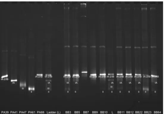
Identification of the main phylotypes byrecA PCR-TTGE
The denaturing gel PCR assay was based on the genetic
diversity of the 3′ part of the recA gene, containing 13
poly-morphic sites in a 333-bp fragment (Table 2). This method
was applicable to complex microbiota without subculturing and allowed the direct detection of the main recA phylotypes
in a single experiment, by comparison to a ladder (Fig.4).
0% 10% 20% 30% 40% 50% 60% 70% 80% 90% 100% III II IA-2/IB IA
Fig. 2 Relative prevalence of the different phylotypes of P. acnes according to clinical origin
Single nucleotide polymorphisms (SNP) in the 3′ part of the recA gene according to the types of sequence and the
isolates are given in Table2. Thirty-five of the 36 isolates
belonging to phylotype IA shared an identical sequence type (IA-3′recA1). The strain PA55, remote from the other IA strains in the recA tree, displayed a T/C mutation in position 275 (IA-3′recA2), which conferred to the corresponding
333-bp fragment a particular migration behavior in TTGE (Fig.4).
Almost all (41/42) of the type IA-2/IB strains shared the same allele (IA-2/IB-3′recA1). PA69 isolated from a neonatal gas-tric fluid was the sole strain with IA-2/IB-3′recA2 because of an SNP (C/A) in position 125. Despite a single SNP (G/A) in position 167 between IA-3′recA1 and IA-2/IB-3′recA1, they
were clearly separated in TTGE (Fig.4). The three clinical
isolates PA47, 48, and 87 belonging to type III in the recA tree shared the same sequence type III-3′recA1 that migrated dif-ferentially from other ST. The type III-3′recA1 TTGE band displayed a fuzzy aspect compared to bands corresponding to
the other phylotypes (Fig.4).
As expected from the recA tree topology, phylotype II was more variable, with four different ST that roughly
corresponded to the four branches in the tree (Fig.1). Allele
II-3′recA1 was shared by 13 of the 19 type II strains (Fig.4),
mostly grouped in the main cluster that contains the reference
sequence of phylotype II in the recA tree (Fig.1). Four clinical
isolates presented the same II-3′recA2, differing from II-3′
recA1 by one SNP (C/T in 98). The three other type II clinical strains PA36, 37, and 57 in the external position in the recA
tree (Fig. 1) displayed two different sequences (Fig. 4).
Finally, phylotype II displayed four migration profiles, each differing from the other phylotypes. The ten bands corre-sponding to ten 3′-recA types were included in a ladder to represent the recA PCR-TTGE diversity of our collection (Fig.4).
P. acnes phylotypes involved in cardiac surgery
We obtained 154 superficial and deep samples from 14 pa-tients during the different stages of hospitalization in cardiac surgery. Seventy samples were positive when they were screened for significant bacterial load using universal 16S rRNA gene PCR. Therefore, these samples constituted the set for the direct detection of the P. acnes phylotypes by recA PCR-TTGE. Among them, 54 were collected during the surgical procedure, 34 superficially (19/34 and 15/34 cutaneous and subcutaneous samples, respectively) and 20 deeply (11/20 and 9/20 sternal edges and mediastinum sam-ples, respectively). Seven samples concerned the skin before hospitalization, seven samples the skin and scar tissues after the surgical intervention, and only two samples involved the skin 3 months after the surgery. The TTGE fingerprints of ten
representative samples are shown in Fig.4. The phylotypes
present in the 70 perioperative samples were identified by comparison to the diversity ladder. Four bands that did not match the ladder were separately analyzed and did not corre-spond to any known sequence type (data not shown).
The P. acnes phylotyping for all perioperative and
peroperative samples from each patient is presented in Table3.
0 1 2 3 4 5 6 no weak strong IA IA-2/IB II III 0 5 10 15 20 no weak strong 0 1 2 3 4 5 6 no weak strong 0 0.5 1 1.5 2 2.5 3 3.5 4 no weak strong
Fig. 3 Hemolytic phenotype according to the phylotype for: (a) the whole P. acnes population, (b) isolates from cardiothoracic infections, (c) isolates from orthopedic infections, and (d) isolates from neurological infections
Ta b le 2 P hyloty p es of the 3′ -end of th e recA gene se quence types (S T ) of P . acnes iso lates with the correspond ing polymor phic sites. * The nomenclature indicate s the phylotype followed by the sequence type o f the 3′ part of the re cA gene Nucleotidic polymorp hic sites Phylotypes* Clin ic al is olat es 1 (62/333) 2 (98/3 33) 3 (116/333) 4 (12 2 /333) 5 (125/333) 6 (164/333) 7 (167/333) 8 (176/333) 9 (202/333) 10 (254/333) 11 (275/333) 12 (281/333) 13 (299/333) IA -3 ′recA1 P A5, 6, 12, 17, 25, 28, 29, 3 1, 38, 41, 42, 45, 46, 49, 5 0, 52, 60, 74, 75, 77, 78, 79, 8 1, 86, 92, 93, 101, 102, 103, 10 5, 106, 107, 1 10, 1 1 1, 1 14 T C C CCG G T G T C A T IA -3 ′recA2 P A5bis T C C C C G G T G T C A T IA -3 ′r ec A 3 P A 5 5 T C C CCG G T G T T A T IA -2/ IB-3′ recA1 P A7, 9, 13, 14, 18, 19, 23, 2 7, 32, 33, 34, 39, 40, 43, 4 4, 53, 54, 58, 64, 67, 68, 70, 7 2, 73, 76, 76b, 80, 82, 83, 83b, 83 t, 88, 89, 94, 95, 99, 104, 109, 1 13, 1 15 T C C CCG A T G T C A T IA -2/ IB-3′ re cA 2 P A69 T C C C A G A T G T C A T II -3 ′recA1 P A15, 22 , 31b, 51, 56, 61, 62 , 65, 85, 96, 98, 100, 108 C C C CCG G CA T T G T II -3 ′recA2 P A84, 90 , 91, 91b C T C C C G G C A T T G T II -3 ′r ec A 3 P A 5 7 T C C CCG G CA T T G T II -3 ′r ec A 4 P A 3 6 T C C CCG G CA T C G T II I-3′ recA P A 47, 48 , 8 7 C C T T C G G C A C T G C
As previously described for clinical isolates, phylotype I pre-dominated in surgical samples, with 32 (46 %) and 55 (78 %) samples sharing types IA and IA-2/IB, respectively, whereas
phylotype III was found in only three samples (Table 3).
Surprisingly, phylotype II was widely represented in surgical samples (n=30, 43 %), principally in superficial and low-depth samples (i.e., cutaneous and subcutaneous tissues). We observed major variations among patients, some of them presenting a wide diversity of P. acnes with three or four phylotypes during the hospitalization pathway (patients AQ
and AR, for example) and others with only one phylotype at only some stages of the surgery (patients AB and AL)
(Table3).
The phylotype diversity curves according to the stage of surgery and hospitalization were constructed for the whole
population of patients (Fig.5). These curves represented the
relative distribution of each P. acnes phylotype according to the surgical stage and depth of sampling in the surgical site. Despite high variability depending on the patient, the diversity curves showed common themes in the patient population and
Table 3 Origins and characteristics of the samples collected during the hospitalization course of 14 patients in cardiac surgery. The empty cells correspond to negative samples in 16S rRNA gene polymerase chain
reaction (PCR). The shaded cells show the detection of P. acnes phylo-types on samples with positive 16S rRNA gene PCR
Fig. 4 recA polymerase chain reaction temporal temperature gel electrophoresis (PCR-TTGE) experiment showing the migration of strains representative of phylotypes IA, IB, II, and III and the migration of ten representative samples from patients in cardiac surgery. The four bands representative of the major 3′ recA types are included in the ladder
an interesting dynamics of the different phylotypes according to the depth of the wound and the step of surgical intervention
(Fig.5). Types IA and II displayed similar evolutions: they
were present in superficial and low–deep tissues both at the beginning and at the end of the surgery, but they decreased in deep samples. Type IA greatly increased in postoperative samples such as scar tissues and in samples taken 3 months after surgery at a greater level than before surgery, while type II returned to its preoperative level. Type IA was also more present in sternal edges samples at the end than at the begin-ning of the intervention, suggesting peroperative colonization. Type IA-2/IB presented an outstanding evolution, with a gradual increase with the depth of sampling, but also with the time of sampling in subcutaneous and cutaneous samples. This result suggested a particular resistance of phylotype IA-2/IB to the antimicrobial agents used in perioperative prophy-laxis. However, both P. acnes IA and IA-2/IB cardiothoracic isolates were susceptible to antibiotics used in perioperative prophylaxis: cefamandole, vancomycin, and clindamycin.
Despite its low prevalence, type III displayed an increase in deep mediastinal tissues and subcutaneous tissues at the core phase of the surgery, but disappeared in superficial samples. Phylotypes IA and IA-2/IB were both predominant in scar tissues and on postoperative skin a long time after intervention, suggesting a specific predisposi-tion to recolonize skin.
Discussion
P. acnes is an usual member of healthy skin microbiota, often
considered a good bacterium devoid of pathogenicity [25].
However, its role in inflammatory processes in acnes vulgaris
and in several infections has been established [11,33–35]. In
opposition to allopatric true pathogens, mutualistic bacteria
are generally organized in species complex [36] and the
comprehension of their opportunistic pathogenic behavior occurs through deciphering of the population structure inside the species complex. Most population genetic studies were focused on healthy skin and acne isolates, despite a wider the pathogenic spectrum of P. acnes. Deep or severe infections such as HAI were rarely studied, except for large collections
of strains from failed hip replacements [26] and orthopedic
implants [11]. Here, we aimed to determine if genetic
subpop-ulations were specifically linked to specific sites or severe infections, as for the IA phylotype to acne. This hypothesis is supported by linkage disequilibrium analysis suggesting that the different phylogroups of P. acnes may occupy distinct
ecological niches [25] and, therefore, may display different
behaviors in different body sites. To meet our objective, we constituted a collection of natural isolates of P. acnes identi-fied during severe infectious processes or from different ori-gins in the routine exercise of hospital microbiology. The collection consisted mainly of strains from orthopedic and 0% 10% 20% 30% 40% 50% 60% 70% 80% 90% 100%
Fig. 5 Dynamics of the P. acnes phylotypes in samples chronologically collected during hospitalization and cardiac surgical procedures of 14 patients. The curves display the percentage of samples positive for each P. acnes phylotype according to the surgical stage and depth of sampling in the surgical site. C preop preoperative cutaneous sample, C cutaneous samples, SC subcutaneous samples, SE sternal edges samples, M medi-astinal samples, beginning at the beginning of the surgical intervention,
end at the end of the surgical intervention, ST scar tissues, C postop postoperative cutaneous samples (3 months postdischarge). Superficial samples correspond to cutaneous, subcutaneous, and scar tissues. Deeper samples correspond to sternal edges and mediastinal samples both at the beginning and at the end of the surgical procedure. The arrow represents the chronological course of patient hospitalization
cardiothoracic surgeries, as well as head and neck infections. Our collection is also original owing to strains from origins rarely represented or unrepresented in previous studies: peri-toneal dialysis, gastric fluid in neonates, and otolaryngologic samples.
Several studies have already analyzed the P. acnes popula-tion structure by different methods, based first on serotyping
and biotyping [37] and later by multilocus genetics and
geno-mics [17,19,22,23,25,26]. Comparative genomics based on
123,223 SNP nucleotides in the core regions highlighted that the recA classification of the strains was consistent with the
major genomic clades [25]. The gene tly also has a recognized
typing power [23,25,26]. In previous studies on large
popu-lations, the recA and tly genes showed good phylogenetic discriminative power and high genetic diversity index (0.67
each), making them good genetic markers [20,25]. Moreover,
despite the use of only two genes, we gained in allele vari-ability because we analyzed larger sequences than that in
major MLST schemes (available in http://pacnes.mlst.net/
and http://pubmlst.org/pacnes/). Consequently, we found a greater genetic polymorphism than in other studies, with some polymorphic sites being present at the extremities of recA and tly sequences not included in previously published studies. The high polymorphism in the 3′ part of the recA gene met our second objective, which was to develop a rapid and efficient tool (recA PCR-TTGE) to identify the major P. acnes phylotypes in complex communities without a first step of isolation by culture. The primary stratification of the P. acnes populations performed herein would be used as a basis for downstream MLST analysis on selected strains or selected phylotypes potentially associated with a particular type of deep infection.
However, deep MLST analyses [25] underscored the lack
of specificity of recA due to the sharing of some type IB alleles with members of clonal complex 4 in phylotype IA-1 and with all type IA-2 members. Moreover, some tly alleles such as
allele 8 were also shared between IA-2 and IB types [25].
Indeed, allele 8 is detected in 100 % of IB isolates and 97 % of
IA-2 isolates [25]. In our phylogenetic tree, all the strains
affiliated to the clade IA presented the tly allele 1. Therefore, considering the global congruence between the tly and recA trees, they certainly do not belong to IA-2 and probably corresponded to IA-1 isolates. For the alleles described herein, types IB and IA-2 could not be differentiated with certainty but type IA-2/IB differed clearly from type IA-1. The MLST schemes showed that IA-2 and IB underwent numerous re-combination events involving most of the genes studies. De-spite general linkage disequilibrium among phylotypes I, II, and III, the clonality of subtypes within phylotype I is less obvious. Particularly, IA-2 and IB phylotypes merged in split
trees [25,38] as the single clade named IA-2/IB with many
parallelogram structures, indicating recombination events. The consequence is that lineages in phylotype I are not clearly
separated; particularly, IA-2 and IB should be considered as an undifferentiated group IA-2/IB whatever the typing scheme considered because of the high level of allele recombinations. In the collection tested, we did not show a strict correlation between origins of infection and phylotypes. While previous studies showed that phylotypes II and III were rarely present
in skin samples [19,22,24], we highlight their higher
preva-lence in deep infection sites. To our knowledge, we describe for the first time among strains from oropharyngeal sphere that about 35 % of them belong to type II. Not enough strains from blood cultures were included in this study but we observed a
high prevalence of IA-2/IB and II. McDowell et al. [25]
underlined the association of types IA-2/IB and II with med-ical devices, which are frequently the source of bacteremia in HAI. Both types IA and IA-2/IB are found in gastric fluids in neonatology. Therefore, we can hypothesize that the transmis-sion from mother microbiota to newborn during delivery involves principally P. acnes type I.
As described previously [11], we found that phylotype I
was the most often involved in orthopedic surgery, and type IA-2/IB (with 8 of the 12 isolates in the recA tree) more frequently than IA. Considering the lack of discrimination of
recA underlined by McDowell et al. in 2012 [25], one can say
that orthopedic surgery isolates more often belong to type IA-2/IB than to IA1. This was also the case for strains involved in cardiac SSI, unlike the data previously published by
Davidsson et al. in 2012 [19], with a majority of type IA (only
ten strains typed). These results suggest a particular tropism of type IA-2/IB for surgical wounds in orthopedic and cardiac surgeries, but not in neurosurgical infections, where phylotype IA was more often isolated.
Besides its genetic typing value, we studied the tly gene because it encodes a putative virulence factor that could be related to a particular pathogenic behavior. Except for three strains that probably encountered recombination events, tly and recA markers give congruent population structures, sug-gesting that tly coevolve with housekeeping genes. In spite of the general role of hemolysins in bacterial pathogenesis, com-parative genomics prove that tly belongs to the core genome of
P. acnes [25], but not to island-like genomic regions, encoding
a variety of traits that differ between phylotypes [39].
Mc-Dowell et al. [25] hypothesized the importance of tly for both
mutualistic and pathogenic lifestyles. Brzuszkiewicz
et al. [39] concluded that the virulence of different P. acnes
strains is not only determined by the phylotype-specific ge-nome content but also by variable gene expression. For in-stance, despite the presence of tly in the P. acnes core genome, all strains do not express a hemolytic
pheno-type [26]. Moreover, tly hemolysin is not the only
putative virulence factor able to induce hemolysis; sev-eral others like the group of CAMP factors and other cyto-toxins, known to have this ability, have been identified in the
Our results showed an interesting expression of hemolytic activity: the absence or very low hemolytic activity within phylotypes II and III, whereas about half of the IA strains and almost all IA-2/IB strains expressed hemolysis. Studies de-scribing the hemolysis activities according to the genetic type
in the P. acnes population are rare [41]. We found 70 % of
phylotype I strains to be hemolytic, suggesting a potential role of hemolysin activity in infectious processes occurring in deep tissues. Concerning phylotype II, acne strains and deeper infection strains display similar hemolytic behavior, with about 10 % of hemolytic isolates in this study versus 12 % in the population of acne strains described by Kasprowicz
et al. [41]. Besides hemolysis, biofilm formation is a virulence
mechanism with potential importance in HAI [42]. P. acnes
biofilm allows a latent growth [43,44] that could lead to
late-onset deep infections like SSI on foreign materials in ortho-pedic surgery or in endocarditis and prosthetic valves in
cardiac surgery [8, 9, 11,35, 43]. No correlation has been
yet found between phylotypes and biofilm production, as all phylotypes contain isolates with different levels of biofilm
production [43]. However, in sebaceous follicles, P. acnes
biofilms are predominantly composed of types IA and II strains [45].
Finally, as for most opportunistic pathogens originating in the host microbiota, the virulence of P. acnes is difficult to decipher. Members of complex microbiota, such as human-associated microbiota, encounter complex relationships with host and other microbes. Therefore, the pathophysiology of the infection should take the microbiota dynamics into ac-count as a major factor involved in infectious process. For this purpose, we propose a follow-up of the P. acnes diversity during the hospitalization courses of patients in cardiac sur-gery. Taking the opportunity for the high variability of the 3′-terminal region of the recA gene (13 variable positions in 333 bp), we developed an original culture-independent ap-proach, the recA PCR-TTGE. This approach appears suitable to survey P. acnes populations during cardiac surgery proce-dures, allowing rapid screening of the main phylotypes in-volved in several samples. Our results showed that different phylotypes coexist in the same sample. This finding, which presented a certain novelty, is difficult to achieve when the P. acnes detection is based on culture because culture-based methods request the isolation of one pure bacterial colony. Phylotype IA-2/IB seems to be particularly adapted to colo-nize deep sternal and thoracic tissues (both sternal edges and mediastinum), as well as scar tissues after surgery. Type II was also very present at the different steps of hospitalization except in deep tissues, and did not increase during surgical interven-tion. These behaviors seemed to be not linked to particular resistance to antimicrobial agents used in cardiac surgical prophylaxis.
This study highlights a particular adaptation of phylotype IA-2/IB to cardiac surgery, in both the wound surgical
colonizing community and SSI. However, the strains isolated from cardiac SSI were obtained by cultural methods and did not reflect the whole P. acnes population initially involved in the sample. To confirm and complete the distribution scheme of P. acnes phylotypes in severe human infections, it would be necessary to increase the number of isolates from the different clinical sites and explore the dynamics of P. acnes phylotypes in diverse complex communities and their pathological varia-tions. For this purpose, the specific culture-independent ap-proach proposed herein would provide an efficient tool. More-over, it also seems relevant to specify the phylotype(s) and/or the clonal complex (CC) present in surgical samples, thereaf-ter to explore potential virulence characthereaf-teristics involved in the pathophysiology of SSI.
Acknowledgments We thank Brigitte Lamy, Hélène Marchandin, and Anne-Laure Michon from the Bacteriology Laboratory of Montpellier Hospital for providing the clinical strains, and all the members of the cardiothoracic surgery team of Montpellier Hospital for the surgical samples collections. We also thank Valérie Macioce from Département d’Information Médicale of Montpellier Hospital for her help in reviewing the English language. This work was supported by a public grant from the French research ministry and by the association ADEREMPHA, Sauzet, France.
Conflict of interest The authors declare that they have no conflict of interest.
References
1. Delgado S, Suárez A, Mayo B (2011) Identification, typing and characterisation of Propionibacterium strains from healthy mucosa of the human stomach. Int J Food Microbiol 149:65–72
2. Whitman WB, Parte A, Goodfellow M, Kämpfer P, Busse H-J, Trujillo ME, Ludwig W, Suzuki K (2012) Bergey’s manual of sys-tematic bacteriology: volume 5: the actinobacteria. Springer, New York
3. Kunishima S, Inoue C, Kamiya T, Ozawa K (2001) Presence of Propionibacterium acnes in blood components. Transfusion 41: 1126–1129
4. Brüggemann H (2005) Insights in the pathogenic potential of Propionibacterium acnes from its complete genome. Semin Cutan Med Surg 24:67–72
5. Qin M, Pirouz A, Kim MH, Krutzik SR, Garbán HJ, Kim J (2014) Propionibacterium acnes induces IL-1β secretion via the NLRP3 inflammasome in human monocytes. J Invest Dermatol 134:381–388 6. Eishi Y (2013) Etiologic link between sarcoidosis and
Propionibacterium acnes. Respir Investig 51:56–68
7. Hrbacek J, Urban M, Hamsikova E, Tachezy R, Heracek J (2013) Thirty years of research on infection and prostate cancer: no conclu-sive evidence for a link. A systematic review. Urol Oncol 31:951– 965
8. Noel W, Hammoudi N, Wegorowska E, D’Alessandro C, Steichen O (2012) Pacemaker endocarditis caused by Propionibacterium acnes: a case report. Heart Lung 41:e21–e23
9. Pan S-C, Wang J-T, Hsueh P-R, Chang S-C (2005) Endocarditis caused by Propionibacterium acnes: an easily ignored pathogen. J Infect 51:e229–e231
10. Zedtwitz-Liebenstein K, Gabriel H, Graninger W (2003) Pacemaker endocarditis due to Propionibacterium acnes. Infection 31:184– 185
11. Sampedro MF, Piper KE, McDowell A, Patrick S, Mandrekar JN, Rouse MS, Steckelberg JM, Patel R (2009) Species of Propionibacterium and Propionibacterium acnes phylotypes associ-ated with orthopedic implants. Diagn Microbiol Infect Dis 64:138– 145
12. Berthelot P, Carricajo A, Aubert G, Akhavan H, Gazielly D, Lucht F (2006) Outbreak of postoperative shoulder arthritis due to Propionibacterium acnes infection in nondebilitated patients. Infect Control Hosp Epidemiol 27:987–990
13. Unemo M, Friberg O, Enquist E, Källman J, Söderquist B (2007) Genetic homogeneity/heterogeneity of Propionibacterium acnes iso-lated from patients during cardiothoracic reoperation. Anaerobe 13: 121–126
14. Jakab E, Zbinden R, Gubler J, Ruef C, von Graevenitz A, Krause M (1996) Severe infections caused by Propionibacterium acnes: an underestimated pathogen in late postoperative infections. Yale J Biol Med 69:477–482
15. Sampedro MF, Huddleston PM, Piper KE, Karau MJ, Dekutoski MB, Yaszemski MJ, Currier BL, Mandrekar JN, Osmon DR, McDowell A, Patrick S, Steckelberg JM, Patel R (2010) A biofilm approach to detect bacteria on removed spinal implants. Spine (Phila Pa 1976) 35: 1218–1224
16. Fitz-Gibbon S, Tomida S, Chiu B-H, Nguyen L, Du C, Liu M, Elashoff D, Erfe MC, Loncaric A, Kim J, Modlin RL, Miller JF, S o d e r g r e n E , C r a f t N , We i n s t o c k G M , L i H ( 2 0 1 3 ) Propionibacterium acnes strain populations in the human skin microbiome associated with acne. J Invest Dermatol 133:2152–2160 17. Tomida S, Nguyen L, Chiu B-H, Liu J, Sodergren E, Weinstock GM, Li H (2013) Pan-genome and comparative genome analyses of Propionibacterium acnes reveal its genomic diversity in the healthy and diseased human skin microbiome. MBio 4:e00003-13 18. Kasimatis G, Fitz-Gibbon S, Tomida S, Wong M, Li H (2013)
Analysis of complete genomes of Propionibacterium acnes reveals a novel plasmid and increased pseudogenes in an acne associated strain. Biomed Res Inter 2013:918320
19. Davidsson S, Söderquist B, Elgh F, Olsson J, Andrén O, Unemo M, Mölling P (2012) Multilocus sequence typing and repetitive-sequence-based PCR (DiversiLab) for molecular epidemiological characterization of Propionibacterium acnes isolates of heteroge-neous origin. Anaerobe 18:392–399
20. Kilian M, Scholz CF, Lomholt HB (2012) Multilocus sequence typing and phylogenetic analysis of Propionibacterium acnes. J Clin Microbiol 50:1158–1165
21. McDowell A, Gao A, Barnard E, Fink C, Murray PI, Dowson CG, Nagy I, Lambert PA, Patrick S (2011) A novel multilocus sequence typing scheme for the opportunistic pathogen Propionibacterium acnes and characterization of type I cell surface-associated antigens. Microbiology 157:1990–2003
22. Lomholt HB, Kilian M (2010) Population genetic analysis of Propionibacterium acnes identifies a subpopulation and epidemic clones associated with acne. PLoS One 5:e12277
23. McDowell A, Perry AL, Lambert PA, Patrick S (2008) A new phylogenetic group of Propionibacterium acnes. J Med Microbiol 57:218–224
24. Niazi SA, Clarke D, Do T, Gilbert SC, Mannocci F, Beighton D (2010) Propionibacterium acnes and Staphylococcus epidermidis isolated from refractory endodontic lesions are opportunistic patho-gens. J Clin Microbiol 48:3859–3869
25. McDowell A, Barnard E, Nagy I, Gao A, Tomida S, Li H, Eady A, Cove J, Nord CE, Patrick S (2012) An expanded multilocus sequence typing scheme for Propionibacterium acnes: investigation of ‘path-ogenic’, ‘commensal’ and antibiotic resistant strains. PLoS One 7: e41480
26. McDowell A, Valanne S, Ramage G, Tunney MM, Glenn JV, McLorinan GC, Bhatia A, Maisonneuve J-F, Lodes M, Persing DH, Patrick S (2005) Propionibacterium acnes types I and II repre-sent phylogenetically distinct groups. J Clin Microbiol 43:326–334 27. Nagy E, Urbán E, Becker S, Kostrzewa M, Vörös A, Hunyadkürti J,
Nagy I (2013) MALDI-TOF MS fingerprinting facilitates rapid dis-crimination of phylotypes I, II and III of Propionibacterium acnes. Anaerobe 20:20–26
28. Kwon HH, Yoon JY, Park SY, Suh DH (2013) Analysis of distribu-tion patterns of Propionibacterium acnes phylotypes and Peptostreptococcus species from acne lesions. Br J Dermatol 169: 1152–1155
29. Mak TN, Yu S-H, De Marzo AM, Brüggemann H, Sfanos KS (2013) Multilocus sequence typing (MLST) analysis of Propionibacterium acnes isolates from radical prostatectomy specimens. Prostate 73: 770–777
30. Shannon BA, Cohen RJ, Garrett KL (2006) Polymerase chain reaction-based identification of Propionibacterium acnes types iso-lated from the male urinary tract: evaluation of adolescents, normal adults and men with prostatic pathology. BJU Int 98:388–392 31. Romano-Bertrand S, Parer S, Lotthé A, Colson P, Albat B,
Jumas-Bilak E (2012) Temporal temperature gel electrophoresis to survey pathogenic bacterial communities: the case of surgical site infections. In: Magdeldin S (ed) Gel electrophoresis—advanced techniques. InTech, pp 291–312
32. Tammelin A, Hambraeus A, Ståhle E (2002) Mediastinitis after cardiac surgery: improvement of bacteriological diagnosis by use of multiple tissue samples and strain typing. J Clin Microbiol 40:2936– 2941
33. Saleh K, Sonesson A, Persson B, Riesbeck K, Schmidtchen A (2011) A descriptive study of bacterial load of full-thickness surgical wounds in dermatologic surgery. Dermatol Surg 37:1014–1022 34. Dessinioti C, Katsambas AD (2010) The role of Propionibacterium
acnes in acne pathogenesis: facts and controversies. Clin Dermatol 28:2–7
35. Farrar MD, Ingham E (2004) Acne: inflammation. Clin Dermatol 22: 380–384
36. Georgiades K, Raoult D (2010) Defining pathogenic bacterial species in the genomic era. Front Microbiol 1:151
37. Kishishita M, Ushijima T, Ozaki Y, Ito Y (1980) New medium for isolating propionibacteria and its application to assay of normal flora of human facial skin. Appl Environ Microbiol 40:1100–1105 38. McDowell A, Patrick S, Eishi Y, Lambert P, Eady A (2013)
Propionibacterium acnes in human health and disease. Biomed Res Int 2013:493564
39. Brzuszkiewicz E, Weiner J, Wollherr A, Thürmer A, Hüpeden J, Lomholt HB, Kilian M, Gottschalk G, Daniel R, Mollenkopf H-J, Meyer TF, Brüggemann H (2011) Comparative genomics and tran-scriptomics of Propionibacterium acnes. PLoS One 6:e21581 40. Valanne S, McDowell A, Ramage G, Tunney MM, Einarsson GG,
O’Hagan S, Wisdom GB, Fairley D, Bhatia A, Maisonneuve J-F, Lodes M, Persing DH, Patrick S (2005) CAMP factor homologues in Propionibacterium acnes: a new protein family differentially expressed by types I and II. Microbiology 151:1369–1379 41. Kasprowicz A, Owczarek MM, Miedzobrodzki J, Białecka A (2012)
A comparison of biochemical and genetic classification of Propionibacterium acnes strains isolated from skin lesions of patients with acne. Med Dosw Mikrobiol 64:203–210
42. Ziebuhr W, Hennig S, Eckart M, Kränzler H, Batzilla C, Kozitskaya S (2006) Nosocomial infections by Staphylococcus epidermidis: how a commensal bacterium turns into a pathogen. Int J Antimicrob Agents 28(Suppl1):S14–S20
43. Holmberg A, Lood R, Mörgelin M, Söderquist B, Holst E, Collin M, Christensson B, Rasmussen M (2009) Biofilm formation by Propionibacterium acnes is a characteristic of invasive isolates. Clin Microbiol Infect 15:787–795
44. Coenye T, Peeters E, Nelis HJ (2007) Biofilm formation by Propionibacterium acnes is associated with increased resistance to antimicrobial agents and increased production of putative virulence factors. Res Microbiol 158:386–392
45. Jahns AC, Lundskog B, Ganceviciene R, Palmer RH, Golovleva I, Zouboulis CC, McDowell A, Patrick S, Alexeyev O (2012) An increased incidence of Propionibacterium acnes biofilms in acne vulgaris: a case–control study. Br J Dermatol 167:50–58