HAL Id: cea-02134831
https://hal-cea.archives-ouvertes.fr/cea-02134831
Submitted on 20 May 2019HAL is a multi-disciplinary open access archive for the deposit and dissemination of sci-entific research documents, whether they are pub-lished or not. The documents may come from teaching and research institutions in France or abroad, or from public or private research centers.
L’archive ouverte pluridisciplinaire HAL, est destinée au dépôt et à la diffusion de documents scientifiques de niveau recherche, publiés ou non, émanant des établissements d’enseignement et de recherche français ou étrangers, des laboratoires publics ou privés.
Following Stimulation by C3a and C5a Anaphylatoxins :
Specific Increase in Interleukin-6 mRNA Expression
Sakina Sayah-Jeanne, Alexander M. Ischenko, Alexander Zhakhov,
Anne-Sophie Bonnard, Marc Fontaine
To cite this version:
Sakina Sayah-Jeanne, Alexander M. Ischenko, Alexander Zhakhov, Anne-Sophie Bonnard, Marc Fontaine. Expression of Cytokines by Human Astrocytomas Following Stimulation by C3a and C5a Anaphylatoxins : Specific Increase in Interleukin-6 mRNA Expression. Journal of Neurochemistry, Wiley, 1999, 72 (6), pp.2426-2436. �10.1046/j.1471-4159.1999.0722426.x�. �cea-02134831�
Expression of Cytokines by Human Astrocytomas Following
Stimulation by C3a and C5a Anaphylatoxins: Specific
Increase in Interleukin-6 mRNA Expression
Sakina Sayah, *Alexander M. Ischenko, *Alexander Zhakhov,
Anne-Sophie Bonnard, and Marc Fontaine
Institut Fe´de´ratif de Recherche Multidisciplinaire sur les Peptides no. 23, INSERM U78, Faculte´ Mixte de Me´decine et Pharmacie, Rouen, France; and *Research Institute of Highly Pure Biopreparations, St. Petersburg, Russian Federation
Abstract: C3a and C5a anaphylatoxins are two
proin-flammatory peptides generated during complement acti-vation that act through distinct Giprotein-coupled recep-tors named C3aR and C5aR, respectively. We have dem-onstrated previously that human astrocytes expressed C3aR and C5aR constitutively and were able to produce a functional complement. In this study, we examined the effect of an anaphylatoxin stimulation on cytokine ex-pression by human astrocyte cell lines. Interleukin (IL)-1, IL-6, tumor necrosis factor-␣, and transforming growth factor- mRNA expression was studied by quantitative RT-PCR. Whereas IL-1, tumor necrosis factor-␣, and transforming growth factor- mRNA levels remained un-changed, stimulation of astrocytoma cells (T98G, CB193, U118MG) by C3a, C5a, and peptidic C3aR and C5aR agonists induced an increase in the IL-6 mRNA level. The amount of IL-6 was markedly increased at 3 and 6 h and returned to the basal level at 9 h of stimulation. This response was specific, because pretreatment of cells with pertussis toxin or with polyclonal C3aR or anti-C5aR antibodies completely blocked the IL-6 mRNA in-crease. The IL-6 response was also investigated at the protein level, but IL-6 protein was detected neither in cell lysates nor in supernatants of stimulated cells. The ana-phylatoxin-mediated transcriptional activation of IL-6 gene suggests that C3a and C5a could play a role in priming glial cells during the inflammatory process in the brain. Key Words: Complement—Anaphylatoxin— Astrocytes—Cytokines—Brain—Inflammation.
J. Neurochem. 72, 2426 –2436 (1999).
The complement system has been involved in the pathogenesis of the CNS. Complement involvement in brain pathologies has been investigated during demyeli-nating disorders such as multiple sclerosis (MS) (Comp-ston et al., 1989) and neurodegenerative pathologies such as Alzheimer’s disease (AD) (Rogers et al., 1992; Walker and McGeer, 1992). C3a and C5a anaphylatoxins are two proinflammatory peptides generated during com-plement activation. C3a and C5a share several biological activities, including mast cell degranulation,
enhance-ment of vasodilatation, smooth muscle contraction, and recruitment of immune cells to the site of inflammation (Frank and Fries, 1991). Several biological effects have been demonstrated in vitro for C3a anaphylatoxin, in-cluding platelet aggregation, enhancement of leukotriene production by basophils, and interleukin (IL)-1 release from monocytes (Haeffner-Cavaillon et al., 1987; Bischoff et al., 1990; Takabayashi et al., 1996). Beyond its powerful chemotactic function in myeloid cells, C5a anaphylatoxin can also induce superoxide radical pro-duction, hydrolytic enzyme release from neutrophils, phagocyte activation, and IL-1 secretion from macro-phages (Goodman et al., 1982). C3a and C5a exert their biological functions through binding to specific mem-brane receptors, respectively named C3aR and C5aR. C3aR and C5aR belong to the seven transmembrane domain receptor superfamily, coupled to a G protein (Rollins et al., 1991; Norgauer et al., 1993). Expression of these receptors, initially thought to be restricted to monocytes, macrophages, and polymorphonuclear cells, appears to be enlarged to several tissues of the body, suggesting new physiological roles for anaphylatoxins in tissues, in addition to their more classical inflammatory functions during pathological conditions (Sayah et al., 1998). Indeed, recent studies have demonstrated C5aR expression in nonmyeloid cells of the liver and lung
Received September 24, 1998; revised manuscript received Decem-ber 28, 1998; accepted January 6, 1999.
Address correspondence and reprint requests to Dr. M. Fontaine at INSERM U78, Faculte´ de Me´decine et Pharmacie, 22 boulevard Gam-betta, 76183 Rouen Cedex, France.
Abbreviations used: AD, Alzheimer’s disease; C3aR, C3a anaphy-latoxin receptor; C5aR, C5a anaphyanaphy-latoxin receptor; GAPDH, glycer-aldehyde-3-phosphate dehydrogenase; IL, interleukin; LPS, lipopoly-saccharide; MAP, multiple-associated peptide; M-MLV, Moloney murine leukemia virus; MS, multiple sclerosis; MTT, 3-(4,5-dimethyl-thiazol-2-yl)-2,5-diphenyltetrazolium bromide; PMA, phorbol myris-tate acemyris-tate; PTX, pertussis toxin; SDS, sodium dodecyl sulfate; SSPE, saline–sodium phosphate–EDTA buffer; TGF, transforming growth factor; TNF, tumor necrosis factor.
(Haviland et al., 1995) and in endothelial and epithelial cells (Foreman et al., 1994; Werfel et al., 1996). The C3aR cDNA cloning being recent (Ames et al., 1996; Crass et al., 1996; Roglic et al., 1996), data on C3aR expression are more limited. However, preliminary stud-ies have reported a large tissue expression of the C3aR mRNA (Crass et al., 1996). Our laboratory has docu-mented the presence of C5aR and C3aR in human astro-cyte cell lines, human fetal astroastro-cytes, and primary cul-ture of rat astrocytes (Gasque et al., 1995a, 1998; Sayah et al., 1997; Ischenko et al., 1998). These observations suggest new biological roles for C3a and C5a anaphyla-toxins in glial cells and strengthen the involvement of complement fragments in the pathogenesis of the CNS. To obtain further clues on the potential roles of C3a and C5a in neurological diseases, we decided to examine the effect of anaphylatoxins on cytokine production by hu-man astrocytes. IL-1, IL-6, tumor necrosis factor (TNF)-␣, and transforming growth factor (TGF)- mRNA expression was studied by quantitative RT-PCR. Here, we report that stimulation of three human glioma cell lines with C3a and C5a anaphylatoxins induced a specific increase in the IL-6 mRNA level through sig-naling by C3aR and C5aR. Cytokine expression was also studied at the protein level. IL-6 protein was detected neither in culture supernatants nor in cell lysates of stimulated cells. Anaphylatoxin-mediated IL-6 induction strengthens the involvement of C3aR and C5aR in the inflammatory process in the brain. The fact that anaphy-latoxins only induced IL-6 expression at the mRNA level suggests that C3a and C5a could play a role in priming glial cells during brain inflammation.
MATERIALS AND METHODS Reagents
The following materials were purchased as indicated: human recombinant C5a, phorbol myristate acetate (PMA), pertussis toxin (PTX), bacterial lipopolysaccharide (LPS), benzamidine, pepstatin A, leupeptin, EGTA, EDTA (Sigma–Aldrich, St. Quentin Fallavier, France), human recombinant IL-6 (2⫻ 105
U/ml), and monoclonal anti-human IL-6 (Boehringer Mann-heim, Biochemica).
Peptides
Multiple-associated peptide (MAP)-C3a and MAP-C5a pep-tides were synthesized by solid-phase synthesis (Applied Bio-system). Peptides were purified by reverse-phase HPLC, and sequences were ascertained by amino-acid analysis. MAP-C3a and MAP-C5a peptides correspond to the C-terminal part of the anaphylatoxins (amino acids 64 –77 for C3a and 61–74 for C5a) attached to a polylysine comb (eight peptidic monomers). Human C3a was generated by activation of complement using Zymosan A (Sigma). One liter of fresh human serum from healthy volunteers (CRTS, Bois-Guillaume, France) was supplemented with EGTA (10 mM), MgCl2(10 mM),
⑀-ami-nocaproic acid (50 mM), and DL-2-(mercaptoethyl)guanidino-pentanoic acid (1.5 mM). Complement was activated by 20 g of washed Zymosan A for 1 h at 37°C with gentle stirring. At the end of incubation, the pH was lowered to 5.2 by addition of HCl. The mixture was diluted with an equal volume of cold distilled water and centrifuged at 18,000 g and 4°C for 1 h. The
supernatant was adjusted to 1 M NaCl and subjected to immu-noaffinity chromatography on a 150-ml Sepharose anti-C3a column. After extensive washing of the column with 1 M NaCl, phosphate-buffered saline, C3a was eluted by 0.1 M glycine-HCl, 1 M NaCl buffer, pH 2.2. Solid urea was added to C3a-containing fractions (8 M final concentration), which were concentrated by ultrafiltration on a YM-5 membrane (Amicon, Paris, France). Purification was achieved by gel filtration on a TSK G2000 SW HPLC column (Beckman, Gagny, France) equilibrated in 20 mM sodium phosphate, 0.2 M NaCl, pH 7.4. The purity of C3a after gel filtration was assessed by sodium dodecyl sulfate (SDS)–polyacrylamide gel electrophoresis and Coomassie Blue staining. The immunoreactivity was checked by a western blotting experiment using monoclonal anti-C3a antibody and rabbit anti-C3a. Contamination of C3a prepara-tions by C5a was checked with a C5a radioimmunoassay kit (Amersham, Les Ulis, France). C5a was undetectable by this method, indicating a putative contamination of⬍1 ppm.
All peptides were diluted in apyrogen distilled water. Con-taminating endotoxin in MAP-C3a, MAP-C5a, and C3a prep-arations was assayed by the Limulus amebocyte lysate assay with a sensitivity of 25 pg/ml. All preparations contained no detectable amount of endotoxin.
Antibodies
A 21-amino acid peptide corresponding to the sequence 3–23 of human C3aR and a 29-amino acid peptide correspond-ing to the region 8 –36 of human C5aR were synthesized by solid-phase synthesis. The peptides were purified to homoge-neity by reverse-phase HPLC. Pure peptides were emulsified in phosphate-buffered saline and complete Freund’s adjuvant. The mixture was injected into the four footpads of rabbits. After 1 month, immunization was repeated with incomplete Freund’s adjuvant instead of complete adjuvant. After two further booster injections (subcutaneously) at a 1-week interval, rabbits were bled. Whole rabbit IgG was purified by protein A–Sepha-rose according to the manufacturer’s instructions (Pharmacia).
Cell culture
Human astrocyte cell lines T98G and U118MG and the human monocyte cell line U937 were obtained from American Type Culture Collection (Rockville, MD, U.S.A.). The human astrocyte cell line CB193 was a gift from Dr. Delpech (Centre H. Becquerel, Rouen, France). The astrocytic origin of the three cell lines was assessed by the expression of their specific marker, the glial fibrillary acidic protein. All cell lines were screened routinely by the Hoechst 33258 DNA staining method to ensure that they were mycoplasma-free. Cells were grown in Ham’s F-12 (astrocytes) or RPMI (U937 cells) culture medium (Biowhittaker, Verviers, Belgium) supplemented with 2 mM glutamine (Biowhittaker), 1% antibiotic–antimitotic solution (Biowhittaker), and 10% decomplemented fetal calf serum (Boehringer-Ingelheim, Bioproducts, Gagny, France) and grown at 37°C in a 5% CO2atmosphere.
Cells were cultured for 2 days before stimulation. Each experiment was carried out on 106cells. In some cases, cells
were incubated either with 200 ng/ml PTX for 48 h or with 100
g/ml anti-C3aR or anti-C5aR antibodies for 25 min before
stimulation with peptides.
RNA extraction
Total RNAs were extracted from cells by the guanidinium isothiocyanate method followed by ultracentrifugation onto a CsCl cushion as described by Sambrook et al. (1989). RNAs
were controlled by electrophoresis onto 1% agarose gel, and concentration was determined by absorbance at 260 nm.
PCR primers
Human IL-1, IL-6, TNF-␣, TGF-, and glyceraldehyde-3-phosphate dehydrogenase (GAPDH) primers were chosen accord-ing to cDNA sequences reported in the EMBL Data Library under accession numbers M15330, M14584, M10988, M19154, and M33197. Their sequences from 5⬘ to 3⬘ were as follows: IL-1 forward (CATATGAGCTGAAAGCTCTCCA), IL-1 reverse (GAGGTGCTGATGTACCAGTT), IL-6 forward (GATGGAT-GCTTCCAATCTGGAT), IL-6 reverse (AGTTCTCCATA-GAGAACAACATA), TNF-␣ forward (GCCAACGCCCTCCT-GGCCAATG), TNF-␣ reverse (CCCTTCTCCAGCTGGAA-GAC), TGF- forward (AGACCCCACATCTCCTGCA), TGF- reverse (TGAGTGTCTGAACTCCATA), GAPDH forward (TGCCATCAACGACCCCTTCA), GAPDH reverse (TGACCT-TGCCCACAGCCTTG).
RT-PCR
Total RNAs (50g) were treated for 20 min at 37°C with 90 U of RQ-1 RNase-free DNase (Promega, Charbonnie`re, France) in 100l of buffer (40 mM Tris-HCl, pH 8, 10 mM NaCl, 6 mM MgCl2, and 10 mM CaCl2) and 200 U of RNasin
ribonuclease inhibitor (Promega) to remove all traces of DNA. Following phenol/chloroform extraction and ethanol precipita-tion, RNAs were resuspended in diethyl pyrocarbonate-treated water.
Prior to PCR steps, the RT was carried out for 60 min at 37°C in 30l (final volume) with 2 g of total RNAs (DNA-free), 40 U of RNasin, 250 pmol of random hexamer primers pdN6 (Pharmacia Biotech, Orsay, France), and 200 U of Molo-ney murine leukemia virus (M-MLV) RT (Promega) in the reaction buffer (50 mM Tris-HCl, 75 mM KCl, 3 mM MgCl2,
and 5 mM dithiothreitol). The absence of contaminant was checked routinely by RT-PCR in negative control samples in which either the RNA samples were replaced with sterile water or the M-MLV RT was omitted. PCR was carried out with 5l of a cDNA mixture, in 50l final volume with 2.5 mM MgCl2
(Promega), 200M dNTPs (Pharmacia), 25 pmol of cytokine primers, and 2.5 U of Taq DNA polymerase (Promega) in the reaction buffer (50 mM KCl, 10 mM Tris-HCl, pH 9, and 0.1% Triton X-100). The PCR stages used were as follows: denatur-ation step for 4 min at 94°C; five cycles of 94°C for 40 s, annealing at 56°C for 50 s, and extension at 72°C for 90 s with ramping 6 s/°C from 56°C to 72°C; 25–30 cycles as above without ramping; and final elongation step at 72°C for 10 min. PCR was performed in a Hybaid Omnigene thermocycler (Schleicher and Schuell, Ce´ra-Labo, Ecquevilly, France). PCR products were analyzed by electrophoresis onto 1% agarose gel.
For quantitative RT-PCR, PCR was realized with 25 pmol of each cytokine primer, 1.25 pmol of each GAPDH primer, and 1Ci of [33P]dATP (Redivue, Amersham, Les Ulis, France).
Primers were used with a GAPDH/cytokine molar ratio of 1:20. The number of PCR cycles was chosen so that plateau phases were not reached (24 cycles). PCR products were loaded onto a 5% polyacrylamide gel and separated by electrophoresis at 200 V for 35 min. Gels were then exposed overnight at room temperature onto Biomax films (Sigma), and autoradiograms were analyzed by scanning using a Lecphor camera densitom-eter coupled to a computer (Biocom, Les Ulis, France). The relative cytokine mRNA amounts were estimated by dividing the peak densitometry area of the cytokine amplicon by the peak densitometry area of the GAPDH amplicon. The value of
cytokine mRNA from unstimulated cells was set equal to 1 unit, and values for the other samples were calculated relative to it.
cDNA probes
The human IL-1, IL-6, TNF-␣, TGF-, and GAPDH cDNA probes were cloned from U937 or T98G RNAs, after RT-PCR using human cDNA-specific primers (see above). The RT-PCR products were purified by the gel extraction kit (Pro-mega) following the manufacturer’s instructions, subcloned in the pGEM-T vector system (Promega), and sequenced using the T7 polymerase sequencing kit (Pharmacia). The probes were 100% homologous with the cDNA sequences reported in the EMBL Data Library under accession numbers M15330 (IL-1), M14584 (IL-6), M10988 (TNF-␣), M19154 (TGF-), and M33197 (GAPDH). The probes were then isolated from pGEM-T vector with SalI and NcoI restriction enzymes (Pro-mega).
Southern blotting
Agarose gel electrophoresis was performed on 5l of RT-PCR products. The gel was treated for 10 min in 0.15 M HCl and neutralized for 30 min in 0.4 M NaOH. Southern blotting was performed using 16-h capillary transfer in 0.4 M NaOH onto Nylon Plus membranes (Qiabrane from Qiagen GmbH, Hilden, Germany). The blot was neutralized for 10 min in 2⫻ SSPE (0.3 M NaCl, 17 mM Na2HPO4, and 50 mM EDTA).
Probes were labeled by random priming with [32P]dCTP
(Re-divue, Amersham) at a specific activity of 2 ⫻ 109cpm/g.
The membranes were prehybridized at 42°C for 4 h in a solution containing 50% formamide, 5⫻ SSPE, 1% SDS, 5⫻ Denhardt’s, 5% dextran sulfate, and 100g/ml herring sperm DNA. Hybridization was performed at 42°C for 16 h in the same solution supplemented with 108 cpm of labeled probe.
The membranes were washed briefly three times at room tem-perature in 2⫻ SSPE/0.1% SDS, 1 h at 68°C in 2⫻ SSPE/0.1% SDS, and 1 h at 68°C in 1⫻ SSPE/1% SDS, and exposed for 10 min at room temperature onto Cronex Films (Du Pont de Nemours–NEN, Les Ulis, France).
Northern blotting
Total RNAs (20 g) were fractionated by electrophoresis onto 0.8% agarose/0.2 M formaldehyde gels at 50 V for 16 h. RNA marker from Promega was used as RNA ladder. RNAs were then blotted to Nylon Plus membrane (Qiabrane, Qiagen) by capillary transfer in 50 mM NaOH for 3 h. Membrane was washed twice in 2⫻ SSPE and allowed to dry at room temper-ature. Hybridization with the IL-6 or GAPDH cDNA probes was performed as described above. Autoradiographic expo-sures onto Cronex films (Du Pont–NEN) were for 2–7 days at⫺80°C.
IL-6 bioassay
The IL-6 activity contained in supernatants was measured using the dose-dependent growth of B-cell-derived mouse hy-bridoma B9 cells. B9 cells were grown in RPMI medium (Biowhittaker) supplemented with 2 mM glutamine, 25 mM HEPES (Biowhittaker), 25 mM sodium pyruvate (Biowhit-taker), 1% antibiotic–antimitotic solution, 10% fetal calf serum, and 50 U/ml human IL-6. Before the assay, B9 cells were rinsed several times in RPMI medium to remove all traces of IL-6. Supernatants were collected at 12, 24, 48, and 72 h after stimulation of astrocyte cell lines by anaphylatoxins and were mixed with a proteinase inhibitor solution (1 mM EDTA, 1 mM EGTA, 1 mM benzamidine, 1 g/ml leupeptin, and 1 g/ml pepstatin A). One hundred microliters of each supernatant
dilution was mixed with 104 B9 cells and incubated in a
microtiter plate for 72 h. The number of cells was measured by the tetrazolium salt [3-(4,5-dimethylthiazol-2-yl)-2,5-diphe-nyltetrazolium bromide (MTT)] assay. Supernatants from T98G cells stimulated with 5 g/ml LPS for 48 h or from PMA-treated U937 cells (2.5 ng/ml PMA for 48 h) were used as positive controls. In some cases, supernatants were incu-bated with 100 ng/ml anti-IL-6 monoclonal antibody for 25 min before each dilution was mixed with B9 cells.
RESULTS
All results presented here were obtained on T98G cells. All data were reproducibly confirmed for CB193 and U118MG cells with only slight variations.
Stimulation of astrocyte cell lines by MAP-C3a and MAP-C5a only markedly increased IL-6 mRNA level
The expression of cytokine mRNAs by astrocytes was first analyzed on unstimulated cells by classical nonra-dioactive RT-PCR. All RNA preparations were pre-treated with DNase to eliminate contamination with genomic DNA. RT-PCR products were visualized by agarose gel electrophoresis (Fig. 1). Unstimulated astro-cytes constitutively expressed IL-1, IL-6, TNF-␣, and
TGF- mRNAs with a weaker level of expression for
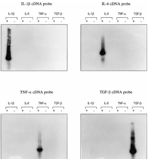
IL-6 and TNF-␣. No amplification product appeared for the negative controls. The specificity of each PCR prod-uct was confirmed by Southern blotting experiments using specific cDNA probes (Fig. 2).
Astrocyte cell lines were stimulated by 10⫺10M
MAP-C3a and MAP-C5a, and total RNAs were extracted at different stimulation times (3, 6, 9, and 20 h). IL-1, IL-6, TNF-␣, and TGF- mRNA expression was studied by quantitative RT-PCR using GAPDH as internal stan-dard. Results are presented as the relative fold increase over control (unstimulated cells). MAP peptide stimula-tion of T98G cells induced an increase in the IL-6 mRNA level, as shown by autoradiography (Fig. 3A). The intensity of the IL-6 amplicon was increased at 3 and 6 h of stimulation, whereas the intensity of the GAPDH amplicon did not vary upon stimulation. The related histogram is presented in Fig. 3B. The IL-6 mRNA was increased by 7.8-fold and sixfold for MAP-C3a and MAP-C5a, respectively, at 3 h of stimulation. The IL-6 mRNA level remained elevated at 6 h (⬃3.5-fold) and
returned to the basal level at 9 h of stimulation. On the contrary, the amount of IL-1, TNF-␣, and TGF- mRNAs did not vary significantly upon stimulation by MAP peptides (Fig. 3B).
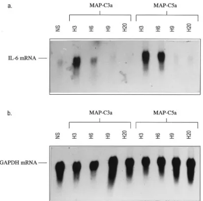
The increase in IL-6 mRNA expression observed by quantitative RT-PCR was confirmed by a northern blot-ting experiment using total RNAs from unstimulated and stimulated T98G cells. The IL-6 cDNA probe hybridized with a 1.1-kb mRNA (Fig. 4a). The signal was weak for RNAs from unstimulated cells, as well as for RNAs derived from T98G cells after 9 and 20 h of stimulation by MAP peptides. On the contrary, the IL-6 cDNA probe showed a strong signal for RNAs derived from T98G cells after 3 and 6 h of stimulation by MAP-C3a and MAP-C5a. The blot was then hybridized with the GAPDH cDNA probe (Fig. 4b). GAPDH transcript (1.2 kb) was highly expressed by RNAs derived from un-stimulated cells, as well as by RNAs derived from stim-ulated astrocytes (3, 6, 9, and 20 h).
Human C3a and C5a anaphylatoxins also induced an IL-6 mRNA increase in astrocyte cell lines
The IL-6 mRNA increase obtained with MAP peptides was confirmed with human purified C3a and recombi-nant C5a. Cells were stimulated with 10⫺8 M C3a and
C5a, and IL-6 mRNA expression was studied by quan-titative RT-PCR. Histograms are reported in Fig. 5. The IL-6 mRNA level was increased after 3 h of stimulation for T98G cells (4.4-fold for C3a and 3.5-fold for C5a). After 9 h of stimulation, the IL-6 mRNA amount was equal to that of unstimulated cells for both C3a and C5a stimulation.
Anaphylatoxin-induced IL-6 mRNA response was dose-dependent and specific
T98G cells were stimulated with different concentra-tions of MAP peptides and of C3a and C5a anaphyla-toxins in the range of 10⫺14to 10⫺6M. Total RNAs were
extracted at 3 h of stimulation, which corresponded to the peak in IL-6 mRNA response (Figs. 3 and 4), and the IL-6 mRNA level was evaluated as described above. The dose–response curves are represented in Fig. 6. The optimal response was observed at 10⫺10M for MAP-C3a
and at 10⫺8 M for purified C3a. The same profile was
obtained with MAP-C5a and recombinant C5a, respec-tively (data not shown).
FIG. 1. Cytokine mRNA expression by T98G cells. RT-PCR was carried out on DNase-treated RNAs derived from unstimulated T98G cells, using spe-cific human IL-1, IL-6, TNF-␣, and TGF- primers. PCR products were submitted to agarose gel elec-trophoresis. IL-1, IL-6, and TGF- amplicons were obtained after 30 PCR cycles and TNF-␣ amplicon after 35 PCR cycles. Lanes⫹, RT-PCR of T98G RNAs with different cytokine primers (IL-1 amplicon, 306 bp; IL-6 amplicon, 443 bp; TNF-␣ amplicon, 289 bp; TGF- amplicon, 246 bp); lanes⫺, RT-PCR on negative controls (without M-MLV).
Cells were preincubated with PTX for 4 h and subse-quently stimulated by peptides for 3 h, and the level of IL-6 mRNA was determined as described above. Cells stimulated with peptides alone were used as positive controls (3 h of stimulation). The IL-6 mRNA level was increased by eightfold and sixfold when T98G cells were stimulated with MAP-C3a and MAP-C5a, respectively. Pretreatment of T98G with PTX reduced the IL-6 mRNA level to that of unstimulated cells (Fig. 7). To confirm that the IL-6 induction was mediated by C3aR and C5aR, cells were incubated with either anti-C3aR or anti-C5aR antibody and subsequently stimulated with MAP-C3a or MAP-C5a, respectively. The incubation with anti-C3aR and anti-C5aR completely abolished the IL-6 mRNA increase induced by MAP-C3a and MAP-C5a, respec-tively (Fig. 7).
Preincubation with PTX completely abolished the IL-6 mRNA increase induced by C3a and C5a (Fig. 7). Pretreatment with anti-C3aR antibody blocked the C3a-induced IL-6 mRNA increase, which amount returned to the basal level. On the contrary, when cells were incu-bated with anti-C3aR antibody, the C5a-induced IL-6 mRNA increase was not blocked. Pretreatment with anti-C5aR antibody abolished the C5a-induced IL-6 mRNA
increase, but not the C3a-induced IL-6 mRNA increase (Fig. 7).
Astrocytes stimulated by anaphylatoxins were unable to produce IL-6 at the protein level
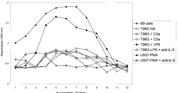
IL-6 induction by anaphylatoxins was also studied at the protein level. The supernatants of stimulated T98G cells were collected at 12, 24, 48, and 72 h of stimulation by either MAP peptides or anaphylatoxins and were analyzed using the dose-dependent growth of B9 cells. The dose–response profile of B9 cells is presented in Fig. 8. The functionality of this assay was tested using the supernatant of the human monocyte cell line U937 stim-ulated with PMA for 48 h. A negative control was composed of B9 cells incubated with RPMI medium alone. B9 cells in the absence of IL-6 did not grow, as represented by the absence of a bell-shaped curve (-✳-). The bell-shaped dose–response curve obtained with U937-PMA supernatant (-Œ-) corresponded typically to an IL-6-containing supernatant. The B9 response to U937-PMA supernatant was abolished almost com-pletely by preincubating the supernatant with a mono-clonal anti-IL-6 antibody (-‚-). B9 cells did not grow in response to unstimulated T98G supernatant as shown by FIG. 2. Southern blotting experiment on amplification products obtained after RT-PCR using cytokine primers. PCR products were blotted onto ny-lon membrane, and the membrane was hybridized with IL-1-, IL-6-, TNF-␣-, or TGF--specific cDNA probes. Autoradiographic exposures were for 10 –15 min at room temper-ature. Lanes ⫹, RT-PCR of T98G RNAs with different cytokine primers; lanes ⫺, RT-PCR on negative con-trols (without M-MLV).
the absence of a bell-shaped curve (-⫹-). Supernatants of T98G stimulated by C3a (- -) and C5a (-E-) showed the same response as supernatants of unstimulated T98G cells. On the contrary, the supernatant of T98G cells stimulated with LPS (-■-) showed a bell-shaped dose– response curve characteristic of an IL-6-containing su-pernatant. This response was abolished by preincubating the T98G-LPS supernatant with a monoclonal anti-IL-6 antibody (-䊐-). The same results were obtained with supernatants collected at 12, 24, and 72 h after stimula-tion by MAP peptides, purified C3a, and recombinant C5a (data not shown).
DISCUSSION
The complement system is a central component of the innate immune system. It has the capacity to mediate cell damage through killing via the membrane attack
com-plex, or to amplify the inflammatory response through generation of chemotactic fragments and opsonins. Ana-phylatoxins C3a and C5a are two proinflammatory sub-stances that activate monocytes and polymorphonuclear cells and induce the recruitment of immune cells through chemotactic gradients. C3a and C5a receptors belong to the seven transmembrane domain receptor family, cou-pled to a G protein (Rollins et al., 1991; Norgauer et al., 1993). The human C5aR cDNA was cloned some years ago by Gerard and Gerard (1991), whereas the C3aR cDNA has been cloned recently by three separate groups (Ames et al., 1996; Crass et al., 1996; Roglic et al., 1996). Cellular expression of these receptors was ini-tially thought to be limited to myeloid and polymorpho-nuclear cells. Recent studies have reported that C3aR and C5aR are expressed widely and constitutively in several tissues (e.g., liver, lung, spleen, intestine, heart, and brain) (Sayah et al., 1998), raising the possibility that anaphylatoxins might have new putative roles in these tissues, in addition to their more classical inflammatory properties. We have reported recently that astrocytes express constitutively a functional C3aR and C5aR (Gasque et al., 1995a; Sayah et al., 1997; Ischenko et al., 1998), but the role of anaphylatoxins in brain cells re-mains unknown. The complement has been shown to be involved in brain pathologies such as AD or MS (Comp-ston et al., 1989; Rogers et al., 1992; Walker and Mc-Geer, 1992), and studies have demonstrated the lethal effects of complement on brain cells in vitro (Wren and Noble, 1989; Piddlesden and Morgan, 1993) and in ex-perimental models of MS (Linington et al., 1989; Piddlesden et al., 1994) and AD (Rogers et al., 1992). If plasma complement can enter the brain following a blood– brain barrier breakdown, there is now evidence that complement can be synthesized locally. Our labora-tory has demonstrated that astrocyte cells have the ca-pacity to produce all the components of the complement in vitro (Gasque et al., 1992, 1993, 1995b), and the expression of some component mRNAs was observed in vivo during experimental brain lesioning and AD (John-son et al., 1992; Pasinetti et al., 1992). Until now, the role of the complement in brain pathogenesis remains unclear. To understand better the involvement of C3a and C5a anaphylatoxins in the CNS, we have set out to examine the effect of C3a and C5a stimulation on cyto-kine production by three astrocyte cell lines (T98G, CB193, and U118MG). We have used these three cell lines in other studies and have shown that they represent excellent models for complement studies (Gasque et al., 1992, 1993, 1995a,b; Sayah et al., 1997; Ischenko et al., 1998).
C3a and C5a anaphylatoxins did not induce obvious effects on morphological change or growth retardation in gliomas. In this article, we report that stimulation of human astrocyte cell lines by anaphylatoxins induced an increased expression of IL-6 mRNA, whereas the levels of other cytokine mRNAs remained unchanged. The experiments were carried out at least twice on three different astrocyte cell lines, and all data presented were FIG. 3. Quantitative RT-PCR analysis of cytokine mRNA
expres-sion following stimulation of T98G cells by MAP peptides. T98G cells were stimulated with 10⫺10M MAP peptides, and RNAs were extracted at 3, 6, 9, and 20 h and analyzed by semiquan-titative RT-PCR, using GAPDH as internal standard. PCR prod-ucts were separated by polyacrylamide gel electrophoresis and exposed onto autoradiographic film. The relative cytokine mRNA amount was estimated by dividing the peak densitometry area of the cytokine amplicon by the peak densitometry area of the GAPDH amplicon. A: Autoradiography of the IL-6:GAPDH RT-PCR products. B: Histograms representing IL-1, IL-6, TNF-␣, and TGF- mRNA expression. Results are presented as relative fold increase over control (unstimulated cells). Top panel: MAP-C3a stimulation; bottom panel: MAP-C5a stimulation. NS, un-stimulated cells; H3, H6, H9, and H20, 3, 6, 9, and 20 h of stimulation, respectively. Values shown are not the means of several determinations, but represent the typical profile of IL-6 mRNA level changes obtained for T98G cell line.
reproducibly obtained with only slight variations. The study was first conducted with peptidic anaphylatoxin analogues, MAP-C3a and MAP-C5a, that bear strong agonist properties (A. Ischenko, unpublished observa-tions). A marked increase in the IL-6 mRNA level was observed at 3 h of stimulation. The amount of IL-6 mRNA returned to the basal level after 9 h of stimula-tion. The results obtained by quantitative RT-PCR were confirmed by a northern blotting experiment using spe-cific IL-6 and GAPDH probes. An increase of IL-6
mRNA expression was also observed at 3 and 6 h of stimulation by MAP peptides, whereas the level of GAPDH mRNA remained unchanged. This latter obser-vation confirms the validity of the quantitative RT-PCR experiment used in this study. A similar IL-6 profile was obtained when cells were stimulated with human purified FIG. 4. Northern blotting experiment on RNAs de-rived from stimulated (10⫺10M MAP peptides) or unstimulated T98G cells. Total RNAs were blotted onto nylon membrane, and the membrane was hy-bridized either with human IL-6 cDNA probe (a) or with human GAPDH cDNA probe (b). Autoradio-graphic exposure times were for 2 days (GAPDH probe) or 7 days (IL-6 probe) at ⫺80°C. NS, un-stimulated cells; H3, H6, H9, and H20, 3, 6, 9, and 20 h of stimulation, respectively.
FIG. 5. Analysis of IL-6 mRNA expression by T98G cells
stimu-lated with human purified C3a and recombinant C5a. T98G cells were stimulated with 10⫺8M purified C3a or recombinant C5a,
RNAs were extracted at 3, 6, and 9 h, and IL-6 mRNA expression was studied by quantitative RT-PCR as described before. Values shown are not the means of several determinations, but repre-sent the typical profile of IL-6 mRNA level changes obtained for T98G cell line. NS, unstimulated cells; H3, H6, and H9, 3, 6, and 9 h of stimulation, respectively.
FIG. 6. Dose–response effect of MAP-C3a and purified C3a.
T98G cells were stimulated with different concentrations of MAP-C3a or C3a, RNAs were extracted at 3 h, and IL-6 mRNA expression was studied as described before. Top panel: MAP-C3a dose–response; bottom panel: MAP-C3a dose–response.
C3a and recombinant C5a, with a peak between 3 and 6 h of stimulation. The IL-6 response was weaker with C3a and C5a compared with that obtained with MAP pep-tides. Several observations confirm that the IL-6 re-sponse was anaphylatoxin-specific. First, the peptides used in this study did not contain detectable amounts of endotoxin as revealed by the Limulus amebocyte lysate assay, so that the response could not have been due to contaminating LPS. Second, anaphylatoxins induced an IL-6 mRNA increase in a dose-dependent fashion, with an optimal concentration of 10⫺8and 10⫺10M for
ana-phylatoxins and MAP peptides, respectively. A dose– dependent curve with an optimal concentration for ana-phylatoxins of 10⫺8M has also been observed for
che-motactic action on monocytes by another group (Arend et al., 1989). Third, pretreatment of cells with PTX completely blocked the IL-6 response and preincubation with anti-C3aR and anti-C5aR antibodies abolished the C3a- and C5a-induced IL-6 mRNA increase, respec-tively. No cross-reaction was detected, because anti-C3aR and anti-C5aR antibodies did not block the C5a and C3a responses, respectively, which confirmed the fact that C3a and C5a acted through distinct specific receptors.
IL-6 production was also investigated at the protein level. Cell lysates and supernatants were collected at 12,
24, 48, and 72 h of stimulation by MAP peptides, C3a, and C5a. Cell lysates were analyzed by western blotting with a specific anti-IL-6 antibody (data not shown), and supernatants were tested using the dose-dependent growth of B-cell-derived mouse hybridoma. IL-6 protein was detected neither in cell lysates nor in supernatants of stimulated cells. On the contrary, astrocytes stimulated with LPS were able to secrete a functional IL-6 protein, which confirmed again that the anaphylatoxin-induced IL-6 mRNA response could not have been attributed to contaminating LPS. The failure to demonstrate IL-6 pro-tein synthesis despite the IL-6 mRNA elevation was not due to an inhibitory effect of C3a and C5a on B9 cell growth, because we observed similar results using a specific ELISA (data not shown). Moreover, IL-6 pro-duction has already been observed in supernatants de-rived from C3a- and C5a-stimulated endothelial cells using the B9 bioassay in our laboratory (T. Monsinjon, personal communication).
This phenomenon of transcriptional activation without protein synthesis has already been observed for anaphy-latoxins by other groups in other systems (Arend et al., 1989; Schindler et al., 1990; Kaspar and Gehrke, 1994). Arend et al. (1989) failed to demonstrate increased IL-1 production from adherent human blood monocytes after stimulation with various complement fragments, includ-ing C3a and C5a. Another study showed that recombi-nant human C5a lacked translational signal for IL-1 and TNF-␣ synthesis by peripheral blood mononuclear cells (Schindler et al., 1990). The regulation of cytokine gene expression and biological activity is complex and can be regulated at different levels. Posttranscriptional regula-tion of protein synthesis includes mRNA stability and translational efficiency. Repeated AUUUA sequences in the 3⬘-untranslated region of mRNA have been shown to increase the susceptibility of mRNA to cytoplasmic deg-radation (Caput et al., 1986). The IL-6 mRNA contains six of these sequences, suggesting instability of mRNA (Tonouchi et al., 1989). The mechanisms responsible for an increased IL-6 mRNA degradation can include inhi-bition of RNase inhibitor or increased production of an AUUUA-specific mRNA binding protein (Malter, 1989). However, it is unlikely that the absence of IL-6 mRNA translation on C3a- and C5a-stimulated astrocytes results from IL-6 mRNA instability or enhanced degradation. Indeed, it has been reported that C5a-induced IL-1 mRNA had the same half-life as LPS-induced mRNA in peripheral blood mononuclear cells, i.e., C5a appeared not to influence IL-1 mRNA stability (Schindler et al., 1990). Kaspar and Gehrke (1994) have compared IL-1 synthesis by peripheral blood mononuclear cells stimu-lated with either LPS or C5a. Although C5a was able to induce IL-1 mRNA synthesis, very little or no protein could be detected by ELISA or western blotting. On the contrary, LPS-treated cells produced both IL-1 mRNA and protein. Polyribosome analysis revealed that the initiation step of protein synthesis is not limited in the C5a-stimulated cells and that the putative translational control mechanism must be acting downstream at the
FIG. 7. Blocking effect of PTX, C3aR, and C5aR
anti-bodies on anaphylatoxin-induced IL-6 mRNA expression. T98G cells were incubated either for 4 h with PTX or for 25 min with anti-C3aR or anti-C5aR, and 10⫺10M MAP-C3a or MAP-C5a
(top panel) or 10⫺8M C3a or C5a (bottom panel) was
subse-quently added to the culture medium. IL-6 mRNA expression was studied at 3 h of stimulation by peptides, as described before. Stimulation of cells with peptides alone served as a positive control. Values shown are not the means of several determinations, but represent the typical profile of IL-6 mRNA level changes obtained for T98G cell line.
level of polypeptide chain elongation and termination (Kaspar and Gehrke, 1994).
The mechanisms interfering in the posttranscriptional process in C3a- and C5a-stimulated astrocytes in our system are under investigation. It is likely that a stimulus like LPS induces IL-6 protein in astrocytes by at least two independent mechanisms: stimulation of transcrip-tion and enhancement of mRNA translatranscrip-tion. It is possible that translation of IL-6 mRNA requires a second signal that could be stimulation of a second messenger or phos-phorylation of a protein interacting with the IL-6 mRNA to enhance translational efficiency. We can speculate that in vivo a second signal is required to obtain the secretion of IL-6 by C3a- and C5a-stimulated astrocytes in the brain. Our results suggest that anaphylatoxins could play an important role in priming astrocytes during inflam-matory processes in the brain. This priming role of anaphylatoxins in peripheral blood mononuclear cells has already been suggested. C3a alone did not induce detectable TNF-␣ and IL-1 protein levels, whereas C3a enhanced LPS-induced mRNA and protein synthesis for TNF-␣ and IL-1 in adherent mononuclear cells (Tak-abayashi et al., 1996). C5a-primed peripheral blood mononuclear cells produce more IL-1 and TNF-␣ than cells incubated with LPS or PMA alone (Schindler et al., 1990). The physiological significance of the C3a- and C5a-induced IL-6 mRNA expression in astrocytes may be relevant in terms of synergism between anaphylatox-ins and other inflammatory stimuli, such as LPS or cytokines. The C3a and C5a stimuli could prime astro-cyte cells for a rapid response of IL-6 synthesis by stimulating IL-6 mRNA transcription, while providing suboptimal conditions for translation of the new mRNA.
The second putative signal would then stimulate IL-6 mRNA translation with subsequent release of the cyto-kine within the brain.
Our article is the first to describe a role for C3a and C5a in astrocytes. IL-6 is a multifunctional cytokine that is released during inflammation. Increased levels of this cytokine have been reported in the brain or the cerebro-spinal fluid of patients with MS, AD, Parkinson’s dis-ease, or ischemia (Maimone et al., 1996; McGeer and McGeer, 1997). IL-6 exerts regulatory activities on im-mune cells and trophic effects on a wide spectrum of tissues, including glial and neural cells (Gadient and Otten, 1997). Indeed, studies have demonstrated neuro-protective properties of IL-6 in vitro and in vivo, and Maeda et al. (1994) have postulated that glial-derived IL-6 may potentially enhance neuronal survival. IL-6 has also been reported to promote astrocyte proliferation and then contribute to astrogliosis (Gadient and Otten, 1997). The fact that C3a and C5a could prime astrocytes to synthesize IL-6 led to the hypothesis that local release of anaphylatoxins within the brain during pathological con-ditions could then have dual effects. C3a and C5a could exert a neurotrophic function, triggering neuronal sur-vival after injury, or increase the astrogliosis process by activating astrocytes, which could lead to neural tissue injury through the release of inflammatory mediators. We have shown that astrocytes are able to produce func-tional complement convertases (Gasque et al., 1992, 1993). The fact that astrocytes could generate anaphyla-toxins, which in turn can stimulate astrocytes through binding to C3aR and C5aR, led to the hypothesis that anaphylatoxins could exert an autocrine function on glial
FIG. 8. IL-6 bioassay. T98G cells were incubated either with 10⫺8M C3a or C5a or with LPS, and supernatants were collected at 48 h.
B9 cells were incubated in a microtiter plate with different dilutions of supernatants for 72 h. In some cases, supernatants were incubated with anti-IL-6 monoclonal antibody for 30 min before the B9 assay. The number of cells was measured by the MTT assay. Numbers on the abscissa represent dilutions of supernatants from 1 (undiluted supernatant) to 12 (1:2,048 dilution). Supernatant of U397 cells stimulated with PMA was used as a positive control.
cells, thus considerably amplifying the inflammatory process in the brain.
Acknowledgment: We gratefully acknowledge Dr. Delpech
for providing us with the CB193 cell line, M. Hiron and P. Chan for excellent technical advice, and J. F. Jeanne and R. Publier for photographic work. This work was supported by the University of Rouen (DRET grant 942616A) and by INSERM grant 4EO11B.
REFERENCES
Ames R. S., Li Y., Sarau H. M., Nuthulaganti P., Foley J. J., Ellis C., Zeng Z., Su K., Jurewicz A. J., Hertzberg R. P., Bergsma D. J., and Kumar C. (1996) Molecular cloning and characterisation of the human anaphylatoxin C3a receptor. J. Biol. Chem. 271, 20231–20234.
Arend W. P., Massoni R. J., Nieman M. A., and Giclas P. C. (1989) Absence of induction of IL-1 production in human monocytes by complement fragments. J. Immunol. 142, 173–178.
Bischoff S. C., Deweck A. L., and Dahinden C. A. (1990) Interleukin-3 and granulocyte/macrophage-colony-stimulating factor render hu-man basophils responsive to low concentrations of complement component C3a. Proc. Natl. Acad. Sci. USA 87, 6813– 6817. Caput D., Beutler B., Hartog K., Thayer R., Brown-Shimer S., and
Cerami A. (1986) Identification of a common nucleotide sequence in the 3⬘-untranslated region of mRNA molecules specifying inflammatory mediators. Proc. Natl. Acad. Sci. USA 83, 1670 – 1674.
Compston D. A. S., Morgan B. P., Campbell A. K., Wilkins P., Dole G., Thomas N. D., and Jasani B. (1989) Immunocytochemical localisation of the terminal complement complex in multiple scle-rosis. Neuropathol. Appl. Neurobiol. 15, 307–316.
Crass T., Raffetseder U., Martin U., Grove M., Klos A., Ko¨hl J., and Bautsch W. (1996) Expression cloning of the human C3a anaphy-latoxin receptor (C3aR) from differentiated U937 cells. Eur. J. Im-munol. 26, 1944 –1950.
Foreman K. E., Vaporciyan A. A., Bonish B. K., Jones M. L., Glovsky M. M., Eddy S. M., and Ward P. (1994) C5a-induced expression of P-selectin in endothelial cells. J. Clin. Invest. 94, 1147–1150. Frank M. M. and Fries L. F. (1991) Complement interactions and functions: the role of complement in inflammation and phagocy-tosis. Immunol. Today 12, 322–326.
Gadient R. A. and Otten U. H. (1997) Interleukin-6 (IL-6), a molecule with both beneficial and destructive potentials. Prog. Neurobiol.
52, 379 –390.
Gasque P., Julen N., Ischenko A., Picot C., Mauger C., Chauzy C., Ripoche J., and Fontaine M. (1992) Expression of complement components of the alternative pathway by glioma cell lines. J. Im-munol. 149, 1381–1387.
Gasque P., Ischenko A., Legoedec J., Mauger C., Schouft M. T., and Fontaine M. (1993) Expression of the complement classical path-way by human glioma in culture. A model for complement ex-pression by nerve cells. J. Biol. Chem. 268, 25068 –25074. Gasque P., Chan P., Fontaine M., Ischenko A., Lamacz M., Go¨tze O.,
and Morgan B. P. (1995a) Identification and characterisation of the complement C5a anaphylatoxin receptor on human astrocytes: relevance to inflammation in the brain. J. Immunol. 155, 4882– 4889.
Gasque P., Fontaine M., and Morgan B. P. (1995b) Complement expression in human brain. Biosynthesis of terminal pathway components and regulators in human glial cells and cell lines. J. Immunol. 154, 4726 – 4733.
Gasque P., Singhrao S. K., Neal J. W., Wang P., Sayah S., Fontaine M., and Morgan B. P. (1998) The receptor for complement anaphy-latoxin C3a is expressed by myeloid cells and nonmyeloid cells in inflamed human central nervous system: analysis in multiple scle-rosis and bacterial meningitis. J. Immunol. 160, 3543–3554. Gerard N. P. and Gerard C. (1991) The chemotactic receptor for C5a
anaphylatoxin. Nature 349, 614 – 617.
Goodman M. G., Chenoweh D. E., and Weigle W. O. (1982) Induction of interleukin-1 secretion and enhancement of humoral immunity by binding of human C5a to macrophages surface C5a receptors. J. Exp. Med. 156, 912–917.
Haeffner-Cavaillon N., Cavaillon J. M., Laude M., and Kazatchkine M. D. (1987) C3a (C3adesarg) induces production and release of interleukin-1 by cultured human monocytes. J. Immunol. 139, 794 –799.
Haviland D. L., MacCoy R. L., Whitehead W. T., Akama H., Molmenti E. P., Brown A., Haviland J. C., Parks W. C., Perlmutter D. H., and Wetsel R. A. (1995) Cellular expression of the C5a anaphy-latoxin receptor (C5aR): demonstration of C5aR on nonmyeloid cells of the liver and lung. J. Immunol. 154, 1861–1869. Ischenko A. M., Sayah S., Patte C., Andreev S., Gasque P., Schouft
M.-T., Vaudry H., and Fontaine M. (1998) Expression of a func-tional anaphylatoxin C3a receptor by astrocytes. J. Neurochem.
71, 2487–2496.
Johnson S. A., Lampert-Etchells M., Pasinetti G. M., Rozovsky I., and Finch C. E. (1992) Complement mRNAs in the mammalian brain: responses to Alzheimer’s disease and experimental brain lesion-ing. Neurobiol. Aging 13, 641– 648.
Kaspar R. L. and Gehrke L. (1994) Peripheral blood mononuclear cells stimulated with C5a or lipopolysaccharide to synthesize equivalent levels of IL-1 mRNA show unequal IL-1 protein accumulation but similar polyribosome profiles. J. Immunol. 153, 277–286. Linington C., Morgan B. P., Scolding N. J., Piddlesden S., Wilkins P.,
and Compston D. A. S. (1989) The role of complement in the pathogenesis of experimental allergic encephalomyelitis. Brain
112, 895–911.
Maeda Y., Matsumoto M., Hori O., Kuwabara K., Ogawa S., Yan S. D., Ohtsuki T., Kinoshita T., Kamada T., and Stern D. M. (1994) Hypoxia/reoxygenation-mediated induction of astrocyte interleu-kin-6: a paracrine mechanism potentially enhancing neuron sur-vival. J. Exp. Med. 180, 2297–2308.
Maimone D., Guazzi G. C., and Annunziata P. (1996) IL-6 detection in multiple sclerosis brain. J. Neurol. Sci. 146, 59 – 65.
Malter J. S. (1989) Identification of an AUUUA-specific messenger RNA binding protein. Science 246, 664 – 668.
McGeer E. G. and McGeer P. L. (1997) Inflammatory cytokines in the CNS. Possible role in the pathogenesis of neurodegenerative dis-orders and therapeutic implications. Pharmacol. Pathophysiol. 3, 214 –228.
Norgauer J., Dobos G., Kownatzki E., Dahinden C., Burger R., Kupper R., and Gierschik P. (1993) Complement fragment C3a stimulates Ca2⫹influx in neutrophils via a pertussis-toxin sensitive G pro-tein. Eur. J. Biochem. 217, 289 –294.
Pasinetti G. M. Johnson S. A., Rozovski I., Lampert-Etchells M., Morgan D. G., Gordon M. N., Morgan T. E., Willoughby D., and Finch C. E. (1992) Complement C1q and C4 mRNA responses to lesioning in rat brain. Exp. Neurol. 118, 117–125.
Piddlesden S. J. and Morgan B. P. (1993) Killing rat glial cells by complement components: deficiency of the rat analogue CD59 is the cause of oligodendrocyte susceptibility to lysis. J. Neuroim-munol. 48, 169 –176.
Piddlesden S. J., Storch M. K., Hibbs M., Freeman A. M., Lassman H., and Morgan B. P. (1994) Soluble recombinant complement recep-tor 1 inhibits inflammation and demyelination in antibody-medi-ated demyelinating experimental allergic encephalomyelitis. J. Immunol. 152, 5477–5484.
Rogers J., Cooper N. R., Webster S., Schultz J., McGeer P. L., Styren S. D., Civin W. H., Brachova L., Bradt B., Ward P., and Lieber-burg I. (1992) Complement activation by-amyloid in Alzheimer disease. Proc. Natl. Acad. Sci. USA 89, 10016 –10020. Roglic A., Prossnitz E. R., Cavanagh S. L., Pan Z., Zou A., and Ye
R. D. (1996) cDNA cloning of a novel G-protein-coupled receptor with a large extracellular loop structure. Biochim. Biophys. Acta
1305, 39 – 43.
Rollins T. E., Siciliano S., Kobayashi S., Cianciarulo D. N., Bonilla-Argudo V., Collier K., and Springer M. S. (1991) Purification of the active C5a receptor from human polymorphonuclear leuko-cytes as a receptor–Gicomplex. Proc. Natl. Acad. Sci. USA 88,
Sambrook J., Fritsch E. F., and Maniatis T. (1989) Molecular Cloning, A Laboratory Manual, 2nd edit., pp. 7.19 –7.22. Cold Spring Harbor Laboratory Press, Cold Spring Harbor, New York. Sayah S., Patte C., Gasque P., Chan P., Ischenko A., Vaudry H., and
Fontaine M. (1997) Characterisation of rat C5a anaphylatoxin receptor (C5aR): cloning of rat C5aR cDNA and study of C5aR expression by rat astrocytes. Mol. Brain Res. 48, 215–222. Sayah S., Leon P., Chan P., and Fontaine M. (1998) C3a and C5a
anaphylatoxin receptors. Med. Sci. 14, 291–299.
Schindler R., Gelfand J. A., and Dinarello C. A. (1990) Recombinant C5a stimulates transcription rather than translation of interleu-kin-1 (IL-1) and tumor necrosis factor: translational signal pro-vided by lipopolysaccharide or IL-1. Blood 76, 1631–1638. Takabayashi T., Vannier E., Clark B. D., Margolis N. H., Dinarello
C. A., Burke J. F., and Gelfand J. A. (1996) A new biological role for C3a and C3a desarg. Regulation of TNF-␣ and IL-1 synthe-sis. J. Immunol. 156, 3455–3460.
Tonouchi N., Miwa K., Karasuyama H., and Matsui H. (1989) Deletion of the 3⬘-untranslated region of human BSF-2 mRNA causes stabilisation of the mRNA and high-level expression in mouse NIH-3T3 cells. Biochem. Biophys. Res. Commun. 163, 1056 – 1062.
Walker D. G. and McGeer P. L. (1992) Complement gene expression in human brain: comparison between normal and Alzheimer dis-ease cases. Mol. Brain Res. 14, 109 –116.
Werfel T., Zwirner J., Opperman M., Sieber A., Begeman G., Drommer W., Kapp A., and Go¨tze O. (1996) CD88 antibodies specifically bind to C5aR on dermal CD117⫹ and CD14⫹ cells and react with a desmosomal antigen in human skin. J. Immunol. 157, 1729 – 1735.
Wren D. R. and Noble M. (1989) Oligodendrocytes and oligodendro-cyte/type-2 astrocyte progenitor cells of adult rats are specifically susceptible to the lytic effects of complement in absence of antibody. Proc. Natl. Acad. Sci. USA 86, 9025–9029.