HAL Id: tel-03162545
https://tel.archives-ouvertes.fr/tel-03162545
Submitted on 8 Mar 2021HAL is a multi-disciplinary open access archive for the deposit and dissemination of sci-entific research documents, whether they are pub-lished or not. The documents may come from teaching and research institutions in France or abroad, or from public or private research centers.
L’archive ouverte pluridisciplinaire HAL, est destinée au dépôt et à la diffusion de documents scientifiques de niveau recherche, publiés ou non, émanant des établissements d’enseignement et de recherche français ou étrangers, des laboratoires publics ou privés.
Diagnostic automatisé des pathologies de la rétine à
l’aide des volumes OCT
Rami Safarjalani
To cite this version:
Rami Safarjalani. Diagnostic automatisé des pathologies de la rétine à l’aide des volumes OCT. Traite-ment des images [eess.IV]. Université Bourgogne Franche-Comté; Université de Balamand (Tripoli, Liban), 2020. Français. �NNT : 2020UBFCK039�. �tel-03162545�
1
THESE DE DOCTORAT DE L’ETABLISSEMENT UNIVERSITE BOURGOGNE FRANCHE-COMTE
PREPAREE au Laboratoire ImVia/IFTIM, Univ. Bourgogne Franche-Comté, Le Creusot 71200, France
En association avec le laboratoire LIA, EDST, Univ. Libanaise, Tripoli, Liban
Ecole doctorale n°37
ÉCOLE DOCTORALE «SPIM»
Doctorat de l’instrumentation et informatique de l'image
Par
M. Rami SAFARJALANI
« Diagnostic automatisé des pathologies
de la rétine à l'aide des volumes OCT »
« Application de classification et de détection de
rétinopathie diabétique et d’œdème maculaire »
Thèse présentée et soutenue à « Le Creusot », le « 15/12/2020 »
Composition du Jury:
Mme Fan YANG
Professeur, Univ. Bourgogne Franche-Comté
Présidente
M. Jocelyn CHANUSSOT
Professeur, Grenoble INP
Rapporteur
Mme Caroline PETITJEAN
Maître de Conférences HDR,
Université de Rouen Normandie
Rapporteuse
M. Ahmad SHAHIN
Professeur, Université Libanaise
Examinateur
Mme Samia AÏNOUZ
Professeur, INSA de Rouen Normandie
Examinatrice
M. Fabrice MÉRIAUDEAU
Professeur, Univ. Bourgogne Franche-Comté
Directeur de thèse
M. Désiré SIDIBÉ
Professeur, Université d'Evry - Paris Saclay
Codirecteur de thèse
Université Bourgogne
Franche-Comté
2
Acknowledgment
This thesis was prepared in the Laboratory ImVia with the IFTIM Team (Functional and Molecular Imaging, Medical Image Processing) under the direction of Prof. Fabrice Meriaudeau, at Univ. Bourgogne Franche-Comté.
First of all, I would like to thank my thesis supervisor, Prof. Fabrice, for welcoming me to his team, for his encouragement, his repetitious help, his relevant advice, and his human spirit. I am also grateful to him for the significant time he has given me, for his support and his understanding. I learned a lot from him and I am honored to work under his surveillance. May he be convinced of my deep respect.
In addition, my appreciation goes to all the professors and doctors who supported me during the accomplishment of this thesis:
To Prof. Desire Sidibe, Co-supervisor of this thesis, for the consideration he brought to me during my thesis. I am very grateful for his aid and his advice.
To Prof. Samia Ainouz, for her care and assistance; her remarks and words are and will be always appreciated. All my appreciation is addressed to her for the trust she has shown in me.
I offer my sincere thanks to Prof. Ahmad Shahin, Professor of Lebanese University for his support and his advice. I will be always appreciated for his assistance.
To Prof. Salah El-Falou and Dr. Mohamad El-Falou for their encouragement and their support.
All my appreciation and gratitude will go also to my mother, for her continuous assistance, her encouragement, her back-up, and her feelings.
I especially thank my brothers and my friends… Finally, I dedicate this work to my late father.
3
Remerciements
Cette thèse a été préparée au Laboratoire ImVia avec l'équipe IFTIM (Imagerie Fonctionnelle et Moléculaire, Traitement d'Images Médicale) sous la direction du Pr. Fabrice Meriaudeau, à l'Univ. Bourgogne Franche-Comté.
Tout d'abord, je tiens à remercier mon directeur de thèse, le Pr. Fabrice, de m'avoir accueilli dans son équipe, pour ses encouragements, son aide répétitive, ses conseils pertinents et son esprit humain. Je lui suis également reconnaissant pour le temps considérable qu'il m'a accordé, pour son soutien et sa compréhension. J'ai beaucoup appris de lui et je suis honoré de travailler sous sa surveillance. Qu'il soit convaincu de mon profond respect.
De plus, mes remerciements vont à tous les professeurs et les maitres de conférences qui m'ont soutenu lors de la réalisation de cette thèse :
Au Pr. Desire Sidibe, codirecteur de cette thèse, pour la considération qu'il m'a apportée lors de ma thèse. Je lui suis très reconnaissant de son aide et de ses conseils.
Au Pr. Samia Ainouz, pour ses soins et son assistance; ses remarques et ses paroles sont et seront toujours appréciées. Toute ma gratitude est adressée à elle pour la confiance qu'elle m'a témoignée.
Je remercie sincèrement le Pr. Ahmad Shahin, professeur à l'Université Libanaise pour son soutien et ses conseils. Je serai toujours apprécié de son aide.
À Pr. Salah El-Falou et Dr. Mohamad El-Falou pour leurs encouragements et leur soutien.
Toute ma reconnaissance et ma gratitude iront aussi à ma mère, pour l'assistance continue, ses encouragements et ses sentiments.
Je remercie particulièrement mes frères et mes amis… Enfin, je dédie ce travail à mon défunt père.
4
Titre : Diagnostic automatisé des pathologies de la rétine à l'aide des volumes OCT. Mots clés : OCT, CNN, DR, DME, GNN.
Résumé :
La principale cause de cécité dans la population pourrait être surtout la détérioration de la rétine causée par les
problèmes liés au diabète et la
complication du vieillissement. La
rétinopathie diabétique (DR) et l'œdème maculaire diabétique (DME) sont les principales causes directes de problèmes de vision chez les citoyens en âge de travailler de la plupart des pays avancés. Le nombre élevé de personnes diabétiques dans le monde indique que le DME et la RD resteront les principaux facteurs de perte de vision partielle ou totale, ce qui affecte la qualité de vie des patients pendant de nombreuses années et menace leur vie. Par conséquent, une détection précoce suivie de procédures de traitement rapide des personnes atteintes de maladies liées au diabète est importante pour prévenir les problèmes optiques et peut réduire le risque de cécité. De plus, les personnes de plus de 50 ans sont exposées à la dégénérescence maculaire liée à l'âge (AMD) qui attaque la rétine. Par conséquent, les chercheurs du monde entier sont attirés par les différences liées à plusieurs maladies rétiniennes.
Plusieurs méthodes automatisées utilisant l'AI ont été appliquées pour la détection et le test des maladies
rétiniennes. Malheureusement, ces
modèles peuvent être confondus avec une incapacité de calcul, ce qui nécessite une intervention supplémentaire de la part de spécialistes. Cette thèse présente une méthode automatique - basée sur des algorithmes de réseaux de neurones d'apprentissage en profondeur - pour détecter DME et DR, ce qui permet de dépasser l'évaluation pratique subjective des ophtalmologistes.
Basé sur "Convolutional Neural Network", un modèle proposé est présenté avec un classificateur soft-max et entrainé de bout en bout pour la classification automatique des images rétiniennes de tomographie par cohérence optique (OCT). Ce modèle a la capacité de détecter des caractéristiques permettant d'identifier la DR et le DME en utilisant ces images rétiniennes avec une précision et une sensibilité améliorées. De plus, un modèle préformé a été affiné et réformé à l'aide d'un ensemble de données qui a été enrichi à l'aide de "Generative Adversarial Networks" (GAN). Contrairement au diagnostic manuel de la maladie rétinienne basé sur un examen clinique personnel et l'analyse des images OCT, cette méthode a
montré la capacité de prédire
automatiquement les cas atteints de DME par rapport aux cas sains. Les expériences ont été évaluées sur plusieurs ensembles
5 de données fournis par différentes institutions.
Le modèle, comparé à d'autres modèles CNN entrainés de bout en bout ou pre-entrainés et affinés, montre des fonctionnalités d'extraction efficaces, avec moins de temps, sur la base d'une étape
de prétraitement efficace des données. Les résultats expérimentaux ont montré une plus grande précision de classification, ce qui est prometteur dans le domaine de la
détection précoce des maladies
diabétiques pour aider les
ophtalmologistes dans les technologies biomédicales.
Title: Automated Diagnostics of Retinal Pathologies Using OCT Volumes. Keywords: OCT, CNN, DR, DME, GNN
Abstract:
The leading cause of blindness in the
population could mostly be the
degeneration of the retina caused by the diabetic-related problems and the aging issue. Diabetic retinopathy (DR) and diabetic macular edema (DME) are the main direct causes of vision problems in the labor age citizens of most advanced countries. The elevated number of diabetic people globally indicates that DME and DR will remain to be the principal factor to partial or total vision loss, which affects the lives quality of patients for many years to come and threaten their lives. Therefore, early detection followed by fast treatment procedures of persons with diabetic-related diseases is significant in preventing optical problems and can decrease the risk of blindness.
In addition, people above 50 are
exposed to age-related macular
degeneration (AMD) disease that hits the retina. Therefore, researchers over the world have attracted to the differences related to several retinal diseases.
Several automated methods using Artificial Intelligence (AI) (varying from traditional computer vision to advanced machine learning algorithms) have been applied for the detection and examination of retinal diseases. Unluckily, these models
are able to be mistaken with
computational inability, which necessitates additional interference from specialist
personal. This thesis presents an
automatic method - based on deep learning neural networks algorithms - to detect DME and DR, which allows
overstepping the subjective handy
evaluation of ophthalmologists.
Based on Convolutional Neural Network, a proposed model is presented with a soft-max classifier and fully trained
from scratch for the automatic
classification of Optical Coherence
Tomography (OCT) retinal images where OCT screening techniques are applied as the current dependable assessment and measurement method to discover the existence of swallow in the retina. This
6 model has the ability to detect patterns for DR and DME using these retinal images with improved accuracy and sensitivity. Moreover, a pre-trained model has been fined-tuned and re-trained using a dataset that has been augmented using Generative Adversarial Networks (GANs). In opposite to manual retinal disease diagnosis based on personal clinical examination and analysis of OCT images, this method showed the capability to automatically predict DME diseased cases versus healthy cases. The experiments have been
evaluated over several datasets provided by different institutions.
The model, compared to other CNN end-to-end or transfer learned models, shows effective extracting features, with less time consumption, based on an efficient data pre-processing stage. The experimental results showed a higher
accuracy of classification which is
promising in the field of early detection of diabetic diseases to aid ophthalmologists
in biomedical technologies.
Université Bourgogne Franche-Comté 32, avenue de l’Observatoire
7
Table of contents
Acknowledgment ... 2 Abstract ... 4 Table of Contents ... 7 List of Acronyms ... 11 List of Figures ... 15 List of Tables ... 18 List of Equations ... 19 Introduction... 20 1- General context ... 20 2- Thesis objective ... 213- Thesis synthetic plan ... 21
Chapter 1 ... 23
Overview of Diabetic Retinopathy pathologies ... 23
1.1. Introduction ... 24
1.2. Overview of eyeball and retinal-related pathologies ... 24
1.2.1. The Eyeball ... 24
1.2.2. Eye structure ... 25
1.2.3. Retinal layers ... 26
1.2.4. Retinal pathologies ... 27
1.2.4.1. Age-related macular degeneration (AMD) ... 28
1.2.4.2. Cataracts ... 29 1.2.4.3. CytoMegaloVirus Retinitis (CMV) ... 30 1.2.4.4. Retinal Detachment ... 30 1.2.4.5. Glaucoma... 31 1.2.4.6. Cardiovascular Disease ... 31 1.2.4.7. Diabetic Retinopathy (DR) ... 32
1.2.4.8. Diabetic macular edema (DME) ... 34
1.3. Optical Imaging Modalities ... 36
1.3.1. Fundus Photography ... 37
1.3.2. Adaptive Optics (AO) ... 37
1.3.3. Scanning laser ophthalmoscopy (SLO) ... 38
1.3.4. Photoacoustic microscopy (PAM) ... 39
8
1.3.6. Magnetic resonance imaging (MRI) ... 40
1.3.7. Optical Coherence Tomography (OCT) ... 41
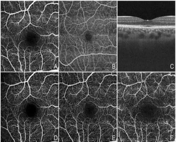
1.3.8. Optical coherence tomography angiography (OCTA) ... 45
1.4. Deep Neural Networks: ... 46
1.4.1. Deep learning applications ... 48
1.5. Conclusion ... 51
Chapter 2 ... 52
Introduction to data pre-processing and classification in machine learning ... 52
2.1. Introduction ... 53 2.2. Data pre-processing in ML ... 53 2.2.1. Dataset acquiring: ... 53 2.2.2. Dataset Cleaning: ... 54 2.2.3. Dataset splitting: ... 54 2.3. Classification tasks in ML ... 54 2.3.1. Unsupervised classification: ... 55 2.3.2. Supervised classification: ... 55
2.3.2.1. Standard terms in classification algorithms ... 56
2.3.2.2. Types of classification: ... 56
2.3.2.3. Types of classification learners: ... 56
2.3.2.3.1. Lazy learners ... 56
2.3.2.3.2. Eager learners ... 56
2.3.2.4. Classification algorithms and techniques ... 56
2.3.2.4.1. Softmax classifier: ... 57
2.3.2.4.2. Artificial Neural Networks: ... 58
2.3.2.4.3. K-Nearest Neighbors (KNN) ... 59
2.3.2.4.4. Support Vector Machine (SVM) ... 59
2.3.2.4.5. Decision Tree ... 61
2.3.2.4.6. Random Forest ... 62
2.3.2.5 Classifier performance evaluation ... 62
2.3.2.5.1. Holdout method ... 62
2.3.2.5.2. Cross-validation ... 63
2.3.2.5.3. Confusion matrix (Precision and Recall) ... 63
2.3.2.5.4. Receiver Operating Characteristics (ROC curve) ... 65
9
Chapter 3 ... 67
State of the Arts ... 67
3.1. Introduction ... 67
3.2. Literature review ... 67
3.2.1. Conventional ML analysis ... 67
3.2.2. Deep learning and Neural networks models ... 70
3.2.2.1. Segmentation approach ... 70
3.2.2.2. Classification approach ... 73
3.3. Perspective and motivation ... 80
3.4. Conclusion ... 81
Chapter 4 ... 82
Diabetic Retinal Tomographical Image Classification using CNN ... 82
4.1. Introduction ... 82
4.2. Image processing materials and methods ... 83
4.2.1. Dataset acquisition: ... 83
4.2.2. Dataset Pre-processing method ... 84
4.2.2.1. Dimension reduction: ... 84
4.2.2.2. Image denoising ... 84
4.2.2.3. Image cropping ... 84
4.3. The proposed method ... 86
4.3.1. CNN architecture ... 86
4.3.2. Setting-Up Hyper parameters: ... 89
4.4. Experimental performance evaluation ... 90
4.5. Discussion ... 94
4.6. Conclusion ... 95
Chapter 5 ... 97
Convolutional Neural Network Classification Model for Diabetic Macular Edema using Spectral Domain Tomographical Scans ... 97
5.1. Introduction ... 97
5.2. Image processing methods ... 98
5.2.1. Dataset acquisition: ... 98
5.2.1.1. SERI Dataset ... 98
5.2.1.2. DUKE Dataset ... 98
10
5.2.2. Dataset Pre-processing technique:... 99
5.2.3. Synthetic data augmentation: ... 101
5.2.3.1. Generator: ... 104
5.2.3.2. Discriminator: ... 105
5.2.3.3. Adversarial Model: ... 106
5.2.3.4. GAN architecture: ... 106
5.2.3.5. Synthetic image evaluation ... 107
5.3. The proposed method ... 110
5.3.1. Fine-tune the pre-trained model ... 110
5.3.2. Re-train the model ... 110
5.4. Performance Analysis ... 113
5.5. Discussion ... 117
5.6. Conclusion ... 119
General Conclusion ... 120
11
List of Acronyms
1. 2D (Two Dimension) 2. 3D (Three Dimension) 3. ACC (Accuracy)
4. Adagrad (Adaptive Gradient)
5. Adam (Adaptive Moment Estimation) 6. AE (Auto-Encoder)
7. AI (Artificial Intelligence)
8. AMD (Age-related Macular Degeneration) 9. ANN (Artificial Neural Networks)
10.AUC (Area Under Curve)
11.BM3D (Block-Matching and 3D-Filtering) 12.BoW (Bag Of Words)
13.CMV (CytoMegaloVirus)
14.CNN (Convolutional Neural Network) 15.CNV (Choroidal NeoVascularization) 16.Convnet (Convolutional Neural Network) 17.CUHK (Chinese University of Hong Kong) 18.CV (Computer Vision)
19.CWS (Cotton Wool Spots)
20.DAGAN (Data Augmentation Generative Adversarial Network) 21.DCGAN (Deep Convolutional Generative Adversarial Network) 22.DenseNet (Densely-Connected Network)
23.DL (Deep Learning)
24.DME (Diabetic Macular Edema) 25.DR (Diabetic Retinopathy) 26.DT (Decision Tree)
27.ECG (Electro-Cardiogram Impulse)
28.EDST (Doctoral School in Science & Technology) 29.ELM (External Limiting Membrane)
12 31.EMG (Electro-Myogram Waves)
32.ERR (Error Rate) 33.EX (Exudates)
34.FC (Fully Connected Layer) 35.FCC (Fully Connected Layer)
36.FD-OCT (Fourier domain Optical Coherence Tomography) 37.FN (False Negative)
38.FP (False Positive)
39.FPR (False Positive Rate) 40.FT (Fine-Tunning)
41.GAN (Generative Adversarial Network) 42.GCL (Ganglion Cell Layer)
43.HC (Hierarchical) 44.HD (High dimension) 45.HEs (Hemorrhages)
46.HoG (Histogram of Oriented Gradients) 47.HR (Hemorrhages)
48.IFCNN (Iterative Fusion CNN)
49.IFTIM (Functional and Molecular Imaging, Medical Image Processing) 50.ILM (Internal Limiting Membrane)
51.ImVia (Artificial Imaging and Vision) 52.INL (Inner Nuclear Layer)
53.IPL (Inner Plexiform Layer) 54.IR (Infra Red)
55.IRF (Intra-Retinal Fluid)
56.IS/OS (photoreceptor rod and cone Inner and Outer Segments) 57.KNN (K-Nearest Neighbors)
58.LBP (Local Binary Pattern)
59.LIA (Computer Science and Applications Laboratory) 60.LOPO-CV (Leave One Patient Out Cross-Validation) 61.LTPO-CV (Leave-Two-Patients-Out Cross-Validation)
13 62.MA (MicroAneurysms)
63.MAs (MicroAneurysms) 64.ML (Machine Learning)
65.MMC (Maximum Margin Classifier) 66.MP (Mega-Pixel)
67.N (Negative)
68.NFL (Nerve Fiber Layer) 69.NHC (Non-Hierarchical) 70.NLM (Non-Local Means)
71.OCT (Optical Coherence Tomography) 72.ONL (Outer Nuclear Layer)
73.OPL (Outer Plexiform Layer) 74.P (Positive)
75.PCA (Principal Component Analysis) 76.PPV (Positive Predictive)
77.PREC (Precision)
78.RBF (Radial Basis Function Kernel) 79.RBF (Radial Basis Function)
80.RBM (Restricted Boltzmann Machine) 81.REC (Recall)
82.RELU (Rectified Linear Activation Unit) 83.ResNet (Residual Neural Networks) 84.RF (Random Forest)
85.RMSProp (Root Mean Square Propagation) 86.ROC (Receiver Operating Characteristics) 87.ROI (Region On Interest)
88.RPE (Retinal Pigment Epithelium) 89.RPE (Retinal Pigmented Epithelium) 90.SAE (Stacked Auto-Encoder)
91.SDA (Stacked Denoising Auto-encoder)
14 93.Se (Sensitivity)
94.SERI (Singapore Eye Research Institute) 95.SEs (Soft Exudates)
96.SGD (Stochastic Gradient Descent) 97.SM (Soft Margin)
98.Sp (Specificity)
99.SPIM (Engineering And Micro-technical Sciences) 100. SVC (Support Vector Classifier)
101. SVM (Support Vector Machine) 102. Tanh (Hyperbolic Tangent Function)
103. TD_OCT (Time Domain Optical Coherence Tomography) 104. TL (Transfer Learning)
105. TN (True Negative)
106. TNR (True Negative Rate) 107. TP (True Positive)
108. TPR (True Positive Rate) 109. US (Ultrasound)
110. UV (Ultra Violet)
15
List of Figures
Fig. 1: Eye's anatomy ... 25
Fig. 2: Anatomy of the retina ... 26
Fig. 3: A representation of the 10 layers in the retina's OCT image ... 27
Fig. 4 : Partial blindness in the central vision caused by AMD ... 28
Fig. 5: Leakage representation caused by AMD ... 29
Fig. 6: An eye infected by cataracts ... 29
Fig. 7: Fundus image with CMV retinitis ... 30
Fig. 8: Retinal Detachment representation... 30
Fig. 9: An Eye representation with Glaucoma ... 31
Fig. 10: Developpement of the Glucoma ... 31
Fig. 11: Retinal degeneration with yellow spots is a sign of heart problem ... 32
Fig. 12: A normal eye vs. DR eye showing abnormal leaking and vessels growth ... 32
Fig. 13: A display of healthy vision vs. same one infected by DR with dark spots. ... 33
Fig. 14: Fundus retinal image representation in the center. Pointing to normal formations (fovea,
vessels and optic disc) and deformations related to DR: The left section (MAs), and HEs and in
right( SEs), and (EXs) . ... 33
Fig. 15: Representative OCT images of the different types of DME ... 34
Fig. 16: DR vs. DME eye where swelling vessels in DR leads into leaking fluid in macula ... 35
Fig. 17: Comparison between several sight being seen by eye infected with DR (Group A) and DME
(Group B) ... 36
Fig. 18: Color Fundus photograph of a healthy person ... 37
Fig. 19: An illustration of 2 AO systems with its corresponding image ... 38
Fig. 20: Images of the normal retinal nerve fiber layer using AO-SLO... 38
Fig. 21: PAM imaging presentation of a Rabbit ... 39
Fig. 22: An Ultrasonography presentation of retinal detachment ... 40
Fig. 23: A presentation of anatomical MRI of a Rat and Cat retina ... 41
Fig. 24: Operating method of TD-OCT: light emitting from the light source is divided into the reference
wave and the middle wave. The echo light received is joined again and recorded by the
detector ... 43
16
Fig. 26: Optical implementation of spectrometer based OCT (SD-OCT) which contains a spectrometer
for wave division ... 44
Fig. 27: The left image is a fundus optic nerve tissue captured by SPECTRALIS machine where the
green line is represented on the right image as OCT cross-sectional B-scan . The Edema area is
presented with blue arrow... 44
Fig. 28: OCTA Fields of View ... 45
Fig. 29: An example of RBM network architecture. ... 47
Fig. 30: An example of AE network architecture ... 47
Fig. 31: An example of CNN model architecture. ... 47
Fig. 32: Example of generated HD face images (Bottom) produced by handy drafts (top) ... 49
Fig. 33: : Softmax Regression ... 57
Fig. 34: A flowchart overview of ANN architecture ... 59
Fig. 35: Decision Tree classification algorithm architecture ... 61
Fig. 36: Random Forest algorithm outline ... 62
Fig. 37: A representation of 10-fold Cross-Validation ... 63
Fig. 38: A representation of ROC Curve plot ... 66
Fig. 39: Flowchart of the Gaussian mixtures model creation ... 68
Fig. 40: Overview of the algorithm for classifying SD-OCT volumes ... 68
Fig. 41: Outline of the algorithm for preprocessing and classifying ... 69
Fig. 42: Flowchart for OCT image ... 71
Fig. 43: Flowchart of multiclass fluid segmentation ... 72
Fig. 44: Details algorithm for preprocessing and classifying ... 74
Fig. 45: The preprocessing steps ... 74
Fig. 46: Experts and CNN gate network are fed by precise scales of the input model ... 74
Fig. 47: VGG16 Layer block model ... 76
Fig. 48: Overview of combined framework of feature extraction using AlexNet, VggNet and GoogleNet
... 77
Fig. 49: Architecture of the CNN model ... 78
Fig. 50: An example of OCT images of diabetic pathologies compared to normal case ... 83
Fig. 51: Example of NLM image denoising for a normal patient ... 84
Fig. 52: Cropping example where red rectangles indicates the area to eliminate 256 rows each (Group
A) leading to a 256×256 pixels images (Group B). ... 85
17
Fig. 54: Flow chart diagram ... 86
Fig. 55: Steps to create a deep learning-based CNN model for DR diagnosis using SD-OCT ... 87
Fig. 56: Architecture of CNN model depth for CUHK dataset: the convnet contains 16 layers. ... 87
Fig. 57: Architecture of the CNN model. The input layer accepts B-scans with 256x256 pixels from
CUHK dataset. The parameters (feature maps and size) are reported beneath every layer. .... 89
Fig. 58: Accuracy and Loss rate ... 91
Fig. 59: curve plot showing the performance of proposed model during testing ... 93
Fig. 60: Example of an OCT image pre-processing phase, from original to flattened, cropped and
mirrored image for a helathy person (Group A) and a diabetic patient with DME (Group B). 100
Fig. 61: (a) Cropping simulation where red rectangles indicate the area to eliminate with a size of
256×256 pixels resulting a flattenend and cropped image with a size of 256×256 (b). ... 101
Fig. 62: A representation of GAN model ... 102
Fig. 63: Discriminator training presentation where the discriminator learns to identify fake images. 103
Fig. 64: Generator training presentation where the generator learns to fool the discriminator. ... 104
Fig. 65: Representation of the proposed generator model ... 104
Fig. 66: Representation of the ... 106
Fig. 67: Generator and Discriminator Loss ... 108
Fig. 68: The proposed approach's workflow diagram for classifying ... 111
Fig. 69: Flowchart of the model trained over CUHK dataset ... 112
Fig. 70: Flowchart of the pre-trained model over SERI dataset in addition to newly classification (red
section) ... 113
Fig. 71: The ROC curve showed the classification performance ... 115
Fig. 72: Example B-scans from Normal, and DME ... 115
18
List of Tables
Table 1: Confusion Matrix for binary classification ... 63
Table 2: State of the art summary for retinal disorders detection methods using conventional ML
analysis ... 70
Table 3: State of the art summary for segmentation methods using neural network ... 72
Table 4: State of the art summary for classification methods using neural network ... 79
Table 5: Layers values for the proposed architecture ... 88
Table 6: The distribution of images used in the system ... 90
Table 7: Evaluation results of the testing dataset ... 91
Table 8: Performance result of proposed model during training ... 92
Table 9 : Performance result of proposed model during validation ... 92
Table 10: Confusin matrix showing the testing rate ... 93
Table 11: Performance ratios of testing set... 94
Table 12: Examples of generated DME images ... 107
Table 13: . Examples of generated images for normal cases... 108
Table 14: Representaton of SERI preparation ... 109
Table 15: Images distribution for the training and validation sets ... 114
Table 16: The confusion matrix of our model’s classification on the validation ... 114
Table 17: Classification report showing the validation ratios... 115
Table 18: A demonstration for all DME volumes where the number of slices showing DME prediction
are colored in "green" compared to false negative colored in "red" ... 118
19
List of Equations
(1) ... 57
(2) ... 57
(3) ... 58
(4) ... 58
(5) ... 59
(6) ... 64
(7) ... 64
(8) ... 64
(9) ... 65
(10) ... 65
(11) ... 65
(12) ... 65
20
Introduction
1- General context
The lifestyle of recent civilization has improved significantly with the evolution of AI in recent years, which is a technology with multiple advanced components like ML and DL algorithms. These algorithms are awaited to supply the ophthalmologists with automated machines for early detection and diagnosis for the treatment of ocular diseases in the next years. DL has been applied in the ophthalmic domain to confirm the diagnosis of pathologies and scan images where the ophthalmic imaging presents a method to diagnose and detect the progress of plenty of diseases including DR, DME, and other ophthalmic.
There are two types of imaging applied as diagnostic techniques in the ophthalmic application: fundus photography and OCT is recently the assessment standard utilized to measure the fluid leaks in the retina in ophthalmology worldwide. Variations of lifestyle in communities, demographics differences, the extended average age, and the growing pattern of diabetic pathologies constitute an increasing need for such images. Moreover, the unavailability of many retina-trained human specialists is a principal obstacle in numerous advanced countries. Therefore, regarding the speed growth of the population, it is sure that examining a large number of images takes time and effort besides labor wages and human mistakes. Consequently, the early detection leading to suitable treatment of diabetic disorders via computerized systems will be assured in the coming future.
Any person with an uncontrolled high sugar level in the blood can lead to diabetes stage 1 or 2. Over time, this patient is likely infected by diabetic retinopathy in both eyes with different infections level. The blood vessels that fulfill blood to the eye's retina will be blocked which may lead to swallow and prevent the retina to get the blood it needs to work normally. Eventually, these blood vessels might begin to bleed a thick fluid into the vitreous, which affects the quality of sharp vision (dark spots) and can lead to permanent or partial vision loss. It is crucial in this stage to get immediate treatment. Otherwise, the patient is in danger of losing his sight.
DR and DME are the most generally diabetic eye pathologies that threat the vision and they are the primary causes of blindness in the most advanced nations. Hence, early detection accompanied by urgent therapy systems is meaningful in limiting vision problems and can minimize the prospect of vision loss.
From old traditional methods to new deep learning methods, numerous papers have addressed the problem of the detection of pathologies in the retina using OCT scans. This retinal medical field has been covered using various feature detectors and
21
classifiers for the classification and segmentation of retinal disease layers. Certain models could be confused and necessitate intervention from ophthalmologists. Deep learning models for retinal disease detection over OCT images have received much attention in many research fields such as medical analysis and computer-aided diagnosis. This continuous growth can be related to the computational performance availability and accessibility of processing materials, which were not generally affordable 10 years ago. Besides, it has shown forefront achievement widely in image processing and computer vision, and especially in image detection object recognition, alongside other research fields.
Moreover, the effectiveness of deep learning methods has a powerful dependence on the structure of the proposed model, which raises the processing burdens. The progress and appropriateness of deep learning methods rely simultaneously on the design of the model and its adaptation. In this thesis, we propose a novel deep learning perspective applied to a medical domain with less processing overheads.
2- Thesis objective
In this thesis, the foremost objective is to detect the pathologies from scanned OCT images. Therefore, the major plan is to propose a classification model with the ability to detect patterns for diabetic macular edema and diabetic retinopathy with no intervention of clinical persons analyzing these retinal scans. The model must provide the capability to automatically detect pathology with high precision for predicting image classes as normal or infected. For that reason, this thesis focused on the 2 principal retinal pathologies (DR and DME) using an augmented dataset in order to train a CNN model which is conceivably considered the most efficient model of deep learning algorithms in computer vision.
3- Thesis synthetic plan
The first covered subject is an overview and description of the eye structure and the commonly diabetic ocular-related diseases that can harm human vision. This topic covers detailed information about the pathology besides its causes and symptoms where the detection requires subjective reviews of professionals to examine patient's volumetric OCT scans. Subsequently, deep learning methods are presented where the advantages of DL are shown regarding this topic, alongside with other domains.
The second chapter will introduce an overview of data pre-processing and classification. The first part will present the method to use for data manipulation as a pre-step in order to prepare it to be fed into ML algorithms, leading to the best result.
22
The second will present definitions of classification types, algorithms, approaches, terms, learners, and categories to be used partially in this thesis.
Based on relevant studies, the third chapter of this thesis will discuss the different methods and models used in related work. The first part covers the bottleneck of traditional ML including the different modeling techniques, the feature extractors used, and the classifiers applied for solving the detection problem. In the second part, we present the concept of neural networks and the performance of CNN when applied to medical images. In the third part, we discuss the effectiveness of data augmentation techniques.
The fourth chapter of this thesis will focus on solving the classification of diabetic retinopathy pathologies and detect particularly DR and DME. In the first part, we present a CNN model being trained from scratch over a pre-processed dataset. We show that the proposed model achieved advanced accuracy in detecting pathologies. The second part presents a well-architected CNN model by using hyper-parameters values that provide better results. The third part discussed the classification results.
The fifth chapter will also focus on solving the classification of DME. Based on the CNN model presented in the fourth chapter, this chapter presents a fine-tuning of the pre-trained model using a similar dataset from other institutions after being enlarged using both classical and GAN augmentation methods. This final model achieves advanced accuracy in detection of DME cases.
Finally, we conclude by a general conclusion presenting different perspectives covered by this thesis.
23
Chapter 1
Overview of Diabetic Retinopathy pathologies
OCT Scanning methods and deep learning applications
Summary
1.1. Introduction ... 24 1.2. Overview of eyeball and retinal-related pathologies ... 24 1.2.1. The Eyeball ... 24 1.2.2. Eye structure ... 25 1.2.3. Retinal layers ... 26 1.2.4. Retinal pathologies ... 27 1.2.4.1. Age-related macular degeneration (AMD) ... 28 1.2.4.2. Cataracts ... 29 1.2.4.3. CytoMegaloVirus Retinitis (CMV) ... 30 1.2.4.4. Retinal Detachment ... 30 1.2.4.5. Glaucoma... 31 1.2.4.6. Cardiovascular Disease ... 31 1.2.4.7. Diabetic Retinopathy (DR) ... 32 1.2.4.8. Diabetic macular edema (DME) ... 34 1.3. Optical Imaging Modalities ... 36 1.3.1. Fundus Photography ... 37 1.3.2. Adaptive Optics (AO) ... 37 1.3.3. Scanning laser ophthalmoscopy (SLO) ... 38 1.3.4. Photoacoustic microscopy (PAM) ... 39 1.3.5. Ultrasound Biomicroscopy (UBM) ... 40 1.3.6. Magnetic resonance imaging (MRI) ... 40 1.3.7. Optical Coherence Tomography (OCT) ... 41 1.3.8. Optical coherence tomography angiography (OCTA) ... 45 1.4. Deep Neural Networks: ... 46 1.4.1. Deep learning applications ... 48 1.5. Conclusion ... 51
24 1.1. Introduction
The eye, which is one of the most important and critical part of a human's body, can be sensitive to different problems that affect visual quality. The leading cause of vision loss is primarily related to age and diabetic pathologies that affect the retina. Therefore, due to the value of vision as the main cause of independent healthy life, a worldwide warning has been declared about retinal diseases.
One of these pathologies is diabetes that affects the retina if it is left untreated. Diabetes is a considerable disease that happened when a human's blood sugar is excessively high. Eventually, extra sugar in the blood can lead to crucial health problems [1], such as heart disease, stroke, blindness, nerve damage, and kidney failure. Moreover, people with diabetes appear to be vulnerable to becoming hardly sick with the COVID-19 virus [2]; When diabetic people reveal infected, it can be tougher to heal due to vacillations in blood glucose levels and the appearance of diabetes complexities; Firstly, because the immune system performance made it stubborner to fight the virus and may lead to a more extended recovery period. Secondly, the virus may flourish in an environment of elevated blood glucose. Globally, 1 in 10 persons have diabetes where about 463 million adults over the age of 20 in 2019 were living with diabetes and more than 4 million are deceased; by 2045 this will increase to 700 million [2]while new diabetes cases have increased over the last decade in people younger than 20 years [3].
This thesis focuses on diabetic retinal-related diseases, specifically DR and DME using Spectral Domain OCT (SD-OCT) high-resolution scans that capture the retinal depth. OCT is considered a new screening modality used in the automatic classification of retinal pathologies.
In this chapter, we described the eye’s structure, diseases related to age and diabetic causing the retinal problems, types of retinal pathologies, and the methods applied to observe the retina. Besides, we reviewed the powerful deep learning applications in other research fields where its success is considered the main reason to choose it as our work methodology for this thesis.
1.2. Overview of eyeball and retinal-related pathologies 1.2.1. The Eyeball
The eye is an organ located within a protective bony cavity in the human head, where the diameter is up to 2.5 cm. The eye is linked to its socket by six muscles connected to the sclera [4]. These extra-ocular muscles are responsible for moving the eye in different directions. The eye picks up the light reflected by objects, which allows
25
a human to see where a sensory receptor interacts with the light coming or reflected from the visual objects, and the retina in the eye converts this light into an electrical signal that is transmitted through the nerves and then towards the visual area in the brain. The anatomy of the eye is shown in Fig. 1 with different layers.
Fig. 1: Eye's anatomy [4] 1.2.2. Eyestructure
The eye consists of a number of different parts working together step-forwardly to make a clear vision, where the vision task for every part is listed as follows:
• Cornea: a lens that captures incoming reflected light. It focuses the entry of light into the eye.
• Anterior Chamber: space filled with a transparent watery fluid inside the eye between the iris and the cornea's surface. It maintains the intraocular pressure, expands the globe of the eye, and provides amino acids and glucose for the ocular tissues.
Pupil: a hole placed in the center of the iris of the eye that permits controlled beams of light to hit the retina.
Iris: a colored division of the eye that aids to determine the entering amount of light by moving the eye muscles depending on the pupil instructions.
26
• Retina: nerve layer that fills the end of the eye, senses, and transforms the lights into electrical impulses that transfer through the optic nerve to the brain.
Macula: a small section in the retina that includes sensitive receptors and enables a clearly detailed view.
Optic nerve: links the eye toward the brain and transfers the electrical impulses formed by the retina to the visual cortex of the brain.
Vitreous: a clear, jelly-like substance that fills the middle of the eye. 1.2.3. Retinal layers
A retina is a thin multi-layered membrane holding light-sensitive receptors that line the internal aspect of the back wall of the eyeball. It is composed of epithelial, glial, and neural cells, which are organized into 10 distinctive layers in which a specialized group of receptors, photoreceptors, can be found [5]. These photoreceptors are localized around an area near the macula, which is the practical center of the retina. The fovea is placed in the center of the macula. The macula is responsible for high-resolution and color vision which is provided by different types of photoreceptors.
Fig. 2: Anatomy of the retina [6]
Fig. 2 presents the parts of the retina. Photoreceptors are a specialized type of neuro-epithelial cells that collects light and transforms it into an electrical signal. Photoreceptors are arranged tightly together, permitting a large mass of light to be captured across a small area on the retina. Photoreceptors are organized into two groups: Rod cells are highly sensitive to light and operate in night vision, whereas
27
Cone cells are responsible for color vision and capable of identifying a wide spectrum of light photons.
The retina measures 0.56 mm and consists of 10 layers presented from the vitreous to the choroid as follows: (1) the internal limiting membrane (ILM); (2) the nerve fiber layer (NFL); (3) the ganglion cell layer (GCL); (4) the inner plexiform layer (IPL); (5) the inner nuclear layer (INL); (6) the outer plexiform layer (OPL); (7) the outer nuclear layer (ONL); (8) the external limiting membrane (ELM); (9) the photoreceptor layer (rod and cone inner and outer segments (IS/OS); (10) the retinal pigmented epithelium (RPE). Fig. 3 presents an organization of retina's layers in an OCT scan.
Fig. 3: A representation of the 10 layers in the retina's OCT image [6] where BM= Bruch's membrane and c= choroid
1.2.4. Retinal pathologies
This part describes the deformations that may occur in the retina caused by different deterioration such as aging and diabetic-related issues. Eye deformities do not have any preliminary warning system since any pain or vision obstacles may be felt or perceived by the person unless the problem improves. Therefore, an urgent detection approach is addressed to early discover the foremost silent retinal-related eye diseases. Thus, the main scope in this thesis is to examine the diabetic-related diseases especially DR and DME.
Internationally, the number of people with DR will develop from about 126 million in 2011 to more than 190 million by 2030 which indicates that more than a third of diabetic citizens are infected with DR and the number keeps on rising [3]. Meanwhile, the incidence over the last decade is 20% in younger diabetic patients versus 40% in older diabetic patients [21].
28
Diabetic eye disease is one of the eye problems that can harm people with diabetes. These ailments involve diabetic retinopathy DR, diabetic macular edema DME, cataracts, and glaucoma. Over time, diabetes can cause harm to people's eyes that may lead to vision loss progressively [1]. Several actions can be taken to prevent, early detect diabetic eye diseases or manage and control diabetes in case of infection. Usually, there are neither symptoms signs of diabetic eye disease nor blindness when the illness first occurs. An entire widened eye exam assists the doctor to detect and treat eye problems at the beginning before serious vision problems can happened.
Diabetes strikes the eyes when glucose in the blood is excessively increased, which doesn't necessarily lead to blindness in the short term, except for blurriness vision sometimes which affects people for several days or weeks, since care plan programs or medicines have been changed. This temporary blurred vision is caused by upraised fluid levels or swelling in the tissues, and vision will be normal and stable as soon as the sugar level reaches closer to average.
Over time, high glucose in the blood can destroy the blood vessels in the eyes. When the blood glucose begins firstly to rise, especially during the pre-diabetes period, the damage can begin without even diagnosed as infected. Damaged vessels commence at the beginning to leak fluid that can cause swelling which makes blood vessels start to expand and provoke a bleed in the eye, lead to scarring, or begin seriously high pressure inside the eye.
The frequently retinal diseases that can endanger the vision are: 1.2.4.1. Age-related macular degeneration (AMD)
Age-related macular degeneration (AMD) is an acute eye disease that affects the macula and can blur the central clear vision of older people above 50. AMD is a condition commonly spread in advanced western countries; it progresses slowly in some people, and for other people it proceeds faster which can lead to central blindness in one eye or both eyes as shown in Fig. 4.
29
AMD is distinguished by degeneration or collapse of the macula. In AMD, blood vessels begin to swell up from the choroid located below the retina into the retina. This loss in central clear vision induced by AMD makes the person suffer to see face details, drive easily, or perform any home improvements. For AMD, there are two categories: dry AMD and wet AMD. In wet AMD, the growing blood vessels behind the retina start to arise, leaking fluid, leading to fast central vision loss. In dry AMD, the macula sensitive cells for lights start slowly breaking down leading to central vision decrease over time. Fig. 5 shows an example of fluid leakage in the retina.
Risk factors for developing macular degeneration include aging, gender (which strikes women more than men), obesity and inactivity, heredity, cardiovascular disease, and smoking.
Fig. 5: Leakage representation caused by AMD [4] 1.2.4.2. Cataracts
A cataract is a painless disease in the eye's lens providing a cloudy-like shape in the crystalline, which creates blurry vision as seen in Fig. 6. It grows slowly with age which causing some changes to the cornea. The cataracts are caused commonly by diabetes, trauma, some medications like steroids, and extreme UV light exposure. Any routine eye exam can observe the cataract while the treatments involve glasses, magnifying lenses, or risky surgery to remove and replace the lens by an artificial one [4]. Patients with cataract afford badly light sensitivity with dimming color; they suffer to see during the night with blurred vision in one or both eyes [8].
30 1.2.4.3. CytoMegaloVirus Retinitis (CMV)
Cytomegalovirus retinitis (CMV retinitis) is a dangerous viral eye disease of the retina founded most often in persons with weakened immune systems [9]. CMV retinitis signs can start with a blurred vision caused by a slow beginning of floaters over a couple of days, or an obstructed spot in the central vision, leading to side vision loss in one eye but often develop to the other eye. CMV retinitis can damage the retina and destroy the optic nerve if not treated properly or enhancing the immune system. People with CMV retinitis will often generate a detached retina. Fig. 7 presents a sample of CMV in a fundus image.
Fig. 7: Fundus image with CMV retinitis [10] 1.2.4.4. Retinal Detachment
Retinal detachment is observed by the existence of fluid under the retina that occurs when the retina detaches from its underlying tissue layers [4]. This painless lift-away issue usually happens when fluid passes through a retinal tear. The accumulation of fluid behind the retina is the reason that separates the retina from the back wall of the eye as seen in Fig. 8. The notable symptoms include the perception of flashing lights, floaters, or a curtain drawn over your visual field.
31 1.2.4.5. Glaucoma
Glaucoma is a combination of eye ailments that damages the optic nerve; it originates due to raised intraocular fluid pressure inside the eye [4] causing headache, dizziness and blurry vision [12]. As seen in Fig. 10, the elevated pressure attacks the optic nerve and may lead to blindness.
Fig. 9: An Eye representation with Glaucoma [13]
Glaucoma is commonly painless and occurs without any symptoms in the early phases, thus it is hard to detect. Glaucoma is classified either as open-angle (a most common painless form that progresses slowly over a long time) or angle-closure glaucoma (a painful form with sudden redness of the eye as seen in Fig. 9. By the time the damage is permanent and cannot be cured which can cause irreversible blindness [13].
Fig. 10: Developpement of the Glucoma [14]
1.2.4.6. Cardiovascular Disease
The cardiovascular condition reveals itself in the retina in a number of ways. Hypertension and atherosclerosis cause variations in the ratio among retinal vessels' diameter and veins. Any ratio diminishing, such as arteries' thinning and veins' widening, is connected with elevated stroke risk and myocardial infarction [15].
32
Direct retinal ischemia can be caused by Hypertension that makes the infarcts of the retina apparent as cotton wool spots (CWS) and the infarcts of the choroid apparent as retinal yellowish spots as seen in Fig. 11.
Fig. 11: Retinal degeneration with yellow spots is a sign of heart problem [16]
1.2.4.7. Diabetic Retinopathy (DR)
DR is one of the leading causes of blindness among adults around the world, and the growth of DR is attached to diabetes mellitus disease duration. DR is a disease that can affect and destroy blood vessels and harm the retina which is the inside part at the end of each eye. A person can see when the retina captures light and transforms it into signals to be decoded by a brain.
a. Healthy Eye b. Eye with DR
Fig. 12: A normal eye vs. DR eye showing abnormal leaking and vessels growth [17]
There are two stages of DR [1]. The first is called non-proliferative DR and the second is called proliferative DR. The non-proliferative stage occurs near the beginning of diabetic retinopathy where blood vessels can be thinner and make a bulge that leads to leaking into the retina. The stage of proliferative happens when DR becomes worse, some blood vessels are blocked causing new blood vessels to expand or increase in the retina leading to dangerous vision difficulties. Fig. 12
33
shows a demonstration how normal eye may look in comparison to proliferative stage where the eye with DR contains bulges, blood vessels swelling leading to abnormal growth, leaking fluid and some changes in retina's appearance [17].
DR is considered as epidemic disease affecting one-third of diabetic patients. It is considering as the most frequent reason for blindness in diabetic persons. Therefore, besides personal caring programs, early exploring and treating can reduce the risk of vision loss by 95% [3]. Fig. 13 (a) depicts an overview of healthy eye seeing 2 boys clearly meanwhile Fig. 13 (b) shows diabetic patient with DR seeing the same boys hardly with hazy and dark spots blocking the sharp vision.
a: scene of normal eye b: scene of eye with DR
Fig. 13: A display of healthy vision vs. same one infected by DR with dark spots [18].
DR directs to progressive changes in vasculature formation (including vascular tortuosity, branching angles, and calibers) and producing malformations (microaneurysms, hemorrhages, and exudates). DR is diagnosed by visually inspecting retinal fundus images for the presence of one or more retinal lesions like microaneurysms (MAs), hemorrhages (HEs), soft exudates (SEs), and hard exudates (EXs) [19] as described in Fig. 14.
Fig. 14: Fundus retinal image representation in the center. Pointing to normal formations (fovea, vessels and optic disc) and deformations related to DR: The left section (MAs), and HEs and in right( SEs), and (EXs) [20].
34 1.2.4.8. Diabetic macular edema (DME)
The macula is the vital partition of a person's retina allowing him to view and controls the ability of detailed sight. Any leak into the macula produces macular edema. This leak is caused by the accumulation of fluid that is called diabetic macular edema (DME). It forms a sort of swelling, and in some cases, cyst formation in the macula, caused by chronic hyperglycemia [21]. Progressively, it may damage the clear vision, leading to partial or total vision loss.
DME is commonly a consequence to people who previously have additional symptoms of DR and is secondary to retinal barrier rupture where up to one-third of diabetic patients are diagnosed with DME. The most significant molecule in the retinal barrier rupture is the vascular endothelial growth factor (VEGF). The initiation of anti-VEGF and steroid medications for treating DME has increased the understanding of pathophysiology [22]. However, the utilization of anti-VEGF drugs has shown that about one-third of patients built an immune system to intra-vitreal therapy [23].
The diagnosis of macular edema is performed clinically. Traditionally, stereoscopic fundus photography was used as a standard screening system for diagnosing DME [24] but has since been replaced by OCT images. DME can be classified into focal and diffuse. Focal macular edema is characterized by the appearance of retinal thickening in local areas, derived from leakage of individual/clusters of microaneurysms.
(a): diffuse retinal thickening (b): cystoid macular edema
(d): serous retinal detachment (e): mixed type Fig. 15: Representative OCT images of the different types of DME [25]
35
Diffuse macular edema is derived from damaged capillaries, microaneurysms, and arterioles. It is distinguished by a more spread thickening of the macula. Cystoid macular edema is usually linked to diffuse macular edema, it occurs from a breakdown of the retinal barrier with a swollen fluid in the outer plexiform and inner nuclear layers. The appearance or absence of cystoid does not affect the diagnosis of DME. Fig. 15 presents different types of DME in OCT scans. If the macular edema has not improved yet, the first sign of this disease will be the hazy vision in the middle of the visual scope amongst the surrounding area of one eye. Another sign is the loss of color brightness with double vision.
Unfortunately, people affected by DME will have symptoms that vary from lightly blurry vision to complete blindness eventually. A person having DR might develop into DME when there is an unnatural accumulation of fluid in the macula left untreated caused by broken blood vessels in the retina. These blood vessels start to increase pressure in the eye and leak fluid. Fig. 16 presents an eye with abnormal vessels growth in a DR person in image (a) which is the principal cause to swollen macula in image (b). This leakage in retina leads to macular edema DME [17].
a: Eye with DR b:Eye with DR and DME
Fig. 16: DR vs. DME eye where swelling vessels in DR leads into leaking fluid in macula [17]
DME can also happen after an eye's operation, which can be related to age macular deterioration, or as a result of inflammatory disorders that impact the eye [26]. Macular edema may occur within a few weeks after any eye surgery related to retinal disease. The infection probably hits the second eye with an incidence of 50% after the first eye is infected. Contrary to macular edema caused by diabetes, this inflammation is temporary and mild, which will be healed using eye-drop treatment.
Diabetes as well as early unsupervised high blood glucose may lead to DR and DME. It is necessary to control the blood sugar otherwise, problems with vision may
36
occur. Fig. 17 presents a diversity of sights where each image indicates the progress of disease. DR in group 'A' may differ from thin to dark spots as seen in image 'b' and 'c'. Meanwhile group 'B' shows multiple develops of vision failure such as blurry vision like image 'd', loss of color brightness in image 'e' or blunt one in image 'f'.
Early detection of DR and DME can save lives and prevent unpleasant consequences. Thus diabetic patients should control their blood sugar initially, then undergo clinical examination to check the retina frequently using a powerful optical scan tool via Spectral-Domain Optical Coherence Tomography SD-OCT [27]. OCT is popularly used in ophthalmology for investigating the morphology of the retina for disease discovery
.
This thesis applies this important optical screening system to help patients with diabetic by detecting diseases in their eyes before any partial or total blindness may occur.Group A: Eye sight with DR
a: vision of a healthy normal eye b: small dark area c : large dark area Group B: Eye sight with DME
d: Blurry vision e: fade vision f: corrugated vision Fig. 17: Comparison between several sight being seen by eye infected with DR (Group A) and DME (Group B) [17]
1.3. Optical Imaging Modalities
Optical imaging (especially for retina) has experienced progress in the past century [28] to provide better knowledge of the eye in wellness and illness. Important developments have occurred in hardware as well as for image analysis software. In this section, we will present different ocular imaging techniques that have been made to improve the visualization of ocular pathophysiology.
37 1.3.1. Fundus Photography
The easy to use fundus photography is a widely available machine that captures pictures of the retina and optic nerve from 30- to 50- degree as shown in Fig. 18. Almost all ophthalmologists own a fundus camera that is very useful at registering the appearance of the optic nerve and presence of blood accumulation, but the treatment decisions made based on the captured views are rare [29].
Although recent cameras produce colored fundus images with high resolution, fundus photography has not encountered any significant transformations since the last 50 years; improvements that are more novel, introduce developed capabilities for generating color photographs of the back and outside pole with computerized software.
The fundus dyeing process is applied using some dyes like indocyanine and fluorescein to build-up a colored fundus. Besides, a new stereo fundus imaging system represents the amount of the reflected light from several view angles for depth resolution.
Fig. 18: Color Fundus photograph of a healthy person [30] 1.3.2. Adaptive Optics (AO)
Adaptive Optics (AO) is an emerging imaging system that attempts to enhance the performance of an ocular system by decreasing the consequences of wave-front deformities [31]. AO uses active ocular components to compensate for irregularities in the visual path between the target and the camera. It includes three main elements: a wave-front sensor, an adjustment element, and a controller system [32].
The efficiency of AO is to produce cellular-level imaging of retina by adjusting ocular irregularities. In recent times AO has been integrated with some primary imaging devices, such as AO-SLO, AO-OCT. Fig. 19 depicts a presentation of 2 clinical AO system prototypes with similar contrast and resolution, where (A) Photograph of the imagine system and (B) its corresponding acquired image. (C) Photo of the scientific integrated system and (D) its corresponding acquired image.
38
Fig. 19: An illustration of 2 AO systems with its corresponding image [33] 1.3.3. Scanning Laser Ophthalmoscopy (SLO)
Scanning Laser Ophthalmoscopy (SLO) uses a single low power, monochromatic confocal laser to acquire an image of the cornea, the retina, and the optic nerve head [34]. The obtained images show higher contrast than regular fundus images since they can decrease the factor of dispersed light.
39
A powerful enhancement in results is achieved by combining SLO to AO since AO-SLO has the capability of real-time observation of photoreceptors and vessels which is essential in diagnosing retinal ailments such as DR, and DME as presented in Fig. 20 where (A) represents a SLO image of an eye. (B): Magnified blue-channel fundus photography image of the region within the red box in (A). (C): Magnified red-free SLO image of the same red region in (A). (D): Magnified red-free AO-SLO image of the red area in (C). (E), Magnified AO-SLO image of the red area in (A). The resolution and contrast are higher in the AO-SLO images (E).
1.3.4. Photoacoustic microscopy (PAM)
Photoacoustic microscopy (PAM) is a safe, non-invasive visual excitation using ultrasonic exposure. A low pulse laser in nanosecond duration illuminates and attracts a spot tissue, thus producing ultrasonic pressure waves that have been focused on the tissue surface and record the signals to generate an image [36]. PAM can image retinal vessels besides the RPE with a better contrast ratio than any other retinal imaging modality. Quantitative ocular information can be obtained using PAM that can determine the anatomic formations of the eye. PAM is important to assess the role of oxidative damage, hypoxia, and ischemia in the pathogenesis of ocular pathologies.
(a): Fundus (b): Max PAM intensity (c): 3D PAM image Fig. 21: PAM imaging presentation of a Rabbit [36]
When PAM is combined with other imaging techniques, (especially SD-OCT), more structural and useful information can be provided [37]. SD-OCT can acquire retinal structural information whereas PAM can provide molecular details of biological tissue. Nowadays, PAM stills in the development process without any licensed eyes clinical system existence [38], and all the imaging work has been performed in vitro. Fig. 21 presents a PAM imaging of eye retinal blood vessels for a rabbit where (a) presents a close-up look of the retinal vessels, while (b) shows a
40
projection of PAM waves with maximum intensity, (c) renders the PAM projection with a 3D volumetric projection. However, its improvement may considerably increase the range of retinal scanning in the future.
1.3.5. Ultrasound Biomicroscopy (UBM)
UBM provides screening of eye pathologies including the anterior and obscured area by overlying opaque pathologic optical structures. It offers diagnostically crucial information in diseases such as cysts, neoplasms, and glaucoma given that it measures the time between each high radiofrequency pulses (35–50 MHz) reflected by the ocular tissues to provide 2D scans as seen in Fig. 22. Furthermore, it presents significant biometric information concerning anterior structures, such as the cornea and its layers and the posterior chambers. Although it has been in use presently for more than a decade, including transducer arrangements, and aggregation of light with ultrasound, provide the potential for meaningful advances in 3D high-resolution diagnostic imaging structure of the eye [39].
Fig. 22: An Ultrasonography presentation of retinal detachment [40]. 1.3.6. Magnetic resonance imaging (MRI)
MRI is a nuclear magnetic resonance-based screening modality that provides deeply detailed high-resolution physiological photographs of the internal structure and anatomical large field of views, without depth limitations of the eye [41]. MRI
41
produces excellent structural details and tissue contrast for clinical analysis considering at a precise radiofrequency pulse, the nuclei particles spin to retrieve signals that involve information about the physical structure of molecules. It can capture quantitative blood-flow variations in the retina. MRI has the capability to offer unique information on how retinal blood-flow is controlled. The density of the nuclei and the internal frame affect the magnitude and the decay of the signal. Fig. 23 presents an MRI scan of both Rat and Cat retina including inner and outer layers.
Fig. 23: A presentation of anatomical MRI of a Rat and Cat retina [41]
1.3.7. Optical Coherence Tomography (OCT)
Optical coherence tomography (OCT) is a non-invasive modality applied for cross-sectional imaging without contact. It captures the eye vessels and the subsurface structures that cannot be reached by different optical systems or operations. Using OCT imaging, ophthalmologists detect notable retinal layers that enable them to map and scale these layers that contribute to treatment [42]. It is a type of diagnostic equipment where a fast infrared laser emission is pointed at the
![Fig. 1: Eye's anatomy [4]](https://thumb-eu.123doks.com/thumbv2/123doknet/14551136.725697/26.918.196.725.212.579/fig-eye-s-anatomy.webp)
![Fig. 3: A representation of the 10 layers in the retina's OCT image [6]](https://thumb-eu.123doks.com/thumbv2/123doknet/14551136.725697/28.918.182.718.376.635/fig-representation-layers-retina-s-oct-image.webp)
![Fig. 12: A normal eye vs. DR eye showing abnormal leaking and vessels growth [17]](https://thumb-eu.123doks.com/thumbv2/123doknet/14551136.725697/33.918.122.827.593.894/fig-normal-eye-showing-abnormal-leaking-vessels-growth.webp)
![Fig. 14: Fundus retinal image representation in the center. Pointing to normal formations (fovea, vessels and optic disc) and deformations related to DR: The left section (MAs), and HEs and in right( SEs), and (EXs) [20]](https://thumb-eu.123doks.com/thumbv2/123doknet/14551136.725697/34.918.147.810.730.1037/fundus-retinal-representation-pointing-formations-vessels-deformations-related.webp)
![Fig. 16: DR vs. DME eye where swelling vessels in DR leads into leaking fluid in macula [17]](https://thumb-eu.123doks.com/thumbv2/123doknet/14551136.725697/36.918.169.754.516.832/fig-where-swelling-vessels-leads-leaking-fluid-macula.webp)
![Fig. 19: An illustration of 2 AO systems with its corresponding image [33]](https://thumb-eu.123doks.com/thumbv2/123doknet/14551136.725697/39.918.294.704.86.442/fig-illustration-ao-systems-corresponding-image.webp)
![Fig. 22: An Ultrasonography presentation of retinal detachment [40].](https://thumb-eu.123doks.com/thumbv2/123doknet/14551136.725697/41.918.268.673.487.915/fig-ultrasonography-presentation-retinal-detachment.webp)
![Fig. 23: A presentation of anatomical MRI of a Rat and Cat retina [41]](https://thumb-eu.123doks.com/thumbv2/123doknet/14551136.725697/42.918.248.745.272.824/fig-presentation-anatomical-mri-rat-cat-retina.webp)
![Fig. 25: Demonstration of 3D OCT-2000 SD-OCT machine and SPECTRALIS OCT device [52, 53]](https://thumb-eu.123doks.com/thumbv2/123doknet/14551136.725697/44.918.262.644.675.922/fig-demonstration-oct-oct-machine-spectralis-oct-device.webp)