RESEARCH OUTPUTS / RÉSULTATS DE RECHERCHE
Author(s) - Auteur(s) :
Publication date - Date de publication :
Permanent link - Permalien :
Rights / License - Licence de droit d’auteur :
Bibliothèque Universitaire Moretus Plantin
Institutional Repository - Research Portal
Dépôt Institutionnel - Portail de la Recherche
researchportal.unamur.be
University of Namur
Preparation and characterisation of mixed oxide (Ce,Zr)O2 thin films on Si(111)
substrates
Galtayries, Anouk; Crucifix, Michel; Blanchard, Gilbert; Terwagne, Guy; Sporken, Robert
Published in:
Applied Surface Science
Publication date:
1999
Document Version
Publisher's PDF, also known as Version of record
Link to publication
Citation for pulished version (HARVARD):
Galtayries, A, Crucifix, M, Blanchard, G, Terwagne, G & Sporken, R 1999, 'Preparation and characterisation of mixed oxide (Ce,Zr)O2 thin films on Si(111) substrates', Applied Surface Science, no. 142, pp. 159-163.
General rights
Copyright and moral rights for the publications made accessible in the public portal are retained by the authors and/or other copyright owners and it is a condition of accessing publications that users recognise and abide by the legal requirements associated with these rights. • Users may download and print one copy of any publication from the public portal for the purpose of private study or research. • You may not further distribute the material or use it for any profit-making activity or commercial gain
• You may freely distribute the URL identifying the publication in the public portal ?
Take down policy
If you believe that this document breaches copyright please contact us providing details, and we will remove access to the work immediately and investigate your claim.
ž
/
Preparation and characterisation of mixed oxide Ce,Zr O thin
2ž
/
films on Si 111 substrates
A. Galtayries
a,), M. Crucifix
a, G. Blanchard
b, G. Terwagne
c, R. Sporken
aa ( ) ( )
Laboratoire Interdisciplinaire de Spectroscopie Electronique LISE , Facultes UniÕersitaires Notre-Dame de la Paix FUNDP ,´
rue de Bruxelles 61, B-5000 Namur, Belgium
b
Rhodia, 52 rue de la Haie Coq, F-93308 AuberÕilliers cedex, France
c
LARN, FUNDP, rue de Bruxelles 61, B-5000 Namur, Belgium
Abstract
Ž .
We have grown thin films of mixed oxides on Si 111 substrates by electron beam evaporation of pressed Ce Zrx 1yxO2 pellets. The growth was initiated under UHV environment, and proceeded then under O2 atmosphere. A multitechnique
Ž .
approach XPS, AES, LEED, XRD, RBS was used to characterise the chemical and structural composition of the film as
˚
well as their interface with the substrate. The films are about 100 A thick and present at least two phases corresponding to ZrO and CeO . XPS depth profiles showed the following structure, starting from the substrate: a region of interdiffused Zr2 2
Ž .
and Si, a CeSiO layer mixed with ZrO and finally the homogeneous Ce,Zr O film, enriched in Ce with respect to thex 2 2 pellet. q 1999 Elsevier Science B.V. All rights reserved.
Keywords: Ceria–zirconia mixed oxide; Thin film; XPS; Interface; Solid solution
1. Introduction
Cerium oxide is widely employed as a promoter for noble metalralumina automotive exhaust
cata-Ž .
lysts three way catalysts . Adding ZrO2 to CeO2 significantly increases its thermal stability and
im-Ž
proves its ability to store and release oxygen oxygen
. w x
storage capacity under reaction conditions 1 . As far as thin films are concerned, the growth of cerium oxide films on various substrates has been reported by several groups, with several techniques as this system has many potential applications: buffer layer
Ž .
on sapphire Al O2 3 for chemically isolating
Ž . w x
YBa Cu O2 3 7yd YBCO films 2–4 , interface
mate-)
Corresponding author. Tel.: 72-45-91; Fax: q32-81-72-45-95; E-mail: [email protected]
Ž .
rial for silicon-on-insulator SOI applications, and
w x
stable capacitor devices 2 . The interesting point in
Ž .
using Si 111 as a substrate is its good lattice match
Ž .
with CeO2 - 0.35% mismatch . Our purpose is to
prepare oriented model surfaces of ceria-based mixed
Ž .
oxides grown on Si 111 substrates, starting from pressed mixed oxides: this article describes these films as well as their interface with silicon.
2. Experimental
2.1. Materials
The source materials are solid solutions of cerium and zirconium mixed oxides with CeO molar con-2
Ž
tent x ranging from 15 to 68% referred to as
0169-4332r99r$ - see front matter q 1999 Elsevier Science B.V. All rights reserved.
Ž .
( ) A. Galtayries et al.r Applied Surface Science 142 1999 159–163
160
.
CZ-xr100-x as well as the reference oxide of pure
Ž .
CeO2 CZ-100r0 . All these materials were prepared
Ž .
by Rhodia. 111 -oriented silicon substrates, 0.40 mm thick, were cut into 80 mm = 80 mm squares and chemically cleaned according to a procedure
w x
established by Kern and Puotinen 5 , including three different baths based on NH OH:H O , pure HF,4 2 2 and HCl:H O2 2 solutions. During the evaporations,
Ž .
pure O2 purity ) 99.9995% was used.
2.2. Thin film preparation
The oxide targets were pressed into pellets before
Ž .
loading into the evaporator. The clean Si 111 sub-strates were introduced into ultra-high vacuum
ŽUHV after rinsing and drying and flashed to re-.
move the oxide layer before evaporation. The films
Ž .
were prepared using a Varian evaporator VT112 equipped with a water-cooled Varian electron gun
Ž989-0003 . The base pressure was 4 = 10. y9 Torr and the substrate-to-target distance about 20 cm. After careful preliminary outgassing of the targets to 10y8 Torr, the evaporation proceeded under vacuum
˚
y7 y6during the first 50 A, then under 5.10 –5.10 Torr O2 with a substrate heated at about 5008C during evaporation. The samples were then ex situ analysed by several techniques.
2.3. Techniques
The XPS spectra were measured with an SSX-100
Ž
spectrometer equipped with a focused spot size 600
˚
. ŽA and monochromatised Al Ka anode hn s 1486.6
.
eV . The X-ray source power was kept around 150 W. The thin films were sufficiently conducting, so that no charge-stabilising technique was needed. The
Ž
energy resolution corresponds to a FWHM full width
.
at half maximum of 1.1 eV for the Au 4f7r2 peak. Binding energies are referred to the C 1s peak at
Ž .
284.6 eV hydrocarbon form contamination and given within an accuracy of "0.2 eV. Depth profiles
Ž
were also recorded using argon sputtering 4 keV,
y8 .
8 = 10 Torr in order to get information on the substraterfilm interface. Ce 3d, Ce 4d, Zr 3d, O 1s, and C 1s core levels have been recorded at each steps, as well as the Si 2p peak when needed.
Ž .
Low energy electron diffraction LEED and
Ž .
Auger electron spectroscopy AES facilities were
used to obtain information about the surface chemi-cal state and the structure of the outermost layer.
Ž . Ž
X-ray diffraction XRD with a Cu K anode 1.5413a
˚
. Ž .A , Rutherford backscattering spectrometry RBS
Ž . 3 q
and nuclear reaction analysis NRA using a He beam of 2.4 MeV have been performed to complete the characterisation of the bulk.
3. Results and discussion
We have made different mixed oxide thin films by evaporating pellets made from 15r85, CZ-50r50, and CZ-68r32 powders, chosen from the first evaporation attempts on polycrystalline Ta
sub-w x
strates 6 . All samples have been analysed after transfer in air, which systematically creates a con-tamination layer detected by both AES and XPS on the as-received samples. The surface composition deduced from the XPS analysis of the films has been
Ž .
used to label them: Film-xr 100-x , by reference to the powder label CZ-xr100-x. First, the XPS CerZr atomic surface ratios point out a zirconium surface enrichment in the contamination layer compared to
Ž .
the bulk of the film Table 1 : the zirconium surface segregation is most probably associated with carbon and oxygen contamination because the higher Orca-tions and CrcaOrca-tions ratios, the higher the ZrrcaOrca-tions proportion compared to the bulk of the film. As for any oxide exposed to the atmosphere, both undisso-ciated and dissoundisso-ciated H O molecules can contribute2 to the saturation of the cationic and anionic
termina-w x
tions in the outer layer 7 , here it seems that zirco-nium oxide is more sensitive to hydroxylation than
Table 1
XPS atomic ratios calculated with the O 1s, C 1s, Ce 4d and Zr 3d core levels
Source OrCe CrCe Film-x r Film-x r Film-x r
Ž . Ž . Ž . materials qZr qZr 100-x a 100-x b 100-x c CZ-15r85 2.80 1.88 F-5r95 F-39r61 – CZ-50r50 2.57 1.62 F-47r53 – – CZ-50r50 2.31 1.04 F-58r42 F-75r25 F-80r20 CZ-68r32 2.25 1.44 F-63r37 F-80r20 – Ž .
Surface cation distribution F-x r100-x is obtained: a from XPS
Ž .
analysis of the as-received surface film; b average value over the
Ž .
XPS depth-profile analysis of the film; c average value from RBSrNRA analysis over the full thickness of the film.
Ž q .
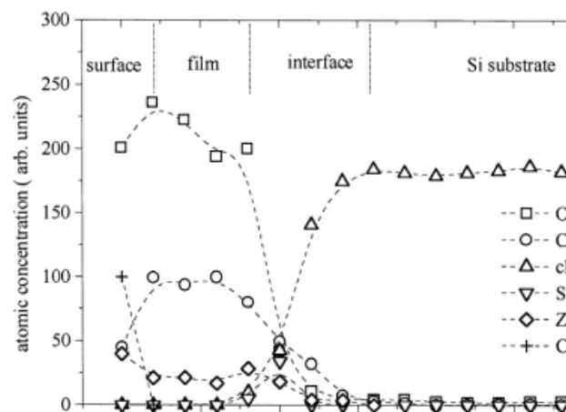
Fig. 1. XPS depth profile Ar sputtering of a film obtained from CZ-50r50 source and labelled FilmXP S-75r25 based on the
˚
composition determination by XPS. The film is 86 A thick according to quartz oscillator.
cerium oxide. Then, in addition to the contamination layer, the XPS depth profiles allow us to divide the thickness of the obtained films into three other re-gions, referred to as: film, interface and substrate
ŽFig. 1 . The following paragraphs describe the cor-.
responding chemical information deduced from XPS. We have been able to obtain homogeneous mixed films with no detectable impurities in all attempts. From the point of view of cation distribution, zirco-nium seems to present some difficulty to be evapo-rated by the electron bombardment of the powder. If we compare the distribution in the films and in the
Ž .
source Table 1 , there is at least a 30% Ce increase in the films compared to the mixed oxide powder. This was already observed in the case of films
w x
prepared on polycrystalline Ta 6 , in similar condi-tions. As an element of comparison, Tomaszewski et
w x
al. 8 observe that the deposition speed is about 10 times lower in the reactive sputtering of ZrO than2 of a metallic Zr target. We had some difficulty comparing our results with data from other ceria– zirconia thin films available in the literature as they
Ž
were prepared by completely different methods laser
w x w x.
ablation 9 or sol–gel dip coating 10 : in both cases, no cerium enrichment was detected by AES and RBS.
To complete the film characterisation, one of our
Ž .
sample FilmXP S-75r25 from a CZ-50r50 source was characterised by RBS and NRA. These
tech-niques allow us to determine the average tion of the film as well as its thickness. The composi-tion is given in Table 1, it is very close to the value
Ž
obtained by XPS FilmRB S-80r20 vs. FilmXPS
-˚
.
75r25 . The thickness is 130 A which is reasonably
Ž
close to the value indicated by quartz oscillator 90
˚
.A . Fig. 2 shows the corresponding RBS spectrum. From a theoretical fit of this spectrum, we conclude that the interface between the mixed oxide layer and the substrate is not sharp. This is confirmed by XPS results which show that the interface is rather com-plicated indeed, as we will see next.
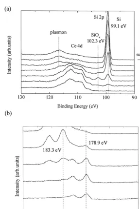
Fig. 3a shows the region of the Ce 4d and Si 2p core levels in the XPS spectra, recorded at various stages during the depth profile. As one approaches the interface, the Ce 4d signal starts to decrease and new feature appear in the Si 2p core levels, at a
Ž .
binding energy B.E. of 102.3 eV. Simultaneously,
Ž .
the O 1s signal decreases see Fig. 1 . Eventually, only the Si 2p peak corresponding to pure Si is seen at 99 eV, with a feature near 117 eV due to a plasmon loss in Si.
The B.E. of Si 2p at 102.3 eV does not corre-spond to silicon in a SiO environment which would2
be expected at 103.4 eV, but rather to another form of oxidised silicon. This may be because another type of oxide, involving Ce andror Zr forms during the growth, or because Arq
sputtering changes the oxide to a form where most of the Si is in a Si3q
stage. In the case of pure CeO films, the presence2 of oxidised silicon has been abundantly described in
Fig. 2. RBS spectra of FilmXP S-75r25 obtained from CZ-50r50 source.
( ) A. Galtayries et al.r Applied Surface Science 142 1999 159–163
162
Ž .
Fig. 3. a Ce 4d and Si 2p core levels from XPS depth profile of
Ž .
FilmXP S-75r25 zoomed at the interface with the substrate. b Zr 3d core level from XPS depth profile of FilmXP S-39r61 zoomed at the interface with the substrate.
w x
the literature, sometimes as CeO2y xrSiOx 11,12 ,
w x
or as an amorphous silicate layer 13,14 , where partial reduction of Ce4q to Ce3q occurs together with partial oxidation of Si.
This growth behaviour of oxides on Si has been
w x
tentatively explained by Nagata 15 . Based on
stan-Ž .
dard free energies of formation DG 8 , they havef shown that CeSiO is more stable than CeO during4 2 the early stages of growth of CeO on Si. Our data2 suggest that a similar phenomenon may occur during the growth of mixed oxides.
The thickness of the interface layer can be esti-mated based on the area of the SiO and Si peaks inx
the Si 2p spectra. In this way, we find a range of
˚
10–15 A, equivalent to two to three initial CeSiOx
planes.
Ž .
The Zr 3d core level spectra Fig. 3b provide us with some information on the role of Zr at the
beginning of the growth. All films obtained during the evaporations of 15r85, 50r50 and CZ-68r32 present the doublet of reduced zirconium at 178.9 eV near the Si substrate. The literature dealing with zirconia thin film mentions formation of a
w x
Zr-silicide 16 during the epitaxial growth of
yttria-Ž . Ž .
stabilised zirconia YSZ on Si 100 by ion beam sputtering of a target of ZrO with 10 mol.% Y O2 2 3 target. In particular, if the oxygen pressure is too low
Ž- 2 = 10y6 Torr during the growth of the films at.
Ž .
7508C, formation of possibly epitaxial ZrSi2 is detected by Auger electron spectroscopy. Two reac-tion pathways can be identified which strongly de-pend on the O2 pressure and on temperature of the substrate under vacuum: an arriving Zr atom may react with the silicon substrate to form Zr-silicide or will be oxidised if there are oxygen atoms available. The latter process is energetically more favourable
ŽDG 8 ZrO < DG 8 ZrSif Ž 2. f Ž 2.., but if a silicide nu-cleus has been formed it remains stable even under oxygen exposure. XPS characterisations of epitaxial
Ž .
growth of YSZrSi 100 confirm this interpretation
w17 . In our experiment, oxygen is added up to ax
pressure of 5 = 10y6
Torr only after a few minutes of evaporation to avoid oxidising the substrate. Therefore, the nucleation conditions are favourable for zirconium silicide formation. Reduced zirconium was not observed in the depth profiles of mixed
w x
films grown on Ta 6 ruling out the hypothesis of reduction of Zr under Arq
sputtering during XPS profiles. Away from the interface, the Zr 3d core
Ž
level shifts gradually towards higher B. E. 183.3
.
eV and a strong signal from cerium oxide is
de-Ž .
tected Fig. 3a,b . The hypothesis of a ZrSiO com-4
pound formed at the interface by analogy with the CeSiOx already described cannot be neglected, though in this case the B.E. of Si 2p would be
w x
observed at 101 eV 17 , which we do not detect. To summarise: at the interface with silicon, we detect first reduced zirconium then oxidised cerium, silicon and zirconium. Early admission of oxygen would avoid forming reduced zirconium but would favour formation of silicon oxide which can induce the undesired complete amorphisation of the ceria-con-taining mixed oxide film.
Finally, we investigated the mixed oxide solid solution character of the film, as well as its structural
Ž .
Ž . Ž .
epitaxial growth of CeO2 111 on Si 111 has been
w
reported in the past for similar conditions 3,11–
x
14,18 . Presently, the e-beam evaporation is not cou-pled to any analytical facilities. Therefore, LEED was not used to characterised the films structure, as it is too difficult to restore atomically clean and well ordered surfaces after transfer through air. To look for preferential growth orientations, XRD measure-ments were made on a pure CeO2 film and on Film-39r61 obtained from CZ-15r85. XRD refer-ence data on CZ-68r32, CZ-50r50 and CZ-15r85 powders have been characterised by XRD at another
w x
laboratory 19 and present the X-ray diffraction patterns of Ce1y xZr Ox 2 solid solution as expected. The structure of this solid solution is cubic for x - 0.5, and tetragonal for x )s 0.5. Only in the case of the CZ-15r85 sample, does the presence of peaks due to monoclinic–ZrO2 indicate a partial segregation of ZrO from the solid solution. In our2
XRD pattern coming from the mixed oxide film, we
Ž .
detect the presence of the 101 diffraction peak of
Ž .
monoclinic ZrO , and the 111 diffraction peak of2 cubic CeO2 in addition to the signal coming from the substrate and little evidence of the formation of a solid solution. More work is still needed to charac-terise the structure of these films, probably using grazing incidence X-ray diffraction in order to min-imise the signal from the substrate, since the films are very thin. However, these preliminary experi-ments are compatible with dual growth of both ZrO2
and CeO starting from the interface after bombard-2
ing the pellet.
4. Conclusion
We have obtained thin mixed ceria–zirconia films on silicon substrates by electron beam evaporation of mixed oxide solid solution pellets. The films show a ceria enrichment compared to the source material, regardless of composition of the source material. The interface of the oxide film with its substrate presents first a Zr-silicide layer and then the simultaneous presence of silicon, cerium and zirconium oxide. In the film, only zirconium and cerium in ZrO2 and CeO2y x environment are detected. Preliminary char-acterisations suggest to prove that the solid solution properties of the mixed oxides have been lost during
evaporation to the benefit of oriented ZrO2 and CeO .2
Acknowledgements
This work has been partly supported by the TMR Programme of the EU under contract ERB
FMRX-Ž .
CT96-0060 CEZIRENCAT Project and by the Bel-gian Federal Office for Scientific Technical and Cul-tural Affairs. RS acknowledge support from the
Bel-Ž .
gian Fund for Scientific Research F.N.R.S. .
References
w x1 P. Fornasiero, R. Di Monte, G. Raga Rao, J. Kaspar, S. Ž .
Meriani, A. Trovarelli, M. Graziani, J. Catal. 151 1995 168.
w x2 C. Tian, Y. Du, S. Chan, J. Vac. Sci. Technol. A 15 1997Ž .
85.
w x3 T. Inoue, Y. Yamamoto, M. Satoh, T. Hoshi, K. Miyoshi, Ž .
Jpn. J. Appl. Phys. 33 1994 L751.
w x4 H. Nagata, T. Tsukahara, S. Gonda, Jpn. J. Appl. Phys. 30 Ž1991 L1136..
w x5 W. Kern, D.A. Puotinen, RCA Rev. 31 1970 187.Ž . w x6 A. Galtayries, R. Sporken, J. Riga, G. Blanchard, R.
Cau-Ž .
dano, J. Electron Spectrosc. Rel. Phenom. 88–91 1998 951.
w x7 G. Cerrato, S. Bordiga, S. Barbera, C. Morterra, Appl. Surf. Ž .
Sci. 115 1997 53.
w x8 H. Tomaszewski, J. Haemers, N. De Roo, J. Denul, R. De Ž .
Gryse, Thin Solid Films 293 1997 67.
w x9 A.G. Akimov, D.B. Bogomolov, A.E. Gorodetskii, L.P.
Kazanskii, A.N. Khodan, I.L. Krylov, J.-P. Langeron, N.A. Melnikova, D. Michel, J.-L. Vignes, J. Perriere, Thin Solid
Ž .
Films 238 1994 15.
w10 R. Di Maggio, L. Fedrizzi, S. Rossi, R. Scardi, Thin Solidx Ž .
Films 286 1996 127.
w11 H. Nagata, M. Yoshimoto, T. Tsukahara, S. Gonda, H.x Ž .
Koinuma, Mater. Res. Soc. Symp. Proc. 202 1991 445.
w12 H. Koinuma, M. Kawasaki, M. Yoshimoto, Mater. Res. Soc.x Ž .
Symp. Proc. 397 1996 145.
w13 T. Inoue, Y. Yamamoto, M. Satoh, A. Ide, S. Katsumata,x Ž .
Thin Solid Films 281–282 1996 24.
w14 F. Sanchez, M. Varela, C. Ferrater, M.V. Garcia-Cuenca, R.x Ž .
Aguiar, J.L. Morenza, Appl. Surf. Sci. 70–71 1993 94.
w15 H. Nagata, Thin Solid Films 224 1993 1.x Ž . w16 T. Koch, P. Ziemann, Appl. Surf. Sci. 99 1996 51.x Ž . w17 H. Behner, J. Wecker, T. Mathee, K. Samwer, Surf. Interf.x
Ž .
Anal. 18 1992 685.
w18 C.E. Guillaume, M. Vermeersch, R. Sporken, J.J. Verbist, S.x Ž .
Mathot, G. Demortier, Surf. Interf. Anal. 22 1994 186.
w19 Laboratory SPIN, Ecole Nationale Superieure des Mines dex ´
Ž . Ž .
Saint Etienne France . Both LISE FUNDP and SPIN are members of a TMR network on model TWC funded by the European Commission.