HAL Id: tel-02901570
https://tel.archives-ouvertes.fr/tel-02901570
Submitted on 17 Jul 2020HAL is a multi-disciplinary open access archive for the deposit and dissemination of sci-entific research documents, whether they are pub-lished or not. The documents may come from teaching and research institutions in France or abroad, or from public or private research centers.
L’archive ouverte pluridisciplinaire HAL, est destinée au dépôt et à la diffusion de documents scientifiques de niveau recherche, publiés ou non, émanant des établissements d’enseignement et de recherche français ou étrangers, des laboratoires publics ou privés.
Splenocyte microvesicles are pro-senescent endothelial
effectors : impact of age and protection by EPA/DHA
6/1, an optimized formulation of nutritional
eicosapentaenoic and docosahexaenoic polyunsaturated
fatty acids
Abdul Wahid Qureshi
To cite this version:
Abdul Wahid Qureshi. Splenocyte microvesicles are pro-senescent endothelial effectors : impact of age and protection by EPA/DHA 6/1, an optimized formulation of nutritional eicosapentaenoic and docosahexaenoic polyunsaturated fatty acids. Pharmacology. Université de Strasbourg, 2019. English. �NNT : 2019STRAJ077�. �tel-02901570�
ÉCOLE DOCTORALE DES SCIENCES DE LA VIE ET DE DA SANTE
Regenerative Nanomedicine
– INSERM UMR 1260
THÈSE
présentée par :Abdul Wahid QURESHI
Soutenue le : 02 OCT 2019
Pour obtenir le grade de :
Docteur de l’université de Strasbourg
Discipline/ Spécialité : DOCTORAT Sciences Pharmaceutiques -Pharmacologie-Pharmacocinétique
THÈSE dirigée par :
Mme Florence TOTI Professeur, Université de Strasbourg RAPPORTEURS :
M. Eduardo ANGLES-CANO Professeur, Université de Paris
M. Paul KERTH Docteur
Examinateur interne :
M. Thierry VANDAMME Professeur, Université de Strasbourg
INVITEE :
Mme Valerie SCHINI-KERTH Professeur, Université de Strasbourg
Les microvésicules splénocytaires effecteurs de
la sénescence endothéliale
: Impact de l’âge et
protection par apport nutritionnel d
’une
formule optimisée
d’acides gras poly-insaturés
eicosapentaenoique et docosahexaenoique,
EPA:DHA 6:1
i
ACKNOWLEDGEMENTS
“Great things are not done by one person. They’re done by a team of people” - Steve Jobs Undertaking this PhD has been a truly life-changing experience for me and it would not have been possible to do without the support and guidance that I received from many people.
I would like to first express special appreciation to my supervisor Prof. Florence TOTI for all the support and encouragement she gave me, during my PhD. I would like to thank her for giving me the freedom to follow my ideas which helped me to grow as a researcher. I appreciate all her contributions of time and ideas to make my Ph.D. experience productive and stimulating. Without her guidance and constant feedback this PhD would not have been achievable.
I am also pleased to say thank you to Prof. Valerie Schini-Kerth, who contributed to my discussions that helped to shape this project. The joy and enthusiasm she has for her research was contagious and motivational for me, even during tough times in the Ph.D. pursuit. I would also like to acknowledge the valuable input of Dr Cyril AUGER. I really appreciate him for sharing his insightful suggestions. I would also like to thank our ex-technician Brigitte POLLET for making the early morning work enjoyable. It wouldn’t have been easy to conduct this research without her precious support.
Sincere thanks to my ex lab colleagues Faraj ZGHEEL, Amisi SAID, Fuong NAGA. I would especially like to give thanks to Malak ABBAS, Raed ALTAMIMI, Sonia KHEMAIS and Zahid Rasool NIAZI for helping me in learning techniques. This acknowledgment would not be complete without mentioning my lab colleagues Ahmed CHAKER, Ali el HABHAB, Christophe BRUKERT, Eugenia BELCASTRO, Hanine el ITAWI, Hira HASAN, Hyunho LEE, Lamia AMOURA, Lamia REMILA, Muhamed KASEEM, Paola algara SUAREZ, Sebastein GAERTNER, Fatiha ZOBAIRI and Sin-hee PARK. It was great pleasure working with them and I appreciate their ideas, help and great humor. I would always remember my lab colleague Akmal FAROOQ, for the fun-time we spent together and the sleepless nights for completing the confocal microscopy.
My time at Strasbourg was enjoyable in large part due to the many friends and groups that became a part of my life. I am grateful for the time spent with Pakistani friends and my Cricket buddies. My time at Strasbourg was also enriched by the Strasbourg Cricket Club.
I gratefully acknowledge the funding received towards my PhD from the Higher Education Commission (HEC) of Pakistan.
Lastly, I would like to thank my family for all their love and encouragement. Special thanks to my parents without them, I would not have had the courage to embark on this journey in the first place. I would also like to say a heartfelt thank you to all my siblings for always believing in me and encouraging me to follow my dreams. I would also like to thank all of my friends who supported me and incented me to strive towards my goal.
Abdul Wahid Qureshi Strasbourg, France
iii
TABLE OF CONTENTS
ACKNOWLEDGEMENT ... i
TABLE OF CONTENTS ... iii
LIST OF FIGURES ... vi
LIST OF TABLES ... viii
LIST OF ABBREVIATIONS ... ix
Résumé en français ... 1
Chapter 1: Ageing and its impact on the immune system ... 9
1.1 Ageing ... 11
1.2 Overview of immune system ... 12
1.2.1 The innate immune system ... 13
1.2.2 The adaptive immune system ... 13
1.2.3 Spleen, a rich source of leukocytes ... 14
1.3 Impact of ageing on immune system ... 15
1.3.1 Immunosenescence ... 16
1.3.2 Inflamm-ageing ... 18
1.4 Cell membrane asymmetry and fluidity: impact of ageing ... 21
Chapter 2: Microparticles: overview and role in vascular damage ... 25
2.1 Overview of Microparticles ... 27
2.2 Mechanisms of formation of microparticles ... 29
2.2.1 Loss of asymmetry of membrane phospholipids and formation of Microparticles ... 29
2.2.2 Reorganization/degradation of cytoskeleton and formation of Microparticles ... 32
2.3 Composition of MPs ... 33
2.3.1 Protein Content of MPs... 34
2.3.2 Lipid content of MPs ... 35
2.3.3 Nucleic acid and miRNA content of MPs ... 36
2.4 Mechanisms of interaction of MPs with recipient cells ... 36
2.5 MPs and vascular damage ... 38
2.5.1 Microparticles and endothelial dysfunction ... 38
2.5.2 Microparticles and inflammation ... 40
2.5.3 Microparticles, coagulation and thrombosis ... 42
2.5.4 Microparticles and endothelial senescence... 44
2.6 Microparticles and atherothrombosis ... 46
iv
3.1 Overview of endothelium ... 51
3.2 Cellular senescence ... 53
3.3 Characteristics of Senescent cells ... 55
3.4 Mechanisms of senescence ... 60
3.4.1 Replicative and premature senescence ... 60
3.4.2 Cell cycle arrest ... 60
3.5 Endothelial senescence ... 62
3.6 Oxidative stress and endothelial senescence ... 64
3.7 Angiotensin system and endothelial senescence ... 65
3.8 Endothelial senescence and endothelial dysfunction ... 67
3.9 Endothelial senescence and age-related cardiovascular diseases ... 68
Chapter 4: Omega-3 PUFAs and vascular health ... 71
4.1 Fatty acids ... 73
4.2 Omega-3 polyunsaturated fatty acids (n-3 PUFAs) ... 73
4.3 Dietary sources and intake of Omega-3 PUFAs ... 74
4.4 Metabolism and bioavailability of Omega-3 PUFAs ... 76
4.5 Mechanisms of Omega-3 PUFAs functions ... 79
4.5.1 Structural and functional alteration of cell membrane ... 79
4.5.2 Modulation of ion channels ... 79
4.5.3 Alteration of nuclear receptors and transcription factors ... 80
4.5.4 Omega-3 derived eicosanoids ... 81
4.6 Omega-3 PUFAs and risk of CVDs ... 84
4.6.1 Blood pressure ... 85 4.6.2 Heart rate ... 87 4.6.3 Plasma lipids ... 88 4.6.4 Inflammation ... 89 4.6.5 Endothelial dysfunction ... 91 4.6.6 Thrombosis ... 92 4.6.7 Arrhythmia ... 93
4.7 Omega-3 PUFAs and clinical trials ... 94
4.7.1 Diet and Reinfarction Trial (DART) ... 94
4.7.2 Gruppo Italia o per lo Studio della Sopravvive za ell’I farto i ardi o Prevenzione (GISSI-Prevenzione) ... 94
4.7.3 GISSI-Heart Failure (GISSI-HF) ... 95
4.7.4 Japan EPA Lipid Intervention Study (JELIS) ... 95
4.7.5 The Reduction of Cardiovascular Events with EPA-Intervention Trial (REDUCE-IT) ... 95
v
Hypothesis and Aims ... 97
Results ... 101
Impact of EPA:DHA 6:1 on vascular ageing and role of microvesicles ... 101
Article ... 109
General discussion, conclusion and further prospect ... 141
General discussion ... 143
Conclusion and further prospects ... 153
References ... 155
Annexes ... 185
vi
LIST OF FIGURES
Figure 1 Timing and progression of ageing-associated phenotypes Figure 2 Hallmarks of ageing
Figure 3 Overview of the immune system Figure 4 Structure of spleen
Figure 5 Age-related changes in the immune system Figure 6 Mechanisms of inflamm-ageing
Figure 7 Inflamm-ageing: a risk for multiple chronic diseases Figure 8 Ageing and plasma membrane remodeling
Figure 9 Cellular microparticles
Figure 10 Schematic representation of the mechanisms of formation of microvesicles, exosomes, and apoptotic bodies
Figure 11 Asymmetrical distribution of phospholipids in red blood cell membrane
Figure 12 Functions of membrane lipid transporters
Figure 13
Floppase activity and facilitated transport of phosphatidylserine by TMEM16-F (ANO-6) and procoagulant MP shedding
Figure 14 Composition of MPs
Figure 15 Mechanisms of interaction of MPs with recipient cells. Figure 16 Cell-type specific MPs and inflammatory responses
Figure 17 Putative mechanisms of MPs induced vascular cell aging and senescence
Figure 18 Processes involved in MPs-mediated endothelial dysfunction Figure 19 Vessel wall and endothelium
Figure 20 Endothelium-related key discoveries and number of publications/year
vii Figure 22 Overview of cellular senescence
Figure 23 Hallmarks of morphological alterations Figure 24 Markers of senescent cells
Figure 25 Functions of senescence-associated secretory phenotype (SASP)
Figure 26 Pathways regulating senescence-mediated cell cycle arrest Figure 27 Characteristics of senescent endothelial cells
Figure 28 Actors of ROS-mediated endothelial senescence
Figure 29 Chemical structures of important dietary omega-3 PUFAs Figure 30 Metabolism of omega-3 and omega-6 PUFAs
Figure 31
A schematic representation of dietary fat digestion and absorption of ethyl ester (EE) and free fatty acid (FFA) forms of eicosapentaenoic acid (EPA) and docosahexaenoic acid (DHA).
Figure 32 Molecular mechanisms of physiological effects of omega-3 PUFAs
Figure 33 Pathways involved in production of SPMs from EPA and DHA Figure 34 Metabolism of EPA and functions of derived metabolites Figure 35 Omega-3 PUFAs targets in cardiovascular diseases Figure 36 Role of omega-3 and omega-6 PUFAs in inflammation
Figure 37 Depiction of some anti-inflammatory pathways triggered by EPA and DHA
viii
LIST OF TABLES
Table 1 Senescence markers: regulation and detection Table 2 Omega-3 PUFAs content in fish oils
Table 3 Omega-3 and -6 derived eicosanoids and their physiological effects
ix
LIST OF ABBREVIATIONS
3H-dT 3-hthymidine 53BP1 P53-Binding Protein-1 5-HT 5-Hydroxy-Tryptamine AA Arachidonic AcidACE Angiotensin Converting Enzyme
ACS Acute Coronary Syndrome
ADMA Asymmetric Dimethyl Arginine
ADP Adenosine Di-Phosphate
ALA α-Linoleic Acid
Ang II Angiotensin II
ANO6 Anoctamine
aPL
Antibodies Anti-Phospholipid Antibodies
ASCEND A Study of Cardiovascular Events iN Diabetes
ATM Ataxia Telangiectasia Mutated
ATP Adenosine Tri-Phosphate
ATR-1 Angiotensin Type 1 Receptor
BCR B-Cell Receptor
BMPs Bone Morphogenetic Proteins
Bp Base Pair
Bpm Beats Per Minute
BrdU Bromodeoxyuridine
x C12FDG 5-dodecanoylaminoFluorescein Di-
-D-Galactopyranoside
Ca Calcium
CCFs Cytoplasmic Chromatin Fragments
CD Cluster Designation
CHD Coronary Heart Disease
COX Cyclooxygenases
cPLAβα Cytosolic Phospholipase aβα
CVD Cardiovascular diseases
DAMPs Damage Associated Molecular Patterns
DAPI 4,6-Diamidino-2-Phenylindole
DART Diet And Reinfarction Trial
DBP Diastolic Blood Pressure
DCs Dendritic cells
DDR DNA Damage Response
DHA Docosahexaenoic acid
DHE Dihydroethidium
DNA Deoxyribonucleic acid
DNA-SCARS
DNA Segments with Chromatin Alterations Reinforcing Senescence
DSBs Double Stranded Breaks
ECM Extracellular matrix
ECs Endothelial Cells
EDCF Endothelium-Derived Contractile Factors
EDH Endothelium-Derived Hyperpolarization
xi
EET Epoxyeicosatrienoic
EGF Epithelial Growth Factor
EMPs Endothelial cell-derived microparticles eNOS Endothelial Nitric Oxide Synthase
EPA Eicosapentaenoic acid
EPCR Endothelial Protein C Receptor
ERK1/2 Extracellular signal–regulated kinase ½
FA Fatty Acids
FAO Food and Agriculture Organization
FD Fine Dust
FFAs Free Fatty Acid
FGF Fibroblast Growth Factor
FOXP3 Forkhead Box Protein P3
Fra-1 Fos-related antigen 1
GISSI Gruppo Italiano per lo Studio della Sopravvivenza nell’Infarto micardico GISSI-HF GISSI-heart failure
GLA Gamma-Carboxyglutamic Acid
GM-CSF Granulocyte-Macrophage Colony-Stimulating Factor
GP Glycoprotein
GPI Glycosylphosphatidylinositol
GPR G-Protein coupled receptors
H2O2 Hydrogen peroxide
HDL High Density Lipoproteins
xii
HNF Hepatic Nuclear Factors
HSC Hematopoietic Stem Cells
HUVEC Human Umbilical Vein Endothelial Cells ICAM-1 Intercellular Adhesion Molecule 1
IgE Immunoglobulin E
IL Interleukin
IMA Internal Mammary Artery
IRF Interferon Regulatory Factor
JELIS Japan EPA Lipid Intervention Study
LA Linoleic Acid
LDL Low Density Lipoproteins
lMPs Lymphocyte-derived microparticles
LMPs Leukocyte-derived microparticles
LOX Lipooxygenases
LPA Lysophosphatidic acid
LPS Lipopolysaccharides
Mac-1 Macrophage antigen-1
MAGs Monoacylglycerols
MAPK Mitogen-Activated Protein Kinase
MaR Maresins
MHC Major Histocompatibility Complex
miR MicroRNA
miRNA Micro RNA
xiii
MMPs Metalloproteinases
mMPs Monocyte/macrophage-derived microparticles
MP Microparticle
mRNA Messenger RNA
mTOR Mammalian Target Of Rapamycin
MV Microvesicle
NAC N-Acetyl Cysteine
NADPH Nicotinamide Adenine Dinucleotide Phosphate NF-κB Nuclear factor kappa-light-chain-enhancer of
activated B cells
NKC Natural Killer Cells
NLRP3 NOD-like receptor family pyrin domain-containing 3
nMPs Neutrophil-derived microparticles
NO Nitric Oxide
Nox NADPH Oxidase
ONOO- Peroxynitrite
OX-LDL Oxidized Low Density Lipoprotein
P Protectin
PALS Peri-Arterial Lymphoid Sheath
PAMPs Pathogen Associated Molecular Patterns
PC Phosphatidylcholine
PCBs Polychlorinated biphenyls
PCNA Proliferating Cell Nuclear Antigen
PCR Polycomb Repressive Complexes
xiv PECAM Platelet Endothelial Cell Adhesion Molecule
PG Prostaglandin
PHA Phytohemagglutinin
PHB1 Prohibitin-1
PI3K Phosphoinositide 3-kinase
PI-3K Phosphatidylinositol-3-kinase
PKC-Ɵ Protein Kinase C Theta
PL Phospholipid
PMA/I Phorbol Myristate Acetate/Ionophore PMPs Platelet-derived microparticles
PPAR Peroxisome Proliferator-Activated Receptors
PS Phosphatidylserine
PUFAs Polyunsaturated fatty acids
RAS Renin-Angiotensin System
REDUCE-IT
The Reduction of Cardiovascular Events with EPA-Intervention Trial
RNA Ribonucleic acid
ROS Reactive Oxygen Species
Rv Resolvins
RXR Retinoid X Receptor
SAHF Senescence-Associated Heterochromatin Foci SASP Senescence-Associated Secretory Phenotype SA- -Gal Senescence-Associated Beta-Galactosidase
SBP Systolic Blood Pressure
xv
SGLT Sodium Glucose Linked Transporter
Shh Sonic hedgehog
SIPs Stress-Induced Premature Senescence
Sirt Sirtuin
SM Sphingomyelin
SOCE Capacitative or Store-Operated Calcium Entry SPMs Specialized pro-resolving lipid mediators SREBP-1c Sterol Regulatory Element Binding Protein-1c STAT Signal Transducer and Activator of Transcription
TAGs Triacylglycerols
TCR T-Cells Receptors
TERT Telomerase reverse transcriptase
TF Tissue Factor
TF+MPs Tissue factor bearing microparticles
TFPI Tissue Factor Pathway Inhibitor
TG Triglycerides
Th cells Helper T-cells
TLRs Toll-Like Receptors
TM Thrombomodulin
TNF Tumor Necrosis Factor
Treg Regulatory T-cells
TRPC Transient Receptor Potential Channel
TX Thromboxane
xvi
VITAL Vitamin D and omega-3 trial
VSMC Vascular Smooth Muscle Cells
X-gal 5-bromo-4-chloro-3-indolyl-D-galactoside
1
3
Introduction et état de l’art :
Sénescence endothéliale et maladies cardiovasculaires :
Les maladies cardiovasculaires (MCV) et le cancer sont les deux principales de mortalité et morbidité dans les pays riches et en voie de développement. L’incidence et la prévalence des de MCV comme l’hypertension, les maladies coronaires et l’insuffisance cardiaque augmentent avec l’âge. Du fait du vieillissement de la population, les MCV représentent un enjeu majeur en santé publique avec un poids important des coûts et des besoins.
L’endothélium, la monocouche cellulaire qui borde la lumière des vaisseaux, joue un rôle pivot dans l’hémostase et dans l’homéostasie vasculaire en contribuant à la régulation fine du de la coagulation et du tonus vasculaire, protégeant ainsi de la thrombose et d’un remodelage vasculaire excessif. La sénescence endothéliale liée à l’âge, caractérisée par un arrêt irréversible du cycle cellulaire, induit un stress oxydant et une dysfonction endothéliale, favorisant l’initiation et la progression des dommages cardiovasculaires.
Les microvésicules
acteurs et effecteurs de l’hémostase et de l’homéostasie
vasculaire
Les microvésicules (MVs) aussi appelées microparticules, sont des vésicules des fluides biologiques émises par la membrane plasmique des cellules en réponse à un stress cellulaire tel que l’inflammation, la sénescence, l’apoptose. Les MVs portent des protéines de la membrane plasmique, utiles pour la caractérisation de leur origine cellulaire, qui leur confèrent des propriétés de marqueurs circulants de dommage cellulaire à valeur diagnostique ou pronostique notamment dans les pathologies vasculaires et cardiaques, mais aussi des molécules bioactives, protéines, lipides, ARN, les transformant en effecteurs pathogéniques des maladies cardiovasculaires. En effet, les MVs sont par essence procoagulantes parce qu’elles exposent de la phosphatidylsérine, un aminophospholipide membranaire qui catalyse les réactions de la coagulation, propriété qui s’ajoute parfois à celle du facteur tissulaire (FT), l’initiateur cellulaire de la cascade de la coagulation. Notre groupe a montré qu’une proportion des MVs endothéliales circulantes chez les patients avec syndrome coronaire aigu se comporte en effecteur pro-sénescent induisant via un stress oxydant, l’activation du système angiotensine local et une réponse inflammatoire exacerbée de l’endothélium.
4
Outre les MVs d’origine endothéliale, les MVs d’origine leucocytaire ont été détectées comme cruciales dans l’homéostasie vasculaire par notre équipe et d’autres. Les MVs leucocytaires circulent en faible proportion chez les individus sains (<10%) et ont un double potentiel procoagulant car elles exposent le facteur tissulaire (cf. ci-dessus). Constitutives des thrombi en formation, ces MVs circulent à des taux élevés chez les patients avec des désordres cardiaques, métaboliques ou une hypercoagulabilité. Les MVs leucocytaires ont une valeur pronostique de risque élevé d’athérothrombose chez les patients asymtomatiques. De fait, elles s’accumulent dans les plaques d’athérome. In vitro et dans les modèles animaux, les MVs leucocytaires apparaissent ainsi comme des effecteurs de la réponse vasculaire et de l’hémostase en véhiculant de nombreuses propriétés pro-inflammatoires pro-coagulantes, pro-angiogéniques, soulignant un rôle possible dans le couplage entre inflammation et thrombose.
Elles ciblent l’endothélium dans des modèles cellulaires et l’injection de MVs leucocytaires émises en réponse à un stress infectieux, apoptotique ou inflammatoire induit une réponse inflammatoire de la paroi vasculaire et du tissu myocardique et réduit la relaxation endothéliale-dépendante des aortes pré-contractée de souris.
Protection cellulaire contre le vieillissement et santé cardiovasculaire
De nombreuses études épidémiologiques ont montré l’impact de la diète sur la santé cardiovasculaire et sur la durée de la vie en bonne santé. Chez les patients à fort risque cardiovasculaire, l’incidence d’évènements cardiovasculaires adverses comme les accidents vasculaires cérébraux, l’infarctus du myocarde est réduit de γ0% chez les sujets suivant un régime méditerranéen riche en acides gras insaturés ingérés sous la forme de noix ou d’huile d’olive. De même, la population japonaise serait protégée des maladies cardiovasculaires par un régime de type Okinawa, riche en produits de la mer contenant des acides gras polyinsaturés (PUFAs), et plus particulièrement en omega-3, PUFAs dont la troisième liaison carbone est insaturée. Ainsi, les acides eicosapentaenoique (EPA) et docosahexaenoique (DHA), diminueraient de 50% la mortalité coronaire chez les hommes sains et de 30% chez ceux ayant récemment subi un infarctus du myocarde. Cependant, une des difficultés majeures dans la démonstration du bénéfice des oméga-3 sur la santé cardiovasculaire au cours des études épidémiologiques ou interventionnelles réside dans la variété des formulations de l’apport nutritionnel ou du degré d’enrichissement en EPA et DHA. De plus, les suspensions d’EPA et DHA ne sont pas utilisables in vitro, du fait de leur5
forte toxicité cellulaire, l’étude de leurs effets nécessite donc des modèles animaux avec gavage. Notre équipe a montré un effet vasoprotecteur de la formulation optimisée EPA:DHA 6:1 qui limite le stress oxydant endothélial grâce à l’induction de la NO-synthase endothéliale via l’activation redox-sensible de la signalisation Src/PI3-kinase/Akt et des MAP-Kinases. Parallèlement, l’ingestion chronique d’EPA:DHA 6:1 réduit significativement la pression systolique dans un modèle d’hypertension induite par l’angiotensine II chez le rat.
Hypothèses et objectifs :
L’ensemble des données précédentes du laboratoire et celles d’autres équipes suggère que la dysfonction endothéliale favorise la perte du contrôle du tonus vasculaire. De plus, les travaux précédents de Ali ElHabhab, doctorant du laboratoire, ont montré que les MVs leucocytaires générées par stimulation de splénocytes de rat ont un effet pro-sénescent sur les cellules endothéliales primaires de coronaires. Nous avons cherché les mécanismes susceptibles d’expliquer le bénéfice cardiovasculaire des omega-3 en nous concentrant sur les interactions des MVs leucocytaires et des cellules endothéliales primaires de coronaires. Nos hypothèses étaient
i) En condition pathologique, les MVs leucocytaires favorisent une sénescence endothéliale accélérée et un phénotype endothélial pro-inflammatoire et pro-coagulant ii) Les propriétés pros-sénescentes des MVs leucocytaires évoluent avec l’âge.
iii) L’ingestion d’omega-3 EPA:DHA 6:1 prévient la sénescence endothéliale en modifiant les propriétés des MVs leucocytaires qui interagissent avec l’endothélium.
Nos objectifs étaient la comparaison des propriétés pro-sénescentes, pro-inflammatoires et pro-coagulantes des MVs leucocytaires de rats jeunes, âgés, ou d’âge moyen et l’étude de l’impact de l’ingestion d’une formulation optimisée d’omega-3 formulation (EPA:DHA 6:1) sur ces propriétés et l’identification des mécanismes moléculaires sous-jacents.
Méthodologie et principaux résultats :
Après prélèvement des rates de rats Wistar mâles, jeunes (1β semaines), d’âge moyen (M, 48 semaines) ou vieux (72 semaines), les splénocytes isolés ont été mis en culture. La capacité des splénocytes à émettre des MVs a été étudiée après 24 heures, leur origine
6
leucocytaire déterminée ainsi que leur propriétés pro-sénescentes des SMVs sur des cellules endothéliales primaires de coronaires de porc en culture. L’induction de la sénescence endothéliale a été mesurée par l’activité de la Senescence-associated -galactosidase activity (SA- -gal) à l’aide d’un substrat fluorogénique le C12FDG, en cytométrie en flux, et par l’expression des marqueurs protéiques de la sénescence p5γ, pβ1, p16 par western blot. La dysfonction endothéliale a été évaluée par la mesure du stress oxydant grâce à la sonde redox-sensible fluorescente dihydroethidium (DHE) et aux marqueurs pro-inflammatoires, pro-coagulants ou potentiellement thrombogéniques par western blot et cytométrie. L’activité procoagulante des SMVs a été mesurée par dosage enzymatique prothrombinase. Parallèlement, l’effet d’une ingestion d’oméga-γ (7 jours, pour les rats d’âge moyen ; 14 jours pour les rats vieux) sur l’émission et les propriétés des SMVs a été mesuré dans chaque fratrie de rat. Les animaux ont ingéré 500 mg/kg/d de EPA:DHA 6:1, ou de EPA:DHA 1:1, ou de l’huile de maïs contrôle.
Impact de l’âge sur l’émission des MVs :
L’émission de SMVs augmente proportionnellement à l’âge, comparativement aux rats jeunes, suggérant une susceptibilité membranaire accrue des splénocytes. De plus, leurs propriétés vis à vis de l’endothélium évoluent. En effet, l’ajout d’une concentration de 10 nM de SMVs aux cellules endothéliales coronaires primaires jeunes (P1 ECs) transforme leur phénotype lorsqu’elles proviennent de splénocytes de rats d’âge moyen ou âgés mais pas de rats jeunes : leur effet pro-sénescent est attesté par l’augmentation progressive de la SA- -gal et une surexpression significative des marqueurs p21 et p16, impliqués dans la régulation du cycle cellulaire avec des réponses endothéliales aussi importantes que celles observées dans des cellules induites en sénescence soit par réplication successive ou à l’aide de peroxyde d’hydrogène (100 µM). La réponse pro-inflammatoire et procoagulante est aussi significativement augmentée en réponse aux SMVs de rats âgés ou d’âge moyen avec une surexpression significative de facteur tissulaire, du récepteur de l’angiotensine et de son récepteur AT1R, des protéines pro-inflammatoires (VCAM-1, ICAM-1, NF-kB, COX-2 mais pas COX-1) comparée aux rats jeunes. De plus, les SMVs de rats âgés ou d’âge moyen mais pas de rats jeunes induisent un stress oxydant détecté par DHE, et caractérisé d’origine cytoplasmique et mitochondriale par inhibition pharmacologique. A l’inverse, la NO synthase endothéliale protectrice est sous-exprimée comparativement aux cellules traitées par des SMVs de rats jeunes. De plus, seules les SMVs7
de rats âgés réduisent la vaso-relaxation endothéliale dépendante d’artères aortiques de porc en réponse à la bradykinine.
L’ensemble des résultats suggère donc fortement que l’émission de SMVs augmente avec l’âge et favorise la sénescence des cellules endothéliales coronaires, déjà particulièrement prédisposées aux mécanismes d’athérothrombose. En effet, les artères coronaires présentent des zones de courbures favorisant des perturbations de flux sanguin et où s’accumulent les plaques d’athérome. Comparativement à l’EPA :DHA 1 :1 ou à l’huile de maïs, l’ingestion d’EPA:DHA 6:1 prévient significativement l’émission des SMVs sénescentes induite par l’âge et réduit leurs effets en limitant leur potentiel proxydant, pro-inflammatoire et prothrombotique, et restent sans effet notoire chez les rats jeunes. En outre, EPA:DHA 6:1 préserve les mécanismes protecteurs de la NO synthase endothéliale.
En conlusion, les MVs leucocytaires d’origine splénique induites par l’âge se comportent en effecteurs endothéliaux nocifs, pro-sénescents et proinflammatoires favorisant la dysfonction endothéliale et une réponse thrombogénique de l’endothélium coronaire spécifiquement contrecarrée par l’ingestion d’EPA:DHA 6:1.
9
11
1.1
Ageing
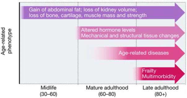
Ageing is defined as an inevitable multifactorial biological process, characterized by progressive loss of physiological functions, allied with frailty and increased probability of death (Panagiotou, Neytchev et al. 2018). Age-related decline begins from third decade onwards and deterioration progresses with advancing age. Ageing-related phenotypes includes loss of muscle and bone mass and strength, abdominal fat gain, systemic functional decline, structural and mechanical alterations, age-related chronic diseases and frailty (Figure 1).
Figure 1. Timing and progression of ageing-associated phenotypes Adapted from (Partridge, Deelen et al. 2018)
About half of the human deaths are attributed to chronic diseases associated with ageing. Ageing-associated increase in cellular oxidative stress, chronic low grade inflammation (inflamm-ageing), senescence, mitochondrial dysfunction, proteasome failure, impaired autophagy contributes to multi-systems loss of reserve and function, ultimately leading to incidence of multimorbidity among elderly (Figueira, Fernandes et al. 2016). Most conspicuous ageing-associated chronic diseases are cardiovascular diseases (CVD), diabetes, neurodegenerative diseases and cancer. CVD contributes for 39.6% of all ageing-related chronic diseases and CVD sharply increases after 40 years of age (Benjamin Emelia, Virani Salim et al. 2018, Fajemiroye, da Cunha et al. 2018).
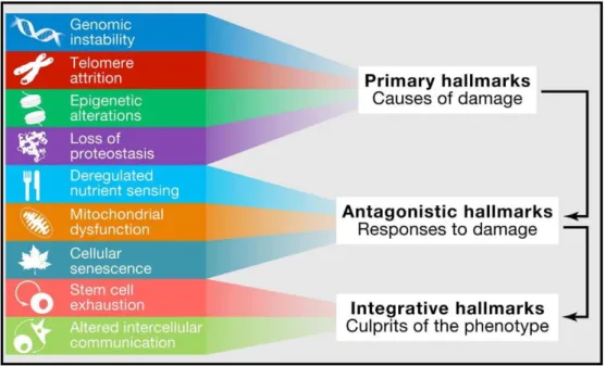
All the mechanisms underlying ageing process are not yet understood. Still, extensive research has defined a set of hallmarks as a ground for better understanding
12
and ameliorate the late-life effects of ageing. This set includes indicators of altered cellular function, antagonistic responses to damage cellular function, and finally integrative hallmarks which are eventual culprits of clinical effects of ageing, mainly loss in organ dysfunction and homeostasis (Figure 2).
Figure 2. Hallmarks of ageing Adapted from (Aunan, Watson et al. 2016)
Since the last two centuries, human life expectancy has doubled in most developed countries due to better quality of food, hygiene, water, lifestyle, immunization, antibiotics and advance medical care. By year β0γ5, one fourth of world’s population is anticipated to reach the age of 65 (Steenman and Lande 2017). However, despite increase in lifespan, there is not much increase in healthy, disease free lifespan. Between 2000 to 2015, on average, total life expectancy raised by 5 years throughout the world whereas healthy life expectancy has been raised by 4.6 years. On average, 16-20% of life is now spent with late-life morbidity. An unhealthy elderly population is a global challenge to society (Partridge, Deelen et al. 2018). Therefore, though it is not possible to abolish ageing, one of the major aims of current research is to reduce the length and severity of late-life morbidity.
1.2
Overview of immune system
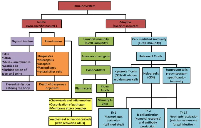
The immune system integrates lymphoid organs, cytokines, cells, humoral factors and is responsible for recognition and eradication/neutralization of pathogens as well as
13
of abnormal self-cells (Parkin and Cohen 2001).Immune system is divided into innate and adaptive immune systems (Figure 3).
1.2.1
The innate immune system
The innate immune system, also called natural or native immune system, is the first line of immediate and non-specific defense mechanism. It comprises physical barriers (e.g. skin, epithelial cell layers etc.), immune cells (neutrophils, monocytes/macrophages, dendritic and natural killer cells) and responds without previous experience of its target. When it is overpowered, adaptive immune system will be activated (Nagaratnam and Adithya Nagaratnam 2019).
1.2.2
The adaptive immune system
The adaptive immune response or acquired immune system, functions via antigen specificity and immune memory. Adaptive responses primarily uses the antigen-specific receptors expressed on the surface of B cells and T cells to initiate targeted effector response. Targeted effector response can be due to activated T cells (cellular immunity) leaving lymphoid tissue and approaching to disease site, or to activated B cells (humoral immunity) releasing antibodies into blood, tissue fluids and finally to the infective focus (Nagaratnam and Adithya Nagaratnam 2019).
14
1.2.3 Spleen, a rich source of leukocytes
In humans, spleen is 7-13 cm in length with a weight of about 150 g which may decrease with age. It is the largest lymphoid organ with important role in both innate and adaptive immune responses. Covered by a capsule of connective tissue, it is composed of branching arterial vessels, with smaller arterioles ending up into a venous sinusoidal system and a functional parenchyma made up of red and white pulp (Figure 4). Red and white pulp are separated by a thin layer of marginal zone. The red pulp is richly vascular specialized area composed of splenic cord, capillaries and venous sinuses that functions as a blood filter. The red pulp contains large aggregates of erythrocytes that favors blood viscosity. Altogether, the human spleen contains 200-250 mL of blood, representing 10% of the total red blood cells and 160 mL red blood cells can be expulsed in response to exercise favoring oxygen fixation through enhanced plasma heamoglobin (Stewart and McKenzie 2002). The marginal zone contains B cells, marginal-zone macrophages, fibroblasts and dendritic cells (DCs). Cells from bloodstream enter and leave the white pulp through the marginal zone. The white pulp, consisting of lymphoid follicles and peri-arterial lymphoid sheath (PALS), is mostly rich in B and T lymphocytes and harbors the immunologic spleen function. Specific cluster designation (CD) and other markers defines the lymphoid cell immunophenotypes of the different spleen regions. (Stewart and McKenzie 2002, Mebius and Kraal 2005, Velasquez-Lopera, Correa et al. 2008, Pernar and Tavakkoli 2019).
15
Altogether, the spleen serves various functions e.g. filtration, destruction of altered or old red blood cells, production of lymphocytes and monocytes, phagocytosis, storage of iron and viable blood cells (Stewart and McKenzie 2002).
1.3
Impact of ageing on immune system
Ageing-related effects on immune system are extensive and stretched from hematopoietic stem cells (HSC) and lymphoid progenitor cells in the thymus and bone marrow, and, to resident mature lymphocytes in secondary lymphoid organs including spleen (Montecino-Rodriguez, Berent-Maoz et al. 2013).
Figure 4: Structure of Spleen:
a) The afferent splenic artery branches into central arterioles, which are sheathed by
white-pulp areas; these white-white-pulp areas consist of the T-cell zone (also known as the periarteriolar lymphoid sheath, PALS), arterioles and B-cell follicles. The arterioles end in cords in the red pulp, from where the blood runs into venous sinuses which collect into the efferent splenic vein. The larger arteries and veins run together in connective-tissue trabeculae, which are continuous with the capsule that surrounds the spleen. b) Comparison of the structure of the white pulp in rodents and primates. The main differences are found in the structure of the marginal zone, which surrounds the white pulp. In contrast to mice, humans have an inner and an outer marginal zone, which is surrounded by a large perifollicular zone. In the perifollicular zone, some blood vessels terminate, and the endings of these capillaries are sheathed by
16
1.3.1
Immunosenescence
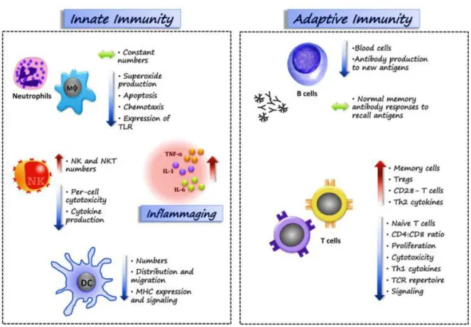
Ageing of the immune system or immunosenescence is characterized by multiple phenotypical and functional alterations in both innate and adaptive immune cells (Figure 5). Immunosenescence is most probably driven by sustained antigenic load (bacteria, virus, fungi, necrotic cell debris, toxins) with ageing, favoring inflammatory status and damage to various organs, at the onset of chronic diseases (Fulop, Dupuis et al. 2016, Nagaratnam and Adithya Nagaratnam 2019).
Neutrophils are the first recruit at the sites of injury and have short life-span until stimulated by some pro-inflammatory stimulus e.g. LPS (lipopolysaccharides). With ageing, the number of neutrophils are reported to be relatively higher with altered effector functions including chemotaxis, production of free radicals production and intracellular killing, while phagocytosis and adhesive ability of neutrophils remain stable during immunosenescence. Neutrophils in quiescent state, with sustained and increased production of cytokines, free-radical, metalloproteinase, concomitant activation of NF-kB (nuclear factor of kappa B) and altered PI3K signaling pathways are indicators of altered effector functions (Elias, Hartshorn et al. 2018, Ray and Yung 2018).
Monocytes/macrophages that are critical regulators and effectors of inflammation also undergo age-related alterations in effector functions, for example, decreased antigen presentation, cytotoxicity and intracellular killing. PAMPs (pathogen-associated molecular patterns)-based activation of TLRs (Toll-like receptors), present on monocytes, macrophages, dendritic, epithelial and endothelial cells, and downstream immune signaling and phagocytosis is altered. With ageing reduced signaling through TLR1/2 heterodimers and decreased TLR-induced costimulatory expression is reported in monocytes (van Duin and Shaw 2007). Conversely, monocyte subpopulations, mainly with an inflammatory phenotype such as CD14+CD16+, are increased, leading to elevated pro-inflammatory cytokines (TNF-alpha, IL-1, IL-6) at the quiescent state. Age-associated DNA methylation and post-translational histone modifications along with transcription factors of forkhead box protein P3 (FOXP3), NF-κB, interferon regulatory factor (IRF), and signal transducer and activator of transcription (STAT) families regulate inflammatory genes in monocytes Oxidation and phosphorylation of transcriptional factor STAT5a that is required for proliferation of macrophages is reduced in ageing. Macrophages exhibit elevated oxidative stress, shortened telomeres (replicative senescence), ultimately leading to impaired GM-CSF-dependent
17
macrophage proliferation. (Sebastian, Herrero et al. 2009, Desai, Grolleau-Julius et al. 2010, Fulop, Dupuis et al. 2016). Natural killer cells that protect from virus infection or cancerous cells are also affected by ageing. Their numbers are increased while their cytotoxic activity is reduced in the elderly (Ray and Yung 2018).
The adaptive immune system undergoes marked functional and phenotypical changes with ageing. Age-related involution of lymphoid tissue, reduced number of dendritic cells, continuous exposure to number of antigens, debilitation of naïve cell and accumulation of memory T cells contributes to overall altered adaptive immune system. Furthermore, the hematopoietic stem cells (HSC) are more inclined towards myeloid lineage at later part of life. Moreover, HSC loose self-renewal capacity due to increased oxidative stress-induced DNA damage.. With ageing, the expression of pro-inflammatory chemokines and chemokines receptors on murine and human T cells is increased, thereby promoting chemotactic responses eventually pivotal in pathogenesis of ageing-related inflammatory diseases; including cardiovascular and autoimmune diseases (Desai, Grolleau-Julius et al. 2010, Ray and Yung 2018). Indeed, pro-inflammatory chemokines and chemokines receptors are important for migration of T cells into site of injury
Ageing-associated shortening of telomeres has been reported in T cells and limit their life-span. All human T cells, at birth, express a costimulatory receptor CD28 which plays a vital role in antigen based activation, division and survival. The ratio of CD28+/CD28 -is, particularly in CD8 subsets, decreases with ageing. Non-expression of CD28 is considered a marker of replicative senescent T cell, though all CD28- cells are not senescent (Yu, Park et al. 2016).
Other than their numbers, most significant change in B cells with age is the narrowing of the diversity of the antibody response leading to impaired capability of the aged immune system to produce high affinity antibodies, clonal B cell expansion (Elias, Hartshorn et al. 2018). In elderly, the dramatic downfall of the B cell repertoire diversity
18
is strongly associated with general health status and an indicator of frailty (Gibson, Wu et al. 2009).
Figure 5. Age-related changes in the immune system (immunosenescence) DC: dendritic cell; MHC: major histocompatibility complex; TLR: Toll-like receptors; NK:
natural killer; Th: helper T cell; TCR: T-cell receptor; Treg: regulatory T cell. (Bauer and Fuente 2016)
All these age-related changes in innate and adaptive immune system favors decreased effector function, constant low-grade inflammation, increased susceptibility to infections and a greater risk for the development of several pathological conditions such as atherosclerosis, cancer, dementia, all sharing an inflammatory pathogenesis (Nagaratnam and Adithya Nagaratnam 2019).
1.3.2
Inflamm-ageing
Ageing is associated with constant low-grade inflammation, known as inflamm-ageing, a term first coined by Claudio Franceschi in 2000. This pro-inflammatory status among the elderly is characterized by high levels of circulating pro-inflammatory cytokines and proteins. High levels pro-inflammatory markers are detected among the majority of older individuals, even in the absence of risk or active clinical conditions.
19
Inflammation, a beneficial defense mechanism working to eliminate pathogens becomes detrimental to health, when sustained and prolonged in later life (Calder, Bosco et al. 2017).
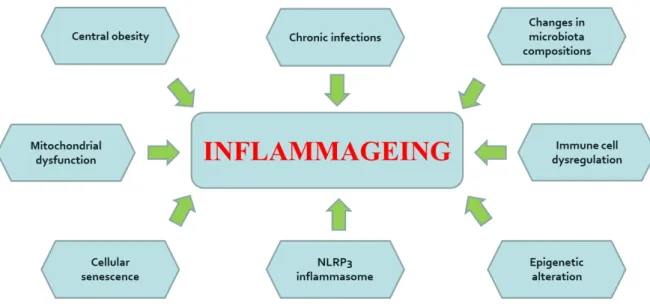
Possible mechanisms of inflamm-ageing include genetic susceptibility, cellular senescence, central obesity, oxidative stress caused by dysfunctional mitochondria, increased gut permeability, changes to microbiota composition, NLRP3 inflammasome activation, immune cell dysregulation, and chronic infections (Figure 6) (Ferrucci and Fabbri 2018).
Figure 6. Mechanisms of inflamm-ageing. (Ferrucci and Fabbri 2018)
Multiple genetic variants have been identified in large populations that affect the concentration of pro-inflammatory mediators in blood. A gene-expression study conducted on whole-blood RNA samples from a large cohort in Europe and USA revealed that immune response and inflammation were the most highly up-regulated pathways in association with ageing. The contribution of miRNAs to inflamm-ageing is an active area of investigation with high translational potential. miRNAs which are non-coding, single-stranded RNAs modulate protein expression by interacting with mRNA. Specific miRNA in circulating cells, plasma or whole blood have been suggested to contribute to inflamm-ageing and related chronic dieases. For example, miR-126–3p inhibits endothelial inflammation, and low levels of miR-126–3p were found in patients with CVD and diabetes. Other miRNAs, such as miR-146 and miR-155, possibly present in microvesicles or other structures carrying miRNAs, promote inflamm-ageing by
20
induction of cellular senescence or modulating immune response (Olivieri, Albertini et al. 2015, Rea, Gibson et al. 2018).
Several studies have shown that ageing is associated with the exponential accumulation of senescent cells in different organs and tissues including skin, T lymphocytes, endothelium, kidney, liver, visceral fat, cardiac muscle, in both animals and humans. Senescent cells acquires a senescence-associated secretory phenotype (SASP) characterized by abnormal secretion of mediators such as interleukins, growth factors, chemokines, metalloproteinases and of other extracellular matrix molecules, all favoring a pro-inflammatory status and the development of cellular senescence in neighboring cells. As senescence is associated with ageing as well as inflammation and CVDs in aged individuals, it appears a strong a candidate for contributor to inflamm-ageing (Fulop, Dupuis et al. 2016, Ferrucci and Fabbri 2018).
With advancing age, stressed cells, undergoing apoptosis, release large number of molecules known as damage associated molecular patterns (DAMPs) which includes ROS from dysfunctional or damaged mitochondria, oxidized LDL, microvesicles, mitochondrial DNA fragments or histones. Late removal of DAMPs can favor inflamm-ageing. For example, accumulated DAMPs activate NLRP3 inflammasome receptors up-regulating pro-inflammatory mediators (IL-1 and IL-18). Indeed, ageing is associated with increased blood levels of IL-18 and blockade of NLRP3 signaling in mouse models extends healthspan by attenuating multiple age-related changes that are associated with inflamm-ageing (Youm, Grant et al. 2013). Moreover, increased levels of age-related ROS can also trigger inflammatory responses by activating NF-kB signaling (Sies, Berndt et al. 2017, Ferrucci and Fabbri 2018).
Inflamm-ageing, indeed, acts as an accelerator for the emergence of age-associated chronic pathologies. For example, inflamm-ageing actively contributes to the initiation and progression of atherosclerosis, which is primary cause of stroke, coronary artery disease and peripheral vascular disease. Inflammation is also involved in pathophysiology of atrial fibrillation and post-myocardial infarction cardiac remodeling (Figure 7) (Fernández-Ruiz 2016, Ferrucci and Fabbri 2018).
21
Figure 7. Inflamm-ageing: a risk factor for multiple chronic diseases. (Ferrucci and Fabbri 2018)
1.4 Cell membrane asymmetry and fluidity: impact of
ageing
The asymmetry of plasma membrane plays a key role in the maintenance of cellular functions. Under resting conditions, in eukaryotic cells, the phosphatidylserine (PS), phosphatidylethanolamine (PE) and aminophospholipids are situated in inner membrane leaflet of the bilayer whereas sphingomyelin and phosphatidylcholine (PC) are located in the outer leaflet. When asymmetry of membrane is lost, under stress conditions, PS detected at the outer leaflet of membrane, increasing the pro-coagulant potential of circulating cells and favoring the shedding of pro-coagulant microvesicles (Morel, Jesel et al. 2011). Cell asymmetry is also essential for cell viability. During apoptosis, exposure of PS at cell surface act as a signal recognition and removal by macrophages (Scott, Heberle et al. 2018).
Plasma membrane fluidity is determined superficially by phospholipid head groups at inner and outer membrane leaflets while in deep hydrophobic area of cell membrane by amount of cholesterol and unsaturated fatty acid side chains. Changes in membrane fluidity is associated with functional alteration of many integral receptors in plasma membrane (Ray, Kassan et al. 2016). In comparison to outer, inner leaflet of membrane has more fluidity because of higher PS content. Thus, alteration of asymmetry distribution of phospholipids in membrane can alter membrane fluidity and also function
22
of fundamental membrane proteins (Noble, Thomas et al. 1999). Membrane lipid rafts, which are microdomains of cholesterol, sphingolipids and scaffolding proteins, are key players in signal transduction, organizing cytoskeleton and vesicular trafficking (Lingwood and Simons 2010). Studies have shown link of immune cell function with functioning lipid raft as multiple immune cell receptors such as T-cell receptor (TCR), B-cell receptor (BCR) and high-affinity IgE receptor are dependent on efficiency of lipid raft for their activation and down-ward signaling (Rajendran and Simons 2005).
Alterations in cellular and intracellular membrane is one of the feature of ageing and many chronic medical conditions. Ageing is accompanied by changes in membrane’s physical properties, lipid composition, receptor function and antigen presentation. Age-associated alteration in plasma membrane lipids including oxidation of lipids and loss of cholesterol leads to the loss of membrane lipids rafts and hence hamper the membrane bioactivity (Egawa, Pearn et al. 2016). Membrane phospholipids and their unsaturated fatty acids are especially sensitive to age-associated oxidative stress. In leukocytes, increased expression of PS on the outer leaflet of membrane in elderly is related to changes in subsets of lymphocytes, higher rate of apoptosis and alteration in the activity of membrane-bound transporters and ion channels (Noble, Thomas et al. 1999). Damaged cells and increased oxidative stress-mediated opening of calcium channels, resulting in increased calcium intracellular concentration, can activate membrane transporter like scramblases that rapidly destroy asymmetrical distribution of the membrane phospholipids (Figure 8) (Kodigepalli, Bowers et al. 2015, Nicolson and Ash 2017). Loss of asymmetry, in old age, is also correlated with increased membrane fluidity. So, age-related structural changes in plasma membrane of leukocytes has a role in altered function of membrane bound receptors and this could partially account for impaired immune responses and hemostasis, as witnessed with advancing age (Noble, Thomas et al. 1999, Nicolson and Ash 2017).
23 Figure 8. Ageing and plasma membrane remodeling
Increased oxidative stress and elevated intracellular accumulation of calcium promotes activation of plasma membrane transporter to alter the asymmetrical distribution of membrane phospholipids. Exposure of PS on surface of plasma membrane and cytoskeleton degradation favours membrane budding and release of microvesicles. PS: phosphatidylserine, ROS: reactive oxygen species
25
Chapter 2: Microparticles: overview and role in
vascular damage
27
2.1
Overview of Microparticles
Microparticles, now commonly referred to as microvesicles (MVs), are small extracellular plasma membrane vesicles, 0.05-1µm in diameter, shed by activated, apoptotic and/or senescent cells (Figure 9) (Ridger, Boulanger et al. 2017). MVs were later identified as coagulant lipid rich particles and characterized by electron microscopy as small bilayer membrane vesicles released from activated platelets in plasma and serum and referred to as “platelet dust” possibly accounting for the “Platelet Factor γ” activity and now studied as vascular effectors extensively (Yáñez-Mó, Siljander et al. 2015, Said, Rogers et al. 2018).
Figure 9. Cellular microparticles: a diffusing storage pool of bioactive effectors. Membrane microparticles are shed from the plasma membrane of stimulated cells. They harbor cytoplasmic proteins as well as bioactive lipids implicated in a variety of fundamental processes. MHC: Major histocompatibility complex, GPI: glycosylphosphatidylinositol (Hugel, Martinez et al. 2005)
28
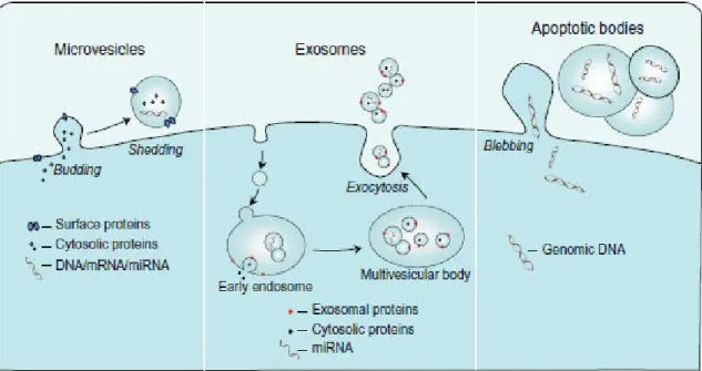
MPs can be differentiated from other extracellular vesicles, including exosomes and apoptotic bodies, on the basis of their size, subcellular origin, content and mechanism of formation (Figure 10) (Ridger, Boulanger et al. 2017).
Figure 10. Schematic representation of the mechanisms of formation of microvesicles, exosomes, and apoptotic bodies (Lawson, Vicencio et al. 2016).
MPs are shed by cells in response to stress, after remodeling of the phospholipids of the plasma membrane, followed by budding of plasma membrane and shedding of microparticles into extracellular environment (Pollet, Conrard et al. 2018). For many decades, MPs were considered as inert debris resulting from the cellular destruction, growth or dynamic renewal. Later, they were recognized as pro-coagulant mediators in coagulation and, owing to their content (active lipids, nucleic acid, miRNA, proteins), as true vectors and mediators of biological messages (Mause and Weber 2010), in intercellular communication initially in cardiovascular issues (Benameur, Osman et al. 2019). Because MPs originate from the plasma membrane, they carry transmembrane and surface protein from parent cells that characterize their cell origin or even the initiator stress of tissue damage (Morel, Toti et al. 2006). MPs are not only markers of activation or cellular and tissue damage, but also actors of major physiological responses such as hemostasis, inflammation, cell survival and apoptosis, endothelial function, vascular remodeling and angiogenesis; some of them also altered by exosomes (Ridder, Sevko et al. 2015, Koenen and Aikawa 2018). The ubiquitous property of MPs is their pro-coagulant phenotype discovered and widely studied in cardiovascular diseases and hemostasis. By exposing phosphatidylserine, and sometimes tissue factor (TF), the MPs
29
constitutes a catalytic surface for the assembly and activation of blood coagulation complexes and thrombin generation (Morel, Jesel et al. 2011). Under normal physiological conditions, MPs do circulate in blood but at low concentration. However, in pathological conditions, the circulating levels of MPs derived from different origins, including platelets, leukocytes, endothelial cells, red blood cells and smooth muscle cells, are increased and are propositional to the severity of disease. Therefore, microparticles have potential use as biomarkers for the diagnosis and prognosis of different pathological conditions. Similarly, they appear as a pivotal target for the therapeutic follow-up in cardiovascular disease or even graft rejection (Amoura, Zobairi El‐Ghazouani et al. 2019).
2.2
Mechanisms of formation of microparticles
The formation and release of microparticles mainly involves the loss of membrane phospholipids asymmetry and limited cytoskeleton cleavage and its re-organization (Figure 13).
2.2.1 Loss of asymmetry of membrane phospholipids and formation of
Microparticles
In resting eukaryotic cells, the distribution of phospholipids in plasma membrane is asymmetrical which is key to maintain important cellular functions including membrane potential and cell activation (Ma, Poole et al. 2017, Ridger, Boulanger et al. 2017). In all eukaryotic cells, sphingomyelin (SM) and phosphatidylcholine (PC) are located in outer membrane leaflet while phosphatidylethanolamine (PE) and the negatively charged lipids phosphatidylserine (PS) and phosphatidylinositol (PI) within the inner leaflet (Figure 11). Other lipids, like cholesterol and ceramides, are present within both membrane leaflets but mostly enriched within inner leaflet (Marquardt, Geier et al. 2015).
30
Figure 11. Asymmetrical distribution of phospholipids in red blood cell membrane
(Marquardt, Geier et al. 2015)
The asymmetrical distribution of phospholipids is not at equilibrium and is, therefore, maintained actively (requiring ATPs) by cells. There are three main classes of proteins involved in the control of asymmetrical distribution of membrane phospholipids i) ATP-dependent floppases, responsible for the outward transfer of ii) ATP-dependent flippases (P4 ATPases), that belong to the family of ABC transporters (ATP binding cassette), responsible for the inverse phospholipid transfer iii) Ca2+-dependent scramblases, which swiftly transports phospholipids across the membrane in both directions (Figure 12) (Hankins, Baldridge et al. 2015). Of note, the ABC transporter (ABCA1) has been identified as occasional plasma membrane phospholipids translocator in vascular wall (Toti, Schindler et al. 1997, Hamon, Broccardo et al. 2000, Quazi and Molday 2011).
31
Figure 12. Functions of membrane lipid transporters (Quazi and Molday 2011)
Disruption or loss of asymmetrical distribution of phospholipid is the driving force for the membrane remodeling and generation of MPs (Ridger, Boulanger et al. 2017). During cell activation or apoptosis, the prominent change is the translocation of PS to the outer leaflet which precedes MP formation (Lee, Meng et al. 2013). Excess of negative charge is created at cell surface due to the rapid egress of PS which imbalances the molecular masses of the two membrane leaflets and is not counterbalanced at equal speed by PC transporters .The surface tension between the two leaflets is abolished by MP shedding. MP shedding is enabled because of the partial loss of the interaction between the cytoskeleton proteins and the inner leaflet resulting from its proteolysis. The link of MP shedding with PS externalization is confirmed in patients with Scott’s syndrome, a rare human hereditary hemorrhagic disorder with defective platelet’s pro-coagulant activity due to defective PS externalization ability, also accompanied by reduced MP shedding (Toti, Satta et al. 1996). Inability to externalize PS by patients with Scott’s syndrome is due to mutated scramblase TMEM16-F (anoctamine, ANO6) but no due to, previously thought, phospholipid scramblase (PLSCR1) as PLSCR1 is not mutated in Scott’s syndrome patients and calcium induced PS externalization is also not affected in mouse cells with knock out
32
PLSCR gene, confirming PLSCR is not responsible for inability to expose PS by Scott’s syndrome patients (Hankins, Baldridge et al. 2015).
Externalization of PS is controlled by calcium-dependent imbalanced activities of flippases, floppases and scramblases. Increased cytosolic levels of calcium results in the inhibition of flippases which is followed by externalization of PS by overwhelming actions of floppases and/or scramblases with a lower phosphatidylcholine and sphingomyeline reverse transport. This initiates local transverse instability of the plasma membrane and shedding of MPs, also associated with lateral membrane reorganization and raft clustering (Benameur, Osman et al. 2019). The increased intracellular calcium levels, under stress or stimulation, results from opening of the membrane calcium ion channels, the depletion of intracellular calcium stocks, a phenomenon known as capacitative or store-operated calcium entry (SOCE) leading to significant increase in cytoplasmic calcium concentration (30-γ50 μM compared to 1 μM at basal state) (Kunzelmann-Marche, Freyssinet et al. 2001). Depolarization of the outer membrane of mitochondria also contributes to increased intracellular calcium levels via the opening of mitochondrial. The SOCE pathway including Orai channel 1 and mitichondrial STIM1 calcium sensors as well as other calcium channels such as TRPC6 (transient receptor potential channel), the P2X1 purinoreceptor in platelets, sustain the cell calcium concentration in the cytoplasm to maintain prolong cell stimulation (VARGA-SZABO, BRAUN et al. 2009).
2.2.2 Reorganization/degradation of cytoskeleton and formation of
Microparticles
Following intracellular calcium influx and externalization of PS, membrane blebbing involves re-organization/degradation of the cytoskeleton constituents (talin, filamin, gelsolin, myosin, α-actinin) by cysteine proteases, like μ calpains and/or caspases. μ calpain is activated in response to elevated levels of cAMP and subsequent activation of protein kinase A, while caspases chiefly act via Rho kinase-dependent phosphorylation of the myosin light chain kinases (MLCK) (Ridger, Boulanger et al. 2017). Calpains are critical for neutrophil or platelet MP shedding, caspases for MP generation in vascular cells under apoptotic and activated conditions. The phosphorylation of MLCK causes actin/myosin-mediated contractile tension, which ultimately results in membrane bleb formation. In apoptotic endothelial cells, the phosphorylation of MLCK is mediated by
33
the serine/threonine kinase Rho-associated protein kinase- I (ROCK-I), one of the dowmstream effectors of the small GTP binding proteins, Rab22A or ARF6 (Ridger, Boulanger et al. 2017, Benameur, Osman et al. 2019). Caspase-3 also triggers Xkr8, a putative scramblase or caspase transducer that promotes PS externalization in the membrane of apoptotic cells. In platelets, inhibition of μ calpain prevented the shedding of MP while αIIb γ-mediated destabilization of cytoskeleton resulted in MP shedding, confirming the importance of cytoskeleton integrity and of its interaction with membrane proteins in shedding of MP (Ridger, Boulanger et al. 2017).
Figure 13. Floppase activity and facilitated transport of phosphatidylserine by TMEM16-F (ANO-6) and procoagulant MP shedding. At rest, phosphatidylserine (PS) is translocated to the inner leaflet by flippase activity. Upon cell activation and calcium-dependent flippase inhibition, PS translocation to the outer leaflet is driven by TMEM-16F and local K+ efflux
prompts cell shrinkage and re-shaping. High calcium concentration promoted by Stored Operated Channels (SOCE) favors the constitution of TMEM16-F platforms by oligomerisation or interaction with other receptors like P2XR in the case of long term exposure to Ca2+. Transient
phospholipid imbalance between leaflets and the proteolysis of cytoskeleton by calpains and/or caspases lead to facilitated procoagulant MP shedding. Putative scramblase transducers as Xkr8 are activated by caspases and would trigger enhanced floppase activity. Exposed PS catalyzes the assembly of blood coagulation complexes at cell and MP surface. (E: Enzyme, S: Substrate, CF cofactor) (Ridger, Boulanger et al. 2017)
2.
3
Composition of MPs
MPs, shed by stimulated/apoptotic cells, carry components from parent’s cells including proteins, lipids and nucleic contents (Figure 14)
34 Figure 14. Composition of MPs.
MPs are loaded with distinct components of genetic material (nucleic acids, mRNAs, microRNAs), lipids (phospholipids and bioactive mediators), and proteins (chemokines, cytokines, membrane receptors, enzymes, adhesion molecules, growth factors, and cytoskeleton-associated and regulatory proteins) to eventually mediating intercellular communication (Koenen and Aikawa 2018).
2.3.1
Protein Content of MPs
Analysis of the MP protein content by proteomics has confirmed its relationship to cell origin and the stimulus initiating their generation (Benameur, Osman et al. 2019). For example, T-lymphocytes stimulated with phytohemagglutinin (PHA) for 72 h, followed by 24h-induction of apoptosis by phorbol-12-myristate-13 (PMA) and actinomycin D, leads to the release of MPs exposing morphogen sonic hedgehog (Shh) (Agouni, Mostefai et al. 2007) while T-lymphocytes only stimulated with actinomycin D, lack Shh expression (Mostefai, Agouni et al. 2008). Shh-MPs reduce NO bioavailability in endothelial cells and promote endothelial dysfunction in mouse aorta. Thereby confirming the close link between the MP inducer and its properties as cellular effector.
As biogenesis of MPs includes remodeling of plasma membrane and cytoskeleton re-arrangement/degradation, MPs carry proteins involved in their generation as well as cytoskeleton components. For example, the MPs from the tumoral LOX cell line harbor
35
ARF6, identified as regulator of their shedding (Muralidharan-Chari, Clancy et al. 2009). Actin is detected in MPs derived from RBCs and neutrophils (Pollet, Conrard et al. 2018).
MPs are enriched in several membrane proteins including adhesion molecules, like, P-selectin, glycoprotein Ib (GPIb) or integrins (GPIIbIIIa). Characteristic membrane proteins can be used MP detection, identification of the cell/stimulus from which they are generated (CD42a for platelets or CD3 for T-cells), and furthermore, on the basis of specific and differential expression of specific patterns between MPs from healthy/abnormal/activated or apoptotic cells, MPs can be used as a tool for the diagnosis and progression of diseases (Distler, Pisetsky et al. 2005). As apoptosis is characterized by nucleus condensation and fragmentation, apoptotic MPs would be identified on the basis of a high proportion of genetic and nuclear content (Saleh and Kabeer 2015). Other proteins includes membrane receptors, transcription factors and bioactive enzymes such as metalloproteinases (MMPs), mitogen-activated protein kinase (MAPK), phosphatidylinositol-3-kinase (PI-3K) and phospholipases A2, C and D, depending on the lineage, stress and species (Benameur, Osman et al. 2019).
2.3.2 Lipid content of MPs
The phospholipid composition of MPs varies with the cell origin, mechanism of its generation as well as the oxidation status of lipids (Pollet, Conrard et al. 2018). For instance, circulating MPs derived from platelets (PMPs) comprise of 20% sphingomyelin, 9% phosphatidylethanolamine (PE), 60% phosphatidylcholine (PC), 5% phosphatidylserine (PS) and minor quantities of other lipids including PAF and inositolphosphate (Weerheim, Kolb et al. 2002, Cognasse, Hamzeh-Cognasse et al. 2015). Furthermore, the MP lipid composition will vary with the parental cell niche and the degree of lipid oxidation (Fourcade, Simon et al. 1995, Huber, Vales et al. 2002). As a matter of proof, MPs from apoptotic or activated ECs have distinct lipid profile (Jimenez, Jy et al. 2003) and circulating MPs or MPs from the atherosclerotic plaque also differs (Leroyer, Isobe et al. 2007). The lipids composition and hence function of MPs from healthy subjects is different from those MPs which are generated and collected from patients with metabolic disturbances and dyslipidemias (Nomura, Inami et al. 2009). Although extensive data are lacking for all MP lineages, it was shown that the pro-coagulant activity of platelet MP is 50-100 folds higher than that of activated platelet. Thereby confirming their pro-coagulant feature (Sinauridze, Kireev et al. 2007).