HAL Id: tel-02466497
https://tel.archives-ouvertes.fr/tel-02466497
Submitted on 4 Feb 2020Drosophila immune response to the endoparasitoid wasp
Leptopilina boulardi : characterization of a resistance
reaction
Chami Kim
To cite this version:
Chami Kim. Drosophila immune response to the endoparasitoid wasp Leptopilina boulardi : char-acterization of a resistance reaction. Parasitology. Université Côte d’Azur, 2019. English. �NNT : 2019AZUR4008�. �tel-02466497�
Réponse immunitaire de la drosophile à la
guêpe endoparasitoïde Leptopilina boulardi:
caractérisation d'une réaction de résistance
Chami KIM
Institut Sophia Agrobiotech
Présentée en vue de l’obtention
du grade de docteur en Sciences de la Vie et de la Santé
d’Université Côte d’Azur Dirigée par : Marylène Poirié Co-encadrée par : Jean-Luc Gatti Soutenue le : 22 mars 2019
Devant le jury, composé de :
Istvan Ando, Professeur, Académie des Sciences de Hongrie
Bernard Charroux, DR CNRS, Institut de Biologie du Développement de Marseille
Michèle Crozatier, DR CNRS, Centre de Biologie Intégrative de Toulouse
Raphaël Rousset, CR CNRS, Institut Sophia Agrobiotech
Drosophila immune response to the
endoparasitoid wasp Leptopilina boulardi:
characterization of a resistance reaction
Jury:
President of the jury
Dr Michèle Crozatier, DR CNRS, Centre de Biologie Intégrative, Toulouse
Reviewers
Dr Istvan Ando, Professor, Biological Research Center of the Hungarian Academy of Sciences, Szeged
Dr Bernard Charroux, DR CNRS, Institut de Biologie du Développement, Marseille
Examiner
Dr Raphaël Rousset, CR CNRS, Institut Sophia Agrobiotech, Sophia Antipolis
Thesis supervisors
Résumé
Drosophila melanogaster est un modèle majeur en biologie, notamment pour l'immunité et
l'évolution.
L’immunité innée de la drosophile a été très étudiée dans le cadre de la réponse contre les bactéries et les champignons, mais on en sait moins sur la défense contre les guêpes endoparasitoïdes dont le développement à l'intérieur de l'insecte hôte entraîne sa mort. L'une des interactions les plus étudiées entre drosophiles et guêpes parasitaires implique Leptopilina boulardi qui pond des œufs à l'intérieur des larves hôtes et se développe à leurs dépens. Une fois que l'oeuf parasitoïde a été reconnu comme un envahisseur étranger, la larve de Drosophila peut déclencher une réponse immunitaire qui mène à l'encapsulation : l'oeuf est entouré de plusieurs couches d'hémocytes. La capsule ainsi formée est mélanisée et il y a formation d’espèces réactives de l’oxygène qui participe à la mort du parasitoïde. Alternativement, la réponse immunitaire peut être contournée grâce aux composants du venin injectés par la guêpe femelle en même temps que l'œuf. Nous avons utilisé deux souches de Drosophila, résistante et sensible à L. boulardi, ne diffèrant que par une région du chromosome 2R contenant un gène de résistance majeur. La résistance s'est révélée être monogénique, avec deux allèles, l'allèle de résistance étant dominant (Rlb+> Rlb-). L'équipe avait précédemment identifié edl / mae (allèles R et S) en tant que gène candidat. Mae (modulateur de l'activité d'ETS) ou edl (ETS-domain lacking) a été décrit comme un médiateur de facteurs de transcription de la famille de l'ETS (E26 transformation-specific) chez la drosophile. Mae interagit avec les facteurs de transcription via son domaine SAM (Steril Alpha Domain), un domaine d'interaction protéine-protéine.
Les objectifs de ma thèse étaient de déchiffrer le rôle possible d’edl et d’identifier les événements moléculaires et cellulaires conduisant au succès ou à l’échec de l’encapsulation. J'ai utilisé diverses approches allant de la génétique des mouches à la cytométrie en flux.
L'implication de edl dans la résistance à Drosophila a été confirmée par surexpression et interférence d'expression de edl La surexpression de l'allèle résistant dans un fond sensible conduit à un phénotype résistant. L'interférence de l'expression de l'allèle sensible entraîne une augmentation du taux d'encapsulation de parasitoïde. Au niveau cellulaire, une augmentation du nombre d'hémocytes après le parasitisme s'est produite plus tôt dans la souche résistante que dans la souche sensible. Il a également été observé que la glande lymphatique des larves résistantes éclate avant celle des larves sensibles. Au niveau moléculaire, des interactants potentiels de edl ont été
identifiés in silico et 2 ont été testés en utilisant la réduction de leur expression qui a conduit à l’observation d’une augmentation de l'encapsulation.
Dans l’ensemble, un acteur clé du mécanisme de résistance de la drosophile à la guêpe parasite a été identifié au cours de ce travail et permet d’ouvrir des pistes pour des travaux futurs sur le mécanisme de régulation de la réponse au niveau moléculaire.
Abstract
Drosophila melanogaster is a main model in biology, notably immunity and evolution.
Although the Drosophila innate immune processes to fight bacteria and fungi have largely been explored, less is known of the defence against endoparasitoid wasps whose successful development inside the insect host leads to its death. One of the most studied Drosophila – parasitoid wasp interaction involves Leptopilina boulardi that lays eggs inside host larvae and develop at their expense. Once the parasitoid egg has been recognized as a foreign invader, the Drosophila larva can mount a successful immune response, the encapsulation: the egg is surrounded by several layers of hemocytes and there is an increase of a specific types of hemocytes, the lamellocytes. The so-formed capsule is melanised and there is formation of reactive oxygen species, which kills the parasitoid. Alternatively, the immune response can be circumvented thanks to the venom components injected by the female wasp together with the egg.
Using two Drosophila strains, resistant and susceptible to L. boulardi, which differ only in a region of chromosome 2R containing a major resistance gene. The resistance was found to be monogenic, with two alleles, the resistance allele being dominant (Rlb+>Rlb). The team previously identified
edl/mae (R and S alleles) as a candidate gene. Mae (Modulator of the Activity of ETS) or edl
(ETS-domain lacking) was described as a mediator of specific transcription factors of the ETS (E26 transformation-specific) family in Drosophila. Mae interacts with transcription factors trough a SAM (Steril Alpha Domain), a protein – protein interaction domain. edl is known to regulate aop and pnt
P2 transcription factors during the eye development and aop and pnt P2 appear to have a role during
haematopoiesis.
The objectives of my thesis were to decipher the possible role of edl and identify the molecular and cellular events leading to success or failure of encapsulation. I used various approaches from fly genetics to flow cytometry.
The involvement of edl in Drosophila resistance was confirmed by using overexpression and interference of edl expression. The overexpression of the resistant allele in a susceptible background leads to a resistant phenotype. The interference of the expression of the susceptible allele results in an increased rate of parasitoid encapsulation. At the cellular level, an increased in the number of hemocytes after parasitism occurred earlier in the resistant strain than in the susceptible strain. It was also observed that the hematopoietic lymph gland of the resistant larvae busted before the one of the susceptible larvae. At the molecular level, potential interactants of edl were identified in silico and 2 were tested using interference of their expression which led to observing an increase of encapsulation.
Overall, a key player in the resistance mechanism of the Drosophila to the parasitic wasp have been identify during this work and it lays the path for future work on regulation mechanism of the response at the molecular level.
Acknowledgments - Remerciements
I warmly thank all the members of the jury, Dr Ando, Dr Charroux, Dr Crozatier and Dr Rousset, for accepting to review my work.
Je remercie les Dr Poirié et Dr Gatti de m’avoir accueillie au sein de leur équipe pour effectuer ma thèse. Je remercie aussi l’équipe ESIM, Laury Arthaud, Dominique Cazes, Dominique Colinet, Severine Lemauf, Chen Luo, Gaurav Pandharikar, Christian Rebuf et Sophie Tares, de m’avoir reçue et d’avoir répondu à toutes les questions qui trainaient régulièrement dans mon esprit.
Merci aux membres de mon comité de thèse, Laurent et Michèle (encore une fois), pour leurs critiques, leurs conseils et leur bienveillance.
Merci à Aurélie, Bin, Fanny et Laurent, mes copains de thèse, vous m’avez tout appris ; surtout Aurélie, à partir du moment où tu m’as appris à sexer des mouches je n’ai fait que ça, et puis Laurent cette histoire de trochanter. Et puis on a bien rigolé quand vous étiez là, vous me manquez énormément les copains.
Merci à Michèle (une dernière fois), Nathalie et toute l’équipe de Toulouse pour leur accueil et leur soutien, leur disponibilité et leur gentillesse.
Merci à Pascaline qui s’est souciée de moi et je sais que je peux compter sur toi si besoin, pour toi c’est juste ton rôle mais pour moi c’était plus que rassurant.
Merci à Corinne, Martine, Marc et Etienne pour leur accueil dans leurs bureaux quand j’avais besoin de changer d’air, je vous ai bien embêtés mais on va avouer que sans moi vous allez être perdus, qui va bien pouvoir tourner vos calendriers ☺.
Merci à Pierre Frendo et à toute l’équipe Symbiose de m’avoir accueillie durant mon exil. Merci à Clémence et Loris pour des heures de rigolades, leur interprétation magistrale de « Libérée, Délivrée » sur la route pour Lyon (j’ai la vidéo pour me remonter le moral) et, quand même, la tête de Clémence en réponse aux blagounettes de Loris, ça reste magique.
Merci à Franck et Mathieu de nous avoir nourrir et abreuver pendant ces 3 dernières années, Vive le canard confit et le vin.
Merci à Julia qui est toujours adorable et souriante, j’aurais acheté beaucoup moins de chocolat sans elle.
Merci à Michael Quentin qui m’a initié au labo, depuis je ne l’ai plus quitté. Et merci à Arnaud Barbary qui m’a fait compter des milliers de galles de nématodes sur des racines de piment, j’ai tellement aimé que je suis allée le faire aux Pays-Bas, et puis grâce à toi j’ai rencontré Cristina et ça c’est cool. Bon accessoirement sans vous, je n’aurais jamais eu mon stage chez Enza et tout ce qui en découle.
Merci à Bruno Favery qui m’a soutenu dans ma démarche de revenir en France et grâce à qui j’ai eu mon financement de thèse, je ne l’oublie pas et j’espère que j’aurais l’occasion de te rendre la pareille.
Thanks to the nice PhD students and researchers community on Twitter, always answering questions and supporting each other #phdchat #phdlife #phdadvice.
And last but certainly not least thank you to Kamila and Hae-Young, you both have been supporting me in applying to PhDs and after in not giving up on my PhD. Kamila you welcomed me into your lab, your home and your heart and I will be forever thankful for that, you have been the best mentor. Hae-Young 언니 despite of the 7-8h time difference between us, you have always been available to talk about anything, you have been the best roommate and now a very dear friend of mine, I couldn’t be there for your wedding but next time I’ll buy you a drink.
Un grand merci à Renaud et à Gilles de m’avoir mise en contact avec Marcio, grâce à vous je peux tourner la page et commencer une nouvelle aventure américaine.
Merci à Marcio de m’avoir permis de tenir le coup en cette fin de thèse, je serais partie avant la fin sans lui.
Immense merci à Renaud qui m’a redonné confiance en moi en une fin de thèse difficile. Merci d’avoir pris le temps et me relire et de discuter avec moi, c’était ce dont j’avais besoin et ce qui était nécessaire pour arriver au bout.
Merci à Antoine de m’avoir soutenu dans mes moments d’égarement, de joie et de désespoir (surtout de désespoir). On aura quand même bien mangé et bien bu pendant cette thèse. Tu reprendras bien une part de flan.
Table of contents
Résumé ... 5
Abstract ... 7
Table of figures and tables ... 15
Table of abbreviations ... 19
I. Introduction ... 21
A. Immunity in Drosophila melanogaster... 27
1. Antibacterial and Antifungal defences ... 29
a) The Toll pathway ... 29
b) The Imd pathway... 31
c) The Jak-Stat pathway ... 31
2. Antiviral immunity ... 33
3. Cellular immunity ... 35
a) Hemocyte types ... 35
b) Hematopoesis ... 39
c) Factors controlling hematopoiesis ... 43
d) Melanisation ... 45
e) Encapsulation ... 47
B. Genetic interaction between Drosophila melanogaster and Leptopilina boulardi ... 51
1. Resistance-virulence in D. melanogaster-L. boulardi populations... 53
2. Fly resistance ... 55
3. Parasitoid virulence factors ... 56
4. Proteomic of the L. boulardi ISm and ISy venom... 57
5. Origin of the D. melanogaster and L. boulardi strains and lines ... 59
C. The Rlb gene ... 60
1. Discovery of edl as the Rbl gene ... 61
2. Edl structure and fonctions ... 63
a) Structure ... 63
b) Fonctions... 63
3. EGF pathway and timing of lamellocytes liberation after parasitism... 65
D. Objectives ... 71
II. Materials and methods ... 73
A. D. melanogaster strains and stocks, L. boulardi lines, rearing conditions ... 75
B. Larval encapsulation assays ... 77
C. Observation of the timing of the lymph glands bursting ... 77
D. Genetic approaches, crossover schemes ... 77
2. Localisation of the UAS-mae insertion in transgenic strains ... 79
a) Test of localization of the insertion on chromosome 1 ... 79
b) Test of localization of the insertion on chromosome 2 ... 79
c) Test of localization of the insertion on chromosome 3 ... 81
3. Rescue experiment ... 81
4. Creation of new Drosophila lines ... 83
E. Immunolabeling of hemocytes ... 83
F. Flow cytometry procedures. ... 85
G. Molecular approaches (DNA, RNA extraction, PCR and qPCR) ... 85
H. Cloning and transformation ... 87
I. Bioinformatics... 87
J. Statistics ... 88
III. Results ... 89
A. New tools for the characterization of the resistance/susceptible phenotype... 91
1. Creation of the new resistance/susceptible lines ... 93
2. Creation of the new resistance/susceptible lines with fluorescent hemocytes ... 93
B. Different timing-dependant cellular response between YS-msnCherry and YR-msnCherry after parasitism ... 95
C. Timing of the lymph gland bursting in parasitized YR and YS lines ... 101
D. Role of edl in the resistance phenotype... 103
1. Expression of the edl resistant allele in hemocytes confers the resistance phenotype 105 2. Effect of the repression of edl, aop and pointed ... 107
E. Potential interactants of edl ... 108
1. Bioinformatic comparison of orthologs between Drosophila species ... 109
2. Bioinformatic search for new edl potential interactants ... 113
3. Ets97D and l(3)mbt in vivo RNAi tests... 117
4. Preliminary tools creation for testing physical interactions with edl ... 117
IV. Discussion ... 119
Bibliography ... 137 Annexes ... 151
Table of figures and tables
Figure 1: Examples of interaction between organisms... 22
Figure 2: Schematics of the activation of immune defences in Drosophila ... 26
Figure 3: Toll pathway... 28
Figure 4: Representation of the Imd pathway ... 30
Figure 5: Jak-Stat pathway ... 30
Figure 6: RNAi machinery in Drosophila ... 32
Figure 7: Origin and function of hemocytes in Drosophila melanogaster ... 36
Figure 8: Ontogenesis of blood cell lines and regulation of hematopoiesis in Drosophila... 38
Figure 9: Hematopoiesis regulators and hemocyte functions in D. melanogaster ... 42
Figure 10: Formation of the multilayer cellular capsule around a parasitoid egg. ... 46
Figure 11: D. melanogaster and L. boulardi life cycle ... 50
Figure 12: Geographical distribution of the encapsulation rate of L. boulardi populations in sympatric populations of D. melanogaster. ... 52
Figure 13: Distribution of Drosophila resistance phenotype ... 52
Figure 14: Geographic distribution of virulence in L. boulardi populations ... 52
Figure 15: Matching interactions in the D. melanogaster – L. boulardi reference system ... 58
Figure 16: Region of chromosome 2R showing the position and distance between CG33136 and edl ... 58
Figure 17: The structure and comparison of edl with other Ets-proteins ... 62
Figure 18: Feedback loop regulation of edl transcription by yan/aop and pointed proteins.... 64
Figure 19: Diagrammatic representation of the EGF signaling pathway... 66
Figure 20: Proposed gene regulatory network that controls lymph gland rupture upon wasp parasitism ... 68
Figure 21: Total numbers of hemocytes in single parasitized larvae ... 68
Figure 22: Picture of a dissected “intact” lymph gland ... 76
Figure 23: Crossover schemes for RNA interference experiments ... 76
Figure 24: Cross-over scheme to test the localization of the insertion on chromosome 1 ... 78
Figure 25: Crossover scheme to test localization of the insertion on chromosome 2... 78
Figure 26: Crossover scheme to test the localization of the insertion on chromosome 3 ... 80
Figure 27: Crossover scheme for the rescue experiment ... 80
Figure 28: Crossover scheme to create resistant and susceptible strains with a double staining of plasmatocytes and lamellocytes (the scheme was used for both YS and YR)... 82
Figure 29: Representation of the sample preparation timeline for the hemocytes counting
experiment ... 84
Figure 30: Acrylamide gel (12%) of DNA from YS and YR lines treated with Mae III Endonuclease ... 86
Figure 31: Alignment of the sequences of the CDS of 2 alleles of edl ... 90
Figure 32: Cross-over scheme to create resistant and susceptible strains with a staining of lamellocytes ... 92
Figure 33: Pictures of lamellocytes from the YR-msnCherry and YS-msnCherry... 92
Figure 34: Flow cytometry plots at representative time points after parasitism ... 94
Figure 35: Total hemocytes count post-parasitism evaluated by flow cytometry ... 96
Figure 36: Lamellocytes count on the resistant and the susceptible lines after parasitism ... 96
Figure 37: Lamellocytes proportion in hemolymph after parasitism. ... 98
Figure 38: State of the lymph gland in control and in parasitized conditions ... 100
Figure 39: Measure Levels of relative gene expression in Drosophila larvae ... 102
Figure 40: Edl structural feature. ... 108
Figure 41: Muscle alignment of the 26 proteins with a SAM-PNT domain of the different Drosophila species ... 109
Figure 42: Alignment of the SAM domain IPR003118. ... 114
Figure 43: The 2 peptides chosen for the antibody production ... 116
Figure 44: Schematic representation of some of the main results from this project ... 134
Table 1: List of the Drosophila strains used ... 74
Table 2: Primers used and their characteristics ... 86
Table 3: Representation of the encapsulation rate of the promotor-Gal4 and UAS-sequence of interest lines before any cross. ... 104
Table 4: Representation of the encapsulation rate of the descendants of the crosses for the overexpression of edl ... 104
Table 5: Encapsulation rate of the F1 from the crosses for the RNAi of edl ... 106
Table 12: Recapitulative table of the effect of edl alleles overexpression and RNAi. ... 126
Table 13: Data from the flow cytometry experiment ... 154
Table 14: Number of total dissected larvae for the lymph gland bursting experiment ... 155
Table of abbreviations
Abbréviation Signification Abbréviation Signification
ADGF adenosine deaminase-related growth factor MAPK Mitogen-activated protein kinases
AdoR Adenosine receptor mRNA messenger RNA
AGO Argonaute MZ medullary zone
AMPs anitmicrobial peptides NF-kB NF-kappa-B
aop anterior open PAMP pathogen-associated molecular pattern
bsk basket PG peptidoglycan
Crq Croquemort PGRP PeptidoGlycan Recognition protein
CZ cortical zone pi piwi-interacting
DAMP Damage-associated molecular pattern pnt pointed
DCV Drosophila C Virus PO Phenoloxidase
die Diedel PPO prophenol-oxidases
Dif Dorsal-related immunity factor PRRs pattern recognition receptors
DNA Deoxyribonucleic acid PSC Posterior signalling center
Dome Domeless PVF platelet-derived growth factor/vascular endothelial growth factor-like factor
Dpp decapentaplegic R2D2 dsRNA binding co-factor of Dicer-2
DRK Downstream of Receptor Kinase RISC RNA-induced silencing complex
dsRNA double strand RNA RNA RiboNucleic acid
EBF early B-cell factors enhancer binding factor RNAi RNA interference
edl ETS binding lacking ROS Reactive Oxygen Species
EGFR Epidermal Growth Factor Receptor RTK Receptor Tyrosine Kinase
ERKA Extracellular-Regulated Kinase A SAM Sterile Alpha Motif
FGFR Fibroblast Growth Factor Receptor SINV sindbis virus
FHV Flock House Virus siRNA small interfering RNA
FOG Friend of GATA SOS Son of Sevenless
gcm missing glial cells SPE Spätzle-processing enzyme
GNBP3 Gram-negative bacteria binding protein 3 Spz Spätzle
hh hedgehog srp serpent
hop hopscotch Stat Signal Transducer and Activator of Transcription
IKK IκB kinase TEL Translocation Ets leukemia
IL-6 Interleukine 6 TLRs Toll-like Receptors
IlS Insulin/IGF TNF tumor necrosis factor
imd immunodeficiency TOR Target of Rapamycin
IMPDH inosine monophosphate dehydrogenase tsr twinstar
Jak Janus Kinase upd unpaired
JNK Januse kinase VSR viral suppressors of RNAi
KO knock-out Wnt wingless
LG Lymph Gland zir Zizimin-related
Figure 1: Examples of interaction between organisms. Predation: a heron caught a fish. Competition: 2 stags fighting. Symbiosis: a clownfish living in symbiosis with a sea anemone. Parasitism: a caterpillar parasitized by Cotesia glomerate. Pictures from National Geographic.
Every living organism interacts with other organism(s) during its life time. These species interactions form the basis for many ecosystem properties and processes such as nutrient cycling and food webs. They can be classified in several categories including predation, competition, symbiosis and parasitism (Figure 1). Predation might be the most obvious direct interaction people think about when asked about species interactions: the predator kills and eats the prey. Competition for the acquisition of a resource (food, reproductive partner) can be direct or indirect and it significantly influences the structuring of communities. Symbiosis refers to a long-term association between 2 different organisms. Parasitism is a form of symbiosis where one individual will thrive and survive at the expense of one or several hosts. Parasites can live in (endoparasite) or on the host (ectoparasite). The interactions’ characterization as a function of the impact on the partner “host” fitness is not as clear as described above. It is admitted that environmental conditions or equilibrium breaks in the interaction can displace an interaction on a scale ranging from mutualism to parasitism or pathogenicity. For instance, commensal intestinal bacteria may behave as pathogens under certain conditions. Finally, we must also highlight the long-term impact of mutualism and parasitism on the evolution of past and present species through selection pressures, interactions of genomes and horizontal gene transfer.
Overall, these interactions may have positive, negative or neutral effects on either specie's ability to survive and reproduce and are widespread causes of natural selection and in fine of species evolution.
Insects are the most successful group of all animals. The origin of insects was dated to the early Ordovician ~479 millions years ago and ~345 millions years ago for major extant lineages (Misof
et al., 2014). They are the most successful group of all animals, considering either the number of
individuals (1019 living simultaneously) (McGavin, 2010) or the number of species (55% of the species biodiversity, 85% of the animal biodiversity). Entomologists estimate the actual number of living insect species could be as high as 5 to 10 millions. The nutrition by consumption of other insects (entomophagy) could be quite old in this taxon and it is found in all the main insect orders. Entomophagous insects are broadly divided into two classes: the parasites and the predators. At least 87 families, in 50 different insects orders contain parasitic species, while those which are predatory are present in 167 families from 14 orders. Allowing for duplications, at least 224 families in 15 orders have adopted entomophagous habit. Between 10 and 20% of known insect species are parasitoids, of which a quarter are Diptera or Coleoptera and three quarters are Hymenopteran wasps.
The parasitoid lifestyle is considered intermediate between parasite and predator. Indeed, a parasitoid develops in interaction with and at the expense of a host (parasite) but consumes the
tissues of the host which usually leads to its death (predator). Thus, parasitoid insects are mainly species with a unique reproductive strategy: they lay eggs in or on other insects (egg, larva, pupa or adult), their larvae develop as parasites while adults have a free life. Therefore, food sources usually differ between larva and adult. A distinction can be made between gregarious parasitoids that can lay up to hundreds of eggs in a host, and solitaries parasitoids for which even if several eggs are laid in a host, a single parasitoid will emerge. During oviposition, some parasitoid females kill or paralyze their host which is immediately consumed by their larvae (idiobiont parasitoids), while others allow the host to continue its development to ensure the proper development of the parasitoid offspring (koinobiont parasitoids). Koinobiont parasitoids, that parasitize host stages with a developed immune system (larval or adult hosts), must then be able to evade or suppress host defences, as well as manipulate host physiology to ensure their survival. This requires a delicate balance between the parasite and the host, the parasite allowing the host to be kept alive while bypassing its immunity and diverting its nutrients. Another alternative to the interaction is the triggering of a successful immune response by the host. The egg/larva of the parasitoid is then "inactivated" and/or killed, which usually allows the survival of the host. These two situations leading to “a single winner” are often described as "resistance" of the host (ability of the host to survive parasitism) and "virulence" (ability of the parasitoid to succeed in parasitism). However, the notion of resistance or virulence in the genetic sense is based on the existence of a variation and therefore presupposes the presence of resistant/susceptible or virulent/avirulent individuals in the population.
Like other insects, Drosophila species (Dipteran) can be parasitized at larval or nymphal stages by many species of parasitoids, including more than 40 Hymenopteran species, which constitute an important factor in the field population regulation (Carton et al., 1986). A large number of laboratory investigations on the biology, behaviour, ecology, genetics, physiology have been performed on these parasitoids, mainly on koinobionts larval parasitoids of the genus Leptopilina (Eucoilidae) and Asobara (Braconidae) (Bouletreau, 1986; Carton et al., 1986; Carton & Nappi, 1997, 2001; Kraaijeveld et al., 1998; Van Dijken & Van Alphen, 1998; Fauvergue et al., 1999; Eijs & Van Alphen, 1999; Eslin & Prévost, 2000; Fellowes & Godfray, 2000; Fleury et al., 2000; Vavre et al.,
with a focus on the cellular aspects of immunity, essential for the response to parasitoids. I will then present data on the interactions between Drosophila and parasitoid wasps. Finally, I will detail our knowledge of the genetic resistance of D. melanogaster to these wasps and outline the issues and objectives of my PhD work.
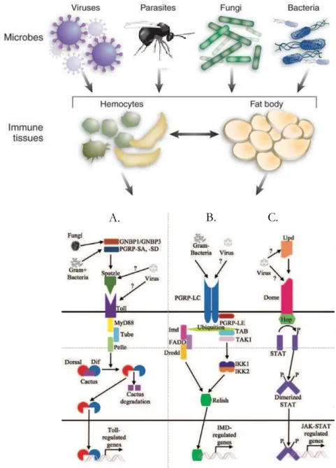
Figure 2: Schematics of the activation of immune defences in Drosophila adapted from (Lopez et
A. Immunity in Drosophila melanogaster
Since the discovery of the “white” mutation and its positioning on the X chromosome by Thomas Hunt Morgan in 1910, D. melanogaster has been a central model organism for studies in genetics, population genetics and other aspects of biology, including immunology. Indeed, the conservation of basic signalling pathways and key transcription factors controlling the development and functions of blood cells from Drosophila to human, makes D. melanogaster a simplified and interesting model to decipher the fundamental mechanisms governing hematopoietic system formation and homeostasis. Therefore, Drosophila is a study model for diseases associated with these mechanisms, such as leukaemia. Like other insects, the fruit fly lacks the mechanisms of adaptive immunity and relies solely on its innate immunity to defend against pathogens and parasites.
Innate immune defences are central mediators of the metazoan immune system and are essential to the health and the success of complex organisms (Hoffmann, 2003). The discovery of antimicrobial peptide responses 40 years ago has established D. melanogaster as a leading system to investigate fundamental components of the innate immune defences: The Nobel Prize in physiology or medicine has been awarded to Jules Hoffmann and Bruce A. Beutler for their discoveries concerning the activation of innate immunity in 2011.
During its life cycle, D. melanogaster flies feed, lay eggs and develop on decaying environments, notably fruits. Therefore, they are more exposed to pathogens than other organisms. This explains that Drosophila produces efficient humoral and cellular mediators to fight against the different pathogenic infections as well as parasitoids wasps. Its immune defences are based on the existence of cellular components, different types of hemocytes, and the production of antimicrobial peptides (AMPs) secreted mainly by fat and hemocytes. At the molecular level, innate immunity has been shown to involve several different pathways, including the Toll-Dorsal (Toll) pathway, the immunodeficiency (imd) pathway, the Janus Kinase (Jak)/Signal Transducer and Activator of Transcription (Stat) pathway, autophagy, and RiboNucleic Acid interference (RNAi) interference (Figure 2)(Mussabekova et al., 2017). The Toll and imd pathways contribute mainly to the antibacterial and antifungal defences even if the Toll pathway may also be an actor of the anti-parasite response (Lemaitre & Hoffmann, 2007; Valanne et al., 2011; Lamiable et al., 2016).
1. Antibacterial and Antifungal defences
a) The Toll pathway
The first immune receptors, the Toll receptors, have been discovered in Drosophila (see Vogel, 2012 for an historical review). Yet, the Toll pathway was first known for its role in the establishment of dorso-ventral polarity in the embryo, thanks to a series of genetic screens for genes involved in early Drosophila embryonic development (Nüsslein-Volhard & Wieschaus, 1980). What is interesting with the Toll pathway is that there is crossing over between the discoveries in humans and Drosophila. The identification of Toll as an activator of the immune response happened in 1995 (Rosetto et al., 1995). Human Toll was found soon after that and it was followed by the discoveries Toll-like Receptors (TLRs) in both Drosophila and mammals, and it was shown that the Toll pathway is conserved across phyla.
In Drosophila innate immunity and embryonic patterning, the Toll signalling pathway (Figure 3) is activated by the endogenous protein ligand Spätzle (Spz) (Valanne et al., 2011). Spz is secreted as
an inactive precursor that during embryogenesis is processed into an active form by the serine protease Easter. During infection, pattern recognition receptors (PRRs) initiate the immune responses when they encounter damage-associated molecular pattern molecules (DAMPs) and pathogen-associated molecular pattern molecules (PAMPs). PAMPs such as microbial Lysine-type peptidoglycan recognized by PeptidoGlycan Recognition Protein (PGRP) or -glucans recognized by Gram-negative bacteria binding protein 3 (GNBP3) activate proteolytic cascades and signal-transduction pathways. Insect PGRPs are classified as short or long: short PGRPs have signal peptides and can be extracellular proteins, whereas long PGRPs can be intracellular, extracellular, and transmembrane proteins. This PGRP-PNG interaction initiates a proteolytic cascade in which Spätzle is processed by a specific serine protease, Spätzle-processing enzyme (SPE). Among, the
Drosophila PGRPs, only PGRP-SA and PGRP-SD are involved in the activation of the Toll
pathway. The Toll receptor activation allows the degradation of the NF-kappa-B (NF-B) inhibitor Cactus and the liberation of transcription factors Dorsal-related immunity factor (Dif) and Dorsal, the Drosophila NF-B homolog. Upon translocation to the nucleus, Dorsal binds to the kappa B-related nucleotide sequences of the antifungal peptide Drosomycin and different antibacterial peptides such as Cecropins, Attacin, and insect Defensin. In larvae and adult, Dorsal is expressed in the fat body, and both its expression level and nuclear localization are enhanced upon microbial challenge.
The gene signalling pathway between Spätzle and Cactus, which includes Toll, Tube, or Pelle, constitute the Toll signalling “cassette” also required for the antifungal response in adult flies.
b) The Imd pathway
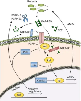
Another evolutionarily conserved signalling cascade regulates Drosophila immunity, the imd pathway (Figure 4), which activates a third NF-κB gene, Relish (Kuraishi et al., 2013; Myllymaki et
al., 2014). Activation of Relish requires phosphorylation and cleavage of the inhibitory C-terminal
part (I-κB like), probably by the caspase 8-homolog Dredd, thus exposing a novel ubiquitin binding site. This leads to activation of the Tab2/Tak1 complex, which in turn phosphorylate the Drosophila IκB kinase (IKK) complex. The phosphorylation of Relish by the IKK complex led to its cleavage and the translocation of its N-terminal part (Rel-68) into the nucleus where it activates the transcription of genes coding for AMPs (Diptericin and Cecropin). IMD, a death domain protein, is part with Dredd of the signalling complex recruited after binding of peptidoglycan (PG) to the receptor. Several PGRPs are involved in the imd pathway activation, the main one being the transmembrane protein PGRP-LC that binds bacterial PG (Tanji & Ip, 2005). PGRP-LE in its short-secreted form binds PG in the hemolymph and presents it to PGRP-LC. The cytoplasmic PGRP-LE can also interact with IMD, independently of PGRP-LC, to activate autophagy, whereas the transmembrane form can activate a prophenoloxidase cascade together with PGRP-LC. PGRP-LE is the only intracellular pathogen receptor identified in Drosophila. The imd pathway is required for expression of most Drosophila AMPs and flies with defects in the pathway die from bacterial infection while resisting fungal infection (Ben-Ami et al., 2009). Interestingly, the pathway initiates the systemic response in the fat body but is also locally activated in various tissues, trachea, brain and gut epithelia and mucosal surfaces.
c) The JAK-STAT pathway
The JAK/STAT pathway (Figure 5) was first discovered and studied in vertebrates. In human, this pathway plays a role in the differentiation of T and B lymphocytes, the control of inflammatory and wound repair in response to interferons, interleukins, other cytokines, and growth factors.
In Drosophila, a conserved JAK/STAT signalling pathway controls segmentation in embryos, as well as blood cell development and many other processes (eye development, gut renewal …) (Morin-Poulard et al., 2013; Zeidler & Bausek, 2013).
`
The Drosophila known ligands in the JAK/STAT pathway consist of three cytokine-like proteins (closely related to vertebrate leptins) called unpaired (upd), upd2 and upd3. The expressions of these 3 ligands are induced in response to tissue damage: upd3 in adult hemocytes after a bacterial infection and upd2 and upd3 in response to viral infection (Myllymäki & Rämet, 2014). Extracellular upds bind to the cytokine receptor Domeless (Dome), which shares similarities with the mammalian IL-6 receptor family. Dome transmits the signal through the only Drosophila JAK kinase hopscotch (hop) which phosphorylate Stat92E (Stat92E/Marelle) which then forms a dimer and translocate to the nucleus where it binds to the promoters of the target genes.
A role for JAK-STAT signalling in cellular immunity was suggested by studies of the hopTum-l
mutant. This mutation leads to an increased number of plasmatocytes and the massive differentiation of lamellocytes ready to encapsulate “self” tissue, leading to the formation of black masses/melanotic pseudo-tumours (Hanratty & Dearolf, 1993; Luo et al., 1995). Plasmatocytes that adhere to injured tissue upon the detection of basement membrane disruption, start a systemic response by producing cytokine (mainly Upd3) that eventually result in hemocyte proliferation (Agaisse et al., 2003). Activation of JAK-STAT signalling in hemocytes is thus required for their increased proliferation in response to both tumours and wound (Asha et al., 2003; Sorrentino et al., 2004). It is interesting to note that upd3 is secreted by the posterior signalling center (PSC) of the larval lymph gland (LG) to maintain the prohemocytes of the medullary zone (MZ) in an undifferentiated state (Makki et al., 2010). In case of parasitism, the JAK/ STAT pathway is switch off to allow lamellocytes differentiation (Makki et al., 2010).
2. Antiviral immunity
Like many organisms, Drosophila can be infected by viruses and is a good model to study the mechanisms of antiviral defence (Kemp & Imler, 2009). The viruses described as able to infect Drosophila are RNA viruses and the best characterized Drosophila immune reactions are for the
Sigma virus, the Drosophila C Virus (DCV) and the Flock House Virus (FHV) (Lopez et al., 2018). Antiviral immunity appears to be mediated by two general mechanisms: RNAi inhibition of viral RNAs, such as the piwi-interacting pathway and an induced response calling on the expression of specific antiviral proteins. As in mammals, the induced response involves the activity of several signalling pathways, among which the JAK/STAT pathway (described in I.A.1.c).
RNAi consists of the formation of an RNA-induced silencing complex (RISC, Figure 6). In the case of a virus infection, the viral mRNA is processed by Dicer-2, a RNAse, into a 21 nucleotides double strands siRNA (small interfering RNA). Dicer-2 is joined by R2D2 (a dsRNA binding
co-factor of Dicer-2), a homodimer of TAR11 and an Argonaute (AGO) protein. The mature RISC complex contains a single strand siRNA and is the guide strand to allow specific degradation of the target RNA by AGO2. This process is essential for the Drosophila survival as the virus need the
Drosophila machinery for multiplication. Indeed, loading of the siRNA duplex onto AGO2 to form
a pre-RISC complex cannot occur solely in the presence of the Dicer-2/R2D2 complex. Three chaperone proteins, Hsc70, Hsp90 and Hop, are essential for pre-RISC formation, whereas two others (Droj2 and p23) further improve the efficiency of AGO2- RISC assembly. Many insect viruses, including those of Drosophila, encode viral suppressors of RNAi (VSRs). Some of them bind to long viral dsRNAs and prevent binding of Dcr-2. For example, the FHV encodes for B2 binds to dsRNA and inhibits the loading of the 21 nucleotides double strands siRNA onto AGO2 (Mussabekova et al., 2017)(Figure 6).
The Toll pathway may play a role in the antiviral response via the AMPs production, silencing of diptericin (Relish-dependent) and attacin (STAT-dependent) resulted in increased of Sindbis virus (SINV) viral load (Lester & Li, 2014). Interestingly, while the Toll pathway is activated in the fat body upon oral infection or direct hemolymph injection of DCV, only the oral infection route gave a phenotype. This suggests that the antiviral action of the Toll pathway targets a specific step of the viral cycle of the oral infection route, which is bypassed when the virus is directly injected in the body cavity.
Genome-wide profiling upon DCV infection identified upregulated genes that contain STAT-binding sites in their promoter like the virus-induced RNA1 (vir-1). In the JAK/STAT signalling pathway, Hop and Dome activity is required for the induction of vir-1 in response to DCV infection and hop mutant flies express low levels of vir-1, have high viral titers and succumb rapidly to DCV infection. Altogether, these data suggest a model in which DCV infected cells produce a cytokine that activates the JAK-STAT pathway and the immune defence in non-infected cells (Morin-Poulard et al., 2013).
Other components of viral particles may be sensed by the fly immune system. For example,
In summary, gene expression profiling studies and experiments with mutant flies point to the involvement of the Toll, imd, and JAK/STAT pathways in the control of viral infections. Apoptosis is also a major response to viral infection, initially characterized in Lepidopteran insects in the context of DNA viruses. Programmed cell death can stop the infection before viral replication is completed. Additionally, apoptosis may promote clearance of infected cells by phagocytes, thus preventing dissemination. Clearly, both the presence of hemocytes and active phagocytosis are required to control some virus in infected flies (Nainu et al., 2015).
3. Cellular immunity
We have seen that the penetration of a pathogen into the Drosophila hemolymph triggers the activation of signalling pathways, especially in the fat body and hemocytes. In healthy larvae, the circulating hemocytes are mainly plasmatocytes, the remainder being crystal cells and a very small number of prohemocytes and lamellocytes. The number of circulating hemocytes in the larval stage increases from a few hundred at the beginning of the first larval stage to about 6000-7000 cells at the end of the third larval stage (Lanot et al., 2001).
a) Hemocyte types
Prohemocytes have been described in the embryonic head mesoderm, in embryo-derived larval hemocytes, in the larval lymph gland and in adult hematopoietic hubs (Williams, 2007; Makki et al., 2010; Gold & Brückner, 2014; Ghosh et al., 2015). They are small cells (4-6 µm in diameter) with a high nuclear/cytoplasmic ratio. So far, there is no known specific marker universally labelling
Drosophila prohemocytes.
Plasmatocytes are the most abundant type of hemocytes in healthy larvae hemolymph, making up to 95% of the circulating hemocytes. These spherical shaped cells of about 8-10µm in diameter are characterized by the presence in their cytoplasm of numerous lysosomes, phagosomes (phagocytosis vacuoles) and resorption bodies, which reflect their phagocytic activity. The phagocytic ability of plasmatocytes depends on the expression of scavenger and pattern recognition receptors on their surfaces such as Croquemort (Crq) for recognition of apoptotic cells and members of the Nimrod family, Eater and NimC1, for Gram-positive and Gram-negative bacteria.
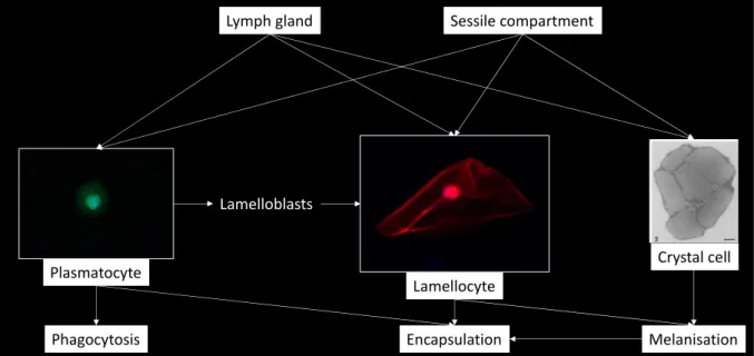
Figure 7: Origin and function of hemocytes in Drosophila melanogaster (in green: plasmatocytes expressing eater-GFP; in red: lamellocytes expressing msnCherry (personal pictures, Axioplan Z1, x400); electronic microscopy picture of crystal cell from (Rizki et al., 1980)
Phagocytosis Encapsulation Melanisation
Lymph gland Sessile compartment
Lamelloblasts
Plasmatocyte
Lamellocyte
They form the primary layer of cells during a response to eliminate bacteria (nodulation (Gandhe
et al., 2007; Dubovskiy et al., 2016)) or to form a cellular "capsule" around parasitic agents such as
nematodes or the egg of a parasitoid too large to be phagocyted (Rizki, 1968; Carton et al., 2008; Bajgar et al., 2015; Dubovskiy et al., 2016). They are involved in wound healing and also synthesize extracellular matrix proteins (Martinek et al., 2008) and antimicrobial peptides (Wang et al., 2014). Plasmatocytes have important functions in the innate cellular immune response, but also in tissue remodelling and homeostasis during embryogenesis. While plasmatocytes are usually considered as a single entity, populations expressing different subsets of markers have been identified (Jung et al., 2005; Honti et al., 2014). Moreover, two plasmatocytes subpopulations with distinct functions in the adult immune response have been identified (Clark et al., 2011).
Crystal cells are non-adhesive round cells of the same size as plasmatocytes and represent 5% of the cells of the hemolymph of healthy larvae (Lemaitre & Hoffmann, 2007). They are named according to the presence of large paracrystalline inclusions in their cytoplasm, which contain prophenol-oxidases (PPO1 and PPO2) which are the inactive form of the enzymes catalysing the melanisation reaction. Upon injury, activation of the JNK pathway and the TNF homolog Eiger leads to crystal cells rupture and release of PPO zymogens into the hemolymph (Bidla et al., 2007). These cells play a major role in clotting, wound healing and antimicrobial defences. The transcription factor Lozenge, expressed only during the development of this cell type (Muratoglu
et al., 2007), constitute a specific marker to identify them.
Lamellocytes are described as large flat (40µm diameter) with adherent cells. Although they are rare in the hemolymph of non-parasitized larvae, their number largely increases at the pre-pupal stage (Holz et al., 2003). They are not observed in the embryo nor the adult stages. During the larval stage, the production and differentiation of lamellocytes can be induced following injury or intrusion of a large foreign body such as a parasitoid egg (Rizki & Rizki, 1992). Lamellocytes form the successive outer layers of the capsule formed around foreign bodies by attaching to the plasmatocyte cells and participate in its melanisation since they contain one specific pro-phenoloxidase (PPO3) (Irving et al., 2005; Dudzic et al., 2015). The lamellocytes origin upon parasitism is still under debate: they can be derived from sessile or circulating plasmatocytes that form intermediate lamelloblasts during the trans-differentiation of into lamellocytes (Honti et al., 2010; Stofanko et al., 2010; Anderl et al., 2016) or release as mature lamellocytes from an accelerated proliferation and bursting of the lymph gland (Lanot et al., 2001). Lamellocytes present several surface markers such as L1/Attila (Kurucz et al., 2007), PS4 (Crozatier et al., 2004) or the integrin myospheroid (Xavier & Williams, 2011).
A B
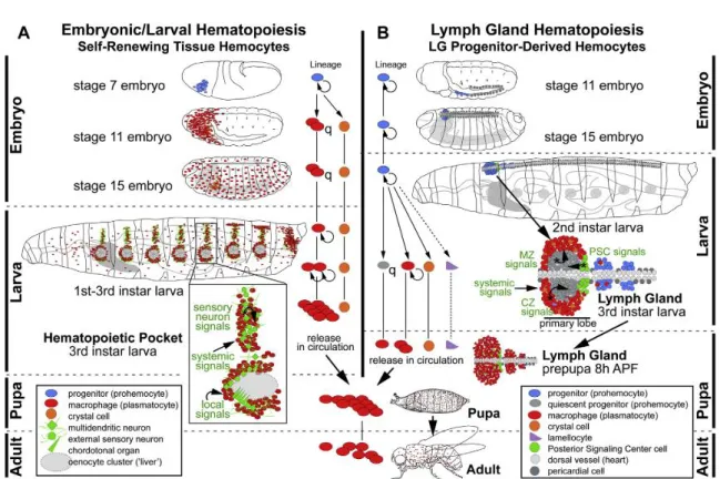
Figure 8: Ontogenesis of blood cell lines and regulation of hematopoiesis in Drosophila. (see text for explanation; from (Gold & Brückner, 2014)).
b) Hematopoesis
The ability to perform genetic manipulations and the conservation of genes and pathways involved with those in mammalian hematopoiesis make D. melanogaster hematopoiesis a powerful model to study the complex biological process involved in the balance between the quiescence of hematopoietic stem cells and their differentiation into lineages. There are many reviews on
Drosophila hematopoiesis (Krzemien et al., 2010; Makhijani & Brückner, 2012; Gold & Brückner,
2014, 2016; Ramond et al., 2015; Hillyer, 2016; El Chamy et al., 2017; Yu et al., 2017) and new findings are published almost every week. The following text will not provide an exhaustive overview of all the work in this field but will highlight the main features of Drosophila hematopoiesis. Until recently in Drosophila, hemocytes were thought to be produced in two successive waves, one during embryogenesis and the other in a specific organ, the lymph gland, during larval stages, with differentiated adult hemocytes derived from embryonic and larval hemocytes (Gold & Brückner, 2014). However, recent studies suggest much higher plasticity, with hematopoietic sites distributed along the larvae and also present in adult flies (Gold & Brückner, 2014; Ghosh et al., 2015).
The Figure 8 from Gold and Bruckner (Gold & Brückner, 2014) summarizes the different stages of embryonic and larval hematopoiesis. In the embryo (panel A), the prohemocytes emerging from the head mesoderm will differentiate into plasmatocytes and crystal cells. At embryonic stage 7, prohemocytic progenitors (in blue) are in the mesoderm of the head. After four cycles of division, these progenitors stop proliferating and differentiate into 600 to 700 plasma cells (in red) and a small number of crystal cells (in orange) that remain grouped around the proventriculus. At stage 11, the differentiated plasmatocytes begin to migrate to the antero-posterior end of the embryo and uniformly colonized the embryo at stage 15, then all cells remain quiescent until the end of embryogenesis. At larval transition, plasmatocytes and embryonic crystal cells persist. Plasmatocytes colonize hematopoietic niches at each larval segment which also contain peripheral sensory neuron clusters (in green) to form the hematopoietic pockets of the sessile compartment. The plasmatocyte Eater protein, a transmembrane receptor of the Nimrod family, is necessary to allow their recruitment as well as that of crystal cells to sessile compartments (Bretscher et al., 2015). Signalling molecule produced by the sensory neurons, such as Activin-b, a TGF-b family ligand, induced the adhesion and proliferation of hemocytes in hematopoietic pockets suggesting strongly that environmental sensory stimuli and neural activity provide another regulatory level (Makhijani
The second wave of hematopoiesis occurs during the larval stages, where two hematopoietic compartments are present: the sessile compartment and the lymph gland (Figure 8; panel B). Lymph gland prohemocytes derive from precursors of the embryo cardiogenic mesoderm (in blue). At that stage, the lymph gland precursors form a single pair of lobes that are localized along the dorsal vessel. This single pair of lobes are designated as the primary lobes (the anterior lobes). At the end of the first larval instar, additional pairs of posterior lobes also emerge along the dorsal vessel, the secondary or posterior lobes. So, in third instar larvae, lymph gland is composed of a large pair of primary anterior lobes organized into three domains: the cortical zone (CZ), the medullary zone (MZ) and the posterior signalling center (PSC), followed by several small pairs of posterior lobes, each separated by a pair of pericardial cells. The posterior lobes are mainly composed of prohemocytes.
In normal condition, the differentiation of lymph gland hemocytes is detected starting from the third larval stage; it develops in a spatiotemporally organized manner. During the third larval stage, the primary lobes of the CZ dilate through proliferation and differentiation of hemocytes into plasmatocytes, a small number of crystal cells and occasionally a few lamellocytes. Then the progenitors present in the MZ become quiescent. As development progresses, almost all hemocytes in the lymph gland differentiate, and 8 hours after puparium formation, all cells in the lymph gland have been released into the circulation (Grigorian et al., 2011). The proliferation and differentiation of hemocytes is controlled by a wide range of signals from the lymphatic gland and systemic sources, such as neurotransmitters and growth factors from the brain, and levels of nutritional compound.
During the larval life, hemocytes in the sessile compartments proliferate and differentiate, so these sites are functional haematopoietic sites (Markus et al., 2009; Makhijani et al., 2011; Leitão & Sucena, 2015). Plasmatocytes proliferate (self-renewal) and also transdifferentiate in crystal cells through a Notch signalling-dependent process (Leitão & Sucena, 2015). They first lose the expression of the differentiation factor Nimrod C1 as well as their phagocytic activity, to acquire the expression of the Lz marker (Leitão & Sucena, 2015). The hemocytes from the sessile compartment can move laterally between the integument and the muscle layer and reach the hemolymph. During normal larval development, sessile hemocytes contribute only gradually to the pool of circulating plasmatocytes and are only released at the beginning of metamorphosis. However, simple stimuli such as light brush strokes are sufficient to induce this release (Makhijani
et al., 2011) indicating that the fate of these sessile hemocytes also depends upon systemic and/or
local signals. Immune challenge, such as Hymenopteran wasp oviposition can cause also their premature mobilization and induce their trans-differentiation into lamellocytes.
Figure 9: Hematopoiesis regulators and hemocyte functions in D. melanogaster from (Williams, 2007). A) At the embryonic level, progenitors in the procephalic mesoderm differentiate into two types of hemocytes (plasmatocytes and crystal cells). B) The lymphatic gland contains many progenitors that differentiate into three types of hemocytes (plasmatocytes, crystal cells and lamellocytes). Many factors and signalling pathways regulating the engagement of these hematopoietic lines in these two compartments have been identified (see text).
c) Factors controlling hematopoiesis
Hematopoietic progenitor maintenance, hemocyte differentiation and the overall homeostasis of the hematopoietic system are finely tuned by intrinsic factors and by environmental stimuli. During embryogenesis, the transcription factor GATA Serpent (srp) (Evans et al., 2014; Spahn et
al., 2014; Shlyakhover et al., 2018) (Figure 9) is required for the specification of the hemocellular
primordium in the head mesoderm at the early embryonic stage and, later, for gene expression during hemocyte maturation. Similarly, the first cells of the lymphatic gland, begin to express Srp well before other differentiation or maturation markers can be detected. The activity of Srp is modulated by the recruitment of Friend Of GATA (FOG), U-Shaped and Lozenge (Lz) co-factors (Figure 9). Srp is necessary for the formation of plasmatocytes and crystal cells populations (Petersen et al., 1999; Waltzer et al., 2003; Muratoglu et al., 2007; Shlyakhover et al., 2018). The transcription factors "missing glial cells" Gcm and Gcm2 are expressed in all prohemocytes and their inhibition in some cells leads to the expression of the transcription factor Lz which induces them to become crystal cells. The continuous expression of Gcm/Gcm2 leads others to transform into plasmatocytes (Waltzer et al., 2002). The binding of the Serrate ligand to the Notch receptor is critical in regulating Lz expression and in specifying crystal cell precursors (Lebestky et al., 2003). In the lymph gland PSC, high levels of Reactive Oxygen Species (ROS) (Owusu-Ansah & Banerjee, 2009), activation of the Wingless signalling pathway (Sinenko et al., 2009) and expression of the EBF transcription factor Collier (Benmimoun et al., 2015; Oyallon et al., 2016) are required for the maintenance of a pool of pluripotent hematopoietic progenitors. The PSC also secretes diffusible signals such as hedgehog (Hh) and the platelet-derived growth factor/vascular endothelial growth factor-like factor (PVF1) to activate different pathways in the lymph gland compartments (Mondal et al., 2011; Tokusumi et al., 2012, 2018; Letourneau et al., 2016). Hh acts directly on the progenitors of the MZ to maintain them from their pluripotent state. PVF1 acts on differentiating hemocytes, stimulating the secretion of adenosine deaminase-related growth factor-A (factor-ADGF-factor-A), which leads to the inactivation of the adenosine/adenosine receptor (factor-AdoR) signalling pathway in MZ cells by modulating the extracellular adenosine level (Lazzaro, 2015). This double control allows the balance between progenitors and differentiated cells to be maintained. Other regulatory pathways are important for PSC cell proliferation such as the decapentaplegic (Dpp) and the wingless (Wnt) pathway. A balance between the signalling levels Wnt and Dpp determines the number of PSC cells. The Insulin/IGF (IlS) and Target of Rapamycin (TOR) signalling pathways, components of the nutrient detection system, also appear to be important for triggering progenitor proliferation (Benmimoun et al., 2012; Shim et al., 2015).
In addition, prohemocyte fate is controlled by local signals from the neighbouring heart tube and through the regulation of PSC morphology (Morin-Poulard et al., 2016). In L3 larvae, PSC cells act to maintain JAK/STAT signalling activity in prohemocytes, thus preserving their multipotency necessary for these cells to adopt a lamellocyte fate in response to parasitism. When pathways such as JAK/STAT or Toll are constitutively activated, for example by expressing active forms of receptors in hemocytes, there are few sessile hemocytes and the majority of blood cells are circulating (Luo et al., 1995; Benmimoun et al., 2012; Shim et al., 2015). The Ras/Raf/MAPK/Toll and Notch signalling pathways are also involved in the proliferation and differentiation of hemocytes (Asha et al., 2003; Valanne et al., 2011; Ferguson & Martinez-Agosto, 2014; Schmid et
al., 2014; Reimels & Pfleger, 2015; Hao & Jin, 2017).
Immune cells produced during the embryonic and larval stages are found in adults and, recently, progenitor cells have been shown to form groups of subcuticular cells (hematopoietic nodes) that can differentiate into crystal and plasmocytic cells (for review(Ramond et al., 2015)).
d) Melanisation
Melanisation covers complex enzymatic and spontaneous reactions that lead to the formation and deposit of black melanin that forms the scar during wound healing and terminates the encapsulation of a large invader. Phenoloxidase (PO) catalyses the first reaction steps by forming indole groups from tyrosine which are then polymerized to form melanin. Enzymatic and spontaneous reactions in turn produce a set of intermediate products such as quinones, diphenols, superoxide, hydrogen peroxide and reactive nitrogen intermediates (Nappi & Christensen, 2005). These reactive species produced during the reaction contributes also to the killing of the aggressors (Nappi & Christensen, 2005; Hillyer, 2016). PO is synthesized intracellularly as an inactive pro-enzyme form, the prophenoloxidase (PPO). The Drosophila genome encodes for three PPOs: PPO1 and PPO2 that are produced in crystal cells (Binggeli et al., 2014) and PPO3 specifically expressed in lamellocytes (Irving et al., 2005; Dudzic et al., 2015). The activation of phenoloxidases is normally tightly controlled to limit the reaction at the lesion site and prevent systemic melanisation. PPO1 and PPO2 require a proteolytic cleavage of their pro-domain to be activated (Chen et al., 2012). The cleavage of PPO1 is mediated by a clip-domain protease serine called Hayan (Nam et al., 2012). Hayan also exists as an inactive zymogen which is itself stimulated by a stepwise process involving other serine proteases, that activities are controlled by serine protease inhibitors (Tang et al., 2006).
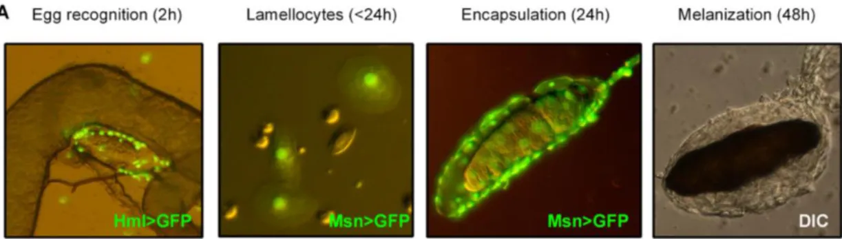
Figure 10: Formation of the multilayer cellular capsule around a parasitoid egg (from Bajgar et al, 2015). The wasp egg is recognized by plasmatocytes (green, Hml>GFP) within 2 h. Lamellocytes, labeled by the Msn>GFP marker appear in circulation less than 24h post-parasitism and are detected on the parasitoid egg 24h after parasitism. Melanisation is observed 48h after the parasitoid egg injection.
The activation of this proteases cascade in response to microbial and fungal attacks can be attributed to PGLP-LC and PGRP-LE expressed by hemocytes and fat body cells (Schmidt et al., 2009). PPO3 in contrast is capable of self-activation without proteolysis (Chen et al., 2012) but it is present only under parasitic conditions and involved in the melanisation of the cell capsule formed around the invader, suggesting a limited diffusion and a short period of activity.
e) Encapsulation
Encapsulation is an important defence mechanism for insects against parasites and other large foreign bodies. It makes it possible to limit the immune response around the intruder and destroy it while avoiding a possible negative effect of the systemic response on the host itself. Encapsulation refers to multiple hemocytes binding to larger invaders, like protozoans, nematodes and parasitoids (eggs and larvae), that cannot be phagocytized by a single cell. The binding of multiple hemocytes to aggregations of bacteria, fungi and protozoans is also sometimes called nodulation (Dubovskiy et al., 2016).
The encapsulation of the parasitoid wasp Leptopilina boulardi egg by the larvae of D. yakuba and
D. melanogaster was described by Russo et al., (1996). It involves three key steps with coordinated
actions of both plasmatocytes and lamellocytes. Oviposition by Leptopilina wasps occurs mainly in second instar larvae. After perforation of the cuticle and basement membrane by the ovipositor, the egg is usually deposited at the abdominal level by the wasp where it floats freely in the hemocoele. Within minutes, it will be recognized as "non-self" by the larva's immune system, which will trigger the various mechanisms leading to encapsulation. The breaching of the basement membrane and the subjacent epithelium releases factors that certainly play a role in the triggerring of the response. The egg is recognized by circulating plasmatocytes which form the first basal cellular layer around the parasitoid egg chorion (Figure 10). This recognition also leads to the increase of the number of circulating hemocytes, mainly plasmatocytes and lamellocytes. These hemocytes may be liberated from an accelerated differentiation and bursting of the lymph gland (Lanot et al., 2001) or mobilized from the sessile compartments and by transdifferentiation of plasmatocytes in lamellocytes (Markus et al., 2009; Honti et al., 2010; Anderl et al., 2016). Indeed, a novel population of infection-induced cells, named lamelloblasts, derived from plasmatocytes, appears in the circulation few hours after parasitism. Lamelloblasts proliferate vigorously and develop into lamellocytes (Anderl et al., 2016). Plasmatocytes transdifferentiation into lamellocyte-like cells was also detected directly on the wasp egg (Anderl et al., 2016). Different signalling pathways are important for lamellocytes proliferation upon parasitism (Zettervall et al., 2004). The
transdifferentiation of plasmatocytes into lamellocytes seems to implicate the Charlatan (Chl) protein, a transcription factor that interacts with CoREST and it is also induced following activation of the JAK / STAT pathway (Stofanko et al., 2010). In accordance with this, genes involved in Toll, Jak-Stat and PO pathways are upregulated 12 hours after L. boulardi wasp infection (Schlenke et al., 2007). In addition, mutations in the Toll and Jak-Stat pathways affect hemocyte counts, lamellocyte differentiation, and the rate of encapsulation of parasitoid eggs (Sorrentino et al., 2004). As the Toll pathway is implicated in both PO (Ligoxygakis et al., 2002) and JAK/STAT (Lagueux et al., 2000) pathways, it might be a central regulator of the response to the parasitism. Edin (elevated during infection) expression is induced after parasitism in the fat body and is required for the encapsulation response trough mobilization of sessile hemocytes leading to an increase in the number of plasmatocytes (Vanha-Aho et al., 2015). The use of Drosophila mutant for the Rac1 GTPase also showed the role of this protein and the involvement of the Jun N-terminal Kinase Basket (Bsk), as well as the stabilization of actin filaments in the recruitment of the population of sessile hemocytes (Williams et al., 2006). This stabilization of actin is necessary for Rac1-induced hemocyte activation by lowering expression of Cofiline (encoded by the twinstar (tsr) gene). Elimination of Bsk by RNAi in hemocytes suppresses the Rac1-induced sessile hemocyte release and the induction of Rac 1 in lamellocytes. Rac1 may thus act on Bsk activity and stable actin formation for cellular immune activation, leading to the release of sessile hemocytes and increasing their number in circulation. The first visible event in encapsulation is the deposition of a dense layer of unknown material on the chorion of the egg six hours after infection. This layer could consist of extracellular matrix deposited by the first plasma cells. Indeed, in mutant Drosophila larvae without Laminin A, an essential component of the extracellular matrix, plasmatocytes are not able to adhere to eggs and there is no encapsulation (Howell et al., 2012). The following plasmatocytes spread on the surface of this layer by emitting filipodia that come into contact, the cells forming tight junctions between them to surround the egg (Williams, 2009). The migration, spreading and formation of tight junctions between plasmatocytes involve the Rho GTPase Rac2 (Williams et al., 2005) and certainly the Rho nucleotide exchange factor protein (RhoGEF) Zizimin-related (Zir) that interacts with Rac2 and CDC42 (Zir KO gives a phenotype very similar to the Rac2 mutant).
After 24h the parasitoid egg is already embedded and at 48h the capsule is melanised (Figure 10). PPO3 is the most important PPO contributing to this melanisation, certainly released upon the lysis of lamellocytes proximal to the egg (Russo et al., 1996; Dudzic et al., 2015). Interestingly, PPO2 is also necessary to achieve the melanisation of the capsule (Dudzic et al., 2015).
Figure 11: D. melanogaster and L. boulardi life cycle (adapted from Carolina Biological Supply Company)