UNIVERSITY MOHAMMED V-RABAT
FACULTY OF MEDICINE AND
PHARMACY RABAT
YEAR: 2020
THESIS N°:
313
MALIGNANT TEMPORAL LOBE GLIOMA
THESIS
Publicly presented in: ………
BY
Mr. TALOUNI AYMENE
Born in 26/08/1994
Royal school of medical military service - Rabat
To obtain the medical doctorate degree
Key words : Malignant gliomas, temporal lobe, surgery, radiotherapy.
JURY
Mr Miloud GAZZAZ
PRESIDENT
Professor of neurosurgery
Mr
CHERIF EL ASRI AbadPROTRACTOR
Professor of neurosurgery
Mr
AdylMELHAOUIProfessor of neurosurgery
Mr Hassan Ennouali
Professor of radiology
Mr
HassanSIFATProfessor of radiotherapy
JUDGES
UNIVERSITY MOHAMMED V
FACULTY OF MEDICINE AND PHARMACY RABAT
HONORQRY DEANS :
1962 – 1969: Professor Abdelmalek FARAJ
1969 – 1974: Professor Abdellatif BERBICH
1974 – 1981: Professor Bachir LAZRAK 1981 – 1989: Professor Taieb CHKILI 1989 – 1997: Professor Mohamed Tahar ALAOUI 1997 – 2003: Professor Abdelmajid BELMAHI 2003 - 2013: Professor Najia HAJJAJ – HASSOUNI
ADMINISTRATION :
Dean Professor Mohamed ADNAOUI
Vice-Dean in charge of academic and student affairs
Professor Brahim LEKEHAL Vice-Dean in charge of research and cooperation
Professor Toufiq DAKKA Vice-Dean in charge of pharmaceutical affairs
Professor Younes RAHALI General secretary
1 - ENSEIGNANTS-CHERCHEURS MEDECINS ET PHARMACIENS PROFESSEURS DE L’ENSEIGNEMENT SUPERIEUR :
Décembre 1984
Pr. MAAOUNI Abdelaziz Médecine Interne – Clinique Royale Pr. MAAZOUZI Ahmed Wajdi Anesthésie -Réanimation
Pr. SETTAF Abdellatif Pathologie Chirurgicale Décembre 1989
Pr. ADNAOUI Mohamed Médecine Interne –Doyen de la FMPR
Pr. OUAZZANI Taïbi Mohamed Réda Neurologie Janvier et Novembre 1990
Pr. KHARBACH Aîcha Gynécologie -Obstétrique
Pr. TAZI Saoud Anas Anesthésie Réanimation
Février Avril Juillet et Décembre 1991
Pr. AZZOUZI Abderrahim Anesthésie Réanimation- Doyen de FMPO
Pr. BAYAHIA Rabéa Néphrologie
Pr. BELKOUCHI Abdelkader Chirurgie Générale Pr. BENCHEKROUN Belabbes Abdellatif Chirurgie Générale
Pr. BENSOUDA Yahia Pharmacie galénique
Pr. BERRAHO Amina Ophtalmologie
Pr. BEZAD Rachid Gynécologie Obstétrique
Méd. Chef Maternité des Orangers
Pr. CHERRAH Yahia Pharmacologie
Pr. CHOKAIRI Omar Histologie Embryologie
Pr. KHATTAB Mohamed Pédiatrie
Pr. SOULAYMANI Rachida Pharmacologie- Dir. du Centre National PV Rabat
Pr. TAOUFIK Jamal Chimie thérapeutique
Décembre 1992
Pr. AHALLAT Mohamed Chirurgie Générale Doyen de FMPT
Pr. BENSOUDA Adil Anesthésie Réanimation
Pr. CHAHED OUAZZANI Laaziza Gastro-Entérologie
Pr. CHRAIBI Chafiq Gynécologie Obstétrique
Pr. EL OUAHABI Abdessamad Neurochirurgie
Pr. FELLAT Rokaya Cardiologie
Pr. JIDDANE Mohamed Anatomie
Pr. ZOUHDI Mimoun Microbiologie Mars 1994
Pr. BENJAAFAR Noureddine Radiothérapie
Pr. BEN RAIS Nozha Biophysique
Pr. CAOUI Malika Biophysique
Pr. CHRAIBI Abdelmjid Endocrinologie et Maladies Métaboliques Doyen de la FMPA
Pr. EL AMRANI Sabah Gynécologie Obstétrique
Pr. ERROUGANI Abdelkader Chirurgie Générale – Directeur du CHIS
Pr. ESSAKALI Malika Immunologie
Pr. ETTAYEBI Fouad Chirurgie Pédiatrique
Pr. IFRINE Lahssan Chirurgie Générale
Pr. RHRAB Brahim Gynécologie –Obstétrique
Pr. SENOUCI Karima Dermatologie
Mars 1994
Pr. ABBAR Mohamed* Urologie Inspecteur du SSM
Pr. BENTAHILA Abdelali Pédiatrie
Pr. BERRADA Mohamed Saleh Traumatologie – Orthopédie Pr. CHERKAOUI Lalla Ouafae Ophtalmologie
Pr. LAKHDAR Amina Gynécologie Obstétrique
Pr. MOUANE Nezha Pédiatrie
Mars 1995
Pr. ABOUQUAL Redouane Réanimation Médicale
Pr. AMRAOUI Mohamed Chirurgie Générale
Pr. BAIDADA Abdelaziz Gynécologie Obstétrique
Pr. BARGACH Samir Gynécologie Obstétrique
Pr. EL MESNAOUI Abbes Chirurgie Générale
Pr. ESSAKALI HOUSSYNI Leila Oto-Rhino-Laryngologie Pr. IBEN ATTYA ANDALOUSSI Ahmed Urologie
Pr. OUAZZANI CHAHDI Bahia Ophtalmologie
Pr. SEFIANI Abdelaziz Génétique
Pr. ZEGGWAGH Amine Ali Réanimation Médicale
Décembre 1996
Pr. BELKACEM Rachid Chirurgie Pédiatrie
Pr. BOULANOUAR Abdelkrim Ophtalmologie
Pr. EL ALAMI EL FARICHA EL Hassan Chirurgie Générale
Pr. OUZEDDOUN Naima Néphrologie
Pr. ZBIR EL Mehdi* Cardiologie Directeur HMI Mohammed V
Novembre 1997
Pr. ALAMI Mohamed Hassan Gynécologie-Obstétrique
Pr. BIROUK Nazha Neurologie
Pr. FELLAT Nadia Cardiologie
Pr. KADDOURI Noureddine Chirurgie Pédiatrique
Pr. KOUTANI Abdellatif Urologie
Pr. LAHLOU Mohamed Khalid Chirurgie Générale
Pr. MAHRAOUI CHAFIQ Pédiatrie
Pr. TOUFIQ Jallal Psychiatrie Directeur Hôp.Ar-razi Salé Pr. YOUSFI MALKI Mounia Gynécologie Obstétrique
Novembre 1998
Pr. BENOMAR ALI Neurologie Doyen de la FMP Abulcassis
Pr. BOUGTAB Abdesslam Chirurgie Générale
Pr. ER RIHANI Hassan Oncologie Médicale
Pr. BENKIRANE Majid* Hématologie
Janvier 2000
Pr. ABID Ahmed* Pneumo-phtisiologie
Pr. AIT OUAMAR Hassan Pédiatrie
Pr. BENJELLOUN Dakhama Badr.Sououd Pédiatrie
Pr. BOURKADI Jamal-Eddine Pneumo-phtisiologie Directeur Hôp. My Youssef Pr. CHARIF CHEFCHAOUNI Al Montacer Chirurgie Générale
Pr. ECHARRAB El Mahjoub Chirurgie Générale
Pr. EL FTOUH Mustapha Pneumo-phtisiologie
Pr. EL MOSTARCHID Brahim* Neurochirurgie
Pr. TACHINANTE Rajae Anesthésie-Réanimation
Pr. TAZI MEZALEK Zoubida Médecine Interne Novembre 2000
Pr. AIDI Saadia Neurologie
Pr. AJANA Fatima Zohra Gastro-Entérologie
Pr. BENAMR Said Chirurgie Générale
Pr. CHERTI Mohammed Cardiologie
Pr. ECH-CHERIF EL KETTANI Selma Anesthésie-Réanimation
Pr. EL HASSANI Amine Pédiatrie - Directeur Hôp.Cheikh Zaid
Pr. EL KHADER Khalid Urologie
Pr. MDAGHRI ALAOUI Asmae Pédiatrie Décembre 2001
Pr. BALKHI Hicham* Anesthésie-Réanimation
Pr. BENABDELJLIL Maria Neurologie
Pr. BENAMAR Loubna Néphrologie
Pr. BENAMOR Jouda Pneumo-phtisiologie
Pr. BENELBARHDADI Imane Gastro-Entérologie
Pr. BENNANI Rajae Cardiologie
Pr. BENOUACHANE Thami Pédiatrie
Pr. BEZZA Ahmed* Rhumatologie
Pr. BOUCHIKHI IDRISSI Med Larbi Anatomie
Pr. BOUMDIN El Hassane* Radiologie
Pr. CHAT Latifa Radiologie
Pr. DAALI Mustapha* Chirurgie Générale
Pr. EL HIJRI Ahmed Anesthésie-Réanimation
Pr. EL MAAQILI Moulay Rachid Neuro-Chirurgie
Pr. EL MADHI Tarik Chirurgie-Pédiatrique
Pr. EL OUNANI Mohamed Chirurgie Générale
Pr. ETTAIR Said Pédiatrie - Directeur Hôp. Univ. Cheikh Khalifa
Pr. GAZZAZ Miloudi* Neuro-Chirurgie
Pr. HRORA Abdelmalek Chirurgie Générale Directeur Hôpital Ibn Sina
Pr. KABIRI EL Hassane* Chirurgie Thoracique
Pr. LAMRANI Moulay Omar Traumatologie Orthopédie
Pr. LEKEHAL Brahim Chirurgie Vasculaire Périphérique
V-D chargé Aff Acad. Est.
Pr. MEDARHRI Jalil Chirurgie Générale
Pr. MIKDAME Mohammed* Hématologie Clinique
Pr. MOHSINE Raouf Chirurgie Générale
Pr. NOUINI Yassine Urologie
Pr. SABBAH Farid Chirurgie Générale
Pr. SEFIANI Yasser Chirurgie Vasculaire Périphérique Pr. TAOUFIQ BENCHEKROUN Soumia Pédiatrie
Décembre 2002
Pr. AL BOUZIDI Abderrahmane* Anatomie Pathologique
Pr. AMEUR Ahmed * Urologie
Pr. AMRI Rachida Cardiologie
Pr. AOURARH Aziz* Gastro-Entérologie Dir.-Adj. HMI Mohammed V
Pr. BAMOU Youssef * Biochimie-Chimie
Pr. BENZEKRI Laila Dermatologie
Pr. BENZZOUBEIR Nadia Gastro-Entérologie
Pr. BERNOUSSI Zakiya Anatomie Pathologique
Pr. CHOHO Abdelkrim * Chirurgie Générale
Pr. CHKIRATE Bouchra Pédiatrie
Pr. EL ALAMI EL Fellous Sidi Zouhair Chirurgie Pédiatrique
Pr. EL HAOURI Mohamed * Dermatologie
Pr. FILALI ADIB Abdelhai Gynécologie Obstétrique
Pr. HAJJI Zakia Ophtalmologie
Pr. JAAFAR Abdeloihab* Traumatologie Orthopédie
Pr. KRIOUILE Yamina Pédiatrie
Pr. MOUSSAOUI RAHALI Driss* Gynécologie Obstétrique
Pr. OUJILAL Abdelilah Oto-Rhino-Laryngologie
Pr. RAISS Mohamed Chirurgie Générale
Pr. SIAH Samir * Anesthésie Réanimation
Pr. THIMOU Amal Pédiatrie
Pr. ZENTAR Aziz* Chirurgie Générale
Janvier 2004
Pr. ABDELLAH El Hassan Ophtalmologie
Pr. AMRANI Mariam Anatomie Pathologique
Pr. BENBOUZID Mohammed Anas Oto-Rhino-Laryngologie
Pr. BENKIRANE Ahmed* Gastro-Entérologie
Pr. BOULAADAS Malik Stomatologie et Chirurgie Maxillo-faciale
Pr. BOURAZZA Ahmed* Neurologie
Pr. CHAGAR Belkacem* Traumatologie Orthopédie
Pr. CHERRADI Nadia Anatomie Pathologique
Pr. EL FENNI Jamal* Radiologie
Pr. EL HANCHI ZAKI Gynécologie Obstétrique
Pr. EL KHORASSANI Mohamed Pédiatrie
Pr. HACHI Hafid Chirurgie Générale
Pr. JABOUIRIK Fatima Pédiatrie
Pr. KHARMAZ Mohamed Traumatologie Orthopédie
Pr. MOUGHIL Said Chirurgie Cardio-Vasculaire
Pr. OUBAAZ Abdelbarre * Ophtalmologie
Pr. TARIB Abdelilah* Pharmacie Clinique
Pr. TIJAMI Fouad Chirurgie Générale
Pr. ZARZUR Jamila Cardiologie
Janvier 2005
Pr. ALLALI Fadoua Rhumatologie
Pr. AMAZOUZI Abdellah Ophtalmologie
Pr. BAHIRI Rachid Rhumatologie Directeur Hôp. Al Ayachi Salé
Pr. BARKAT Amina Pédiatrie
Pr. BENYASS Aatif Cardiologie
Pr. DOUDOUH Abderrahim* Biophysique
Pr. HAJJI Leila Cardiologie (mise en disponibilité)
Pr. HESSISSEN Leila Pédiatrie
Pr. JIDAL Mohamed* Radiologie
Pr. LAAROUSSI Mohamed Chirurgie Cardio-vasculaire
Pr. LYAGOUBI Mohammed Parasitologie
Pr. SBIHI Souad Histo-Embryologie Cytogénétique
Pr. ZERAIDI Najia Gynécologie Obstétrique
AVRIL 2006
Pr. ACHEMLAL Lahsen* Rhumatologie
Pr. BELMEKKI Abdelkader* Hématologie
Pr. BENCHEIKH Razika O.R.L
Pr. BIYI Abdelhamid* Biophysique
Pr. BOUHAFS Mohamed El Amine Chirurgie - Pédiatrique
Pr. BOULAHYA Abdellatif* Chirurgie Cardio – Vasculaire. Directeur Hôpital Ibn Sina Marr. Pr. CHENGUETI ANSARI Anas Gynécologie Obstétrique
Pr. DOGHMI Nawal Cardiologie
Pr. FELLAT Ibtissam Cardiologie
Pr. FAROUDY Mamoun Anesthésie Réanimation
Pr. HARMOUCHE Hicham Médecine Interne
Pr. IDRISS LAHLOU Amine* Microbiologie
Pr. JROUNDI Laila Radiologie
Pr. KARMOUNI Tariq Urologie
Pr. KILI Amina Pédiatrie
Pr. KISRA Hassan Psychiatrie
Pr. KISRA Mounir Chirurgie – Pédiatrique
Pr. LAATIRIS Abdelkader* Pharmacie Galénique Pr. LMIMOUNI Badreddine* Parasitologie
Pr. MANSOURI Hamid* Radiothérapie
Pr. OUANASS Abderrazzak Psychiatrie
Pr. SAFI Soumaya* Endocrinologie
Pr. SOUALHI Mouna Pneumo – Phtisiologie
Pr. TELLAL Saida* Biochimie
Octobre 2007
Pr. ABIDI Khalid Réanimation médicale
Pr. ACHACHI Leila Pneumo phtisiologie
Pr. ACHOUR Abdessamad* Chirurgie générale
Pr. AIT HOUSSA Mahdi * Chirurgie cardio vasculaire
Pr. AMHAJJI Larbi * Traumatologie orthopédie
Pr. AOUFI Sarra Parasitologie
Pr. BAITE Abdelouahed * Anesthésie réanimation
Pr. BALOUCH Lhousaine * Biochimie-chimie
Pr. BENZIANE Hamid * Pharmacie clinique
Pr. BOUTIMZINE Nourdine Ophtalmologie
Pr. CHERKAOUI Naoual * Pharmacie galénique
Pr. EHIRCHIOU Abdelkader * Chirurgie générale
Pr. EL BEKKALI Youssef * Chirurgie cardio-vasculaire
Pr. EL ABSI Mohamed Chirurgie générale
Pr. EL MOUSSAOUI Rachid Anesthésie réanimation
Pr. EL OMARI Fatima Psychiatrie
Pr. GHARIB Noureddine Chirurgie plastique et réparatrice
Pr. HADADI Khalid * Radiothérapie
Pr. ICHOU Mohamed * Oncologie médicale
Pr. ISMAILI Nadia Dermatologie
Pr. KEBDANI Tayeb Radiothérapie
Pr. LOUZI Lhoussain * Microbiologie
Pr. MADANI Naoufel Réanimation médicale
Pr. MAHI Mohamed * Radiologie
Pr. MARC Karima Pneumo phtisiologie
Pr. MASRAR Azlarab Hématologie biologique
Pr. MRANI Saad * Virologie
Pr. OUZZIF Ez zohra * Biochimie-chimie
Pr. RABHI Monsef * Médecine interne
Pr. RADOUANE Bouchaib* Radiologie
Pr. SEFFAR Myriame Microbiologie
Pr. SEKHSOKH Yessine * Microbiologie
Pr. SIFAT Hassan * Radiothérapie
Pr. TABERKANET Mustafa * Chirurgie vasculaire périphérique
Pr. TACHFOUTI Samira Ophtalmologie
Pr. TAJDINE Mohammed Tariq* Chirurgie générale
Pr. TANANE Mansour * Traumatologie-orthopédie
Pr. TLIGUI Houssain Parasitologie
Mars 2009
Pr. ABOUZAHIR Ali * Médecine interne
Pr. AGADR Aomar * Pédiatrie
Pr. AIT ALI Abdelmounaim * Chirurgie Générale
Pr. AKHADDAR Ali * Neuro-chirurgie
Pr. ALLALI Nazik Radiologie
Pr. AMINE Bouchra Rhumatologie
Pr. ARKHA Yassir Neuro-chirurgie Directeur Hôp.des Spécialités Pr. BELYAMANI Lahcen * Anesthésie Réanimation
Pr. BJIJOU Younes Anatomie
Pr. BOUHSAIN Sanae * Biochimie-chimie
Pr. BOUI Mohammed * Dermatologie
Pr. BOUNAIM Ahmed * Chirurgie Générale
Pr. BOUSSOUGA Mostapha * Traumatologie-orthopédie
Pr. CHTATA Hassan Toufik * Chirurgie Vasculaire Périphérique
Pr. DOGHMI Kamal * Hématologie clinique
Pr. EL MALKI Hadj Omar Chirurgie Générale
Pr. EL OUENNASS Mostapha* Microbiologie
Pr. ENNIBI Khalid * Médecine interne
Pr. FATHI Khalid Gynécologie obstétrique
Pr. HASSIKOU Hasna * Rhumatologie
Pr. KABBAJ Nawal Gastro-entérologie
Pr. KABIRI Meryem Pédiatrie
Pr. KARBOUBI Lamya Pédiatrie
Pr. LAMSAOURI Jamal * Chimie Thérapeutique
Pr. MARMADE Lahcen Chirurgie Cardio-vasculaire
Pr. MESKINI Toufik Pédiatrie
Pr. MESSAOUDI Nezha * Hématologie biologique
Pr. MSSROURI Rahal Chirurgie Générale
Pr. NASSAR Ittimade Radiologie
Pr. OUKERRAJ Latifa Cardiologie
Pr. RHORFI Ismail Abderrahmani * Pneumo-Phtisiologie Octobre 2010
Pr. ALILOU Mustapha Anesthésie réanimation
Pr. AMEZIANE Taoufiq* Médecine Interne Directeur ERSSM
Pr. BELAGUID Abdelaziz Physiologie
Pr. CHADLI Mariama* Microbiologie
Pr. CHEMSI Mohamed* Médecine Aéronautique
Pr. DARBI Abdellatif* Radiologie
Pr. DENDANE Mohammed Anouar Chirurgie Pédiatrique
Pr. EL HAFIDI Naima Pédiatrie
Pr. EL KHARRAS Abdennasser* Radiologie
Pr. EL MAZOUZ Samir Chirurgie Plastique et Réparatrice
Pr. EL SAYEGH Hachem Urologie
Pr. ERRABIH Ikram Gastro-Entérologie
Pr. LAMALMI Najat Anatomie Pathologique
Pr. MOSADIK Ahlam Anesthésie Réanimation
Pr. MOUJAHID Mountassir* Chirurgie Générale
Pr. NAZIH Mouna* Hématologie
Pr. ZOUAIDIA Fouad Anatomie Pathologique
Decembre 2010
Pr. ZNATI Kaoutar Anatomie Pathologique
Mai 2012
Pr. AMRANI Abdelouahed Chirurgie pédiatrique Pr. ABOUELALAA Khalil * Anesthésie Réanimation Pr. BENCHEBBA Driss * Traumatologie-orthopédie
Pr. DRISSI Mohamed * Anesthésie Réanimation
Pr. EL ALAOUI MHAMDI Mouna Chirurgie Générale Pr. EL OUAZZANI Hanane * Pneumophtisiologie
Pr. ER-RAJI Mounir Chirurgie Pédiatrique
Pr. JAHID Ahmed Anatomie Pathologique
Pr. RAISSOUNI Maha * Cardiologie
Février 2013
Pr. AHID Samir Pharmacologie
Pr. AIT EL CADI Mina Toxicologie
Pr. AMRANI HANCHI Laila Gastro-Entérologie
Pr. AMOR Mourad Anesthésie Réanimation
Pr. AWAB Almahdi Anesthésie Réanimation
Pr. BELAYACHI Jihane Réanimation Médicale
Pr. BELKHADIR Zakaria Houssain Anesthésie Réanimation
Pr. BENCHEKROUN Laila Biochimie-Chimie
Pr. BENKIRANE Souad Hématologie
Pr. BENNANA Ahmed* Informatique Pharmaceutique
Pr. BENSGHIR Mustapha * Anesthésie Réanimation
Pr. BENYAHIA Mohammed * Néphrologie
Pr. BOUABID Ahmed Salim* Traumatologie orthopédie
Pr. BOUTARBOUCH Mahjouba Anatomie
Pr. CHAIB Ali * Cardiologie
Pr. DENDANE Tarek Réanimation Médicale
Pr. DINI Nouzha * Pédiatrie
Pr. ECH-CHERIF EL KETTANI Mohamed Ali Anesthésie Réanimation Pr. ECH-CHERIF EL KETTANI Najwa Radiologie
Pr. ELFATEMI Nizare Neuro-chirurgie
Pr. EL GUERROUJ Hasnae Médecine Nucléaire
Pr. EL HARTI Jaouad Chimie Thérapeutique
Pr. EL JAOUDI Rachid * Toxicologie
Pr. EL KABABRI Maria Pédiatrie
Pr. EL KHANNOUSSI Basma Anatomie Pathologique
Pr. EL KHLOUFI Samir Anatomie
Pr. EL KORAICHI Alae Anesthésie Réanimation
Pr. EN-NOUALI Hassane * Radiologie
Pr. ERRGUIG Laila Physiologie
Pr. FIKRI Meryem Radiologie
Pr. GHFIR Imade Médecine Nucléaire
Pr. IMANE Zineb Pédiatrie
Pr. IRAQI Hind Endocrinologie et maladies métaboliques
Pr. KABBAJ Hakima Microbiologie
Pr. KADIRI Mohamed * Psychiatrie
Pr. LATIB Rachida Radiologie
Pr. MAAMAR Mouna Fatima Zahra Médecine Interne
Pr. MEDDAH Bouchra Pharmacologie
Pr. MELHAOUI Adyl Neuro-chirurgie
Pr. MRABTI Hind Oncologie Médicale
Pr. NEJJARI Rachid Pharmacognosie
Pr. OUBEJJA Houda Chirugie Pédiatrique
Pr. OUKABLI Mohamed * Anatomie Pathologique
Pr. RAHALI Younes Pharmacie Galénique Vice-Doyen à la Pharmacie
Pr. RATBI Ilham Génétique
Pr. RAHMANI Mounia Neurologie
Pr. REDA Karim * Ophtalmologie
Pr. REGRAGUI Wafa Neurologie
Pr. RKAIN Hanan Physiologie
Pr. ROSTOM Samira Rhumatologie
Pr. ROUAS Lamiaa Anatomie Pathologique
Pr. ROUIBAA Fedoua * Gastro-Entérologie
Pr. SAYAH Rochde Chirurgie Cardio-Vasculaire
Pr. SEDDIK Hassan * Gastro-Entérologie
Pr. ZERHOUNI Hicham Chirurgie Pédiatrique
Pr. ZINE Ali * Traumatologie Orthopédie
AVRIL 2013
Pr. EL KHATIB MOHAMED KARIM * Stomatologie et Chirurgie Maxillo-faciale MARS 2014
Pr. ACHIR Abdellah Chirurgie Thoracique
Pr. BENCHAKROUN Mohammed * Traumatologie- Orthopédie
Pr. BOUCHIKH Mohammed Chirurgie Thoracique
Pr. EL KABBAJ Driss * Néphrologie
Pr. EL MACHTANI IDRISSI Samira * Biochimie-Chimie
Pr. HARDIZI Houyam Histologie- Embryologie-Cytogénétique
Pr. HASSANI Amale * Pédiatrie
Pr. HERRAK Laila Pneumologie
Pr. JANANE Abdellah * Urologie
Pr. JEAIDI Anass * Hématologie Biologique
Pr. KOUACH Jaouad* Génycologie-Obstétrique
Pr. LEMNOUER Abdelhay* Microbiologie
Pr. MAKRAM Sanaa * Pharmacologie
Pr. OULAHYANE Rachid* Chirurgie Pédiatrique
Pr. RHISSASSI Mohamed Jaafar CCV
Pr. SEKKACH Youssef* Médecine Interne
Pr. TAZI MOUKHA Zakia Génécologie-Obstétrique
DECEMBRE 2014
Pr. ABILKACEM Rachid* Pédiatrie
Pr. AIT BOUGHIMA Fadila Médecine Légale
Pr. BEKKALI Hicham * Anesthésie-Réanimation
Pr. BENAZZOU Salma Chirurgie Maxillo-Faciale
Pr. BOUABDELLAH Mounya Biochimie-Chimie
Pr. BOUCHRIK Mourad* Parasitologie
Pr. DERRAJI Soufiane* Pharmacie Clinique
Pr. DOBLALI Taoufik Microbiologie
Pr. EL AYOUBI EL IDRISSI Ali Anatomie
Pr. EL GHADBANE Abdedaim Hatim* Anesthésie-Réanimation
Pr. EL MARJANY Mohammed* Radiothérapie
Pr. FEJJAL Nawfal Chirurgie Réparatrice et Plastique
Pr. LAKHAL Zouhair* Cardiologie
Pr. OUDGHIRI NEZHA Anesthésie-Réanimation
Pr. RAMI Mohamed Chirurgie Pédiatrique
Pr. SABIR Maria Psychiatrie
Pr. SBAI IDRISSI Karim* Médecine préventive, santé publique et Hyg. AOUT 2015
Pr. MEZIANE Meryem Dermatologie
Pr. TAHIRI Latifa Rhumatologie
PROFESSEURS AGREGES : JANVIER 2016
Pr. BENKABBOU Amine Chirurgie Générale
Pr. EL ASRI Fouad* Ophtalmologie
Pr. ERRAMI Noureddine* O.R.L
Pr. NITASSI Sophia O.R.L
JUIN 2017
Pr. ABBI Rachid* Microbiologie
Pr. ASFALOU Ilyasse* Cardiologie
Pr. BOUAYTI El Arbi* Médecine préventive, santé publique et Hyg.
Pr. BOUTAYEB Saber Oncologie Médicale
Pr. EL GHISSASSI Ibrahim Oncologie Médicale
Pr. HAFIDI Jawad Anatomie
Pr. OURAINI Saloua* O.R.L
Pr. RAZINE Rachid Médecine préventive, santé publique et Hyg.
Pr. ZRARA Abdelhamid* Immunologie
NOVEMBRE 2018
Pr. AMELLAL Mina Anatomie
Pr. SOULY Karim Microbiologie
Pr. TAHRI Rajae Histologie-Embryologie-Cytogénétique
NOVEMBRE 2019
Pr. AATIF Taoufiq * Néphrologie
Pr. ACHBOUK Abdelhafid * Chirurgie Réparatrice et Plastique Pr. ANDALOUSSI SAGHIR Khalid * Radiothérapie
Pr. BABA HABIB Moulay Abdellah * Gynécologie-obstétrique
Pr. BASSIR RIDA ALLAH Anatomie
Pr. BOUFETTAL MONSEF Anatomie
Pr. BOUCHENTOUF Sidi Mohammed * Chirurgie Générale
Pr. BOUZELMAT Hicham * Cardiologie
Pr. BOUKHRIS Jalal * Traumatologie-orthopédie
Pr. CHAFRY Bouchaib * Traumatologie-orthopédie
Pr. CHAHDI Hafsa * Anatolmie Pathologique
Pr. CHERIF EL ASRI Abad * Neurochirugie
Pr. DAMIRI Amal * Anatolmie Pathologique
Pr. DOGHMI Nawfal * Anesthésie-réanimation
Pr. ELALAOUI Sidi-Yassir Pharmacie Galénique
Pr. EL ANNAZ Hicham * Virologie
Pr. EL HASSANI Moulay EL Mehdi * Gynécologie-obstétrique Pr. EL HJOUJI Aabderrahman * Chirurgie Générale
Pr. EL KAOUI Hakim * Chirurgie Générale
Pr. EL WALI Abderrahman * Anesthésie-réanimation
Pr. EN-NAFAA Issam * Radiologie
Pr. HAMAMA Jalal * Stomatologie et Chirurgie Maxillo-faciale
Pr. HEMMAOUI Bouchaib * O.R.L
Pr. HJIRA Naoufal * Dermatologie
Pr. JIRA Mohamed * Médecine Interne
Pr. JNIENE Asmaa Physiologie
Pr. LARAQUI Hicham * Chirurgie Générale
Pr. MAHFOUD Tarik * Oncologie Médicale
Pr. MEZIANE Mohammed * Anesthésie-réanimation Pr. MOUTAKI ALLAH Younes * Chirurgie Cardio-vasculaire
Pr. MOUZARI Yassine * Ophtalmologie
Pr. NAOUI Hafida * Parasitologie-Mycologie
Pr. OBTEL Majdouline Médecine préventive, santé publique et Hyg.
Pr. OURRAI Abdelhakim * Pédiatrie
Pr. SAOUAB Rachida * Radiologie
Pr. SBITTI Yassir * Oncologie Médicale
Pr. ZADDOUG Omar * Traumatologie Orthopédie
Pr. ZIDOUH Saad * Anesthésie-réanimation
2 - ENSEIGNANTS-CHERCHEURS SCIENTIFIQUES PROFESSEURS/Prs. HABILITES
Pr. ABOUDRAR Saadia Physiologie
Pr. ALAMI OUHABI Naima Biochimie-chimie
Pr. ALAOUI SLIMANI Lalla Naïma Histologie-Embryologie
Pr. ANSAR M’hammed Chimie Organique et Pharmacie Chimique
Pr .BARKIYOU Malika Histologie-Embryologie
Pr. BOUHOUCHE Ahmed Génétique Humaine
Pr. BOUKLOUZE Abdelaziz Applications Pharmaceutiques Pr. CHAHED OUAZZANI Lalla Chadia Biochimie-chimie
Pr. DAKKA Taoufiq Physiologie
Pr. FAOUZI Moulay El Abbes Pharmacologie
Pr. IBRAHIMI Azeddine Biologie moléculaire/Biotechnologie
Pr. KHANFRI Jamal Eddine Biologie
Pr. OULAD BOUYAHYA IDRISSI Med Chimie Organique
Pr. REDHA Ahlam Chimie
Pr. TOUATI Driss Pharmacognosie
Pr. YAGOUBI Maamar Environnement, Eau et Hygiène
Pr. ZAHIDI Ahmed Pharmacologie
Updated en 11/06/2020 KHALED Abdellah
Head of the human resources department FMPR
To
HIS LATE MAJESTY KING HASSAN II
To
HIS MAJESTY THE KING MOHAMED VI
Supreme Chief and Chief of the state-major
To
HIS ROYAL HIGHNESS
CROWN PRINCE MOULAY EL HASSAN
To
HIS ROYAL HIGHNESS
PRINCE MOULAY RACHID
To
To
Mr. the General of the army-corps
Abdelfatah LOUARAK
General Inspector of the FAR and Commander of the South Zone
As a token of our great respect
Our deepest consideration and sincere admiration
To
Mr. Brigadier General Doctor
Mohammed ABBAR
Professor of Urology.
Inspector of the Health Service of the Royal Armed Forces.
As a token of our great respect,
And our deepest consideration
To
Mr. Doctor Colonel Major El Mehdi ZBIR
Professor of Cardiology Director of HMIMV -Rabat.
As a token of our great respect
And our deepest consideration
To
Mr. Brigadier General Doctor
Abdelatif BOULAHYA
Professor of Cardiovascular Surgery Director of the Avicenne Military
Hospital of Marrakech
As a token of our great respect
And our deepest consideration
To
Mr. Doctor Colonel Major Mohammed EL BAAJ
Professor of Internal Medicine, Director of the HMMI-Meknes.
As a token of our great respect
And our deepest consideration
To
Mr Doctor Colonel AMEZIANE Taoufiq
Professor of Internal Medicine
Director of the R.S.M.M.S
As a token of our great respect
And our deepest consideration
To
Mr. Doctor Colonel Abderrahmane ELMATAR Commander of the
training and instruction group R.S.M.M.S
As a token of our great respect
And our deepest consideration
This work marks the end of an era, and the beginning of a hopefully better and brighter one.
I would like to thank the members of my small family; my mom Tourya, my dad Mostapha, and my little brother Zakaria who have always been there for me, and supported me greatly.
I would also like to thank a special group of my friends; the king of juicing, Rbiai, Sakri, Imzil, Ennaimi, Biyat, Zouhri, Khairi, Bijbij, Anouar, Adil, Salmane, Naoufal, Ilias, the godfather achraf, sgt Burdett, Fanoré, and the list goes on for sharing with me some of my best and worse memories and experiences during these last eight years of medical studies.
I want to also thank all of my teachers and friends from Misbah, Bismillah, and my entire medical class of 2012, and I want to share my deep appreciation and gratitude to Professor Abad sheriff el Assri and professor Miloud Gazzaz for providing me with this wonderful opportunity and support to make this humble peace of work possible.
Special thanks to one of my mightiest rivals of all time, you know who you are, without you I would’ve probably became strong anyways so don’t flatter yourself too much :) but thanks for the little push, it has been a pleasure and an honor.
Abbreviations
LTL: left temporal lobe
RTL: right temporal lobe
TP53: tumor protein 53
NF1/2: neurofibromatosis 1/2
DNA: deoxyribonucleic acid
APC: argon plasma coagulation’
EGFR: epidermal growth factor receptor
TERT: telomerase reverse transcriptase
RTEL1: regulator of telomere length 1
T-reg cells: regulatory T cells
CMV : cytomegalovirus
TPO junction: temporo-parieto-occipital junction
MTG: middle temporal gyrus
STG: superior temporal gyrus
NSPC: neural stem and progenitor cell
GB: glioblastoma
SLF: superior longitudinal fasciculus
ILF: inferior longitudinal fasciculus
GBM: glioblastoma multiform
NADPH: nicotinamide adenine dinucleotide phosphate (reduced form)
MGMT: methylguanine methyltransferase
PCR: polymerase chain reaction
GFAP: glial fibrillary acidic protein
NOS: not otherwise specified
CT: computerized tomography
MRI: magnetic resonance imaging
fMRI: functional magnetic resonance imaging
ADC maps: apparent diffusion coefficient
FA maps: fractional anisotropy
DWI: diffusion-weighted imaging
VEGF: vascular endothelial growth factor
CRF: Corticotropin-releasing factor
DTI: diffusion tensor imaging
IFOF: inferior fronto occipital fasciculus
OD: oligodendroglioma
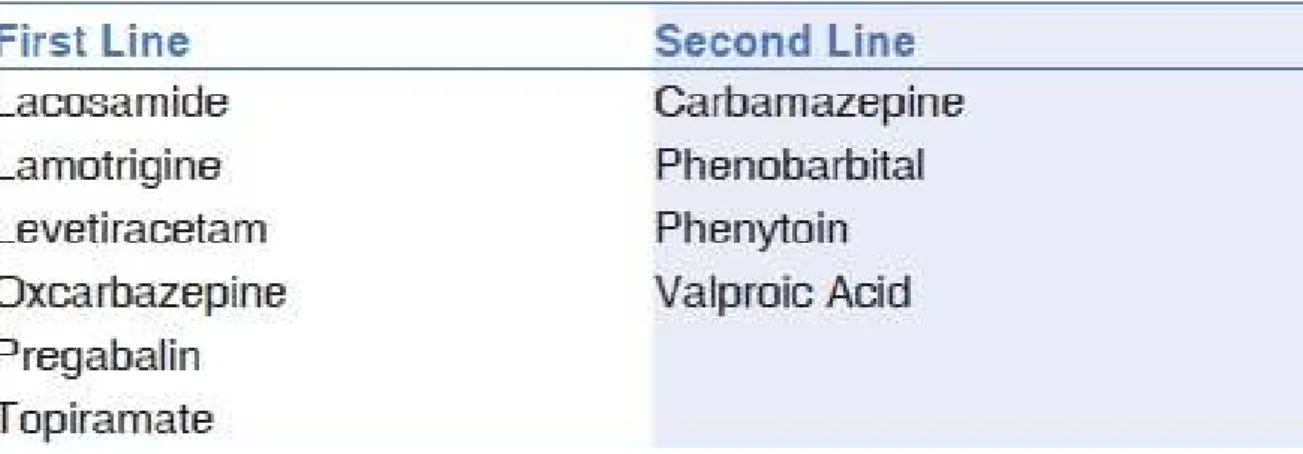
AED: antiepileptic drugs
MdLF: the middle longitudinal fascicle
GPS: global positioning system
iUS: intraoperative ultrasound
iMRI: intraoperative magnetic resonance imaging
PS: performance status
Gy: the gray: unit of absorbed dose of ionizing radiation
RT: radiotherapy
INTRODUCTION ... 1 EPIDEMIOLOGY ... 3 Incidence and prevalence ... 3 Age ... 5 Sex ratio ... 5 Risk factors ... 6 Endogenous Risk Factors ... 6 Exogenous Risk Factors ... 7 Electromagnetic radiation ... 7 ionizing irradiation... 7 Infection with cytomegalovirus ... 7 ANATOMY OF THE TEMPORAL LOBE ... 8 Anatomy of the surface: ... 8 Lateral surface ... 9 Medial surface ... 10 Basal surface ... 14 White Matter ... 16 The superior longitudinal fasciculus ... 16 The uncinate fasciculus ... 16 The inferior longitudinal fasciculus ... 16 The fornix and stria terminalis ... 16 The corpus callosum ... 17 Projection fibers afferent to the temporal cortex ... 17 The thalamocortical projections ... 17 The temporopontine or temporoparietopontine tract ... 17 Important Distinctions in Temporal Lobe Glioma Anatomy ... 19 PHYSIOLOGY OF THE TEMPORAL LOBE ... 20
Visual memories ... 20 Processing sensory input ... 20 Auditory ... 20 Visual ... 20 Language recognition ... 20 new memories... 20 PATHOLOGY ... 22 Pathogenesis and topography ... 22 Macroscopic Features of GBs ... 23 Anatomic classification of Temporal lobe high grade gliomas ... 24 Microscopic features ... 25 Molecular Profiles of Diffuse Astrocytic and Oligodendroglial Tumors ... 25 Isocitrate Dehydrogenase (IDH)Mutations ... 25 Co-deletion of 1p/19q ... 25 MGMT Methylation Status ... 26 Role of Epidermal Growth Factor Receptor (EGFR) Pathway Aberrations ... 26 The new classification of glioma WHO 2016 ... 27 DIAGNOSIS ... 30 Clinical manifestations of temporal lobe gliomas ... 30 Imaging techniques ... 30 Morphological imaging: CT AND MRI ... 30 Metabolic imaging ... 31 Magnetic Resonance Spectroscopy ... 31 Positron Emission Tomography ... 32 Functional imaging in preoperative planning... 32 Diffusion Tractography ... 32 Arcuate Fasciculus ... 33 Cortico-Spinal Tract ... 33 Visual Projections ... 33
Limitations ... 34 Functional MRI Task ... 34 Based fMRI ... 34 Resting State fMRI ... 34 Language fMRI ... 34 Language Localization ... 35 Language Lateralization ... 35 Technical Considerations and limits of the fMRI in high grade glioma ... 35 TREATMENT ... 36 Goals ... 36 Means ... 36 Medical treatment... 36 Intracranial hypertension ... 36 Management of perilesional edema ... 37 Corticosteroids ... 37 Osmotherapy ... 38 Novel treatments ... 38 Management of epilepsy ... 38 Surgical approach of Temporal high-grade gliomas ... 40 Principles of surgery in Temporal Lobe Gliomas ... 40 Preoperative Planning ... 40 Awake or asleep surgery? ... 40 Surgical stages for removing temporal lobe gliomas: the 3Ds Aids ... 41 The “Define” Stage ... 41 The Approach ... 41 Cortical Mapping ... 42 How to Map the Cortex ... 42 How High Do You Set the Current? ... 42 The “Divide” Stage ... 43
Posterior Temporal Division ... 43 Lateral Temporal and Junctional Disconnections ... 45 The “Destroy” Stage ... 47 The en Bloc Anterior Temporal Lobectomy ... 47 The Medial Temporal Structures ... 48 TPO Junction Gliomas ... 50 Fluorescence-Guided Resection of Malignant Gliomas ... 52 Intraoperative imaging: ... 53 Stereotactic Neuronavigation ... 53 Intraoperative Ultrasound (iUS) ... 54 Intraoperative MRI ... 55 The role of intraoperative stimulation mapping ... 55 The Role of Laser-Induced Thermal Therapy in the Management of Malignant Gliomas ... 56 Adjuvant therapy ... 56 Chemoradiotherapy ... 56 Chemotherapy ... 57 Radiation Therapy ... 57 Management at Recurrence ... 59 Disease monitoring ... 59 Role of repeat surgery ... 59 Role of radiotherapy ... 59 Role of chemotherapy ... 60 Guidelines summary ... 60 OUTCOMES AND FOLLOW-UP ... 62 Recurrence ... 62 Mortality ... 62 Age ... 62 WHO grade ... 63 Surgery ... 63
Prognostic value of molecular profiles ... 63 IDH prognosis ... 63 1p/19q prognosis ... 64 MGMT prognosis ... 64 ILLUSTRATIVE CASES ... 65 Case 1 ... 65 Case 2 ... 68 Case 3 ... 71 CONCLUSION ... 73 References ... 75
1
2
Gliomas comprise a heterogeneous group of neoplasms that differ in location within the central nervous system, in age and sex distribution, in growth potential, in extent of invasiveness, in morphological features, in tendency for progression, and in response to treatments. High grade Gliomas are by far the most common glial tumors and it represent over 60% of primary central nervous malignant tumors. In adults, It occur most often in the cerebral hemispheres, with the temporal lobe being the second most commonly affected region right behind the frontal lobe. The temporal lobe glioma is particularly interesting due to its unique accessible location, which may provide more valuable information about risk factors, anatomical relationships and the efficiency of the multidisciplinary treatments and approaches.
Indeed, we focus our study on the temporal lobe tumor because it seems to offer a good ability for extend tumor resection, without impairing a patient’s functionality in the non-dominant side, compared to other central nervous parts.
We discuss the epidemiology, histology and imaging’s advances for central nervous malignant gliomas, and specifically in the temporal lobe location which provide a better understanding of the extent of the lesion as well as its relationship with critical neuroanatomic function. We focus on the evolution of intraoperative imaging, functional brain mapping, and technology and how it participates to identify tumor from brain and how it has significantly improved the ability of surgeons for safer and more aggressive tumor removal.
In this study, we present some illustrative cases of temporal lobe malignant gliomas that were treated in our department. Therefore, we review the more recent literature’s update in managing high grade gliomas in this location in the aim of addressing all the aspects of patient evaluation and care.
This study will be informative for surgeons, oncologists, neurologists, residents, and students who treat these patients and those who are training for a career in managing patients with these challenging tumors in such location.
3
EPIDEMIOLOGY
Incidence and prevalence[1]
The incidence of gliomas has increased worldwide since the late 1970s but it remains relatively low with 5 per 100,000. There are several possible causes for this increase, including improved diagnostic methods, such as modern radiologic imaging, and better access to neurosurgical services. Incidental findings of brain neoplasms increased with the introduction of CT and MRI technology in the 1980s. However, it has also been suggested that the overall increase in incidence is leveling off, whereas the increasing trend continues in the older age groups. There is no major geographical differences in the distribution of new diagnoses of glioblastoma. .
Figure1: Incidence by tumors location for high grade gliomas
A study conducted by Florian and associates in Romania showed that the most frequent locations of the high-grade gliomas were in the cerebral lobes, especially the right and the left frontal lobes, followed by the left temporal for single lobe location. For multiple lobe locations the left fronto-parietal and left temporo-parietal were the most frequent locations[2].
4
histologic type Incidence (/100.000)
Glioblastoma 2.0
Diffuse astrocytoma 0.7
Anaplastic astrocytoma 0.5
Pilocytic astrocytoma 0.3
All other astrocytomas 0.03
Oligodendroglioma 0.5
Mixed glioma 0.5
Table1: Number and incidence of gliomas by histologic type[3]
In a study conducted in Finland by larjavaara and associates; a total of 331 incident gliomas were diagnosed during our study period. The majority were glioblastomas followed by Diffuse and anaplastic astrocytomas, pilocytic astrocytomas and Oligodendroglial tumors.
Temporal lobe region % in LTL % in RTL
Anterior 17.8 20.0
Posterior 30.1 23.3
Medial 37.0 46.7
Multi 15.1 10.0
Table2: comparison of percentage of gliomas between the regions of the right and the left temporal lobe[4].
a study conducted by Noll and associates in New York on 103 patients diagnosed between the years 2001 and 2010 with temporal lobe glioma showed a higher percentage of gliomas in the medial region of both the right and the left temporal lobe followed by the posterior and the anterior regions.
5
Age[1]
Glioblastoma is present at all ages, but the incidence is higher in the 5th and 6th decades of life. Age-adjusted annual incidence rates of glioblastoma peak in patients aged 75–84.
Sex ratio[1]
Males are affected 1.6 times more than females.
Figure2: age-adjusted and age-specific incidence rates for glioblastoma at diagnosis and gender between 2006–2010[5]
A study done in the United States over the course of 4 years (2006-2010) studying incidence rates per 100.000 showed that glioblastomas are interestingly frequent in males over females and concerns mostly the population over the age of 74 in both males and females while being fairly rare amongst younger population.
6
Risk factors
Endogenous Risk Factors[1]
Risk factors contributing to the malignant transformation of these cells remain elusive. Endogenous risk factors for glioblastoma other than age is observed in less than 1% of glioblastomas that arise in patients with hereditary cancer syndromes, like the Li-Fraumeni syndrome usually caused by mutations in the tumor suppressor gene TP53, neurofibromatosis both types I and II caused respectively by mutations in the NF1 and NF2 genes, or the Turcot syndrome resulting from mutations in the DNA repair gene APC.
These Glioblastomas in mostly young patients are usually preceded by the diagnosis of World Health Organization grade II or III.
Families with more than one member affected by glioblastoma is an exceptional situation, thus challenging the searching process for susceptibility gene loci.
Although a risk for glioma has been reported to be doubled in first-degree relatives of affected patients, the familial risk association is extremely low in consideration of the low overall glioma risk. As a result, no high penetrance gene variants linked with glioma risk have been identified among family members.
Population-based genome-wide association studies pinpointed five single nucleotide polymorphism risk alleles for gliomagenesis in the following four genes: TP53, EGFR(Epidermal growth factor receptor), TERT, and RTEL1.
telomerase-associated pathomechanism is associated with gliomagenesis in elderly patients. The low penetrance of risk alleles suggests that the pathophysiology of gliomas follows polygenic patterns and that in there turn follow distinct sequences of evolution.
Atopic disease is associated with reduced risk for gliomas by roughly 40%. The physiologic function of regs is to limit cell responses in order to prevent autoimmunity, and lower reg levels are observed in patients with atopic disease. In comparison, higher frequencies of T-regs decrease survival and contribute to the immunosuppressive microenvironment that stops immunologic antitumor responses.
7
Exogenous Risk Factors
Exogenous risk factors for the development of glioblastomas are very debatable and controversial. No association with exposure to cancerogenic agents or smoking has been reported[1].
Electromagnetic radiation:[6][7][8][9]
A link between gliomas and electromagnetic radiation from cell phones has not been conclusively proven. It was considered possible though several large studies have found no conclusive evidence, as summarized by the NIH's National Cancer Institute review of the topic .
However, further research is still being pursued to obtain more concrete evidence and verify that there is no relationship.
ionizing irradiation:[1]
Risk associations of gliomas with ionizing irradiation have been studied, but data on the more specific causality relationship with glioblastoma have not been reported.
Infection with cytomegalovirus[1][10][11][12]
Viral oncogenesis role for cytomegalovirus in gliomagenesis have been postulated, because some of the CMV gene products interact with glioblastoma core signaling pathways, but experimental evidence to confirm this role for CMV is scarce.
However, most glioblastomas are infected with cytomegalovirus, which speeds the development of tumors.
8
ANATOMY OF THE TEMPORAL LOBE
Anatomy of the surface:
The temporal lobe is one of the six lobes of the brain that largely occupies the middle cranial fossa. it is the second largest lobe, after the larger frontal lobe, accounting 22% of the total neocortical volume. The lobe extends superiorly to the Sylvian fissure, and posteriorly to the lateral parietotemporal line, which separates the temporal lobe from the inferior parietal lobule of the occipital lobe inferiorly and the parietal lobe superiorly. The middle cranial fossa forms its anterior and inferior boundaries[13].
Figure 3: Anatomical landmarks of the cortex of the temporal lobe. Photographs are of the lateral (a), inferior (b), superior (c) surfaces and medial (d) on fixed cadaver[13].
9
Lateral surface[14]
The lateral temporal surface is made of three parallel gyri, the superior, middle, and inferior temporal gyri, divided by two sulci, the inferior and superior temporal sulci. Both the gyri and sulci parallel the sylvian fissure.
The superior temporal gyrus is located between the sylvian fissure and the superior temporal sulcus and is continuous around the lip of the fissure with the transverse temporal gyri, which extend obliquely backwards and medially toward the posterosuperior angle of the insula forming the lower wall of the posterior part of the floor of the sylvian fissure. The middle temporal gyrus is located between the superior and inferior temporal sulci. the ambient, the temporal horn and the crural cisterns are positioned deep to the middle temporal gyrus. The inferior temporal gyrus lies under the inferior temporal sulcus and continues around the inferior border of the hemisphere to form the lateral side of the basal surface. The angular gyrus, a parietal lobe structure, caps the upturned posterior end of the superior temporal sulcus. The inferior temporal gyrus is most of the time made of multiple fragmented gyri and may blend into the middle temporal gyrus without a clear sulcal demarcation.
Figure 4: Lateral view of the left temporal lobe showing boundaries and limits with different gyrus and sulcus.
10
Medial surface[13]
The medial surface of the temporal lobe is very complex. It is formed predominantly by the rounded medial surfaces of the parahippocampal gyrus and uncus. This medial surface is the combination of three longitudinal strips of neural tissue, one located above the other, which are interlocked with the hippocampal formation. The most inferior strip is formed by the rounded medial edge of the parahippocampal gyrus, the site of the subicular zones; the middle strip is formed by the dentate gyrus, located on the medial surface of the hippocampal formation; and the superior strip is formed by the fimbria of the fornix, a white band comprised of the fibers emanating from the hippocampal formation and posteriorly directed into the crus of the fornix. The parahippocampal and dentate gyri are separated by the hippocampal sulcus, and the dentate gyrus and the fimbria are separated by the fimbriodentate sulcus. The amygdala and the hippocampal formation lie just beneath and are so intimately related to the mesial temporal cortex that they are considered in this section. The dentate gyrus blends posteriorly behind the splenium into the fasciolar gyrus, which is continuous with the indusium griseum.
The parahippocampal gyrus deviates medially at the site of the uncus that projects medially above the tentorial edge. The parahippocampal gyrus also extends around the lower border to form the medial part of the basal surface of the temporal lobe, where it is separated from the medially projecting uncus by the rhinal sulcus. Posteriorly, the part of the parahippocampal gyrus below the splenium of the corpus callosum is intersected by the anterior end of the calcarine sulcus, which divides the posterior portion of the parahippocampal gyrus into an upper part that is continuous above and posteriorly with the isthmus of the cingulate gyrus and continuous below and posteriorly with the lingual gyrus.
The uncus, the medially projecting anterior part of the parahippocampal gyrus has an angular shape with anterior and posterior segments that meet at a medially directed apex. The medial face of the anterior segment faces the proximal part of the sylvian, the carotid cistern, and the internal carotid and proximal middle cerebral arteries. The posterior segment faces the cerebral peduncle and, with the peduncle, forms the lateral and medial walls of the crural cistern through which the posterior cerebral, anterior choroidal, and medial posterior choroidal arteries pass.
11
The optic tract passes above the medial edge of the posterior segment in the roof of the crural cistern. The amygdaloid nucleus forms almost all of the interior and comes to the medial surface of the upper part of the anterior segment. The upper part of the posterior segment is formed largely by the medial aspect of the head of the hippocampus.
The apex, where the anterior and posterior segments meet, points medially toward the oculomotor nerve and posterior communicating artery. The head of the hippocampus reaches the medial surface in the upper part of the posterior segment at the anterior end of the dentate gyrus. Within the ventricle, a small medially projecting space, the uncal recess, situated between the ventricular surface of the amygdala and hippocampal head, is located lateral to the uncal apex.
The inferior choroidal point, the lower end of the choroidal fissure along which the choroid plexus is attached, is located just behind the upper edge of the posterior uncal segment, immediately behind the head of the hippocampus, at the site where the anterior choroidal artery passes through the choroidal fissure to enter the temporal horn. The anterior choroidal artery arises near the midlevel of the anterior segment and hugs its surface, sloping gently upward, unless extremely tortuous. It continues to ascend as it courses posteriorly around the uncal apex and reaches the upper part of the posterior segment, where it passes through the fissure at the inferior choroidal point. The dentate gyrus, named for its characteristic tooth-like elevations, extends posteriorly from the upper part of the posterior segment and has the most prominent denticulations anteriorly.
The amygdala can be considered as being entirely located within the boundaries of the uncus. It forms the anterior wall of the temporal horn. Superiorly, the amygdale blends into the claustrum and Globus pallidus without any clear demarcation. The upper posterior portion of the amygdala tilts back above the hippocampal head and the uncal recess to form the anterior portion of the roof of the temporal horn. Medially, it is related to the anterior and posterior segments of the uncus. The amygdala gives rise to the stria terminalis, which courses between the thalamus and caudate nucleus deep to the thalamostriate vein.
The hippocampus, which blends into and forms the upper part of the posterior uncal segment, is a curved elevation, approximately 5 cm long, in the medial part of the entire length of the floor of the temporal horn. It has the dentate gyrus along its medial edge and a curved collection
12
of gray matter in its interior that is referred to as Ammon’s horn. It sits above and is continuous below with the rounded medial surface of the parahippocampal
gyrus referred to as the subicular surface. Ammon’s horn is characterized in transverse sections of the hippocampal formation by its reversed C- or comma-shaped orientation and by its tightly packed pyramidal cell layer.
The hippocampus is divided into three parts: head, body, and tail. The head of the hippocampus, the anterior and largest part, is directed anteriorly and medially, and forms the upper part of the posterior uncal segment. Superiorly, the head of the hippocampus faces the posterior portion of the amygdala. Anterior to the hippocampal head is the uncal recess, a cleft, located between the head of the hippocampus and the amygdala. The body of the hippocampus extends along the medial part of the floor of the temporal horn, narrowing into the tail that disappears as a ventricular structure at the anterior margin of the calcar avis.
The fimbria of the fornix arises on the ventricular surface of the hippocampus behind the head and just behind the choroidal fissure. The temporal horn extends into the medial part of the temporal lobe to just anterior to the hippocampal head and to just behind the amygdala. The temporal horn ends approximately 2.5 cm from the temporal pole. The inferior choroidal point, at the lower end of the choroidal fissure, is located just behind the head of the hippocampus and immediately lateral to the lateral geniculate body.
13
Figure 5: Schematic and cadaveric specimen showing the medial part of the temporal lobe and its relationship to the adjacent structures.
The superior surface, along with the insula, was exposed by removal of parts of the frontal and parietal lobes above the lateral sulcus (A). The hippocampus, dentate gyrus and fimbria as they appear after removal of the roof of the temporal horn of the lateral ventricle and of the choroid plexus.
14
Basal surface[13]
The basal surfaces of the temporal and occipital lobes are formed by the same gyri that continue from anterior to posterior across their uninterrupted border. They are traversed longitudinally by the longer collateral and occipitotemporal sulci and the shorter rhinal sulcus that divide the region from medial to lateral into the parahippocampal and occipitotemporal gyri and the lower surface of the inferior temporal gyrus. The basal surface of the parahippocampal gyrus forms the medial part of the inferior surface.
It extends backward from the temporal pole to the posterior margin of the corpus callosum. Its anterior end projects medially to form the uncus. It is continuous anteriorly with the uncus without a limiting border and continues posteriorly to blend into the isthmus of the cingulate gyrus and lingula.
The collateral sulcus, one of the most constant cerebral sulci, begins near the occipital pole and extends anteriorly, parallel and lateral to the calcarine sulcus. Posteriorly, it separates the lingula and occipitotemporal gyrus, and anteriorly, it courses between the parahippocampal and the occipitotemporal gyri.
The collateral sulcus may or may not be continuous anteriorly with the rhinal sulcus, the short sulcus extending along the lateral edge of the uncus. The collateral sulcus is located below the temporal horn and indents deeply into the basal surface producing a prominence, the collateral eminence, in the floor of the temporal horn on the lateral side of the hippocampus.
Posteriorly, in the area below the atrial floor, the collateral sulcus also indents deeply to produce a prominence in the triangular atrial floor called the collateral trigone. The temporal horn can be exposed from below by opening through the deep end of the collateral sulcus. The occipitotemporal sulcus courses parallel and lateral to the collateral sulcus and separates the occipitotemporal sulcus and basal surface of the inferior temporal gyrus.
15 Figure 6: Inferior view of the left temporal lobe.
The basal surface the temporal lobe consists of, from lateral to medial, the inferior margin of the inferior temporal gyrus, the fusiform gyrus, and the parahippocampal gyrus. The fusiform gyrus is separated laterally from the inferior temporal gyrus by the occipitotemporal sulcus, and medially from the parahippocampal gyrus by the collateral posteriorly and rhinal sulci anteriorly, which are not continuous in every case. The basal parietotemporal line connecting the preoccipital notch and inferior end of parietooccipital sulcus separates the temporal and occipital lobes at the basal surface.
16
White Matter[15][16][17][18][19][20][21]:
Subcortical white matter comprises three populations of axons. Association fibers connect cortical areas within the same cerebral hemisphere. Commissural fibers connect mainly but not exclusively symmetrical cortical areas. Projection fibers connect cortical areas with subcortical nuclei of grey matter. The three types of fiber intersect extensively, but certain bundles can be demonstrated by dissection. The same bundles also can be imaged in the living brain by diffusion tensor imaging, a nuclear magnetic resonance technique.
The temporal cortex is connected by association fibers with all the other lobes of the forebrain.
The superior longitudinal fasciculus:
The largest named bundle is the arcuate fasciculus, whose anterior end is in the frontal lobe. The arcuate fasciculus passes above the insula and lentiform nucleus, where it is also named the superior longitudinal fasciculus and follows a curved course into the temporal lobe, thus providing two-way communication between frontal cortex, including Broca’s expressive speech area, and Wernicke’s receptive language area in the posterior part of the superior temporal gyrus. The condition of conduction aphasia is traditionally attributed to a destructive lesion that interrupts the arcuate fasciculus.
The uncinate fasciculus:
Another frontotemporal association bundle is the uncinate fasciculus, named for its hook-like shape, which passes around the stem of the lateral sulcus and connects the cortex of the temporal pole with the prefrontal cortex. The ventral amygdalofugal pathway is more dorsally and posteriorly located, above the anterior perforated substance.
Visual association cortex extends from the occipital lobe to the middle and inferior temporal and fusiform gyri.
The inferior longitudinal fasciculus:
is in the white matter inferolateral to the temporal horn connects these visual areas with one another and with the temporal polar cortex, an important source of fibers afferent to the amygdala.
The fornix and stria terminalis:
Already discussed in connection with the hippocampal formation and amygdala, respectively, can also be considered association fasciculi.
17
The corpus callosum:
The largest group of commissural fibers is the corpus callosum. Degeneration studies indicate that axons from the middle and posterior parts of the temporal cortex cross the midline in the central part of the body of the corpus callosum. The temporal poles, transverse temporal gyri, and amygdalae may be interconnected mainly by fibers of the anterior commissure.
Projection fibers afferent to the temporal cortex:
It includes those from the medial geniculate body to the primary auditory area of the transverse temporal gyri. These travel in the sublenticular limb of the internal capsule, where they probably are accompanied by fibers from the medioventral thalamic nucleus, which is connected with the amygdala, hypothalamus, hippocampal formation, and parahippocampal gyrus. For much of the temporal cortex, the sources of thalamic afferents have not been determined. All thalamocortical projections are accompanied by reciprocal corticothalamic fibers.
The thalamocortical projections:
An important thalamocortical pathway that passes through the temporal lobe is Meyer’s loop of the geniculocalcarine tract, which is drawn into the anterior temporal white matter with the growth of the nearby temporal horn of the lateral ventricle. This loop carries signals derived from the upper quadrants of the contralateral visual fields to the corresponding primary visual cortex of the anterior half of the inferior bank of the calcarine sulcus.
Some efferent temporal cortical projection fibers go to the amygdala and hippocampus and are thus confined to the temporal lobe.
The temporopontine or temporoparietopontine tract:
Most textbooks of neuroanatomy show a large temporopontine or temporoparietopontine tract occupying the lateral quarter of the basis pedunculi in the midbrain. Degeneration studies following temporal lobe lesions in monkeys, however, show only a small temporopontine projection, originating in the superior temporal gyrus and ending in the most lateral of the pontine nuclei. In the absence of comparable information for the human brain, we must guess that the projection is similar to that of the monkey, and that the temporal cortex does not have a large, direct influence on the workings of the cerebellum.
18
Figure 7: lateral view of the cerebrum, after dissection of the perisylvian region, showing a schematic representation of the Arcuate fasciculus and its relationship with the other white matter tracts.
Figure 8: schematic reconstruction of the main White matter tracts, with superior (A) and Lateral (B) views, based on reported findings of decreased fractional anisotropy on diffusion tensor imaging.
19
Important Distinctions in Temporal Lobe Glioma Anatomy[22]:
Misinterpreting the exact anatomy of a glioma in the temporal lobe is one of the most common pitfalls in failed surgical efforts in glioma surgery. Specific areas of the temporal lobe can be greatly enlarged and distorted which can give the false impression that there is tumor in a part of the brain where there is not. Failure to persist in your resection until validated by the identification of clear anatomic boundaries is a common reason why a large amount of tumor is missed in some cases.
A common mistake is to misjudge the exact position of the Sylvian fissure in cases expanding the lateral temporal lobe. The fissure can be pushed upward by pure tumor bulk or the superior temporal gyrus can be distorted by tumor involvement. Careful attention to this detail, and observation of the position of the superficial middle cerebral vein can combat this common issue.
Involvement of the fusiform gyrus can be difficult to distinguish from hippocampal involvement, and they often occur together making this a moot point. However, fusiform gyrus tumors tend to extend preferentially posteriorly along the inferior longitudinal fasciculus, whereas hippocampal tumors follow the fornix or the cingulum.
Hippocampal gliomas are common and often misinterpreted. The hippocampus can be massive, and often compresses the descending motor fibers superiorly. A study of the coronal images is often helpful at determining the exact location of the deep structures. The tumor tends to extend along the fornix or parahippocampal gyrus or both. Some very enlarged fornices can give the untrained eye the impression that there is invasion of the thalamus or internal capsule, as seen in, but awareness of the tumor’s nature as a hippocampal glioma, and not a TPO junctional glioma makes this pattern more obvious.
Tumors at the TPO junction are far more complex. Because of the complex anatomy of the supramarginal gyrus, and the narrow depth of the insula at this posterior limit, it can be quite hard to tell which lobe these tumors arise from. This is discussed in more detail after discussing the insula and parietal lobe.
Much of the temporal lobe is well known to be removable without severe neurologic problems. The specific functions of the amygdala and hippocampus in memory and emotion are well known.
20
PHYSIOLOGY OF THE TEMPORAL LOBE
Visual memories[23]:
The temporal lobe communicates with the hippocampus and plays an important role in the formation of explicit long-term memory modulated by the amygdala.
Processing sensory input[23][24][25][26]: Auditory
Adjacent areas in the superior, lateral, and posterior parts of the temporal lobes are involved in high-level auditory processing. The temporal lobe is involved in hearing, and holds the primary auditory cortex. The primary auditory cortex receives sensory information from the ears and secondary areas process the info into meaningful units like words and speech. The superior temporal gyrus includes an area where auditory signals from the cochlea reach the cerebral cortex first and then, are processed by the primary auditory cortex in the left temporal lobe.
Visual
The areas correlated with vision in the temporal lobe interpret the meaning of visual stimuli and establish object recognition. The ventral part of the temporal cortices appear to be involved in high-level visual processing of complex stimuli such as scenes (parahippocampal gyrus)and faces (fusiform gyrus).Anterior parts of this ventral stream for visual processing are involved in object recognition and perception.
Language recognition
The temporal lobe holds the primary auditory cortex, which is crucial for the processing of semantics in both vision and speech. Wernicke's area, which spans the region between parietal and temporal lobes, plays an important role in speech comprehension. The functions of the left temporal lobe are not limited to low-level perception but also include naming, verbal memory , and comprehension.
new memories
The medial temporal lobes are thought to be involved in encoding declarative memory. The medial temporal lobes include the hippocampi, which are essential for memory storage, therefore damage to this area can result in impairment in new memory formation leading to permanent or temporary anterograde amnesia.
21
Figure 9: Schematic demonstrating the anatomic relationships of various structures within the temporo-parieto-occipital (TPO) junction.
the lateral surface of the posterior temporal lobe is like a semantic network. Semantic memory is a collection of known informations about the world, such as, the function of tools, the meaning of nonverbal expressions, the name of objects, and on the right side, probably the visual appearance of various objects as necessary to define one object as different from the other. Thus, the semantic network is as a library of varying extent which roughly extends outward from Heschl’s gyrus, involving the MTG, the STG, and in most people into the supramarginal gyrus, and even the angular gyrus. This library which may cover a 1– 3 discrete areas of the superior temporal gyrus, or which may extend over much of the superior and middle temporal gyrus. Destruction of the entire semantic network, will cause problems with comprehension; however, anything short of this usually causes anomia, expressive speech problems or both. The idea that there is a small discrete area of cortex which can be damaged to cause pure receptive aphasia is a neurosurgical fantasy which will cause a wrong approach to brain mapping.
22
PATHOLOGY
Pathogenesis and topography[1][3]:
Glioblastoma originates from a cell that is thought to be a neural stem and progenitor cell. In adults, this type of cells have been identified in the hippocampi of the temporal lobes, in the subventricular zones lining the lateral ventricles and in the subcortical white matter.
In line with the hypothesis that NSPC are the cells from which originates gliomagenesis, the majority of adult gliomas arise in supratentorial brain areas, particularly the frontal (25.8%), temporal (19.7%), and parietal (12.2%) lobes.
The occurrence of bilateral gliomas was toward the frontal lobes, because most gliomas involving both hemispheres are bifrontal. Also, there is a more frequent involvement of the right hemisphere.
The non-uniform anatomic distribution of gliomas with frontal and temporal pre-dominance may reflect the participation of neurochemical, developmental, or functional factors in the pathogenesis of gliomas. It is also postulated that tumors in different parts of the brain arise from different precursor cells or that differences in the extracellular environment may account for the differences between lobes.
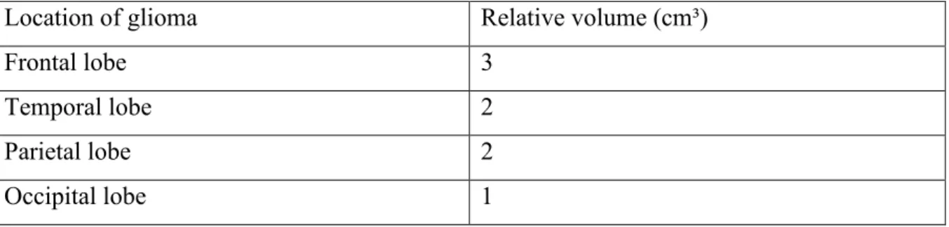
Table 3: volume of tumor tissue depending on cerebral lobe
A study conducted in Finland concerning the incidence and the median volume of tumors in different lobes showed that the median volume of the tumor tends to be bigger in the frontal followed by the temporal lobe.
Location of glioma Relative volume (cm³)
Frontal lobe 3
Temporal lobe 2
Parietal lobe 2
23
Macroscopic Features of GBs[27]:
Macroscopically GBs is heterogeneous, featuring cystic and gelatinous areas, multifocal hemorrhage and necrosis. A characteristic feature of GBs is the variation in gross appearance of the tumor from a region to another. Some of the regions as a result of tissue necrosis appear soft and yellow, whereas some of the tumor areas are firm and white and some regions show marked cystic degeneration and hemorrhage. The tumor is mostly represented by a single, relatively large, irregular lesion which usually arises in the white matter.
Figure 10: A coronal section of a post mortem brain harboring an infiltrative temporal malignant glioma which invade the insular and deep diencephalic structures[1].
![Figure 3: Anatomical landmarks of the cortex of the temporal lobe. Photographs are of the lateral (a), inferior (b), superior (c) surfaces and medial (d) on fixed cadaver[13]](https://thumb-eu.123doks.com/thumbv2/123doknet/15062630.699250/43.892.114.778.368.805/figure-anatomical-landmarks-temporal-photographs-inferior-superior-surfaces.webp)


![Figure 10: A coronal section of a post mortem brain harboring an infiltrative temporal malignant glioma which invade the insular and deep diencephalic structures[1]](https://thumb-eu.123doks.com/thumbv2/123doknet/15062630.699250/58.892.251.645.356.654/figure-coronal-harboring-infiltrative-temporal-malignant-diencephalic-structures.webp)