HAL Id: hal-01932373
https://hal.univ-lorraine.fr/hal-01932373
Submitted on 23 Nov 2018
HAL is a multi-disciplinary open access archive for the deposit and dissemination of sci-entific research documents, whether they are pub-lished or not. The documents may come from teaching and research institutions in France or abroad, or from public or private research centers.
L’archive ouverte pluridisciplinaire HAL, est destinée au dépôt et à la diffusion de documents scientifiques de niveau recherche, publiés ou non, émanant des établissements d’enseignement et de recherche français ou étrangers, des laboratoires publics ou privés.
Glaucome après chirurgie précoce de la cataracte
congénitale et implantation primaire : étude
rétrospective de 167 enfants
Pauline Beaujeux
To cite this version:
Pauline Beaujeux. Glaucome après chirurgie précoce de la cataracte congénitale et implantation primaire : étude rétrospective de 167 enfants. Sciences du Vivant [q-bio]. 2016. �hal-01932373�
AVERTISSEMENT
Ce document est le fruit d'un long travail approuvé par le jury de
soutenance et mis à disposition de l'ensemble de la
communauté universitaire élargie.
Il est soumis à la propriété intellectuelle de l'auteur. Ceci
implique une obligation de citation et de référencement lors de
l’utilisation de ce document.
D'autre part, toute contrefaçon, plagiat, reproduction illicite
encourt une poursuite pénale.
Contact : ddoc-thesesexercice-contact@univ-lorraine.fr
LIENS
Code de la Propriété Intellectuelle. articles L 122. 4
——————————————————————————————————————-
UNIVERSITÉ DE LORRAINE
FACULTÉ DE MÉDECINE DE NANCY
2016
N°
THÈSE
Pour obtenir le grade de
DOCTEUR EN MÉDECINE
Présentée et soutenue publiquement
dans le cadre du troisième cycle de Médecine Spécialisée
par
Pauline BEAUJEUX
Le 1
erjuillet 2016
GLAUCOME APRES CHIRURGIE PRECOCE DE LA CATARACTE
CONGENITALE ET IMPLANTATION PRIMAIRE :
Etude rétrospective de 167 enfants
Examinateurs de la thèse :
Monsieur le Professeur Jean-Luc GEORGE
Président
Monsieur le Professeur Jean-Michel HASCOET
Juge
Monsieur le Professeur Jean-Paul BERROD
Juge
1e r avril 2016
Président de l’Université de Lorraine : Professeur Pierre MUTZENHARDT Doyen de la Faculté de Médecine Professeur Marc BRAUN
Vice-doyens
Pr Karine ANGIOI-DUPREZ, Vice-Doyen Pr Marc DEBOUVERIE, Vice-Doyen
Assesseurs :
Premier cycle : Dr Guillaume GAUCHOTTE
Deuxième cycle : Pr Marie-Reine LOSSER Troisième cycle : Pr Marc DEBOUVERIE
Innovations pédagogiques : Pr Bruno CHENUEL Formation à la recherche : Dr Nelly AGRINIER Animation de la recherche clinique : Pr François ALLA
Affaires juridiques et Relations extérieures : Dr Frédérique CLAUDOT Vie Facultaire et SIDES : Dr Laure JOLY
Relations Grande Région : Pr Thomas FUCHS-BUDER Etudiant : M. Lucas SALVATI
Chargés de mission
Bureau de docimologie : Dr Guillaume GAUCHOTTE
Commission de prospective facultaire : Pr Pierre-Edouard BOLLAERT Orthophonie : Pr Cécile PARIETTI-WINKLER
PACES : Dr Chantal KOHLER Plan Campus : Pr Bruno LEHEUP International : Pr Jacques HUBERT
==========
DOYENS HONORAIRES
Professeur Jean-Bernard DUREUX - Professeur Jacques ROLAND - Professeur Patrick NETTER Professeur Henry COUDANE
==========
PROFESSEURS HONORAIRES
Jean-Marie ANDRE - Daniel ANTHOINE - Alain AUBREGE - Jean AUQUE - Gérard BARROCHE - Alain
BERTRAND Pierre BEY - Marc-André BIGARD - Patrick BOISSEL – Pierre BORDIGONI - Jacques BORRELLY - Michel BOULANGE Jean-Louis BOUTROY - Jean-Claude BURDIN - Claude BURLET - Daniel BURNEL - Claude CHARDOT Jean-François CHASSAGNE - François CHERRIER Jean-Pierre CRANCE - Gérard DEBRY - Emile de LAVERGNE Jean-Pierre DESCHAMPS - Jean DUHEILLE - Jean-Bernard DUREUX - Gérard FIEVE - Jean FLOQUET - Robert FRISCH Alain GAUCHER - Pierre GAUCHER - Alain GERARD - Hubert GERARD - Jean-Marie GILGENKRANTZSimone GILGENKRANTZ - Gilles GROSDIDIER - Oliéro GUERCI - Philippe
3
PROFESSEURS ÉMÉRITESProfesseur Gérard BARROCHE - Professeur Pierre BEY - Professeur Marc-André BIGARD - Professeur Jean-Pierre CRANCE - Professeure Michèle KESSLER Professeur Jacques LECLÈRE - Professeur Alain LE FAOU - Professeur Jean-Marie GILGENKRANTZ Professeure Simone GILGENKRANTZ – Professeur Gilles
GROSDIDIER - Professeur Philippe HARTEMANNProfesseur Pierre MONIN - Professeur Jean-Pierre
NICOLAS Professeur Luc PICARD - Professeur François PLENAT - Professeur Jacques POUREL - Professeur Daniel SIBERTIN-BLANC Professeur Paul VERT - Professeur Michel VIDAILHET
PROFESSEURS DES UNIVERSIT ÉS - PRATICIENS HOSPITALIERS
(Disciplines du Conseil National des Universités)
42ème Section : MORPHOLOGIE ET MORPHOGENÈSE
1ère sous-section : (Anatomie) Professeur Marc BRAUN
2ème sous-section : (Cytologie et histologie)
Professeur Christo CHRISTOV – Professeur Bernard FOLIGUET
3ème sous-section : (Anatomie et cytologie pathologiques)
Professeur Jean-Michel VIGNAUD
43ème Section : BIOPHYSIQUE ET IMAGERIE MÉDECINE
1ère sous-section : (Biophysique et médecine nucléaire)
Professeur Gilles KARCHER – Professeur Pierre-Yves MARIE – Professeur Pierre OLIVIER
2ème sous-section : (Radiologie et imagerie médecine)
Professeur René ANXIONNAT - Professeur Alain BLUM - Professeur Serge BRACARD - Professeur Michel CLAUDON Professeure Valérie CROISÉ-LAURENT - Professeur Jacques FELBLINGER
44ème Section : BIOCHIMIE, BIOLOGIE CELLULAIRE ET MOLÉCULAIRE, PHYSIOLOGIE ET NUTRITION
1ère sous-section : (Biochimie et biologie moléculaire)
Professeur Jean-Louis GUÉANT – Professeur Bernard NAMOUR – Professeur Jean-Luc OLIVIER
2ème sous-section : (Physiologie)
Professeur Christian BEYAERT– Professeur Bruno CHENUEL – Professeur François MARCHAL
4ème sous-section : (Nutrition)
Professeur Didier QUILLIOT - Professeure Rosa-Maria RODRIGUEZ-GUEANT – Professeur Olivier ZIEGLER
45ème Section : MICROBIOLOGIE, MALADIES TRANSMISSIBLES ET HYGIÈNE
1ère sous-section : (Bactériologie – virologie ; hygiène hospitalière) Professeur Alain LOZNIEWSKI – Professeure Evelyne SCHVOERER
2ème sous-section : (Parasitologie et Mycologie) Professeure Marie MACHOUART
3ème sous-section : (Maladies infectieuses ; maladies tropicales)
Professeur Thierry MAY– Professeure Céline PULCINI– Professeur Christian RABAUD
46ème Section : SANTÉ PUBLIQUE, ENVIRONNEMENT ET SOCIÉTÉ
1ère sous-section : (Épidémiologie, économie de la santé et prévention)
Professeur François ALLA – Professeur Serge BRIANÇON - Professeur Francis GUILLEMIN Professeur Denis ZMIROU-NAVIER
2ème sous-section : (Médecine et santé au travail) Professeur Christophe PARIS
3ème sous-section : (Médecine légale et droit de la santé) Professeur Henry COUDANE
4ème sous-section : (Biostatistiques, informatique médicale et technologies de communication) Professeure Eliane ALBUISSON – Professeur Nicolas JAY
47ème Section : CANCÉROLOGIE, GÉNÉTIQUE, HÉMATOLOGIE, IMMUNOLOGIE
1ère sous-section : (Hématologie ; transfusion) Professeur Pierre FEUGIER
2ème sous-section : (Cancérologie ; radiothérapie)
Professeur Thierry CONROY Professeur – François GUILLEMIN - Professeur Didier PEIFFERT Professeur Frédéric MARCHAL
3ème sous-section : (Immunologie)
Professeur Marcelo DE CARVALHO-BITTENCOURT – Professeur Gilbert FAURE
4ème sous-section : (Génétique)
48ème Section : ANESTHÉSIOLOGIE, RÉANIMATION, MÉDECINE D’URGENCE, PHARMACOLOGIE ET THÉRAPEUTIQUE
1ère sous-section : (Anesthésiologie - réanimation ; médecine d’urgence)
Professeur Gérard AUDIBERT– Professeur Hervé BOUAZIZ –Professeur Thomas FUCHS-BUDER – Professeure Marie-Reine LOSSER– Professeur Claude MEISTELMAN
2ème sous-section : (Réanimation ; médecine d’urgence)
Professeur Pierre-Édouard BOLLAERT – Professeur Sébastien GIBOT – Professeur Bruno LÉVY
3ème sous-section : (Pharmacologie fondamentale ; pharmacologie clinique ; addictologie)
Professeur Pierre GILLET – Professeur J.Y. JOUZEAU (pharmacien) – Professeur Patrick NETTER
4ème sous-section : (Thérapeutique ; médecine d’urgence ; addictologie)
Professeur François PAILLE - Professeur Patrick ROSSIGNOL– Professeur Faiez ZANNAD
49ème Section : PATHOLOGIE NERVEUSE ET MUSCULAIRE, PATHOLOGIE MENTALE,
HANDICAP ET RÉÉDUCATION
1ère sous-section : (Neurologie)
Professeur Marc DEBOUVERIE –Professeur Louis MAILLARD – Professeur Luc TAILLANDIER – Professeure Louise TYVAERT
2ème sous-section : (Neurochirurgie)
Professeur Jean AUQUE – Professeur Thierry CIVIT – Professeure Sophie COLNAT-COULBOIS – Professeur Olivier KLEIN - Professeur Jean-Claude MARCHAL
3ème sous-section : (Psychiatrie d'adultes ; addictologie) Professeur Jean-Pierre KAHN – Professeur Raymund SCHWAN
4ème sous-section : (Pédopsychiatrie ; addictologie) Professeur Bernard KABUTH
5ème sous-section : (Médecine physique et de réadaptation) Professeur Jean PAYSANT
50ème Section : PATHOLOGIE OSTÉO-ARTICULAIRE, DERMATOLOGIE ET CHIRURGIE PLASTIQUE
1ère sous-section : (Rhumatologie)
Professeure Isabelle CHARY-VALCKENAERE – Professeur Damien LOEUILLE
2ème sous-section : (Chirurgie orthopédique et traumatologique)
Professeur Laurent GALOIS – Professeur Didier MAINARD – Professeur Daniel MOLE - Professeur François SIRVEAUX
3ème sous-section : (Dermato-vénéréologie)
Professeure Annick BARBAUD – Professeur Jean-Luc SCHMUTZ
4ème sous-section : (Chirurgie plastique, reconstructrice et esthétique ; brûlologie) Professeur François DAP - Professeur Gilles DAUTEL - Professeur Etienne SIMON
51ème Section : PATHOLOGIE CARDIO-RESPIRATOIRE ET VASCULAIRE
1ère sous-section : (Pneumologie ; addictologie)
Professeur Jean-François CHABOT – Professeur Ari CHAOUAT – Professeur Yves MARTINET
2ème sous-section : (Cardiologie)
Professeur Etienne ALIOT – Professeur Edoardo CAMENZIND – Professeur Christian de CHILLOU DE CHURET Professeur Yves JUILLIERE – Professeur Nicolas SADOUL -
3ème sous-section : (Chirurgie thoracique et cardiovasculaire) Professeur Thierry FOLLIGUET – Professeur Juan-Pablo MAUREIRA
4ème sous-section : (Chirurgie vasculaire ; médecine vasculaire)
Professeur Sergueï MALIKOV – Professeur Denis WAHL
52ème Section : MALADIES DES APPAREILS DIGESTIF ET URINAIRE
1ère sous-section : (Gastroentérologie ; hépatologie ; addictologie)
Professeur Jean-Pierre BRONOWICKI – Professeur Laurent PEYRIN-BIROULET
3ème sous-section : (Néphrologie)
Professeur Luc FRIMAT – Professeure Dominique HESTIN
4ème sous-section : (Urologie)
5
54ème Section : DÉVELOPPEMENT ET PATHOLOGIE DE L'ENFANT,GYNÉCOLOGIE-OBSTÉTRIQUE, ENDOCRINOLOGIE ET REPRODUCTION
1ère sous-section : (Pédiatrie)
Professeur Pascal CHASTAGNER - Professeur François FEILLET – Professeur Jean-Michel HASCOET – Professeur Emmanuel RAFFO – Professeur Cyril SCHWEITZER
2ème sous-section : (Chirurgie infantile)
Professeur Pierre JOURNEAU – Professeur Jean-Louis LEMELLE
3ème sous-section : (Gynécologie-obstétrique ; gynécologie médicale)
Professeur Philippe JUDLIN – Professeur Olivier MOREL
4ème sous-section : (Endocrinologie, diabète et maladies métaboliques ; gynécologie médicale)
Professeur Bruno GUERCI – Professeur Marc KLEIN – Professeur Georges WERYHA
55ème Section : PATHOLOGIE DE LA TÊTE ET DU COU
1ère sous-section : (Oto-rhino-laryngologie)
Professeur Roger JANKOWSKI – Professeure Cécile PARIETTI-WINKLER
2ème sous-section : (Ophtalmologie)
Professeure Karine ANGIOI –Professeur Jean-Paul BERROD – Professeur Jean-Luc GEORGE
3ème sous-section : (Chirurgie maxillo-faciale et stomatologie) Professeure Muriel BRIX
==========
PROFESSEURS DES UNIVERSITÉS
61ème Section : GÉNIE INFORMATIQUE, AUTOMATIQUE ET TRAITEMENT DU SIGNAL Professeur Walter BLONDEL
64ème Section : BIOCHIMIE ET BIOLOGIE MOLÉCULAIRE Professeure Sandrine BOSCHI-MULLER
==========
PROFESSEUR ASSOCIÉ DE MÉDECINE GÉNÉRALE
Professeur associé Paolo DI PATRIZIO
==========
MAÎTRES DE CONFÉRENCES DES UNIVERSITÉS - PRATICIENS HOSPITALIERS 42ème Section : MORPHOLOGIE ET MORPHOGENÈSE
1ère sous-section : (Anatomie)
Docteur Bruno GRIGNON – Docteure Manuela PEREZ
2ème sous-section : (Histologie, embryologie et cytogénétique) Docteure Chantal KOHLER – Docteure Françoise TOUATI
3ème sous-section : (Anatomie et cytologie pathologiques) Docteur Guillaume GAUCHOTTE
43ème Section : BIOPHYSIQUE ET IMAGERIE MÉDECINE
1ère sous-section : (Biophysique et médecine nucléaire) Docteur Jean-Marie ESCANYE
2ème sous-section : (Radiologie et imagerie médecine) Docteur Damien MANDRY – Docteur Pedro TEIXEIRA
44ème Section : BIOCHIMIE, BIOLOGIE CELLULAIRE ET MOLÉCULAIRE, PHYSIOLOGIE ET
NUTRITION
1ère sous-section : (Biochimie et biologie moléculaire)
Docteure Shyue-Fang BATTAGLIA – Docteure Sophie FREMONT - Docteure Isabelle GASTIN – Docteure Catherine MALAPLATE-ARMAND – Docteur Marc MERTEN – Docteur Abderrahim OUSSALAH
2ème sous-section : (Physiologie)
Docteure Silvia DEMOULIN-ALEXIKOVA – Docteur Mathias POUSSEL
3ème sous-section : (Biologie Cellulaire)
Docteure Véronique DECOT-MAILLERET
45ème Section : MICROBIOLOGIE, MALADIES TRANSMISSIBLES ET HYGIÈNE
1ère sous-section : (Bactériologie – Virologie ; hygiène hospitalière)
Docteure Corentine ALAUZET – Docteure Hélène JEULIN – Docteure Véronique VENARD
2ème sous-section : (Parasitologie et mycologie)
46ème Section : SANTÉ PUBLIQUE, ENVIRONNEMENT ET SOCIÉTÉ
1ère sous-section : (Epidémiologie, économie de la santé et prévention)
Docteure Nelly AGRINIER – Docteur Cédric BAUMANN – Docteure Frédérique CLAUDOT – Docteur Alexis HAUTEMANIÈRE
2ème sous-section (Médecine et Santé au Travail) Docteure Isabelle THAON
3ème sous-section (Médecine légale et droit de la santé) Docteur Laurent MARTRILLE
47ème Section : CANCÉROLOGIE, GÉNÉTIQUE, HÉMATOLOGIE, IMMUNOLOGIE
1ère sous-section : (Hématologie ; transfusion) Docteur Aurore PERROT
2ème sous-section : (Cancérologie ; radiothérapie) Docteure Lina BOLOTINE
4ème sous-section : (Génétique)
Docteure Céline BONNET – Docteur Christophe PHILIPPE
48ème Section : ANESTHÉSIOLOGIE, RÉANIMATION, MÉDECINE D’URGENCE,
PHARMACOLOGIE ET THÉRAPEUTIQUE
2ème sous-section : (Réanimation ; Médecine d’Urgence) Docteur Antoine KIMMOUN (stagiaire)
3ème sous-section : (Pharmacologie fondamentale ; pharmacologie cliniqu ; addictologie) Docteur Nicolas GAMBIER – Docteure Françoise LAPICQUE – Docteur Julien SCALA-BERTOLA
4ème sous-section : (Thérapeutique ; Médecine d’Urgence ; Addictologie) Docteur Nicolas GIRERD (stagiaire)
50ème Section : PATHOLOGIE OSTÉO-ARTICULAIRE, DERMATOLOGIE ET CHIRURGIE PLASTIQUE
1ère sous-section : (Rhumatologie) Docteure Anne-Christine RAT
3ème sous-section : (Dermato-vénéréologie) Docteure Anne-Claire BURSZTEJN
4ème sous-section : (Chirurgie plastique, reconstructrice et esthétique ; brûlologie) Docteure Laetitia GOFFINET-PLEUTRET
51ème Section : PATHOLOGIE CARDIO-RESPIRATOIRE ET VASCULAIRE
3ème sous-section : (Chirurgie thoracique et cardio-vasculaire) Docteur Fabrice VANHUYSE
4ème sous-section : (Chirurgie vasculaire ; médecine vasculaire)
Docteur Stéphane ZUILY
52ème Section : MALADIES DES APPAREILS DIGESTIF ET URINAIRE
1ère sous-section : (Gastroentérologie ; hépatologie ; addictologie)
Docteur Jean-Baptiste CHEVAUX
53ème Section : MÉDECINE INTERNE, GÉRIATRIE et CHIRURGIE GÉNÉRALE
1ère sous-section : (Médecine interne ; gériatrie et biologie du vieillissement ; médecine générale ;
addictologie)
Docteure Laure JOLY
3ème sous-section : (Médecine générale)
Docteure Elisabeth STEYER
55ème Section : OTO-RHINO-LARYNGOLOGIE
1ère sous-section : (Oto-Rhino-Laryngologie)
Docteur Patrice GALLET (stagiaire)
7
MAÎTRES DE CONFÉRENCES5ème Section : SCIENCES ÉCONOMIQUES Monsieur Vincent LHUILLIER
7ème section : SCIENCES DU LANGAGE : LINGUISTIQUE ET PHONETIQUE GENERALES Madame Christine DA SILVA-GENEST
19ème Section : SOCIOLOGIE, DÉMOGRAPHIE Madame Joëlle KIVITS
60ème Section : MÉCANIQUE, GÉNIE MÉCANIQUE, GÉNIE CIVIL Monsieur Alain DURAND
61ème Section : GÉNIE INFORMATIQUE, AUTOMATIQUE ET TRAITEMENT DU SIGNAL Monsieur Jean REBSTOCK
64ème Section : BIOCHIMIE ET BIOLOGIE MOLÉCULAIRE
Madame Marie-Claire LANHERS– Monsieur Nick RAMALANJAONA – Monsieur Pascal REBOUL
65ème Section : BIOLOGIE CELLULAIRE
Madame Nathalie AUCHER – Madame Natalia DE ISLA-MARTINEZ – Monsieur Jean-Louis GELLY – Madame Céline HUSELSTEIN – Madame Ketsia HESS – Monsieur Hervé MEMBRE – Monsieur Christophe NEMOS
66ème Section : PHYSIOLOGIE Monsieur Nguyen TRAN
==========
MAÎTRES DE CONFÉRENCES ASSOCIÉS Médecine Générale
Docteur Pascal BOUCHE – Docteur Olivier BOUCHY –- Docteur Arnaud MASSON – Docteure Sophie SIEGRIST
==========
DOCTEURS HONORIS CAUSA
Professeur Charles A. BERRY (1982)
Centre de Médecine Préventive, Houston (U.S.A)
Professeur Pierre-Marie GALETTI (1982)
Brown University, Providence (U.S.A)
Professeure Mildred T. STAHLMAN (1982)
Vanderbilt University, Nashville (U.S.A)
Professeur Théodore H. SCHIEBLER (1989)
Institut d'Anatomie de Würtzburg (R.F.A) Université de Pennsylvanie (U.S.A)
Professeur Mashaki KASHIWARA (1996)
Research Institute for Mathematical Sciences de Kyoto (JAPON)
Professeure Maria DELIVORIA-PAPADOPOULOS (1996)
Professeur Ralph GRÄSBECK (1996)
Université d'Helsinki (FINLANDE)
Professeur Duong Quang TRUNG (1997)
Université d'Hô Chi Minh-Ville (VIÊTNAM)
Professeur Daniel G. BICHET (2001)
Université de Montréal (Canada)
Professeur Marc LEVENSTON (2005)
Institute of Technology, Atlanta (USA)
Professeur Brian BURCHELL (2007)
Université de Dundee (Royaume-Uni)
Professeur Yunfeng ZHOU (2009)
Université de Wuhan (CHINE)
Professeur David ALPERS (2011)
Université de Washington (U.S.A)
Professeur Martin EXNER (2012)
A notre Président de Thèse
Monsieur le Professeur Jean-Luc George
Professeur d’Ophtalmologie
Vous nous faites l’honneur de présider cette thèse et de juger ce travail.
Votre bienveillance nous a accompagnée tout au long de notre internat. Vous avez révélé notre intérêt pour l’orbito-palpébral et la strabologie, et partagé votre vaste expérience dans ces domaines.
Nous admirons votre insatiable curiosité, votre savoir et votre humilité. C’est un honneur de compter parmi vos élèves.
9
A notre Juge
Monsieur le Professeur Jean-Michel Hascoët
Professeur de Pédiatrie
Vous nous faites l’honneur de juger ce travail, et nous vous remercions pour le vif intérêt que vous y avez porté.
Votre gentillesse et votre disponibilité nous font espérer que nous pourrons de nouveau collaborer sur d’autres projets à l’avenir.
A notre Juge
Monsieur le Professeur Jean-Paul Berrod
Professeur d’Ophtalmologie
Vous nous faites l’honneur de juger ce travail.
Vos qualités humaines et votre adresse chirurgicale sont pour nous un exemple.
Soyez assuré de nos respectueux sentiments et notre grande admiration.
11
A notre Directeur de Thèse
Monsieur le Docteur Pascal Dureau
Ophtalmologiste
Vous nous avez fait l’honneur de diriger notre travail de thèse.
Nous vous remercions de la confiance que vous nous avez accordée tout au long de l’élaboration de ce travail.
Votre disponibilité, votre rigueur en toute décontraction et l’envie permanente de transmettre sont autant de qualités qui rendent l’ophtalmologie pédiatrique encore plus passionnante à vos côtés.
A tous ceux que j’ai eu la chance de rencontrer au cours de mon internat :
A tout le service de Neurochirugie de Nancy :
Jean Auque, Thierry Civit, Olivier Klein, Jean Claude Marchal, Isabelle Merlot, Anthony Joud, Catherine Pinelli, Sophie Colnat, mes co-internes fétiches et tout le personnel… pour ce premier semestre haut en couleurs à vos côtés.
Merci à Nicolas Salaun d’avoir si parfaitement encadré mes débuts en ophtalmologie. A mes maîtres d’internat :
Karine Angioi, Marie-Christine Bazard, Toufic Maalouf, Pierre Lesure pour tout ce que vous m’avez transmis tout au long de ces années passées à vos côtés. Je ne vous remercierai jamais assez… A mes chefs :
Jérôme Selton, Véronique Cloché, Jean-Baptiste Conart, François Ameloot, Benjamine Batta, Audrey Baudot, Fanny Tréchot, Bertrand Leroy, merci pour tous les souvenirs partagés ensemble, à l’hôpital ou ailleurs.
Merci à tout le service d’Ophtalmologie du CHU de Nancy, les secrétaires, orthoptistes, infirmières de consultation et de bloc opératoire, sans oublier mes nombreux co-internes.
Merci au personnel du service d’Ophtalmologie d’Epinal, pour votre incroyable enthousiasme, et tous nos fou-rires…
A Georges Caputo et toute la Caputo’s Team :
Pascal Dureau, Catherine Edelson, Amandine Barjol, Ana Clément, Aude Affortit, Florence Metge, Alexis Pinel, Sabine Derrien… et Adrien Sarfati ! Vous m’avez accueillie parmi vous avec beaucoup de bienveillance et m’avez fait adorer l’ophtalmologie pédiatrique sous tous ses aspects... Je suis fière d’avoir fait partie de votre service, si propice à l’épanouissement professionnel… et personnel !
Merci à Chantal Evin pour son aide précieuse au cours de cette étude.
Un merci inconditionnel à Hervé Picard pour sa disponibilité, son talent et ses onomatopées… Enfin, à Edouard Koch, pour la confiance que tu m’accordes et toutes les opportunités que tu m’offres… un immense merci !
13
A mes Parents et mon Frère chéris… pour absolument tout…
15
SERMENT
«
A
u moment d'être admise à exercer la médecine, je promets et je jure d'être fidèle aux
lois de l'honneur et de la probité. Mon premier souci sera de rétablir, de préserver ou de
promouvoir la santé dans tous ses éléments, physiques et mentaux, individuels et sociaux. Je
respecterai toutes les personnes, leur autonomie et leur volonté, sans aucune discrimination
selon leur état ou leurs convictions. J’interviendrai pour les protéger si elles sont affaiblies,
vulnérables ou menacées dans leur intégrité ou leur dignité. Même sous la contrainte, je ne
ferai pas usage de mes connaissances contre les lois de l'humanité. J'informerai les patients
des décisions envisagées, de leurs raisons et de leurs conséquences. Je ne tromperai jamais
leur confiance et n'exploiterai pas le pouvoir hérité des circonstances pour forcer les
consciences. Je donnerai mes soins à l'indigent et à quiconque me les demandera. Je ne me
laisserai pas influencer par la soif du gain ou la recherche de la gloire.
Admise dans l'intimité des personnes, je tairai les secrets qui me sont confiés. Reçue à
l'intérieur des maisons, je respecterai les secrets des foyers et ma conduite ne servira pas à
corrompre les mœurs. Je ferai tout pour soulager les souffrances. Je ne prolongerai pas
abusivement les agonies. Je ne provoquerai jamais la mort délibérément.
Je préserverai l'indépendance nécessaire à l'accomplissement de ma mission. Je
n'entreprendrai rien qui dépasse mes compétences. Je les entretiendrai et les
perfectionnerai pour assurer au mieux les services qui me seront demandés.
J'apporterai mon aide à mes confrères ainsi qu'à leurs familles dans l'adversité.
Que les hommes et mes confrères m'accordent leur estime si je suis fidèle à mes promesses ;
que je sois déshonorée et méprisée si j'y manque ».
TABLE DES MATIERES
Partie I. PRESENTATION ET CONTEXTE DE L’ETUDE ... 17
Partie II. ARTICLE ORIGINAL : Glaucoma after early congenital cataract surgery and primary implantation: a retrospective study of 167 children ... 23
1. Introduction ... 23 2. Methods ... 25 3. Results ... 28 4. Discussion ... 32 5. Conclusion ... 37 6. References ... 38
17
Partie I. PRESENTATION ET CONTEXTE DE L’ETUDE
Cette étude a été réalisée au sein du service d’Ophtalmologie Pédiatrique du Docteur Caputo à la Fondation Ophtalmologique Adolphe de Rothschild (FOR).
Son objectif principal était de rapporter le risque de survenue du glaucome secondaire après chirurgie de la cataracte congénitale, dans une population d’enfants pseudophaques opérés précocement.
La cataracte congénitale (CC) est définie par une opacification uni (CU) ou bilatérale (CB) du cristallin chez l’enfant avant l’âge d’un an (1). Son incidence varie entre 1 et 5/10 000 enfants (2) selon les études.
Sur le plan physiopathologique, il s’agit d’une anomalie cristallinienne malformative, dont l’étiologie est variable en fonction de la latéralité de l’atteinte : le plus souvent héréditaire ou secondaire à une pathologie oculaire.
Induisant une déprivation visuelle, les cataractes obturant l’axe visuel sont fortement amblyogènes. A ce titre, elles relèvent d’une prise en charge chirurgicale précoce pour optimiser le pronostic fonctionnel : idéalement avant 6 et 8 semaines de vie respectivement pour les CU (3) et les CB (4,5).
Dans la littérature, la procédure chirurgicale concernant la CC est relativement consensuelle depuis le début des années 2000 (5,6). Cette intervention comprend une phacoaspiration par abord cornéen limbique, suivie de la réalisation systématique d’un capsulorhexis postérieur et d’une vitrectomie antérieure afin de prévenir l’opacification capsulaire secondaire constante et précoce chez l’enfant. Par ailleurs, la question du choix de l’implantation primaire versus l’aphakie reste toujours débattue et relève essentiellement des habitudes propres à chaque équipe. Dans notre service d’Ophtalmologie Pédiatrique, l’implantation primaire est la règle. Elle est pratiquée tant pour les CU
que pour les CB et quelque soit l'âge de l’enfant, dès lors que la taille de l’oeil et l’existence d’un plan capsulaire postérieur permettent la mise en place d’un implant dans le sac ou le sulcus.
Le choix de cette pratique, il y a plusieurs années, repose sur plusieurs arguments en faveur de l’implantation primaire :
l’augmentation théorique des chances de réussite de la rééducation de l’amblyopie par suppression du problème de compliance lié au port de lentilles rigides perméables au gaz (LRPG) corrigeant l’aphakie,
la diminution des complications inflammatoires imputées à l’implant intra oculaire (ICP) grâce à l’utilisation d’implants acryliques hydrophobes (7,8),
le potentiel rôle protecteur de l’implantation primaire sur l’apparition d’un glaucome secondaire (9).
Le glaucome secondaire (GS) est la complication la plus redoutable après chirurgie de la cataracte congénitale, que les enfants soient aphaques ou pseudophaques (5,10–13). Sa prévalence varie considérablement selon les différentes études (9), de 3 à 41 %. Plusieurs explications peuvent être soulevées :
l’absence de définition consensuelle du GS, rendant le diagnostic d’autant plus difficile en pratique. Ce dernier repose sur un faisceau d’arguments cliniques :
l’hypertonie oculaire, en tenant compte du fait que la pression intraoculaire (PIO) physiologique de l’enfant est inférieure à celle de l’adulte. Il convient également de ne pas négliger le fait que les mesures de PIO chez les jeunes enfants sont réalisées sous anesthésie générale, ce qui sous-estime les valeurs de PIO d’environ 30 % en raison de l’utilisation de gaz halogénés (2),
19
les modifications morphologiques du globe pendant la période de croissance oculaire (jusqu’à 3 ans) : augmentation anormale de la longueur axiale et/ou du diamètre cornéen, myopisation importante en rapport avec un myopic shift (MS) élevé ou asymétrique,
l’œdème cornéen cliniquement significatif ou pachymétrie augmentée.
la différence entre les populations étudiées dans ces différentes études ; en effet, la prévalence du GS semble être fortement corrélée et inversement proportionnelle à l’âge de l’enfant lors de l’intervention chirurgicale de CC (9,10,14),
le suivi post-opératoire des études, puisque le délai d’apparition du GS est typiquement retardé de plusieurs années après l’intervention de CC, bien que des cas de GS plus précoces existent également (5).
La physiopathologie du GS reste incertaine et multifactorielle (2) comprenant des mécanismes à angle ouvert (altération de la filtration trabéculaire par la présence de vitré au niveau de l’angle iridocornéen ou par réaction inflammatoire post-opératoire, trabéculum immature lésé par l’intervention chirurgicale, modification de la matrice extracellulaire trabéculaire induite par une corticothérapie, toxicité des cellules cristaliniennes résiduelles) et d’autres, plus rares, par fermeture de l’angle (modification de la dynamique du plan iridocristallinien, synéchies dans l’angle).
Plusieurs facteurs de risque de survenue d’un GS ont été suspectés dans la littérature. Les plus consensuels sont : l’âge de l’enfant lors de la chirurgie de CC, notamment avant 1 an (5,12,13), la microphtalmie (15,16), l’existence d’une persistance de la vascularisation foetale (PVF) ou encore le recours à des chirurgies intraoculaires additionnelles (17) . L’un des plus débattu de ces facteurs de risques présumés reste l’implantation primaire (9,11–13,17). Toutefois, l’Infant Aphakia Treatment Study (IATS) (15), première étude prospective sur le sujet, a récemment conclu à l’absence de différence significative de la survenue d’un GS entre enfants aphaques et pseudophaques à 5 ans.
L’objectif principal de notre étude était de déterminer le risque de survenue du glaucome secondaire dans notre population d’enfants opérés avant l’âge d’un an de cataracte congénitale uni ou bilatérale avec implantation primaire, et de la comparer aux données de la littérature. Les objectifs secondaires étaient de rechercher d’éventuels facteurs prédisposant au glaucome secondaire, et de décrire la prise en charge thérapeutique et le pronostic fonctionnel de ces yeux glaucomateux.
21
REFERENCES
1. Tartarella MB, Britez-Colombi GF, Milhomem S, Lopes MCE, Fortes Filho JB. Pediatric cataracts: clinical aspects, frequency of strabismus and chronological, etiological, and morphological features. Arq Bras Oftalmol. 2014 Jun;77(3):143–7.
2. Dureau P. Ophtalomologie pédiatrique et strabismes. In: Médecine Sciences Publications. Lavoisier; 2014.
3. Birch EE, Stager DR. The critical period for surgical treatment of dense congenital unilateral cataract. Invest Ophthalmol Vis Sci. 1996 Jul;37(8):1532–8.
4. Lundvall A, Kugelberg U. Outcome after treatment of congenital bilateral cataract. Acta Ophthalmol Scand. 2002 Dec;80(6):593–7.
5. Vishwanath M, Cheong-Leen R, Taylor D, Russell-Eggitt I, Rahi J. Is early surgery for congenital cataract a risk factor for glaucoma? Br J Ophthalmol. 2004 Jul;88(7):905–10.
6. Vasavada A, Desai J. Primary posterior capsulorhexis with and without anterior vitrectomy in congenital cataracts. J Cataract Refract Surg. 1997;23 Suppl 1:645–51.
7. Trivedi RH, Wilson ME, Bartholomew LR, Lal G, Peterseim MM. Opacification of the visual axis after cataract surgery and single acrylic intraocular lens implantation in the first year of life. J AAPOS Off Publ Am Assoc Pediatr Ophthalmol Strabismus Am Assoc Pediatr Ophthalmol Strabismus. 2004 Apr;8(2):156–64.
8. Trivedi RH, Wilson ME. Single-piece acrylic intraocular lens implantation in children. J Cataract Refract Surg. 2003 Sep;29(9):1738–43.
9. Asrani S, Freedman S, Hasselblad V, Buckley EG, Egbert J, Dahan E, et al. Does primary intraocular lens implantation prevent “aphakic” glaucoma in children? J AAPOS Off Publ Am Assoc Pediatr Ophthalmol Strabismus Am Assoc Pediatr Ophthalmol Strabismus. 2000 Feb;4(1):33–9. 10. Lambert SR. Treatment of congenital cataract. Br J Ophthalmol. 2004 Jul;88(7):854–5.
11. Sahin A, Caça I, Cingü AK, Türkcü FM, Yüksel H, Sahin M, et al. Secondary glaucoma after pediatric cataract surgery. Int J Ophthalmol. 2013;6(2):216–20.
12. Trivedi RH, Wilson ME, Golub RL. Incidence and risk factors for glaucoma after pediatric cataract surgery with and without intraocular lens implantation. J AAPOS Off Publ Am Assoc Pediatr Ophthalmol Strabismus Am Assoc Pediatr Ophthalmol Strabismus. 2006 Apr;10(2):117–23.
13. Wong IBY, Sukthankar VD, Cortina-Borja M, Nischal KK. Incidence of early-onset glaucoma after infant cataract extraction with and without intraocular lens implantation. Br J Ophthalmol. 2009 Sep;93(9):1200–3.
14. Rabiah PK. Frequency and predictors of glaucoma after pediatric cataract surgery. Am J Ophthalmol. 2004 Jan;137(1):30–7.
15. Freedman SF, Lynn MJ, Beck AD, Bothun ED, Örge FH, Lambert SR, et al. Glaucoma-Related Adverse Events in the First 5 Years After Unilateral Cataract Removal in the Infant Aphakia Treatment Study. JAMA Ophthalmol. 2015 Aug;133(8):907–14.
16. Wallace DK, Plager DA. Corneal diameter in childhood aphakic glaucoma. J Pediatr Ophthalmol Strabismus. 1996 Oct;33(5):230–4.
17. Mataftsi A, Haidich A-B, Kokkali S, Rabiah PK, Birch E, Stager DR, et al. Postoperative glaucoma following infantile cataract surgery: an individual patient data meta-analysis. JAMA Ophthalmol. 2014 Sep;132(9):1059–67.
23
Partie II. ARTICLE ORIGINAL : Glaucoma after early congenital cataract
surgery and primary implantation: a retrospective study of 167 children
1.
Introduction
Dense congenital or early developmental cataracts represent a serious threat to the development of the visual system in a young child (1,2). The priority is prompt surgery after the diagnosis has been made, to clear the visual axis and to achieve a good visual outcome (3–8).
Current methods to compensate for the refractive power of the lens include glasses, aphakic contact lenses and posterior chamber intraocular lenses (IOL). The primary implantation of a posterior chamber IOL is now increasingly admitted, even in younger children and for both monocular and bilateral cataracts (9–13).
Despite advances in congenital cataract management, the occurrence of secondary glaucoma (SG) is the major sight-threatening complication of cataract removal in infancy (2). Aphakic or pseudophakic glaucoma typically include a delayed intraocular pressure (IOP) elevation (14–16) with an open-angle (17). Rarely, it happens as an early angle closure glaucoma (18).
Although the pathogenesis of glaucoma after cataract surgery is still unclear, several ocular and surgical risk factors for its occurrence are strongly suspected and could impact on clinical practice, like early surgery (5), chronic postoperative inflammation (19), primary posterior capsulotomy (20), persistence of fetal vasculature (PFV) (21), or microphthalmia (18). Furthermore, a protective role of IOL implantation has been suggested (22,23) by mechanical support of the trabecular meshwork and by blocking a potentially toxic component of the vitreous. However, a recent prospective study
(24,25) stated that primary IOL placement had no influence on the risk of glaucoma after cataract surgery.
The incidence of SG, ranging from 3% to 41% (26,27) depending on the series, remains unclear because there is no universal definition of aphakic or pseudophakic glaucoma, making the diagnosis difficult in children. Furthermore, as in most paediatric glaucomas, patient care remains complex and the long-term visual outcomes are still not well known.
The primary goal of this study was to report on the incidence of glaucoma after early monocular or bilateral cataract removal with primary IOL implantation.
The secondary objectives were to identify the risk factors for the development of glaucoma in these eyes, the treatments used and the visual outcomes.
25
2.
Methods
Subjects:
We conducted a retrospective review of all consecutive children who underwent unilateral or bilateral paediatric cataract surgery with primary implantation in their first year of life, between January 2003 and December 2012 at the Fondation Ophtalmologique Adolphe de Rothschild (Paris, France). This hospital is a tertiary centre in paediatric ophthalmology, receiving patients from all over the country.
A least 1 month of follow-up after surgery was required for inclusion in the study. Anterior form of persistent of fetal vasculature (PFV) that did not require posterior vitrectomy, and relative microphthalmia allowing primary IOL implantation were included in the study. Exclusion criteria were patients with other ocular malformations, especially anterior segment dysgenesis, pre-existing IOP elevation or congenital glaucoma, syndromic cataract (ie Lowe, congenital Rubella), history of trauma or uveitis, and premature children born before 32 weeks’ gestation.
Appropriate approval was granted from ethic committees of the institution.
Surgical technique and postoperative aftercare:
One surgeon performed 68% of the lensectomies and 7 other paediatric ophthalmologists operated on the remaining cases. However, the surgical procedure was consensual as follows: clear corneal incision, anterior capsulorhexis, lensectomy using the vitrectome, posterior capsulorhexis and anterior vitrectomy. Primary posterior chamber implantation was performed into the capsular bag or the sulcus based on anatomical conditions. The type of IOL varied according to the year of the intervention. Two types of material were used: Poly Methyl MethAcrylate (PMMA) in the older cases, or acrylic hydrophobic IOL in the more recent years.
A topical postoperative treatment was prescribed for a period of one month, including a mydriatic (atropine) and an antibiotic/steroids combination (dexamethasone-neomycin-polymixin B). An oral steroid treatment (betamethasone) was added for one week.
The postoperative visual rehabilitation was started within one to two weeks after cataract surgery, with glasses correction and patching when indicated.
Every child had an examination under general anaesthesia one month after surgery to control the refraction, the axial length, the cornea, the anterior chamber, the IOP and the fundus. Then, regular examinations under general anaesthesia were performed until the age of the children allowed a full and reliable ophthalmologic examination at the slit lamp.
Glaucoma diagnosis and follow-up:
In the study, the primary outcome measure was the diagnosis of secondary glaucoma, based on the ophthalmologist‘s decision to initiate a permanent medical treatment or a glaucoma specific surgery at least one month after cataract surgery, relevant to the end of the steroid treatment.
Several criteria were used by the physician to suspect the onset of a secondary glaucoma, as a body of clinical arguments: ocular hypertension, asymmetric optic nerve cupping (>0.2) or large cup-to-disk ratio (>0.4), corneal oedema or pachymetric elevation, enlargement of corneal diameter, abnormal asymmetric axial length elongation or myopic shift. The IOP was measured during general anaesthesia by a portable applanation tonometer (Perkins). Considering that the physiologic IOP in children is lower than in adults and that some anaesthetic drugs (as sevoflurane) falsely decrease by about 30% the IOP, an elevation ≥ 14 mmHg was considered as suspicious. Corneal diameter was not uniformly available, that’s why an eye was considered as microphthalmic if it was consigned on the
27
Statistical analysis:Data were analysed using the R statistical package (R Core Team 2014 R: A language and environment for statistical computing. R Foundation for Statistical Computing, Vienna, Austria. URL http://www.R-project.org/). A statistically significant association was defined by a p-value inferior to 0.05.
Inter-groups differences between quantitative variables were tested using Student test (if the variable distribution could be considered normal after a Shapiro test) or exact corrected Wilcoxon test (if the normality assumption could not be made). Inter-groups differences between qualitative variables were tested using Fisher exact test.
Time-related occurrence of glaucoma after surgery was analysed using a Kaplan-Meier estimator. Effect on glaucoma occurrence was tested using log-rank tests (univariate analysis) and then a multivariate Cox model. As for the final visual acuity, only eyes with an evaluation after 5 years of age were taken into consideration.
3.
Results
Study population
One hundred and ninety six children underwent unilateral or bilateral congenital cataract surgery with primary IOL implantation in their first year of life, between January 2003 and December 2012. Twenty nine children were excluded, among which 10 posterior PFV, 5 anterior segment dysgenesis, 2 pre-existing IOP elevations, 2 Lowe syndromes, 1 congenital Rubella, 1 premature child, and 8 because of unavailable data.
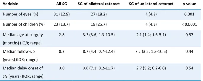
Eventually, 167 children (241 eyes) were included in the study: 90 females (53.9%) and 77 males (46.1%). The median age at surgery was 4.2 months (interquartile range (IQR) 4,1; range 0.9-12.0 months). The median follow-up period was 4.6 years (IQR 6.6; range 1 month-12.4 years) and the median age at the last visit was 5.2 years (IQR 6.3; range 4.3 months-12.8 years).
Laterality of cataracts
Seventy four patients (44.3%) had a bilateral cataract and 93 patients (55.7%) an unilateral one. These two groups were significantly different : the age at lensectomy was lower for bilateral cataracts (mean 3.8 vs. 5.1 months, p = 0.04) and their follow-up time was longer (mean 5.3 vs. 3.8 years, p = 0.04).
Cases of secondary glaucoma
29
The median time to glaucoma diagnosis was 3.0 years (IQR 5.8; range 2.4 months-11.7 years). In 11/31 eyes (35%), the SG appeared in the first postoperative year. The SG eyes were significantly younger at the time of cataract surgery than those who did not developed glaucoma (mean 3.8 vs. 4.8 months, p = 0.04), and had a significantly longer follow-up (mean 8.0 vs. 4.5 years, p < 0.0001). A Kaplan-Meier survival curve of eyes remaining glaucoma-free over time is presented on Figure 1. In our cohort, the cumulative risk of developing a SG after cataract removal increased from 11.2% at 5 years (95%CI [6.3-15.9%]) to 26.4% at 10 years (95% CI [15.2-36.1%]). The estimated linear incidence of SG was 2.6 new cases per 100 eyes per year (Figure 2).Predictive factors of secondary glaucoma
The results of the univariable analyses of the associations between possible predictive factors and the occurence of SG are shown in Table 2.
When a multivariate model was constructed that adjusted for all the dignificant factors listed in Table 2, the independent factors associated with SG were age at cataract surgery before 3 months (HR 2.3, 95% CI [1.1-5.1], p = 0.03), bilateral cataract (HR 3.9, 95% CI [1.3-11.7], p = 0.01), microphthalmia (HR 3.4, 95% CI [1.5-8.0], p = 0.004), and family history of SG (HR 3.4, 95% CI [1.1-10.2], p = 0.03).
Diagnostic criteria at the time of diagnosis
At the time of SG diagnosis, the average initial IOP was 22 mmHg (± 6.5; 8-35). However, the average IOP was lower when measured under general anaesthesia than at the slit lamp in consultation (19 mmHg vs. 28 mmHg). The average optic nerve cupping was 0.4 (± 0.2; 0.1-1.0). The average pachymetry was 630μm (± 76; 484-750). Among the 31 eyes diagnosed as having a SG, 13 (42%) showed an initial IOP > 21mmHg, 12 (39%) an increased optic nerve cupping > 0.4, and 15 a pachymetry > 630 μm.
Although detailed gonioscopic informations were not collected, 30 of 31 eyes with glaucoma were assumed to be open angle, while 1 eye had circumferential angle synechiae.
Therapeutic care in glaucoma eyes
Therapeutic choices depended on the surgeon and on its appreciation of the gravity of the glaucoma. During their follow-up, 17/31 eyes (55%) received at least one surgical treatment, whereas only glaucoma medications were prescribed for 14/31 eyes (45%) (Table 3).
The SG surgical procedures included trabeculectomy, Ahmed glaucoma drainage implant, and diode laser (Table 4). Among the 17 SG eyes that required a surgical treatment, 9 (53%) had more than one procedure and the average time between the diagnosis and the first surgical procedure was 1.0 year (min 0-max 5.7 years). Glaucoma surgery was associated with the early onset of SG during the first postoperative year (p = 0.007).
Among the 17 eyes requiring glaucoma surgery, 8 (47%) developed sight-threatening complications: 3 phtysis bulbi, 2 bullous kerathopathies, 2 retinal detachments and one IOL dislocation. One eye developed a retinal detachment in the glaucoma medications group. The occurrence of sight-threatening complications was significantly associated with a glaucoma surgery (p = 0.02), but not with the early onset of SG (p = 0.3).
At the last visit, SG was considered as controlled if IOP was ≤ 21mmHg, and represented 20/31 eyes (65%) : 10/14 eyes (71%) in the medication group (all with still glaucoma medications) and 10/17 eyes (59%) in the surgery group (whose 5 eyes without any medication). The treatment escape was no significantly associated with the surgical group (p = 0,7) neither with the early onset of SG (p = 0,2).
The average decrease IOP at the end of the follow-up in GS eyes was 8.5 mmHg (95% CI [3.2-10.8]), whereas the average optic nerve cupping increase was 0.15 (95% CI [0.05-0,2]).
31
Final visual outcomesVisual acuity (VA) data were collected from children older than 5 years old at the last visit (n = 130 eyes, 54 %).
There was no difference in terms of functional prognosis between uni and bilateral cataracts (median VA 0.9 LogMAR, IQR 1.8 vs. 0.7 LogMAR, IQR 0.8, p = 0.68). There was no difference in the median VA in children operated on in their first 3 months of life versus the others (p = 0.06). However, there was some suggestions of effect modification by cataract laterality. For the bilateral cataracts subgroup, there was a trendency towards worsening vision in children operated before 3 months of age (median VA 0.6 vs. 0.4 LogMAR, p = 0.03), but not for the unilateral cataracts subgroup (p = 0.7). Median VA of eyes with SG was 1.0 LogMAR (IQR 1.45), and 0.3 LogMAR (IQR 0.6) in those without. Glaucoma was associated with worsening vision in the affected eye (p = 0.001) (Figure 3). The same significant association was observed when uni and bilateral cataracts were taken into consideration separately (Figure 4). The visual outcomes were not different between the two groups of treatment (p = 0.08) (Table 3).
The final rate of severe visual impairment was two times greater in glaucomatous eyes than in eyes without SG (Figure 5).
4.
Discussion
Although recent advances in congenital cataract surgery techniques and materials have brought better visual outcomes, the occurrence of a secondary glaucoma (28,29) remains a severe complication (2).
Age at surgery is increasingly recognized as a major risk factor in the occurrence of SG. We focused our study on early cataract surgery in the first year of life, associated with the highest risk of SG (25,30,31). In our cohort, the incidence of SG was comparable to other studies involving a similar paediatric population. Especially, the Infant Aphakia Treatment Study (IATS) (25), with a prospective design, has recently describe 17% of SG after 4.5 years of follow-up, in a 113 children cohort operated before the age of 1 year. Comer et al. (32) reported 24% of SG (18/75 eyes) with a mean follow-up of 6.5 years whereas Ruddle et al. (33) found a most important risk of 33% (48/147 eyes) with a longer median follow-up of 10 years. Finally, a recent meta-analysis reported SG in 80/470 eyes (17%) after an early congenital cataract surgery, with median onset 4.3 years after surgery (26).
As suspected in Ruddle, Lambert and Solebo’s studies (10,33,34), we found that the long-term risk of SG follows a linear increase since the cataract surgery. However, there seems to be a higher incidence of SG during the first postoperative year (18,34). We think that our own incidence of SG is likely to increase substantially over the years of follow-up, while some authors suggest that most SG manifest in the first few postoperative years (35,36).
33
and anaesthetic gas (sevoflurane) underestimate the IOP of 30% (37,38). In our practice, an IOP > 15mmHg under general anaesthesia must be considered as suspect in children.As we know, the pachymetry modifies the interpretation of the IOP measured. Indeed, the central corneal thickness (CCT) is reported as being thicker after congenital cataract surgery, for aphakic and pseudophakic eyes (39–41). Lim et al.(39) reported a 30μm increasing of mean CCT value after congenital cataract surgery with a significantly higher increasing for SG eyes group (+ 59μm). The IATS (42) recently reported a significantly lower endothelial cells density for SG eyes than those without glaucoma (3289 vs. 3783 cells/mm2), but no difference in mean CCT value (623 vs. 612μm). Because of the difficulties to appreciate IOP for eyes with high CCT value, it might appear that glaucoma may be suspected for all patients with postoperative important CCT increase.
Repeated ophthalmologic examinations under general anaesthesia are relevant in children as soon as glaucoma is suspected, to evaluate all the eye parameters (43): IOP, pachymetry, cup/optic ratio, gonioscopy, axial length, corneal diameter and myopic shift by refraction. In practice, the diagnosis is based on a panel of arguments and on the progression of all these parameters.
Among presumed risk factors previously evaluated for the occurrence of SG after congenital cataract surgery, a young age at surgery has been frequently identified (4,5,30,44). Our practice is based on previous studies suggesting an increased risk of glaucoma for surgery performed in the first 4 weeks of life (3–5,45,46). For this reason, no child has been operated before this age in our study. However, we also noted that young age at surgery (30-90 days) were associated with a higher risk for developing a SG than those operated after 3 months of age. Furthermore, Ruddle et al. (33) reported an approximate 20% reduction of the risk of SG with each month that surgery can be differed.
We also report a significant influence of microphthalmia on SG occurrence. An hypothesis would be that microphthalmic eyes are precociously diagnosed and operated, therefore subject to SG (47,48). Moreover, we suggest that microphthalmic eyes have narrow angles probably associated with malformations of the trabecular meshwork and ciliary body.
In addition, the risk of SG was increased for bilateral cataracts according to Ruddle or Rabiah’s studies (33,49), whereas Swamy et al. (21) found a similar risk for uni and bilateral cataract. This observation could be explained by the younger age at surgery for bilateral than unilateral cataracts, the fewer microphthamic eyes in bilateral cataracts (23% vs. 40% in unilateral), the exclusion of severe unilateral PVF (46), and a possible higher degrees of anterior segment dysgenesis involving the trabecular meshwork in bilateral cataracts.
Anterior PVF, sulcus placement of the IOL, and surgery complications were not criteria associated with SG occurrence whereas postoperative complications and use of blue colorant where only significant in univariate analysis.
The pathophysiology of SG is not clearly understood (50). The age at surgery, as an important risk factor, could be explained by the stress of surgery interfering with the maturation of trabecular meshwork. In addition, a chronic trabeculitis due to increased postoperative inflammation in children could reduce the trabecular filtration. Increased outflow resistance due to lens remnants in paediatric cataracts or toxic substances from the vitreous (35,46) could also be involved. Michaelides et al. (20) suggest that an intact posterior capsule may be associated with a lower rate of SG. Michael
et al.(19) note that the changes observed in the trabecular meshwork after exposure to child lens
epithelial cells are similar to pathological changes in the primary open-angle glaucoma. Besides, we suggest that eyes with congenital cataracts and microphthalmia presents others infra clinical anterior segment dysgenesis like trabeculum dysgenesis, according to Bayoumi et al. (43).
Actually, the role of primary implantation is discussed in the occurrence of SG. Several studies demonstrated a reduced incidence of SG in pseudophakic versus aphakic eyes (26,35,22,51,23).
35
according to randomised IATS results, failed to demonstrate the potential protective effect of IOL implantation.Even if IOL implantation effect on SG is not clearly demonstrated, it presents the advantage to dispense with aphakic rigid contact lenses (25,53,55) and could improved visual outcomes in bilateral cataracts (10, 54).
Several authors (10,46,56) noted that IOL implantation increased the risk of postoperative complications requiring reintervention. Nevertheless, considering all surgical revisions (glaucoma, visual axis opacification, IOL exchange or explantation, secondary IOL implantation and strabismus), Tadros (57) reported that reoperations were common in both primary IOL and secondary IOL groups.
The therapeutic management of SG associates medical and surgical treatments. In our cohort, surgery was performed for 57% of glaucoma eyes, with a close proportion to these reported on the IATS (46%) (25). However, we noted a relatively poor success rate for the initial glaucoma surgical procedure, with only 7/17 (41%) eyes having only one procedure, and 8/17 (48%) developed sight-threatening complications. It is hypothesized that the surgical failure was related to the important postoperative inflammation particularly in young children. Other studies described a decline of benefits of surgery over time, requiring additional glaucoma procedures (43,58). Using initial combined angle and filtering surgery (trabeculotomy-trabectulectomy), Bayoumi (43) reported that an only one surgery was necessary in almost 75% of SG eyes, with only 3/41 serious complications. Bothum (58) proposed the angle surgeries as a safe and successful alternative for glaucoma surgery. In this study reporting 14 angle surgeries (goniotomy or trabeculotomy alone), the procedure was successful in 57% of eyes. In addition, the non-penetrating deep sclerectomy has emerged as a efficient surgical option (59), as well as endolaser cyclophotocoagulation (60).
Analysis of visual acuity in children may be limited by several factors, especially the observance of amblyopia therapy. Only VA of children older than 5 years old was analysed in order to provide objective functional results. We found a significantly lower VA for SG eyes than for those without glaucoma. This difference doesn’t exist in Ruddle’s study and in the IATS (25,33). Furthermore, all this studies reported a poor visual prognosis in SG eyes with a median VA of 0.7 LogMAR for Ruddle, 1.1 LogMAR in the IATS and 1.0 LogMAR in our study.
In our study, surgery performed before the age of 3 months was associated with a worse VA in bilateral cataracts whereas there was no difference in the unilateral group. The IATS (25) found a positive correlation between visual acuity at 4.5 years old and age at surgery performed before 7 weeks.
For dense congenital cataracts, we suggest to perform surgery during the critical period of visual development to minimize the visual deprivation. However, we discuss to differ the surgery, especially for bilateral cataracts whose the lens opacification is not total in order to decrease the SG risk.
In our study, secondary glaucoma was developed in 31/241 eyes (13%), with a median follow-up of 5 years. Patients with short follow-up were not excluded, and were considered as not developing SG, induced a potential underestimation bias. Moreover, some ocular hypertension cases may have been included due to the selection criteria considering only IOP for glaucoma diagnosis (61,62).
The strong points of this study is the large population analysed with primary IOL implantation before the age of 1 year and the patient follow-up in a national reference centre.
37
5.
Conclusion
Secondary glaucoma is a sight-threatening complication of the congenital cataract surgery whose the occurrence can occur at any time of evolution, describing a linear relationship.
The SG diagnosis is difficult and remains a challenge for clinicians. Better understanding and identification of risk factors associated with SG could be very helpful for the early screening of this disease. Thus, an extended follow-up after surgery is of major importance.
A significant proportion of children require a surgical treatment to control IOP and the functional prognosis remains uncertain.
In future, prospective studies should be performed to determine the better age of surgery in order to optimize the visual outcomes and minimize the visual deprivation and the occurrence of SG.
6.
References
1. Dureau P. Ophtalomologie pédiatrique et strabismes. In: Médecine Sciences Publications. Lavoisier; 2014.
2. Wang M, Xiao W. Congenital Cataract: Progress in Surgical Treatment and Postoperative Recovery of Visual Function. Eye Sci. 2015 Mar;30(1):38–47.
3. Birch EE, Stager DR. The critical period for surgical treatment of dense congenital unilateral cataract. Invest Ophthalmol Vis Sci. 1996 Jul;37(8):1532–8.
4. Lundvall A, Kugelberg U. Outcome after treatment of congenital bilateral cataract. Acta Ophthalmol Scand. 2002 Dec;80(6):593–7.
5. Vishwanath M, Cheong-Leen R, Taylor D, Russell-Eggitt I, Rahi J. Is early surgery for congenital cataract a risk factor for glaucoma? Br J Ophthalmol. 2004 Jul;88(7):905–10.
6. Speeg-Schatz C. [Results and complications of surgery of congenital cataract]. J Fr Ophtalmol. 2011 Mar;34(3):203–7.
7. Magli A, Forte R, Rombetto L. Long-term outcome of primary versus secondary intraocular lens implantation after simultaneous removal of bilateral congenital cataract. Graefes Arch Clin Exp Ophthalmol Albrecht Von Graefes Arch Für Klin Exp Ophthalmol. 2013 Jan;251(1):309–14.
8. Hartmann EE, Lynn MJ, Lambert SR, Infant Aphakia Treatment Study Group. Baseline characteristics of the infant aphakia treatment study population: predicting recognition acuity at 4.5 years of age. Invest Ophthalmol Vis Sci. 2015 Jan;56(1):388–95.
9. Hussin HM, Markham R. Long-term visual function outcomes of congenital cataract surgery with intraocular lens implantation in children under 5 years of age. Eur J Ophthalmol. 2009 Oct;19(5):754–61.
10. Solebo AL, Russell-Eggitt I, Cumberland PM, Rahi JS, British Isles Congenital Cataract Interest Group. Risks and outcomes associated with primary intraocular lens implantation in children under 2 years of age: the IoLunder2 cohort study. Br J Ophthalmol. 2015 Nov;99(11):1471–6.
11. Lundvall A, Zetterström C. Primary intraocular lens implantation in infants: complications and visual results. J Cataract Refract Surg. 2006 Oct;32(10):1672–7.
12. Bhusal S, Ram J, Sukhija J, Pandav SS, Kaushik S. Comparison of the outcome of implantation of hydrophobic acrylic versus silicone intraocular lenses in pediatric cataract: prospective randomized
39
15. Egbert JE, Wright MM, Dahlhauser KF, Keithahn MA, Letson RD, Summers CG. A prospective study of ocular hypertension and glaucoma after pediatric cataract surgery. Ophthalmology. 1995 Jul;102(7):1098–101.16. Lloyd IC, Ashworth J, Biswas S, Abadi RV. Advances in the management of congenital and infantile cataract. Eye Lond Engl. 2007 Oct;21(10):1301–9.
17. Al-Dahmash S, Khan AO. Pediatric pseudophakic glaucoma following surgery for isolated childhood cataract. Ophthalmic Surg Lasers Imaging Off J Int Soc Imaging Eye. 2010 Aug;41(4):463–6. 18. Koc F, Kargi S, Biglan AW, Chu CT, Davis JS. The aetiology in paediatric aphakic glaucoma. Eye Lond Engl. 2006 Dec;20(12):1360–5.
19. Michael I, Shmoish M, Walton DS, Levenberg S. Interactions between trabecular meshwork cells and lens epithelial cells: a possible mechanism in infantile aphakic glaucoma. Invest Ophthalmol Vis Sci. 2008 Sep;49(9):3981–7.
20. Michaelides M, Bunce C, Adams GGW. Glaucoma following congenital cataract surgery--the role of early surgery and posterior capsulotomy. BMC Ophthalmol. 2007;7:13.
21. Swamy BN, Billson F, Martin F, Donaldson C, Hing S, Jamieson R, et al. Secondary glaucoma after paediatric cataract surgery. Br J Ophthalmol. 2007 Dec;91(12):1627–30.
22. Asrani S, Freedman S, Hasselblad V, Buckley EG, Egbert J, Dahan E, et al. Does primary intraocular lens implantation prevent “aphakic” glaucoma in children? J AAPOS Off Publ Am Assoc Pediatr Ophthalmol Strabismus Am Assoc Pediatr Ophthalmol Strabismus. 2000 Feb;4(1):33–9. 23. Astle WF, Alewenah O, Ingram AD, Paszuk A. Surgical outcomes of primary foldable intraocular lens implantation in children: understanding posterior opacification and the absence of glaucoma. J Cataract Refract Surg. 2009 Jul;35(7):1216–22.
24. Infant Aphakia Treatment Study Group, Lambert SR, Buckley EG, Drews-Botsch C, DuBois L, Hartmann E, et al. The infant aphakia treatment study: design and clinical measures at enrollment. Arch Ophthalmol Chic Ill 1960. 2010 Jan;128(1):21–7.
25. Freedman SF, Lynn MJ, Beck AD, Bothun ED, Örge FH, Lambert SR, et al. Glaucoma-Related Adverse Events in the First 5 Years After Unilateral Cataract Removal in the Infant Aphakia Treatment Study. JAMA Ophthalmol. 2015 Aug;133(8):907–14.
26. Mataftsi A, Haidich A-B, Kokkali S, Rabiah PK, Birch E, Stager DR, et al. Postoperative glaucoma following infantile cataract surgery: an individual patient data meta-analysis. JAMA Ophthalmol. 2014 Sep;132(9):1059–67.
27. Kirwan C, O’Keefe M. Paediatric aphakic glaucoma. Acta Ophthalmol Scand. 2006 Dec;84(6):734–9.
28. Papadopoulos M, Cable N, Rahi J, Khaw PT, BIG Eye Study Investigators. The British Infantile and Childhood Glaucoma (BIG) Eye Study. Invest Ophthalmol Vis Sci. 2007 Sep;48(9):4100–6.
29. Whitman MC, Vanderveen DK. Complications of pediatric cataract surgery. Semin Ophthalmol. 2014 Nov;29(5-6):414–20.