https://doi.org/10.1007/s12576-018-0598-4
ORIGINAL PAPER
Enhancement of pain inhibition by working memory with anodal
transcranial direct current stimulation of the left dorsolateral
prefrontal cortex
Zoha Deldar1,2 · Nabi Rustamov1,2 · Suzie Bois1,2 · Isabelle Blanchette2,3 · Mathieu Piché1,2 Received: 7 December 2017 / Accepted: 6 February 2018 / Published online: 15 February 2018
© The Physiological Society of Japan and Springer Japan KK, part of Springer Nature 2018
Abstract
The aim of this study was to examine whether transcranial direct current stimulation (tDCS) of the dorsolateral prefrontal cortex (DLPFC) enhances pain inhibition by improving working memory (WM). Forty healthy volunteers participated in two tDCS sessions. Pain was evoked by electrical stimulation at the ankle. Participants performed an n-back task (0-back and 2-back). The experimental protocol comprised five counterbalanced conditions (0-back, 2-back, pain, 0-back with pain and 2-back with pain) that were performed twice (pre-tDCS baseline and during tDCS). Compared with the pre-tDCS baseline values, anodal tDCS decreased response times for the 2-back condition (p < 0.01) but not for the 0-back condition (p > 0.5). Anodal tDCS also decreased pain ratings marginally in the 2-back with pain condition, but not the 0-back with pain condi-tion (p = 0.052 and p > 0.2, respectively). No effect was produced by sham tDCS for any condicondi-tion (p > 0.2). These results indicate that tDCS of the left DLPFC may enhance pain inhibition by improving WM.
Keywords Neuromodulation · Nociceptive · Cognition · Descending modulation · Anxiety
Introduction
Limited-capacity models of cognition posit that a sensory signal must be selected for optimal perception because mul-tiple sensory sources from the environment overload cogni-tive processing capacities [1–5]. In line with these models, executive functions allow the selection of stimuli depending on their priority in order to uphold the execution of a task or to promote most adapted goal-directed behaviors [6–12]. For example, a nociceptive stimulus may be selected to prior-itize protective behaviors at the expense of task performance [1, 8, 13–18]. Conversely, pain perception can be inhibited by cognitive processes if task execution is prioritized, in
accordance with contextual demands [6, 12, 17, 19–22]. The balance between these bottom–up and top–down processes is critical for optimal behavioral performance, behavioral adaptation and survival [1–3, 7, 10, 23].
Bottom–up processes give salient stimuli a stronger neu-ronal representation. For example, nociceptive stimuli are intrinsically salient and capture attention [13, 15, 18]. How-ever, attentional capture may be influenced by top–down processes [22, 24, 25]. Top–down selection is determined by cognitive goals represented in the working memory (WM) [1, 23, 26–29]. Cognitive goals determine which stimuli are task relevant (attentional set) [1, 30] and the amount of attentional resources allocated to achieve the task (atten-tional load) [31–34]. This process is supported, in part, by activation of the dorsolateral prefrontal cortex (DLPFC), which is involved in WM and in the allocation of attentional resources [1, 4, 6, 11, 26, 27, 31, 33, 35–38]. According to the model of Baddeley and Hitch, WM comprises a central executive component and slave components that include rehearsal and storing functions [39]. The central executive component of WM determines the attentional set, while the attentional load is determined and limited by WM capac-ity. During pain perception, effective attentional control not only depends on the disengagement of attention from pain * Mathieu Piché
[email protected] http://www.uqtr.ca/cognac
1 Department of Chiropractic, Université du Québec à
Trois-Rivières, 3351 Boul. Des Forges, C.P. 500, Trois-Trois-Rivières, QC G9A 5H7, Canada
2 CogNAC Research Group, Université du Québec à
Trois-Rivières, Trois-Rivières, QC, Canada
3 Department of Psychology, Université du Québec à
but also on the allocation of cognitive resources to main-tain attention on the processing of task-relevant information unrelated to pain [1, 6, 12, 29, 30, 40]. Consistent with this notion, WM allows the selection of task-relevant information and allows attention to be directed towards task execution [1,
6, 12, 34, 38, 41–45]. This results in a top–down regulation of attention in line with current goals, while nociceptive activity and subsequent pain perception are inhibited.
Top–down inhibition of nociceptive activity and pain may be altered in patients with chronic pain [46–49] and in the normal aging process [50–53] due to decreased WM. How-ever, to date, no therapeutic intervention has been proposed to alleviate this reduction of WM performance. Transcranial direct current stimulation (tDCS) is a promising method in this regard since anodal tDCS of the left DLPFC has been shown to improve WM performance [54–63]. However, whether this improvement in WM may enhance top–down regulation of nociceptive activity and pain has not yet been studied. Thus, the aim of the study reported here was to investigate whether pain inhibition by WM engagement can be enhanced by tDCS in healthy volunteers. We hypoth-esized that anodal tDCS of the left DLPFC would improve WM performance, which in turn would improve top–down pain inhibition during a cognitive task. We also examined whether pain inhibition by WM and its enhancement depend on descending inhibitory pathways, using the nociceptive flexion reflex (NFR) as an index of spinal nociceptive transmission.
Methods
Ethics approval
All experimental procedures in this study conformed to the standards set by the latest revision of the Declaration of Hel-sinki and were approved by the Research Ethics Board of Université du Québec à Trois-Rivières. All participants gave written informed consent, acknowledging their right to with-draw from the experiment without prejudice, and received a compensation of $50 for their travel expenses, time and commitment.
Participants
Forty healthy volunteers (23 women, 17 men; age range 19–38 years, mean ± standard deviation 25.77 ± 4.61 years) were recruited by advertisement on the campus of Univer-sité du Québec à Trois-Rivières. Participants were included if they were between 18 and 40 years old with normal or corrected-to-normal vision. They were excluded if they had taken any medication within 2 weeks before the experiment and if they had a history of chronic pain, suffered from acute
or chronic neurological illness or had a psychiatric disorder. Two participants could not complete the experimental proce-dures. In one participant, the NFR could not be evoked at a stimulus intensity that was tolerable for the participant in the context of this study. The other participant could not perform the n-back task. Therefore, data from these two participants were not collected, leaving a sample of 40 participants with the characteristics reported above.
Experimental design
This experiment is based on a double-blind sham-controlled design to determine the effect of a single anodal tDCS ses-sion applied over the left DLPFC on WM and pain inhibition by WM. A modified n-back task was used and consisted of color discrimination of blue and yellow squares by pressing the corresponding button [29]. In order to obtain two differ-ent levels of WM load, the n-back task was either 0-back, discriminating the color of the presented stimulus, or 2-back, discriminating the color of the stimulus presented two tri-als earlier. WM load is greater in the 2-back task because stimuli have to be remembered for two trials while subse-quent stimuli are presented. This leads to stimulus prop-erty storage, rehearsal and updating, in addition to stimulus selection and discrimination, the latter being also required in the 0-back task. The painful stimuli were delivered alone or concurrently to the n-back task to test the interaction between WM and pain.
Transcutaneous electrical stimulation on the foot
Transcutaneous electrical stimulation (trains of 10 × 1-ms pulses at 333 Hz) was delivered with two isolated DS7A constant current stimulators (Digitimer Ltd., Welwyn Gar-den City, Hertfordshire, UK) triggered by a Grass S88 train generator (Grass Medical Instruments, Quincy, MA, USA) that was controlled by a stimulus presentation program (E-Prime2; Psychology Software Tools, Sharpsburg, PA, USA). The degreased skin was stimulated by two adjacent pairs of custom-made surface electrodes (1 cm2;
inter-elec-trode distance 2 cm) placed over the retromalleolar path of the right sural nerve for the painful stimulus and on the dorsum of the foot for the tactile stimulus. For the pain-ful stimulus, the NFR threshold was determined using the staircase method [64–67], including four series of stimuli of increasing and decreasing intensity. Each series began with a stimulus intensity of 1 mA, with subsequent step-wise increments of 1 mA, reaching a suprathreshold level between 15 and 25 mA (clearly above the threshold but adjusted individually to avoid severe pain). Stimulus inten-sity was then decreased in steps of 1 mA. After four of those series were completed, NFR amplitude was plotted against the stimulus intensity (recruitment curve), and threshold
was defined as the intensity producing a clear response that exceeded background EMG in at least 50% of trials. Back-ground EMG was defined as the maximum artefact-free EMG activity observed in the same post-stimulus interval of 90–180 ms across all sub-threshold stimuli. For the tactile stimulus, the detection threshold was determined as the first stimulus intensity that produced a tactile sensation under the electrodes. The painful and tactile stimuli were always delivered with the same pair of electrodes. In both sessions, stimulus intensity was adjusted individually to 120% of the NFR threshold for painful stimulation and to 150% of the detection threshold for tactile (non-painful) stimulation.
NFR measure and analysis
Electromyography of the short head of the right biceps femoris was recorded with a pair of surface electrodes (EL-508; Biopac Systems, Inc., Goleta, CA, USA). It was ampli-fied 2000 times, band-pass filtered (10–500 Hz), sampled at 1000 Hz (Biopac Systems, Inc.) and stored on a personal computer for off-line analyses. The raw EMG recordings were full-wave rectified, and the resulting signal was used to quantify the amplitude of NFR to each shock by extract-ing the integral value between 90 and 180 ms after stimulus onset. This amplitude was standardized using a within-sub-ject Z-transformation. For group analyses, the mean response to ten painful stimuli was calculated for each condition.
Pain and pain‑related anxiety ratings
Participants verbally rated pain intensity and pain-related anxiety using numerical rating scales (NRS) with two anchors on the left and right extremities (0 = no pain/ anxiety; 100 = extreme pain/anxiety). These scales were displayed horizontally on a computer screen after each condition.
Transcranial direct current stimulation
A direct current of 2 mA was generated by a battery-driven stimulator (NeuroConn GmbH, Ilmenau, Germany) and delivered continuously using a pair of rubber electrodes (sur-face area 35 cm2) covered by conductive sponges moistened
with saline. To enhance the activity of the left DLPFC, the anodal electrode was placed on the scalp over the F3 site in accordance with the International 10–20 system of electrode placement. The cathode was placed over the right deltoid muscle to ensure that tDCS effects were due only to anodal stimulation [55]. During the first 30 s of stimulation, the cur-rent was ramped up to 2 mA and then delivered for 22 min. The first 3 min allowed participants to get used to the tDCS before beginning the task. At the end of stimulation, the current was ramped down to 0 mA over 30 s. For the sham
stimulation, electrodes were placed in the same positions, but the current was only applied for 46 s. Predefined codes assigned to either sham or anodal stimulation were used to start the stimulator. These codes allowed for a double-blind study design. The order of tDCS and sham sessions was counterbalanced across participants, with a 1-week inter-session interval. Participants were unaware that the tDCS stimulation was different between sessions, and they were not informed that we were testing the effects of two different types of tDCS stimulation. The participants were informed that they may feel itching or burning but that this was vari-able between individuals. Participants reported slight itching with the tDCS stimulation in both sessions, especially at the beginning of the protocol, but although they may have felt more itching with anodal tDCS, they did not know if this was to produce greater effects on pain or task performance.
n‑Back task
A modified n-back task was used [6, 29] in which the par-ticipant had to discriminate between blue and yellow squares with two levels of WM load (0-back and 2-back). WM performance was examined with two measures, including response time (RT) and response accuracy (RA; percentage of correct responses). The mean RT was calculated for each condition by including RT from each trial with a correct response. Trials with incorrect responses, trials defined as anticipated responses (RT < 150 ms), or trials with missed responses were excluded from the mean RT calculation.
For conditions with electrical stimulation during the
n-back task (0-back or 2-back), a series of task-relevant
stimuli (blue or yellow squares presented for 500 ms) were preceded briefly by a task-irrelevant tactile stimulus (see Fig. 1). Occasionally, the tactile stimulus was replaced by a painful stimulus, as described in a previous study [6], in order to keep the novelty of painful stimuli. The inter-stimu-lus interval (ISI) between the onset of the electrical stimuinter-stimu-lus and the onset of the task-relevant stimulus was 220 ms for tactile trials and 300 ms for painful trials in order to account for the conduction velocity of tactile and nociceptive fibers [6]. The inter-trial interval (ITI) between the onsets of two consecutive task-relevant stimuli was 3000 ms.
Experimental procedure
Participants completed two 180-min sessions on separate days with a 1-week interval. All participants received anodal brain stimulation and sham stimulation in a counterbalanced session order. The same protocol was carried out for both sessions. After individual adjustment of stimulus intensi-ties, the tDCS electrodes were placed as described above, and participants were allowed to familiarize themselves with the n-back task. Familiarization included 20 trials for each
condition (0-back and 2-back) during which participants received visual feedback (correct or incorrect response). After this practice, the experimental protocol began with the pre-tDCS baseline conditions (0-back, 2-back, pain, 0-back with pain, 2-back with pain) followed by the same five conditions during tDCS (see Fig. 2). Each condition included 60 trials. For the 0-back and 2-back conditions, the 60 trials were presented without any electrical stimula-tion. For the pain condition, the 60 trials included 50 tactile stimuli and 10 painful stimuli without the n-back task. For the 0-back and 2-back with pain conditions, 50 trials of the
n-back task were preceded by tactile stimulus while ten
tri-als were preceded by the painful stimulus. The order of the five conditions was counterbalanced between subjects, but
the same order was kept for the pre-tDCS baseline and tDCS conditions. In addition, the order was kept the same within participant for both sessions (anodal and sham).
Statistical analysis
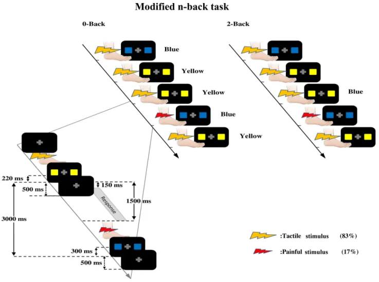
Data analysis was conducted using Statistica v13 (Dell Inc.., Tulsa, OK, USA). All results are expressed as the mean ± standard error of the mean, and the statistical thresh-old was set to p ≤ 0.05 (two-tailed). A priori hypotheses were tested with planned contrasts, and the type I error rate was controlled for using the Bonferroni correction for mul-tiple comparisons, based on the number of comparisons for each independent analysis. All reported p values are Fig. 1 Modified n-back task. Participants performed a modified
n-back task in which they had to discriminate the color of each vis-ual stimulus constituted of two squares which were either both blue or both yellow. In the 0-back condition, participants discriminated the color of the current stimulus directly after its presentation; in the 2-back condition, they responded to the stimulus presented two trials before. The visual stimulus was preceded by a tactile stimulus in 83% of trials or by a painful stimulus in the remaining trials (17%). Bot-tom left panel Sequential timings of stimuli in each trial. A fixation
cross was present at the center of the screen during the entire trial. Electrical stimuli were followed by a visual stimulus of 500 ms dura-tion. The interval between the somatosensory and visual stimuli (ISI) was 220 ms for the tactile trials and 300 ms for the painful trials. Per-formance in the modified n-back task was measured in the time win-dow running from 150 to 1500 ms after onset of the visual stimulus. The next trial started at a latency set so that the inter-trial interval (ITI) measured between the onsets of two consecutive visual stimuli was 3000 ms
therefore corrected for multiple comparisons for all varia-bles, including RT, RA, pain, pain-related anxiety and NFR amplitude. Effect sizes are reported based on partial eta-squared values ( 𝜂2
p).
Results
Manipulation checks
Pre-tDCS baseline values are presented in Table 1. To con-firm that experimental effects crucial to test our hypotheses were observed prior to the tDCS intervention, we performed Bonferroni-corrected planned contrasts to show that WM
performance was unaffected by painful stimuli and that pain was inhibited by the engagement of WM. Accordingly, in the anodal tDCS session, pain did not significantly affect RT or RA for either the 0-back or the 2-back tasks (RT: p > 0.6,
𝜂2p < 0.01; RA: p > 0.2, 𝜂p2 < 0.01). Likewise, in the sham
tDCS session, pain did not significantly alter RT or RA for either the 0-back or 2-back tasks (p > 0.7, 𝜂2
p ≤ 0.04; p > 0.2,
𝜂2p = 0.11 and < 0.01, respectively).
As expected based on prior studies [68], pain perception was decreased by WM for both the 0-back and 2-back tasks in the anodal tDCS session (p = 0.04 and p = 0.003,
𝜂2p = 0.16 and 0.26, respectively) and the sham tDCS session
Fig. 2 Experimental design. The experimental protocol comprised five counterbalanced conditions, including the 0-back, 2-back, pain, 0-back with pain and 2-back with pain conditions. This experimental protocol was performed twice during each session, once to establish a pre-transcranial direct current stimulation (tDCS) baseline and once
during tDCS. The same order was used for the sham and anodal tDCS sessions for a given participant. Each condition contained 60 trials within 3.5 min. Participants had to verbally rate their average pain and their pain-related anxiety using a numerical rating scale (NRS; range 0–100) after each condition comprising painful stimuli
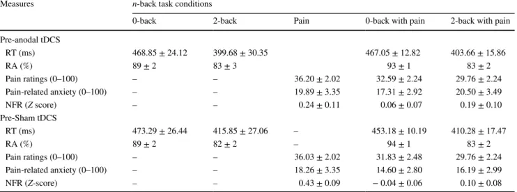
Table 1 Manipulation checks
Values in table are presented as the mean ± standard error of the mean
tDCS, Transcranial direct current stimulation; RT, response time; RA, response accuracy; NFR, nociceptive flexion reflex Measures n-back task conditions
0-back 2-back Pain 0-back with pain 2-back with pain Pre-anodal tDCS RT (ms) 468.85 ± 24.12 399.68 ± 30.35 467.05 ± 12.82 403.66 ± 15.86 RA (%) 89 ± 2 83 ± 3 93 ± 1 83 ± 2 Pain ratings (0–100) – – 36.20 ± 2.02 32.59 ± 2.24 29.76 ± 2.24 Pain-related anxiety (0–100) – – 19.89 ± 3.35 17.31 ± 2.92 20.50 ± 3.49 NFR (Z score) – – 0.24 ± 0.11 0.06 ± 0.07 0.19 ± 0.10 Pre-Sham tDCS RT (ms) 473.29 ± 26.44 415.85 ± 27.06 – 453.18 ± 10.19 410.28 ± 17.47 RA (%) 89 ± 2 82 ± 2 – 94 ± 1 83 ± 2 Pain ratings (0–100) – – 36.03 ± 2.02 31.83 ± 2.48 29.76 ± 2.24 Pain-related anxiety (0–100) – – 18.26 ± 3.35 14.60 ± 2.80 16.19 ± 2.99 NFR (Z-score) – – 0.43 ± 0.09 − 0.04 ± 0.06 0.10 ± 0.08
(both p < 0.001, 𝜂2
p = 0.31 and 0.58, respectively). In
con-trast, pain-related anxiety was not significantly altered by WM for either the 0-back or 2-back tasks in either the anodal or sham tDCS sessions (all p > 0.2, 𝜂2
p ≤ 0.09). Regarding
spinal nociceptive activity, NFR amplitude was not signifi-cantly altered by WM (all p > 0.12, 𝜂2
p ≤ 0.11) except for the
0-back task of the sham condition in which it was decreased (p = 0.001, 𝜂2
p = 0.29).
Taken together, these results confirm that WM perfor-mance was not altered by the task-irrelevant painful stimuli. In addition, engagement of WM produced the expected decrease in pain perception, indicative of top–down regula-tion of pain by cognitive processes.
Effects of anodal tDCS
Working memory
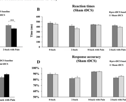
Anodal tDCS significantly reduced RT in the 2-back task with or without pain compared with the respective pre-tDCS baseline values (both p < 0.01, 𝜂2
p = 0.25 and 0.32,
respec-tively; see Fig. 3a), while no difference was observed for the 0-back task with or without pain compared with their respec-tive pre-tDCS baseline values (both p > 0.5, 𝜂2
p = 0.06
and < 0.01, respectively; see Fig. 3a). In addition, no signifi-cant effect was produced by sham tDCS for either task, with or without pain (all p > 0.4, all 𝜂2
p < 0.09; see Fig. 3b).
Con-sistent with the reduction of RT, RA tended to improve with anodal tDCS in the 2-back with pain task compared with its pre-tDCS baseline value (p = 0.057, 𝜂2
p = 0.20), but no effect
was observed for the other tasks (all p > 0.4, all 𝜂2
p ≤ 0.06).
In contrast, the sham tDCS did not produce any significant change in RA for any task compared with the respective pre-tDCS baseline values (all p > 0.3, all 𝜂2
p ≤ 0.06; see
Fig. 3c, d). The between-session comparisons for RT and RA revealed no significant difference between anodal and sham tDCS (all p’> 0.4, 𝜂2
p < 0.07). Pain ratings
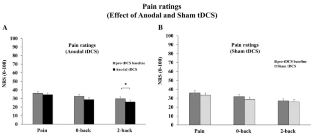
Anodal tDCS marginally improved pain inhibition by WM in the 2-back with pain task compared with its pre-tDCS
Fig. 3 Effect of tDCS on working memory (WM). a Reaction times (RT) during anodal tDCS. Anodal tDCS significantly reduced RT in the 2-back task with or without pain, compared with pre-tDCS baseline values, while no difference was observed for the 0-back task with or without pain compared with pre-tDCS baseline values (both p > 0.5). b RT during sham tDCS. No significant effect was produced by sham tDCS for either task, with or without pain (all p > 0.4). c Response accuracy (RA) during anodal tDCS. Consistent
with the reduction of RT, RA tended to improve with anodal tDCS in the 2-back with pain task compared with the pre-tDCS baseline value (p = 0.057), but no effect was observed for the other tasks (all p > 0.4). d RA during sham tDCS. Sham tDCS did not produce any significant change in RA for any task compared with their respective pre-tDCS baseline values (all p > 0.3). Error bars Standard error of the mean (SEM). Double asterisks indicate significant difference at p ≤ 0.01
baseline value (p = 0.052, 𝜂2
p = 0.16; see Fig. 4a). In
con-trast, pain and pain inhibition by WM in the 0-back task were not significantly different from their respective pre-tDCS baseline values (both p > 0.2, 𝜂2
p = 0.05 and 0.11,
respectively; see Fig. 4a). Also, sham tDCS produced no significant change in pain intensity for any of the three tasks (all p > 0.2, 𝜂2
p = 0.13, 0.10 and 0.01, respectively; see
Fig. 4b). The between-session comparisons revealed no sig-nificant difference between anodal and sham tDCS (all
p > 0.3, 𝜂2
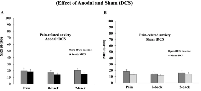
p ≤ 0.03). Pain‑related anxiety ratings
Pain-related anxiety and the inhibition of pain-related anxi-ety by WM were not significantly altered by anodal tDCS compared with their respective pre-tDCS baseline values (all
p > 0.1, 𝜂2
p = 0.01, 0.12 and 0.13, respectively; see Fig. 5a).
Similar results were observed for the sham tDCS session (all
p > 0.3, 𝜂2
p = 0.08, 0.08 and 0.02, respectively; see Fig. 5b). NFR amplitude
NFR amplitude was significantly decreased during anodal tDCS compared with the pre-tDCS baseline value (p = 0.04,
𝜂p2 = 0.17; see Fig. 6). NFR inhibition by WM in the 0-back
with pain and the 2-back with pain tasks were not signifi-cantly changed during anodal tDCS, although it tended to decrease compared with the respective pre-tDCS baseline
values (p = 0.067 and p = 0.057, 𝜂2
p = 0.15 and 0.16,
respec-tively; see Fig. 6). In addition, NFR amplitude was signifi-cantly decreased during sham tDCS compared with the pre-tDCS baseline value (p < 0.001; 𝜂2
p = 0.38; see Fig. 6) while
inhibition of NFR by WM was significantly greater than its pre-tDCS baseline value in the 0-back with pain task (p = 0.036, 𝜂2
p = 0.18) but not the 2-back with pain task
(p = 0.13, 𝜂2
p = 0.13; see Fig. 6).
Discussion
The novel finding of this study is that pain inhibition by WM was enhanced by anodal tDCS of the left DLPFC, especially when WM engagement was stronger (2-back task). In con-trast, pain perception was unchanged by anodal tDCS when painful stimuli were administered alone without the con-current cognitive task. In addition, anodal tDCS improved WM but not NFR inhibition by WM, suggesting that anodal tDCS enhances pain inhibition by improving WM but not by increasing descending inhibition of spinal nociceptive activity.
Enhancement of WM and pain inhibition by tDCS
To have effective attentional control during pain percep-tion, both the disengagement of attention from pain stimuli and the direction of attention to task-related information are essential [1, 12, 30, 69]. In order to test the specific
Fig. 4 Effect of tDCS on pain ratings (NRS: 0–100). a Pain ratings during anodal tDCS. Anodal tDCS marginally improved pain inhi-bition by WM in the 2-back with pain task compared with its pre-tDCS baseline value. In contrast, pain and pain inhibition by WM in the 0-back task were not significantly different from their respective
pre-tDCS baseline values (both p > 0.2). b Pain ratings during sham tDCS. Sham tDCS produced no significant change in pain intensity for any of the three tasks (all p > 0.2). Error bars SEM. Single aster-isk indicates significant difference at p = 0.052
inhibitory effect of WM engagement on pain perception, we used a modified n-back task with different WM loads (0-back and 2-back) [6, 29]. We compared conditions in which the n-back task was performed with or without painful distractors. To make painful distractors more salient and novel, they were applied rarely and randomly among frequent non-painful stimuli [6, 29]. Moreover,
to determine the specific effect of anodal tDCS compared with sham tDCS and to control non-specific between-ses-sion effects, we performed the experimental protocol twice during each session, once as pre-tDCS baseline and once during tDCS. This allowed a within-session assessment of anodal tDCS and sham tDCS effects.
Fig. 5 Effect of tDCS on pain-related anxiety. a Pain-related anxiety during anodal tDCS. Pain-related anxiety and the inhibition of pain-related anxiety by WM were not significantly modulated by anodal tDCS compared with their respective pre-tDCS baseline values (all
p > 0.1). b Pain-related anxiety during sham tDCS. Pain-related anxi-ety and the inhibition of pain-related anxianxi-ety by WM were not sig-nificantly modulated by sham tDCS compared with their respective pre-tDCS baseline values (all p > 0.3). Error bars SEM
Fig. 6 Effect of tDCS on nociceptive flexion reflex (NFR) amplitude. a NFR modulation during anodal tDCS. NFR amplitude was signifi-cantly decreased during anodal tDCS compared with the pre-tDCS baseline value. NFR inhibition by WM in the 0-back with pain and the 2-back with pain tasks was not significantly changed by anodal tDCS, although it tended to decrease compared with their respective pre-tDCS baseline values (p = 0.067 and p = 0.057, respectively).
b NFR modulation during sham tDCS. NFR amplitude was signifi-cantly decreased during sham tDCS compared with the pre-tDCS baseline value while inhibition of NFR by WM was significantly greater than the pre-tDCS baseline value in the 0-back with pain task (p = 0.036) but in not the 2-back with pain task (p = 0.13). Error bars SEM. Single and triple asterisks indicate significant difference at p < 0.05 and p < 0.001, respectively
During the n-back task, mean RT was decreased during anodal tDCS over the left DLPFC. This effect was particu-larly observed in the high WM load condition (2-back task), while no effect was observed in the low WM load condition (0-back task). These results are consistent with improve-ment of WM by anodal tDCS of the left DLPFC [54–58,
61, 70] and with involvement of the DLPFC in the central executive system of WM [1, 11, 31, 33, 35, 36, 42, 55, 71]. They also extend these findings by showing that this WM improvement may contribute to the enhancement of pain inhibition. Indeed, pain inhibition by WM was enhanced by anodal tDCS in the high WM load condition. In con-trast, pain perception was not affected by anodal tDCS when there was no engagement of WM (no n-back task), suggest-ing that in the present conditions, anodal tDCS of the left DLFPC may produce indirect effects on pain inhibition, through cognitive processes, without affecting pain percep-tion directly. Our study also provides novel findings showing that increased pain inhibition by WM during tDCS is not associated with significant inhibition of the NFR, suggesting that tDCS effects on pain inhibition by WM are mediated by supraspinal processes independent of descending pain inhibition processes.
tDCS neuromodulation may affect various brain networks depending on the positioning of the stimulating electrodes and on the state of the stimulated network [72, 73]. As a result, the outcome of the stimulation protocol depends on task characteristics, including WM load, as well as the state of the neural network [72, 74]. Coherent with this notion, some tDCS studies indicate that the effects of anodal tDCS are affected by task difficulty [75, 76]. The availability of cognitive resources for optimal task performance is criti-cal, and the effects of tDCS may depend on increasing the availability of cognitive resources, especially when WM is highly loaded or saturated. In conditions with low WM load, cognitive resources are available as they are not monopo-lized by the task, so tDCS may not bring any gain in perfor-mance. This may explain some of the discrepancies observed between studies examining the effect of tDCS. The lack of tDCS effect may be due to the use of cognitive tasks that are not sufficiently demanding [74]. Based on our findings, we propose that anodal tDCS of the DLPFC may be more effec-tive during more demanding tasks, in accordance with the state-dependant or load-dependent effects reported earlier [74, 77]. This also leads to the inference that anodal tDCS of the DLPFC may be especially useful in clinical conditions in which WM and other cognitive functions are reduced.
Interactions between pain and WM
A nociceptive stimulus may be selected to prioritize a protective behavior in response to pain perception at the expense of task performance [1, 8, 13–18]. Conversely,
pain perception can be inhibited by cognitive processes if task execution is prioritized, in accordance with contextual demands [6, 12, 17, 19–22]. In the present experiment, the protocol was designed to favor the execution of a cogni-tive task and the inhibition of pain. The comparison of WM performance (RT) during the pre-tDCS baseline showed no difference between conditions with or without pain. These results established that in our protocol, WM performance was not affected by salient painful distractors for either task difficulty (0-back or 2-back). WM engagement by rehears-ing the features of visual targets was sufficient to avoid a bottom–up shift of attention to the salient painful distrac-tors [1, 6, 12, 27, 29, 44]. In addition, accuracy in WM was consistent across all conditions. These findings have also been observed in previous pain studies [12, 29, 68]. Indeed, nociceptive signals compete with other sensory signals for entering the attentional network and being processed further by this network [1–3, 5]. This neural response to specific stimuli can be biased by stimulus saliency (bottom–up fil-ter) [13, 15, 18] or by the relevance of stimuli for the task (top–down bias) [12, 22, 24, 25]. The central executive com-ponent of WM that maintains task-relevant target features (attentional set) [1, 30] and the maximal attentional load of WM capacity [31–34] can be one source of bias [1, 29, 39]. Our results indicate that the present experimental paradigm is adapted to favor top–down inhibition of salient nocicep-tive signals. Moreover, the 0-back and 2-back conditions produced the expected decrease in the pain ratings during the pre-tDCS baseline. These results are consistent with RT being unaltered by pain and are also in line with results from previous studies [17, 40, 68, 78]. In summary, salient painful stimuli have the potential to disrupt WM, but this disrup-tion is determined by the balance between bottom–up and top–down processes according to experimental conditions, including the WM task [40, 79], the type and intensity of painful distractors as well as their novelty [6, 12, 40]. In the present study, task performance was maintained, and pain was inhibited in conditions involving both WM engagement and painful distractors, thereby allowing us to examine the effect of anodal tDCS of the DLPFC on pain inhibition by WM.
We also investigated the modulation of spinal nocic-eptive activity by WM with the NFR. NFR amplitude was reduced during low WM load (0-back) but not during high WM load (2-back) compared with the pain alone condition. The reduction of NFR amplitude suggests that descending pain inhibitory pathways were activated. However, the lack of inhibition in the high WM load condition was somewhat unexpected. Increased WM load and decreased pain per-ception should be associated with decreased NFR ampli-tude [80–82], although dissociation between spinal activity and pain perception has been reported in previous studies [83–87]. In the context of the present study, we postulate
that the more demanding task produces a disinhibition of spinal nociceptive activity to maintain protective reflexes while WM shields cognition from nociceptive signals in the brain, with the aim to allow optimal task performance.
Limitations and future directions
Participants were asked to rate pain after each painful con-dition. This pain rating task has the potential to make the painful stimuli relevant for participant’s goals [7], which could reduce the inhibitory effects of WM by altering the balance between bottom–up and top–down processes. Also, although experimental conditions and sessions were coun-terbalanced between participants, the same participants performed all conditions in both sessions, which has the potential to increase the effect of sham tDCS and decrease the relative effect of anodal tDCS compared with sham tDCS. However, this within-subject design is a fair com-promise to avoid inter-subject variability, which may be larger than the within-subject counfounding. Nevertheless, this design remains to be assessed in future studies. It could also be argued that within-subjects designs limit blinding of participants because they may feel a different sensation between the sham and anodal sessions. However, partici-pants were not aware that two different types of stimula-tion were used, and they could feel electrical current in both sessions. Although the sensation may have been different, they received no instructions that may have induced a bias. Lastly, it should be emphasized that in spite of significant differences between sessions, we show significant effects for anodal stimulation and no significant effect for sham stimu-lation, which indicates that anodal tDCS produces signifi-cant effects that cannot be explained by non-specific effects only, including placebo.
Conclusion
The results of our study are consistent with top–down sup-pression of pain by WM and with its improvement by anodal tDCS of left DLPFC, especially with more important WM engagement. In addition, anodal tDCS improved WM but not NFR inhibition by WM, implying that increased pain inhibition by WM improvement is independent of descend-ing inhibition of spinal nociception.
Author contributions ZD contributed to all aspects of the research. SB contributed to data acquisition, analyses and interpretation. NR contributed to data acquisition and interpretation. IB contributed to experimental design, data interpretation and manuscript preparation. MP contributed to all aspects of the research and obtained funding for the study.
Funding This study was supported by a grant from the Natural Sciences and Engineering Research Council of Canada to MP (402176). The contribution of MP was supported by the research Chair in Pain Neuro-physiology from “Université du Québec à Trois-Rivières” (UQTR), the “Fonds de Recherche du Québec en Santé (FRQS) and the “Fondation de Recherche en Chiropratique du Québec. The contribution of Zoha Deldar was supported by the department of Anatomy at UQTR.
Compliance with ethical standards
Ethical approval All experimental procedures conformed to the stand-ards set by the latest revision of the Declaration of Helsinki and were approved by the Research Ethics Board of Université du Québec à Trois-Rivières. All participants gave written informed consent, acknowledging their right to withdraw from the experiment without prejudice.
Conflict of interest Zoha Deldar reports no financial or other relation-ship that may lead to any conflict of interest. Nabi Rustamov reports no financial or other relationship that may lead to any conflict of interest. Suzie Bois reports no financial or other relationship that may lead to any conflict of interest. Isabelle Blanchette reports no financial or other relationship that may lead to any conflict of interest. Mathieu Piché reports no financial or other relationship that may lead to any conflict of interest.
References
1. Legrain V, Damme SV, Eccleston C, Davis KD, Seminowicz DA, Crombez G (2009) A neurocognitive model of attention to pain: behavioral and neuroimaging evidence. Pain 144(3):230–232 2. Legrain V, Mancini F, Sambo CF, Torta DM, Ronga I,
Valen-tini E (2012) Cognitive aspects of nociception and pain: bridging neurophysiology with cognitive psychology. Neurophysiol Clin 42(5):325–336
3. Berti S, Roeber U, Schröger E (2004) Bottom–up influences on working memory: behavioral and electrophysiological distraction varies with distractor strength. Exp Psychol 51(4):249–257 4. Barcelo F, Escera C, Corral MJ, Periáñez JA (2006) Task
switch-ing and novelty processswitch-ing activate a common neural network for cognitive control. J Cognit Neurosci 18(10):1734–1748 5. McCaul KD, Malott JM (1985) Distraction and coping with pain.
Pain 23(3):315
6. Legrain V, Crombez G, Plaghki L, Mouraux A (2013) Shield-ing cognition from nociception with workShield-ing memory. Cortex 49(7):1922–1934
7. Torta DM, Legrain V, Mouraux A, Valentini E (2017) Attention to pain! A neurocognitive perspective on attentional modulation of pain in neuroimaging studies. Cortex
8. Legrain V, Perchet C, Garcia-Larrea L (2009) Involuntary ori-enting of attention to nociceptive events: neural and behavioral signatures. J Neurophysiol 102(4):2423–2434
9. Verhoeven K, Van Damme S, Eccleston C, Van Ryckeghem DM, Legrain V, Crombez G (2011) Distraction from pain and executive functioning: an experimental investigation of the role of inhibition, task switching and working memory. Eur J Pain 15(8):866–873
10. Corbetta M, Shulman GL (2002) Control of goal-directed and stimulus-driven attention in the brain. Nat Rev Neurosci 3(3):201–215
11. Awh E, Vogel EK, Oh SH (2006) Interactions between attention and working memory. Neuroscience 139(1):201–208
12. Legrain V, Crombez G, Mouraux A (2011) Controlling atten-tion to nociceptive stimuli with working memory. PLoS ONE 6(6):e20926
13. Egeth HE, Yantis S (1997) Visual attention: control, representa-tion, and time course. Annu Rev Psychol 48(1):269–297 14. Escera C, Corral MJ (2007) Role of mismatch negativity and
novelty-P3 in involuntary auditory attention. J Psychophysiol 21(3–4):251–264
15. Knudsen EI (2007) Fundamental components of attention. Annu Rev Neurosci 30:57–78
16. Downar J, Mikulis DJ, Davis KD (2003) Neural correlates of the prolonged salience of painful stimulation. Neuroimage 20(3):1540–1551
17. Bingel U, Rose M, Glascher J, Buchel C (2007) fMRI reveals how pain modulates visual object processing in the ventral visual stream. Neuron 55(1):157–167
18. Yantis S, Jonides J (1990) Abrupt visual onsets and selective atten-tion: voluntary versus automatic allocation. J Exp Psychol Hum Percept Perform 16(1):121–134
19. Seminowicz DA, Davis KD (2007) Interactions of pain inten-sity and cognitive load: the brain stays on task. Cereb Cortex 17(6):1412–1422
20. Seminowicz DA, Davis KD (2007) Pain enhances functional con-nectivity of a brain network evoked by performance of a cognitive task. J Neurophysiol 97(5):3651–3659
21. Legrain V, Guérit J-M, Bruyer R, Plaghki L (2002) Attentional modulation of the nociceptive processing into the human brain: selective spatial attention, probability of stimulus occurrence, and target detection effects on laser evoked potentials. Pain 99(1):21–39
22. Hopfinger JB, West VM (2006) Interactions between endogenous and exogenous attention on cortical visual processing. Neuroim-age 31(2):774–789
23. Miyake A, Friedman NP, Emerson MJ, Witzki AH, Howerter A, Wager TD (2000) The unity and diversity of executive functions and their contributions to complex “frontal lobe” tasks: a latent variable analysis. Cognit Psychol 41(1):49–100
24. Van Damme S, Legrain V, Vogt J, Crombez G (2010) Keeping pain in mind: a motivational account of attention to pain. Neurosci Biobehav Rev 34(2):204–213
25. Folk CL, Remington RW, Johnston JC (1992) Involuntary covert orienting is contingent on attentional control settings. J Exp Psy-chol Hum Percept Perform 18(4):1030–1044
26. Miller EK, Cohen JD (2001) An integrative theory of prefrontal cortex function. Annu Rev Neurosci 24:167–202
27. Soto D, Hodsoll J, Rotshtein P, Humphreys GW (2008) Automatic guidance of attention from working memory. Trends Cognit Sci 12(9):342–348
28. Tracey I, Mantyh PW (2007) The cerebral signature for pain per-ception and its modulation. Neuron 55(3):377–391
29. Legrain V, Crombez G, Verhoeven K, Mouraux A (2011) The role of working memory in the attentional control of pain. Pain 152(2):453–459
30. Crombez G, Eccleston C, Baeyens F, Eelen P (1998) Attentional disruption is enhanced by the threat of pain. Behav Res Ther 36(2):195–204
31. Lavie N, Hirst A, de Fockert JW, Viding E (2004) Load theory of selective attention and cognitive control. J Exp Psychol Gen 133(3):339–354
32. Legrain V, Bruyer R, Guerit JM, Plaghki L (2005) Involuntary orientation of attention to unattended deviant nociceptive stimuli is modulated by concomitant visual task difficulty. Evidence from laser evoked potentials. Clin Neurophysiol 116(9):2165–2174 33. Lavie N, Fockert JD (2006) Frontal control of attentional capture
in visual search. Vis Cognit 14(4–8):863–876
34. SanMiguel I, Corral M-J, Escera C (2008) When loading working memory reduces distraction: behavioral and electrophysiological evidence from an auditory-visual distraction paradigm. J Cognit Neurosci 20(7):1131–1145
35. D’Esposito M, Postle BR, Rypma B (2000) Prefrontal cortical contributions to working memory: evidence from event-related fMRI studies. Exp Brain Res 133(1):3–11
36. Levy R, Goldman-Rakic PS (2000) Segregation of working mem-ory functions within the dorsolateral prefrontal cortex. Exp Brain Res 133(1):23–32
37. Szmalec A, Verbruggen F, Vandierendonck A, Kemps E (2011) Control of interference during working memory updating. J Exp Psychol Hum Percept Perform 37(1):137–151
38. Hester R, Garavan H (2005) Working memory and executive func-tion: the influence of content and load on the control of attention. Mem Cognit 33(2):221–233
39. Baddeley A (2003) Working memory: looking back and looking forward. Nat Rev Neurosci 4(10):829–839
40. Buhle J, Wager TD (2010) Performance-dependent inhibition of pain by an executive working memory task. Pain 149(1):19–26 41. Awh E, Jonides J (2001) Overlapping mechanisms of attention
and spatial working memory. Elsevier, London, pp 119–126 42. Wager TD, Spicer J, Insler R, Smith EE (2014) The neural bases
of distracters resistant working memory. Cognit Affect Behav Neurosci 14(1):90–105
43. Berti S, Schröger E (2003) Working memory controls involuntary attention switching: evidence from an auditory distraction para-digm. Eur J Neurosci 17(5):1119–1122
44. Soto D, Heinke D, Humphreys GW, Blanco MJ (2005) Early, involuntary top–down guidance of attention from working mem-ory. J Exp Psychol Hum Percept Perform 31(2):248–261 45. Jan WDF, Rees G, Frith CD, Lavie N (2001) The role of
working memory in visual selective attention. Science 291(5509):1803–1806
46. Berryman C, Stanton TR, Jane Bowering K, Tabor A, McFarlane A, Lorimer Moseley G (2013) Evidence for working memory defi-cits in chronic pain: a systematic review and meta-analysis. Pain 154(8):1181–1196
47. Baker KS, Gibson S, Georgiou-Karistianis N, Roth RM, Gium-marra MJ (2016) Everyday executive functioning in chronic pain: specific deficits in working memory and emotion control, pre-dicted by mood, medications, and pain interference. Clin J Pain 32(8):673–680
48. Ferreira KDS, Oliver GZ, Thomaz DC, Teixeira CT, Foss MP (2016) Cognitive deficits in chronic pain patients, in a brief screening test, are independent of comorbidities and medication use. Arq Neuropsiquiatr 74(5):361–366
49. Moriarty O, McGuire BE, Finn DP (2011) The effect of pain on cognitive function: a review of clinical and preclinical research. Prog Neurobiol 93(3):385–404
50. Sammer G, Brück C, Haberkamp A, Bischoff M, Blecker CR (2009) Visuospatial working memory, executive functioning, language comprehension and aging. Neuroimage 47:S109 51. Mitchell KJ, Johnson MK, Raye CL, Mather M, D’Esposito M
(2000) Aging and reflective processes of working memory: bind-ing and test load deficits. Psychol Agbind-ing 15(3):527–541 52. Gazzaley A, Rissman J, Cooney JW, D’Esposito M (2005) top–
down suppression deficit underlies working memory impairment in normal aging. Nat Neurosci 8(10):1298–1300
53. Sambataro F, Murty VP, Callicott JH, Tan H-Y, Das S, Weinberger DR, Mattay VS (2010) Age-related alterations in default mode network: impact on working memory performance. Neurobiol Aging 31(5):839–852
54. Brunoni AR, Vanderhasselt MA (2014) Working memory improvement with non-invasive brain stimulation of the
dorsolateral prefrontal cortex: a systematic review and meta-analysis. Brain Cognit 86:1–9
55. Wolkenstein L, Plewnia C (2013) Amelioration of cognitive con-trol in depression by transcranial direct current stimulation. Biol Psychiatry 73(7):646–651
56. Andrews SC, Hoy KE, Enticott PG, Daskalakis ZJ, Fitzgerald PB (2011) Improving working memory: the effect of combining cog-nitive activity and anodal transcranial direct current stimulation to the left dorsolateral prefrontal cortex. Brain Stimul 4(2):84–89 57. Mylius V, Jung M, Menzler K, Haag A, Khader PH, Oertel WH,
Rosenow F, Lefaucheur JP (2012) Effects of transcranial direct current stimulation on pain perception and working memory. Eur J Pain 16(7):974–982
58. Mariano TY, Van’t Wout M, Garnaat SL, Rasmussen SA, Green-berg BD (2016) Transcranial direct current stimulation (tDCS) targeting left dorsolateral prefrontal cortex modulates task-induced acute pain in healthy volunteers. Pain Med 17(4):737–745 59. Boggio PS, Ferrucci R, Rigonatti SP, Covre P, Nitsche M, Pas-cual-Leone A, Fregni F (2006) Effects of transcranial direct cur-rent stimulation on working memory in patients with Parkinson’s disease. J Neurol Sci 249(1):31–38
60. Jo JM, Kim YH, Ko MH, Ohn SH, Joen B, Lee KH (2009) Enhancing the working memory of stroke patients using tDCS. Am J Phys Med Rehabil 88(5):404–409
61. Hill AT, Fitzgerald PB, Hoy KE (2016) Effects of anodal transcra-nial direct current stimulation on working memory: a systematic review and meta-analysis of findings from healthy and neuropsy-chiatric populations. Brain Stimul 9(2):197–208
62. Berryhill ME, Jones KT (2012) tDCS selectively improves work-ing memory in older adults with more education. Neurosci Lett 521(2):148–151
63. Park S-H, Seo J-H, Kim Y-H, Ko M-H (2014) Long-term effects of transcranial direct current stimulation combined with com-puter-assisted cognitive training in healthy older adults. NeuroRe-port 25(2):122–126
64. Willer JC (1977) Comparative study of perceived pain and noci-ceptive flexion reflex in man. Pain 3(1):69–80
65. Piché M, Bouin M, Arsenault M, Poitras P, Rainville P (2011) Decreased pain inhibition in irritable bowel syndrome depends on altered descending modulation and higher-order brain processes. Neuroscience 195:166–175
66. Ladouceur A, Rustamov N, Dubois JD, Tessier J, Lehmann A, Descarreaux M, Rainville P, Piche M (2017) Inhibition of pain and pain-related brain activity by heterotopic noxious counter-stimulation and selective attention in chronic non-specific low back pain. Neuroscience. https ://doi.org/10.1016/j.neuro scien ce.2017.09.054
67. Ladouceur A, Tessier J, Provencher B, Rainville P, Piche M (2012) top–down attentional modulation of analgesia induced by hetero-topic noxious counterstimulation. Pain 153(8):1755–1762 68. Coen SJ, Aziz Q, Yágüez L, Brammer M, Williams SCR,
Greg-ory LJ (2008) Effects of attention on visceral stimulus intensity encoding in the male human brain. Gastroenterology 135(6):2065. e1–2074.e1
69. Oliveira JF, Zanao TA, Valiengo L, Lotufo PA, Bensenor IM, Fregni F, Brunoni AR (2013) Acute working memory improve-ment after tDCS in antidepressant-free patients with major depres-sive disorder. Neurosci Lett 537:60–64
70. Kuo M-F, Nitsche MA (2012) Effects of transcranial electrical stimulation on cognition. Clin EEG Neurosci 43(3):192–199 71. Duncan J (2001) An adaptive coding model of neural function in
prefrontal cortex. Nat Rev Neurosci 2(11):820–829
72. Miniussi C, Harris JA, Ruzzoli M (2013) Modelling non-invasive brain stimulation in cognitive neuroscience. Neurosci Biobehav Rev 37(8):1702–1712
73. Paulus W (2011) Transcranial electrical stimulation (tES-tDCS; tRNS, tACS) methods. Neuropsychol Rehabil 21(5):602–617 74. Roe JM, Nesheim M, Mathiesen NC, Moberget T, Alnaes D,
Sneve MH (2016) The effects of tDCS upon sustained visual attention are dependent on cognitive load. Neuropsychologia 80:1–8
75. Bikson M, Name A, Rahman A (2013) Origins of specificity dur-ing tDCS: anatomical, activity-selective, and input-bias mecha-nisms. Front Hum Neurosci 7:688
76. Jones KT, Berryhill ME (2012) Parietal contributions to visual working memory depend on task difficulty. Front Psychiatry 3:81 77. Wu Y-J, Tseng P, Chang C-F, Pai M-C, Hsu K-S, Lin C-C, Juan
C-H (2014) Modulating the interference effect on spatial working memory by applying transcranial direct current stimulation over the right dorsolateral prefrontal cortex. Brain Cognit 91:87–94 78. Buhle JT, Stevens BL, Friedman JJ, Wager TD (2012)
Distrac-tion and placebo: two separate routes to pain control. Psychol Sci 23(3):246–253
79. Moore DJ, Keogh E, Eccleston C (2013) The effect of threat on attentional interruption by pain. Pain 154(1):82–88
80. Sprenger C, Eippert F, Finsterbusch J, Bingel U, Rose M, Büchel C (2012) Attention modulates spinal cord responses to pain. Curr Biol CB 22(11):1019–1022
81. Eippert F, Finsterbusch J, Bingel U, Buchel C (2009) Direct evi-dence for spinal cord involvement in placebo analgesia. Science 326(5951):404
82. Bushnell MC, Duncan GH, Dubner R, Jones RL, Maixner W (1985) Attentional influences on noxious and innocuous cutaneous heat detection in humans and monkeys. J Neurosci 5(5):1103–1110
83. Danziger N, Fournier E, Bouhassira D, Michaud D, De BT, San-tarcangelo E, Carli G, Chertock L, Willer JC (1998) Different strategies of modulation can be operative during hypnotic anal-gesia: a neurophysiological study. Pain 75(1):85–92
84. Bouhassira D, Danziger N, Attal N, Guirimand F (2003) Compari-son of the pain suppressive effects of clinical and experimental painful conditioning stimuli. Brain 126(Pt 5):1068–1078 85. Terkelsen AJ, Andersen OK, Molgaard H, Hansen J, Jensen TS
(2004) Mental stress inhibits pain perception and heart rate vari-ability but not a nociceptive withdrawal reflex. Acta Physiol Scand 180(4):405–414
86. Defrin R, Peleg S, Weingarden H, Heruti R, Urca G (2007) Differential effect of supraspinal modulation on the nocic-eptive withdrawal reflex and pain sensation. Clin Neurophysiol 118(2):427–437
87. Piche M, Arsenault M, Rainville P (2009) Cerebral and cerebro-spinal processes underlying counterirritation analgesia. J Neurosci 29(45):14236–14246