HAL Id: tel-03019506
https://tel.archives-ouvertes.fr/tel-03019506
Submitted on 23 Nov 2020
HAL is a multi-disciplinary open access archive for the deposit and dissemination of sci-entific research documents, whether they are pub-lished or not. The documents may come from teaching and research institutions in France or abroad, or from public or private research centers.
L’archive ouverte pluridisciplinaire HAL, est destinée au dépôt et à la diffusion de documents scientifiques de niveau recherche, publiés ou non, émanant des établissements d’enseignement et de recherche français ou étrangers, des laboratoires publics ou privés.
Co-occurrence of DON and Emerging Mycotoxins in
Worldwide Finished Pig Feed and their Combined
Toxicity in Intestinal Cells and Liver Slices
Abdullah Khoshal
To cite this version:
Abdullah Khoshal. Co-occurrence of DON and Emerging Mycotoxins in Worldwide Finished Pig Feed and their Combined Toxicity in Intestinal Cells and Liver Slices. Toxicology and food chain. Université Paul Sabatier - Toulouse III, 2020. English. �NNT : 2020TOU30017�. �tel-03019506�
THÈSE
En vue de l’obtention du
DOCTORAT DE L’UNIVERSITÉ DE TOULOUSE
Délivré par l'Université Toulouse 3 - Paul Sabatier
Présentée et soutenue par
Abdullah Khan KHOSHAL
Le 4 mars 2020
Le déoxynivalénol et des mycotoxines émergentes dans
l'alimentation du porc: co-occurrence et toxicité combinée sur
cellules intestinales et explants hépatiques
Ecole doctorale : SEVAB - Sciences Ecologiques, Vétérinaires, Agronomiques et
Bioingenieries
Spécialité : Pathologie, Toxicologie, Génétique et Nutrition Unité de recherche :
TOXALIM - Laboratoire de Toxicologie Alimentaire
Thèse dirigée par
Isabelle OSWALD
Jury
Mme Siska CROUBELS, Rapporteure
Mme Ariane VETTORAZZI ARMENTAL, Rapporteure M. Philippe PINTON, Examinateur
Mme Viviane Mayumi MARUO, Examinatrice Mme Isabelle OSWALD, Directrice de thèse
Résumé Français
Les mycotoxines sont des métabolites secondaires toxiques produits par certains champignons filamenteux. Selon leur toxicité et leur occurrence, certaines d'entre elles dont le déoxynivalénol (DON), l'une des toxines les plus répandues dans l'alimentation humaine et animale, sont été réglementées au sein de l'Union Européenne. D'autres métabolites secondaires découverts récemment ou encore peu étudiés sont appelées mycotoxines émergentes et ne sont ni détectés en routine ni réglementés. Les denrées alimentaires destinées à l'homme et à l'animal peuvent être naturellement contaminées par plusieurs mycotoxines et le risque lié à une exposition à des mélanges de mycotoxines est préoccupant.
Parmi les animaux d'élevage, le porc est une espèce très sensible aux mycotoxines. De par son alimentation riche en céréales, il peut être exposé à de fortes concentrations de ces contaminants. 524 échantillons d'aliments complets pour porcs prélevés dans le monde entier ont été analysés par une technique de chromatographie en phase liquide couplée à la spectrométrie de masse en tandem (LC-MS/MS) pour plus de 800 métabolites. 88 % des échantillons étaient co-contaminés avec du DON et d'autres mycotoxines réglementées et émergentes.
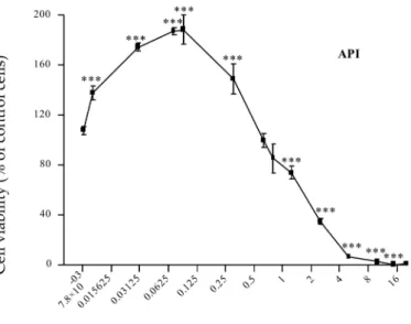
La toxicité du DON et des 10 mycotoxines émergentes les plus répandues a été évaluée en mesurant la viabilité de cellules épithéliales intestinales porcines (IPEC-1) après 48 h d'exposition. Trois mycotoxines émergentes (brevianamide F, cyclo-(L-Pro-L-Tyr) et tryptophol) n'ont pas eu d’impact sur la viabilité cellulaire. Les autres toxines ont été classées dans l'ordre de toxicité suivant: apicidine> enniatine A1> DON> beauvéricine> enniatine B> enniatine B1> émodine> aurofusarine.
La toxicité combinée du DON et des 10 mycotoxines émergentes a été évaluée en fonction de leurs concentrations réelles dans les aliments analysés. Nous avons observé que malgré la très forte fréquence des co-contaminations, la corrélation entre les concentrations de DON et des mycotoxines émergentes étudiées était faible. Nous avons donc évalué les effets toxiques de trois mélanges correspondant à des situations auxquelles les animaux peuvent être exposés. Le ratio n°1 a été calculé en utilisant la concentration P25 (1er quartile) de la mycotoxine émergente et la concentration P75 (3ème quartile) du DON. Le ratio n°3 correspondait au scénario inverse du ratio n°1. Le ratio n°2 a été calculé en utilisant la concentration médiane (2ème quartile) du DON et de chaque mycotoxine émergente.
Pour la plupart des mélanges, la cytotoxicité combinée était similaire à celle du DON seul. Pour ce qui concerne la santé des animaux, ces résultats ont montré que lorsque ces mycotoxines émergentes sont présentes avec le DON, elles n'exacerbent pas la toxicité du DON.
Outre l'intestin, le foie est le principal site de détoxification des xénobiotiques, y compris des mycotoxines, et représente un organe cible des contaminants alimentaires. Nous avons donc mis au point un nouvel outil, les Precision Cut Liver Slices (PCLS), des explants de foie d’épaisseur définie. Elles ont été utilisées pour évaluer la toxicité du DON (3 et 10 µM) à différents temps d'incubation (0 à 20 h), en étudiant l'expression génique, le contenu en ATP et en protéines totales.
Le milieu d'incubation a permis d'évaluer la qualité des PCLS en mesurant les marqueurs de dommage hépatique (phosphatase alcaline, lactate déshydrogénase, alanine aminotransférase, aspartate aminotransférase et protéines totales). Nous avons montré que ces marqueurs n’étaient impactés ni par le temps d'incubation, ni par le traitement. Les PCLS traitées avec 10 µM de DON pendant 4 h ou plus, montrent une altération de l’expression de certains gènes.
Ces expériences préliminaires ont montré que les PCLS représentent un modèle prometteur pour évaluer la toxicité hépatique des mycotoxines ou d'autres contaminants alimentaires.
Mots-clés: Mycotoxines émergentes, DON, Toxicité, Aliments pour porcs, IPEC-1, Slice de foie, Expression de gènes
Summary
Mycotoxins are toxic secondary metabolites, produced by several filamentous fungi. Depending on their toxicity and occurrence, some of them, including deoxynivalenol (DON), one of the most common toxin in food and feed, have been regulated in the European Union. Other secondary metabolites, which neither routinely determined nor regulated, are called emerging mycotoxins because they have been recently discovered or poorly investigated. Food and feed can be naturally contaminated by several mycotoxins and concern about the hazard of exposure to mycotoxin mixtures is increasing.
Among farm animals, pig is one of the most sensitive farm animal to mycotoxins and it can be exposed, through its rich cereal diet, to high concentrations of mycotoxins. In total, 524 finished pig feeds samples from worldwide were analyzed for more than 800 metabolites using, LC-MS/MS (liquid chromatography tandem-mass spectrometry) method. Eighty-eight percent of the samples were co-contaminated with DON and other regulated and emerging mycotoxins. The toxicity of DON and the 10 most common emerging mycotoxins was analyzed on the viability of porcine intestinal epithelial cells (IPEC-1) over a 48 h period. Among the emerging mycotoxins, 3 of them (brevianamide F, cyclo-(L-Pro-L-Tyr), and tryptophol) did not alter cells viability. The other mycotoxins were ranked in the following order of toxicity: apicidin> enniatin A1> DON> beauvericin> enniatin B> enniatin B1> emodin> aurofusarin.
The combined toxicity of DON and the 10 emerging mycotoxins was assessed based on their actual ratios found in pig feed. We observed that, despite the very high frequency of co-contamination, there was a poor correlation between the concentrations of DON and emerging mycotoxins. Thus, we assessed the toxic effects of three mixtures corresponding to situations to which animals may be exposed. Ratio #1 was calculated using the P25 (1st quartile) concentration of the emerging mycotoxin and P75 (3rd quartile) concentration of DON. Ratio #2 was calculated using the median (2nd quartile) concentration of DON and each emerging mycotoxin. Ratio #3 was the reverse scenario of ratio #1.
Cytotoxicity analyses showed that, in most of the mixtures, the combined toxicity was similar to the one of DON alone. These results demonstrated that, when these emerging mycotoxins are present with DON, in terms of pig health, it does not exacerbate the problem of the toxicity of DON.
In addition to intestine, liver is the main site of detoxification for xenobiotics, including mycotoxins and represents a target organ for food contaminants. Hence, we developed a new tool the Precision Cut Liver Slices (PCLs) an ex vivo explants of liver with a well-defined thickness. This tool was used to assess the toxicity of DON (3 and 10 µM) at different incubation times (0 to 20 h), by studying gene expression, ATP and total protein contents. The incubation medium was used to assess the quality of PCLS by measuring liver damage markers (alkaline phosphatase, lactate dehydrogenase, alanine transaminase, aspartate transaminase and total proteins). We showed that these markers were not affected by either incubation time or treatment.
PCLS treated with 10 µM DON for 4 h or more, showed an alteration in the expression of certain genes. These preliminary experiments demonstrated that PCLS represent a promising model for assessing the hepatic toxicity of mycotoxins or other food contaminants.
Keywords: Emerging Mycotoxins, DON, Toxicity, Pig Feed, IPEC-1, Liver Slices, Gene Expression
Acknowledgements
The work presented in this thesis would not have been possible without many people in so many ways. I take this opportunity to extend my sincere gratitude and appreciations to all those who made this Ph.D thesis possible.
First and foremost, I would like to extend my sincere gratitude and deepest appreciation to my Ph.D director “Dr. Isabelle Oswald”, as well research guide and Ph.D supervisor “Dr. Philippe Pinton” for their full support, expert guidance, understanding and encouragement throughout these three years research project. Without their incredible patience and timely wisdom and counsel, my thesis work would have been a frustrating and overwhelming pursuit. I would like to say a heartfelt thanks to both of them.
Second, I would like to express my deepest gratitude to “Dr. Gerd Schatzmayr” and “Ms. Barbara Novak” for their coordination, and regular monitoring of my research work and taking time to talk with me on many occasions. I also greatly appreciate the support received through the collaborative work undertaken with the BIOMIN, for providing the raw data for our article. Many of my statistical work would not have been completed without the assistance and cooperation of “Dr. Pascal G. P. Martin” and “Dr. Timothy Jenkins”. I would like to thank them for statistical analysis and providing some figures and tables to our article.
Many of my experimental work would not have been completed without cooperation of “Ms. Manon Neves”, “Ms. Elodie Person” and “Mrs. Sandrine Bruel”, I would like to thank for all of their time, help and kind cooperation.
I am grateful to Team-2 Metabolism of Xenobotic (MeX) who gave me permission to work in their laboratory and use the facilities available for the liver slices.
My thanks also go out to the support I received from the collaborative work with my dear friends and colleagues: Joelle Laffitte and Sylvie Puel for their availability, introductory in technique, assistance and contribution to my data collection. I am also very grateful to all those our members of the team: Dr. Olivier Puel, Dr. Laura Soler Vasco, Dr. Chloe Terciolo, Dr. Sophie Lorber, Tarek Lahjouji, Dr. Alix Pierron, Dr. Shuai Wang, Carine Al-Ayoubi, Su LUO ... who contribute to the perfect atmosphere of this laboratory so welcoming which I found each time with much pleasure.
This PhD study would not have been possible without the corporation and support extended by the doctoral school “SEVAB”. A very special thanks to Dominique Pantalacci and Professor Claud Maranges for their patience during the numerous focus group discussions as well for the administrative organization.
My deep appreciation also goes out to my M.Sc. internship supervisor, “Dr. Alain Pinton” for being so supportive, encouragement and his valuable advices to pursue my PhD.
I gratefully acknowledge the funding received towards my PhD from the BIOMIN GmbH and Région Occitanie “ANR grants “EmergingMyco” (ANR-18-CE34-0014) and “Newmyco” (ANR-15-CE21-0010). I am also grateful to the Campus France for providing the first 18 months Security Social Scholarship “BCS” to undertake my PhD.
Last but not least, the best outcome from these past three years is finding my best friend, soul-mate, and my wife. There are no words to thank for accompanying me during my PhD, and staying away from the family and country. I would like to say a heartfelt thank you to my wife. I would also like to especially thank my parents. My hard-working parents have sacrificed their lives for us and provided unconditional love and care.
Table of Contents
1. Introduction ... 13
1.1. Generalities on mycotoxins ... 13
1.2. Deoxynivalenol (DON) ... 21
1.3. Fungal toxic secondary metabolites (Emerging Mycotoxins) ... 26
1.3.1. Enniatins (ENNs) ... 27 1.3.2. Beauvericin (BEA) ... 30 1.3.3. Aurofusarin (AFN) ... 33 1.3.4. Apicidin (API) ... 35 1.3.5. Emodin (EMO) ... 36 1.3.6. Brevianamid-F (BRV-F) ... 37 1.3.7. Cyclo-(L-Pro-L-Tyr) (Cyclo) ... 38 1.3.8. Tryptophol (TRPT) ... 39
1.4. Structure and function of the small intestine ... 41
1.5. Structure and function of the liver ... 49
2. Objectives of the study ... 58
2.1. Contribution of this doctoral project ... 59
3. Article ... 61
4. Supplementary Data of Article ... 79
5. Development of an ex vivo model for studying the effects of mycotoxins on liver: the Precision Cut Liver Slices ... 91
5.1. Materials and Methods ... 91
5.2. Statistical Analysis ... 93
5.3. Analysis of Liver damage markers in culture medium ... 93
5.3.1. Methods ... 94
5.3.2. Results and discussion ... 94
5.4. Analysis of total Proteins and ATP content in PCLS ... 95
5.4.1. Methods ... 95
5.4.2. Results and discussion ... 96
5.5. Assessment of the time and dose dependent effects of DON on the expression of selected genes ... 98
5.5.1. Methods ... 98
5.5.1.1. Extraction ... 98
5.5.1.2. Reverse transcription and PCR ... 99
5.5.2. Results and discussion ... 100
5.5.2.2. Assessment of gene expression ... 101
5.6. Discussion on PCLS ... 109
5.7. Discussion on Experimental models ... 111
5.7.1. In silico models ... 111
5.7.2. In vitro models ... 112
5.7.3. Ex vivo models ... 113
5.7.4. In vivo models ... 113
5.7.5. Use of intestinal cells to assess the toxicity of emerging mycotoxins ... 115
5.7.6. Development of PCLS model to assess toxicity of mycotoxins ... 116
6. General Conclusion ... 118 7. Perspectives ... 121 8. References ... 123 9. Publications/Communications ... 146 9.1. Publications ... 146 9.2. Oral Presentations ... 146
Abbreviations List
ABCG-8: ATP Binding CassetteAFB1:Aflatoxin B1 AFN: Aurofusarin AFs: Total Aflatoxins
AGS: Human Gastric Adenocarcinoma Cell Line
ALP: Alkaline Phosphatase ALT: Alanine Transaminase
ANOVA: One-Way Analysis of Variance API: Apicidin
AST: Aspartate Transaminase ATP: Adenosine Tri Phosphate
B2-Microglobulin: Beta-2-Microglobulin BEA: Beauvericin
BEAS-2B: Human Normal Lung Epithelial Cells
BRV-F: Brevianamide-F
CAST: Council For Agricultural Science and Technology
CAT: Catalase
CCL20: C-C Motif Chemokine Ligand CCS: Copper Chaperone
CLDN-3: Claudin-3 CTG: Cell-Titer-Glo®
CONTAM: Panel On Contaminants in the Food Chain
Cyclo: Cyclo-(Pro-L-Tyr) CYP: Cytochrome P450 Family
DMEM: Dulbecco's Modified Eagle Medium DNA: Deoxyribonucleic Acid
DOM-1: Deepoxy-deoxynivalenol
DON: Deoxynivalenol DUOX: Dual Oxidase e.g: Example
EFSA: European Food Safety Authority EMO: Emodin
ENN: Enniatins EU: European Uniton FAO: Food And Agriculture Organization
FDA: Food And Drug Administration FOS: Fos Proto-Oncogene
FUM: Fumonisins
g: Gravitational Acceleration GAP: Good Agricultural Practices GLC-82: Lung Carcinoma Cell Line GPx: Glutathione Peroxidase h : Hour
HBL-100: Human Breast Cancer Cell Line
HCEC-1CT: Non-Tumorigenic Colon Cells
HEK: Human Embryonic Kidney Cells HeLa: Human Cervix Cancer Cell Line HepG2: Human Liver Cell Line
HKG: Housekeeping Gene
HT29: Adenocarcinoma Cell Line HUVEC: Human Normal Endothelial Cells
IC50: Half Maximal Inhibitory
Concentration
LC-MS/MS: Liquid Chromatography Mass Spectrometry/Mass Spectrometry
IL: Interleukin
IPEC-1: Intestinal Porcine Epithelial Cells IPEC-J2: Intestinal Porcine Epithelial Cells from Jejunum
JRC: Joint Research Center JUN: Jun Proto-Oncogene Kg: kilogram
LD50: Lethal Dose of 50% LDH: Lactate Dehydrogenase
LDLR: Low Density Lipoprotein Receptor LOAEL: Lowest Observed Adverse Effect Level
LXR: Liver X Receptor
MCF-7: Human Breast Cancer Cell mg: milligram
MAPK: Mitogen-Activated Protein Kinases mn: Minute
mL: milliliter
MRC-5: Human Fibroblast-Like Cell Line mRNA: Messenger RNA
MT: Metallothionein Member MTL: Maximum Tolerable Level N87: Human Gastric Cell Line
NFKB:Nuclear Factor
Kappa-Light-Chain-Enhancer of Activated B cells nm: Nanometer
NOAEL: No Observable Adverse Effect Level NR1H3: Nuclear Receptor Group H Member 3 NTC: Non-Template Control
OCLN: Occludin
PCTS: Precision-cut Tissue Slices
PDI: Probable Daily Intake
qRT-PCR: Quantitative Real Time-Polymerize Chain Reaction
RASFF: Rapid Alert System for Food and Feed
RIN: RNA Integrity Number RNA: Ribonucleic Acid
RPL32: Ribosomal Protein L32 RPM: Round Per Minute
SCD1: Stearoyl-CoA Desaturase SCRAB2: Scavenger Receptor Class B SOD: Superoxide Dismutase
SP: Species
TBP: TATA-Box Binding Protein TDI: Tolerable Daily Intake TEER: Transepithelial Electrical Resistance
TNF-Α: Tumor Necrosis Factor Alpha: TRPT: Tryptophol
UK: United Kingdom
USA: United States Of America WME: Williams' Medium E ZEN: Zearalenone
$: Symbol of Dollar
‘n’: Total Number of Samples £: Symbol of Pound
€: Symbol of Euro
°C: Symbol of Degree Celsius µg: Microgram
µL: Microliter µm: Micrometer µM: Micromole Ø: Symbol of diameter
List of Figures (Excluding Figures of Article)
Figure Number Details Page Number
1 General schematization of physical, chemical and
microbial strategies used for mycotoxin reduction in food and feed chains
19
2 Layers of the Alimentary Canal. The mucosa,
submucosa, muscularis, and serosa
42
3 Anatomy of different lob of pig liver 49
4 Four steps (an overview) of PCLS preparation 92
5 Preparation and incubation of liver slices 93
6 Total protein in PCLS 96
7 Total ATP contents in PCLS 97
8 RIN obtained from an individual control PCLS in
different time of incubation
100
9 RIN curve of three controls animals over 20 h of
incubation
101
10 PCLS treated with DON 10 µM. Inflammatory
cytokines, gene modulation
104
11 PCLS treated with DON 10 µM. Lipids
metabolisms, gene modulation
105
12 PCLS treated with DON 10 µM. Oxidative Stress,
gene modulation
106
13 PCLS treated with DON 10 µM. Protein junction,
gene modulation
106
14 PCLS treated with DON 3 µM and 10 µM at 20 h
incubation
108
List of Tables (Excluding Tables of Article)
Table Number Details Page Number
1 EU recommendation of mycotoxins in pig feed 15
2 Characteristics of DON. 21
3 EU regulation for DON in foodstuff 25
4 EU regulation for DON in feedstuff 25
5 Characteristics of ENNs 27
6 Effect of ENNs in different cell types 29
7 Characteristics of BEA 31 8 Characteristics of AFN 33 9 Characteristics of API 35 10 Characteristics of EMO 36 11 Characteristics of BRV-F 38 12 Characteristics of Cyclo 39 13 Characteristics of TRPT 40
14 The roles of the cells in the small intestinal mucosa 42
15 Effect of DON on the intestine in vitro 44
16 Effect of emerging mycotoxins on the intestine in
vitro
47
17 Cellular microanatomy and their function 51
18 Effect of DON on the liver cells in vitro 51
19 Effect of DON on the liver in vivo 53
20 PCLS damage markers (ALP, AST, ALT, total
protein, LDH) level in the incubation medium
56
21 ATP contents in PCLS. Samples were treated by
DON 10 µM during 2 h and 20 h of incubation
94
22 List of selected genes assessed in PCLS in response
of DON 10 µM
1. Introduction
1.1. Generalities on mycotoxins
In today’s changing world, safety and security remains basic human needs. Ensuring food safety has been a major focus of national and international actions over the last years. Nowadays, international trade in agricultural commodities such as wheat, rice, barley, corn, sorghum, soybeans, groundnuts and oilseeds reaches hundreds of millions of tons each year (FAO 2019). In 2014, worldwide feed production was 964 million tons and among them 153 million tons were produced just in Europe (Kovalsky et al. 2016). As the occurrence of mycotoxins is widespread throughout the world, the global trade of agricultural commodities (e.g., animal feed) requires to monitor fungal toxins (Kovalsky et al. 2016). The worldwide contamination of food and feed with mycotoxins is a significant problem (Hussein and Brasel 2001). Recent surveys on the occurrence of mycotoxins have shown that 60 – 88% of the world's cereal grains are contaminated with mycotoxins (Gruber-Dorninger et al. 2019; Eskola et al. 2019). Mycotoxins can resist to high temperature and to chemicals like acids, both during storage/milling and cooking process and they are stable during food/feed processing like brewing, melting, hot drying or ensiling (Rodríguez-carrasco et al. 2016). Their presence in food and feed products is an important threat to human and animal health (Heshmati et al. 2017).
Origin of mycotoxins
Mycotoxins are toxic secondary metabolites, produced by toxigenic moulds in the Aspergillus, Alternaria, Claviceps, Fusarium, Penicillium and Stachybotrys genera occurring in food and feed commodities both pre- and post-harvest (Milićević et al. 2010) under appropriate environmental conditions (Jestoi 2008). Mycotoxins represent a potential health risk for humans and/or livestock. (García & Jarque, 2014). The exact number of mycotoxins is not known, but the number of potential toxic fungal metabolites has been estimated to be in the thousands (Council for Agricultural Science and Technology 2003). Traditionally, toxigenic fungi contaminating agricultural grains have been conventionally divided into two groups: those invading seed crops have been described as “field” fungi (e.g., Cladosporium, Fusarium, Alternaria spp.), which reputedly gain access to seeds during plant development, and those proliferating during storage, “storage” fungi, (e.g., Aspergillus; Penicillium spp) (Ismaiel and Papenbrock 2015; Alshannaq and Yu 2017). Among the field occurring mycotoxins, Fusarium
mycotoxins are the most frequently identified in grain and animal feed and it is important for farmers to manage cereals contamination by Fusarium species (Steffen and Graham 2017).
Occurrence and contamination
Over the last years, mycotoxins have been regularly classified in the top ten and top one causes (European Union (EU) members and non-members countries, respectively) for the Rapid Alert System for Food and Feed (RASFF) notifications. RASFF is an European tool that ensures the inter-countries flow of information when risks to public health are detected in the food chain (RASFF 2018). The evidence suggests that mycotoxins are occurring increasingly in agricultural products including grains destined for human and animal consumption (Steffen and Graham 2017). Food can be contaminated with mycotoxins at various stages of the food chain, in field, during storage, milling or at later points (Bennett and Klich 2003). In fact, there is a notable length of time between the harvest of agricultural commodity at the exporting country and its arrival at the distribution center of the importing country. Furthermore, storage conditions at the farm level as well as during transport under adverse weather conditions may not always be satisfactory. Therefore, there is considerable opportunity for mycotoxin contamination of agricultural commodities to take place throughout the food system (FAO and Miller 1991). On the other hands, feeding animal with contaminated feeds can lead to contaminated animal products (eggs, meat, milk…). For example, aflatoxin B1 in cattle feed can be metabolized by cows into aflatoxin M1, which is then secreted in milk (Younis et al. 2016). Furthermore, ochratoxin A in pig feed can accumulate in porcine tissues (Rutqvist 1978). Human exposure to mycotoxins also results from the consumption of several sources of food such as plant-derived foods, animal products (meat, eggs and milk) and/ or the exposure to contaminated air and dust (Council for Agricultural Science and Technology 2003).
Regulations
Regulations on mycotoxins have been established in many countries in order to protect the consumers from the harmful effects of mycotoxins. Two factors are mainly taken into account in the decision-making process of setting limits for mycotoxins: (i) the toxicity and (ii) the exposure (van Egmond et al. 2007). International survey on mycotoxins legislation in foodstuffs and feedstuffs have shown that approximately 100 countries (covering approximately 85% of the world’s inhabitants) had specific regulations or detailed guidelines for some mycotoxins (FAO 2004), whereas no data were available for about 50 countries, many of them were in
Africa (van Egmond 2002). In EU, mycotoxins regulation concern, ochratoxin A, patulin, deoxynivalenol (DON), zearalenone (ZEN), fumonisins (FUM) B1 & B2, T-2 & HT-2 toxin, aflatoxin B1 (AFB1), total aflatoxins (AFs) and patulin (Lerda 2011). In the following (Table 1), the maximum level of each above mentioned mycotoxins for pig feed is stated based on the EU recommendations.
Table 1: EU recommendation of mycotoxins in pig feed
Mycotoxins Feedstuff
Guidance value in mg/kg (ppm) relative to pig
feedingstuff with a moisture content of 12 %
DON Complementary and complete feedingstuffs for pigs 0.9
ZEN
Complementary and complete feedingstuffs for piglets and
gilts (young sows) 0.1
Sows and fattening pigs 0.25
The sum of FUMs (B1 +
B2)
Complementary and complete feedingstuffs for pig 5
The sum of T2
& HT2 Compound feed for pig 0.25
Ochratoxin A Complementary and complete feedingstuffs for pigs 0.05
(European-Commission 2016)
Economical losses
The economic consequences of mycotoxins contamination are important. Commodities over the regulation should be destroyed. In addition, contaminated feed can induce losses in animal production. Due to the insufficient information, the economic costs of mycotoxins are impossible to determine accurately. However, the U.S. Food and Drug Administration (FDA)
has utilized a computer model to estimate the losses due to selected mycotoxins such as AFs, FUM and DON. Only for United States, the mean economic annual costs of crop losses from the above mentioned mycotoxins are estimated to be $932 million (Council for Agricultural Science and Technology 2003), whereas in UK they represent approximately £200/hectare and €450/hectare in Germany (Steffen and Graham 2017).
Toxicity
Acute and chronic dietary exposure to mycotoxins can induce a variety of adverse health effects in humans and animals, making these chemically diverse substances highly relevant agricultural contaminants (Eskola et al. 2019). Mycotoxins can exhibit acute toxicity as well as carcinogenic, mutagenic, teratogenic, immunotoxic, hepatotoxic, nephrotoxic, neurotoxic or estrogenic effects in human and animals (van Egmond et al. 2007). The European Food Safety Authority (EFSA), Panel on Contaminants in the Food Chain (CONTAM) calculated the acute and chronic exposure across the 22 European countries, using the highest reliable percentile of concentrations and individual consumption data for different age groups. Toddlers were in general the age group with the highest dietary chronic and acute exposure to the mycotoxins (EFSA 2014, 2018).
Unlike bacterial toxins, fungal toxins are not proteins and are usually barely detectable by the immune system of humans and animals. Most illnesses caused by mycotoxins are not reported to the doctors, whereas low levels can be dangerous to humans health (Steffen and Graham 2017). On the other hands, diagnosis of mycotoxicoses in human and animals is difficult as they may be similar to diseases with other causations. This is even more difficult in cases where more than one mycotoxin is involved (Alassane-Kpembi et al. 2017b).
Mycotoxins are more thoroughly studied in animals. Studies on animals have demonstrated that among farm animals, pig is one of the most sensitive species to the deleterious effects of mycotoxins. It can be exposed to high concentration of mycotoxins due to its rich cereal diets (Pinton et al. 2010). Previous studies on pig species have shown that, mycotoxins affect intestinal function. The gastrointestinal tract is the first physiological barrier against food contaminants, as well as the first target for these toxicants (Pinton and Oswald 2014). Indeed, after oral intake, the gastrointestinal tract is the first possible site of interaction (Fraeyman et al. 2018). In pigs, mycotoxins can decrease the feed consumption, reduce weight gain and alter reproduction and immunity (Korosteleva et al. 2007). Ruminants are less susceptible to
mycotoxins than monogastrics, because of the rumen microbiota. Ruminant’s digestive system and especially resident bacteria and protozoa, are able to detoxify mycotoxins. Nevertheless, there is some evidence that ruminants can be poisoned by mycotoxins, causing lower animal production or even death of animals. Recent in vitro research showed that at conditions of rumen acidosis and lower microbial activity, a reduced detoxification in the rumen can take place (Debevere et al. 2020). Recent insights have generated an entirely new perspective where a bi-directional relationship exists between mycotoxins and gut microbiota, thus suggesting that gut microbiota might be involved in the development of mycotoxicosis (Jouany et al. 2009; Liew and Mohd-Redzwan 2018). Moreover, most of the toxicant can be detoxified in the liver, which is the main site of metabolism and detoxification of toxins and also a target organ of mycotoxins (Zain 2011; Pongratz and Bergander 2011). Nevertheless, the contamination of animal feed can have a major impact on dairy farming. Mycotoxins can be found in dairy products from two origins, indirect contamination, which results when dairy cows ingest feed that contains mycotoxins which pass into the milk such as aflatoxin M1 and direct contamination, which results from accidental growth of moulds secreting aflatoxins (Younis et al. 2016).
Mitigation strategies
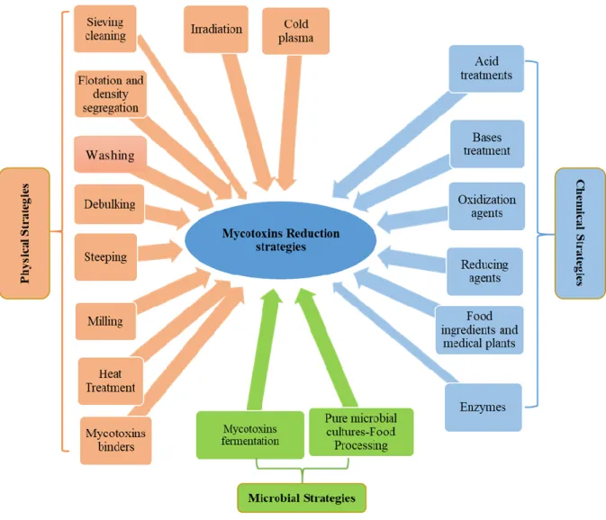
Mycotoxins are still considered unavoidable contaminants in foods and feeds, because agronomic technology has not yet advanced to the stage at which pre-harvest contamination of susceptible crops by fungi can be eliminated (Wood 1992). In order to avoid harmful effects of contamination of food and feed caused by mycotoxins, three principles could be implemented; (i) prevention of contamination; (ii) decontamination of mycotoxin containing food and feed; and (iii) inhibition of absorption of mycotoxin from the digestive tract (Karlovsky 1999). There are three common strategies to mitigate toxicity of mycotoxins (Figure 1). One of the methodologies employed for mycotoxins detoxification is the physical strategy. Even though mycotoxins are stable compounds, some food processes including sorting, trimming, cleaning, milling, brewing, cooking, baking, frying, roasting, canning, flaking, nixtamalization and extrusion may affect their chemical structure (Bretz et al. 2006). Another common mitigation strategy is biotransformation. It consists in the use of enzymes that have been found to be effective in transforming mycotoxins into less toxic metabolites or completely inactivating them (Varga et al. 2010). For example, a mixed culture of two soil bacteria Pseudomonas sp and Lysobacter sp through formation of 3-keto-DON an enzymatic epimerization was capable of transforming DON into the non-toxic 3-epi-DON (Zhai et al. 2019). Likewise, a
de-epoxidase first isolated from Eubacterium isolated from bovine rumen fluid, is capable of converting DON into the nontoxic compound DOM-1 (Pierron et al. 2016c; Loi et al. 2017). Patulin was converted by Saccharomyces cerevisiae into two isomers of ascladiol, E-ascladiol and Z-ascladiol which are nontoxic to human cell lines (Tannous et al. 2017; Wang et al. 2019). Detoxification of fumonisin B1 was carried out in two steps. Initial step was de-esterification reaction followed by deamination of the resulting hydrolyzed fumonisin B1 (Heinl et al. 2010; Grenier et al. 2012). Furthermore, isothiocyanates are natural reactive compounds that were found to reduce the toxicity of some mycotoxins (Varga et al. 2010). In addition, yeast cell wall, probiotics, prebiotics, fibers and protein ingredients have been used to reduce mycotoxins bioaccessibility in the gastrointestinal tract (Mallebrera et al. 2013) (Figure 1).
Control should begin as early as possible, starting at the farm level with primary agricultural production. Good agricultural practices (GAP) is the first line against contamination of cereals with mycotoxins (Awad et al. 2010). Furthermore, preventive strategies in storage level such as; storage at low moisture levels and prevention of grain damage during processing and prevention of the formation of mycotoxins in feed help to reduce mycotoxins content (Dawson, 2001).
Figure 1: General schematization of physical, chemical and microbial strategies used for mycotoxin reduction in food and feed chains (Luz et al. 2017).
Co-contamination
Another issue that pays attention of researchers is the co-contamination of food and feed by several mycotoxins. Food commodities are commonly contaminated with various fungal species at a time. In addition, most fungi are able to simultaneously produce number of mycotoxins. Therefore feed commodities can be contaminated by several mycotoxins, and completed feed is made from various commodities (Streit et al. 2012). The simultaneous presence of mycotoxins in products intended for human consumption is of high importance, because mycotoxins could interact with each other, potentially enhancing their toxic effects (Ruiz et al. 2011). Maize is an example where several mycotoxins have been reported to occur simultaneously (Gonçalves and Cubero-leon 2017).
There are reports of a combination of many mycotoxins, such as DON, AF (B1, B2 and M1), FUM (A, B1, B2 and B3), ZEN and other fungal secondary metabolites in maize seeds and
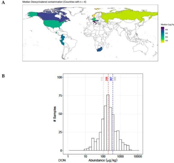
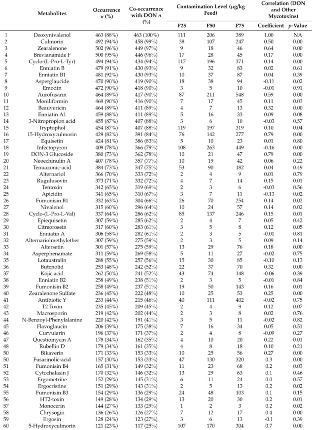
grains, as well as in animal feed formulated (Streit et al. 2013; Anjorin et al. 2016; Abdallah et al. 2017). DON co-occurs with other regulated mycotoxins as well as with emerging mycotoxins (Borutova et al. 2012; FVaclavikova et al. 2013; Streit et al. 2013). Multi-mycotoxin studies have reported that 75%–100% animal feed was co-contaminated by DON and other mycotoxins (Streit et al. 2012; Kovalsky et al. 2016; Novak et al. 2019). In food and feed ingredients sourced in Southern Europe, DON was found in 94% of the samples at maximum 365 µg/kg concentration (total number of samples ‘n’ = 416) (Griessler et al. 2010).
1.2. Deoxynivalenol (DON)
Origin
Deoxynivalenol (DON) is a regulated mycotoxin belonging to the group of type B trichothecenes (Knutsen et al. 2017), mainly produced by the fungi of the Fusarium genus, (Lewczuk et al. 2016). DON is one of the most common contaminants of wheat, corn, and barley worldwide. Second, DON is a very stable compound, during both storage and the processing/cooking of food, and does not degrade at high temperatures. On the other hands, DON is chemically also stable and to some extent resistant to food and feed processing (Table 2) (Wood 1992; Kabak 2009). DON occurs in cereal grains alone or in combination with its most relevant acetylated derivatives, such as 3-acetyl-DON, 15-acetyl-DON and DON-3-glucoside (10–20% of the DON-levels) and either with other fungal secondary metabolites so called emerging mycotoxins (Eskola et al. 2019; Schatzmayr and Streit 2013). It is produced in fields prior to harvest and its occurrence in food cannot be completely avoided due to the major impact of weather conditions as well as the high chemical and thermal resistance, both during storage/milling and processing/cooking of food (Lewczuk et al. 2016). DON is an undesirable substance in animal feed; in particular pigs were identified as the most sensitive animal species (EFSA 2013). In addition, DON can affect all animal species, with the following rank order of sensitivity: pigs > mice > rats > poultry ≈ ruminants (Rotter 1996).
Table 2: Characteristics of DON
Property Information
Name Deoxynivalenol (DON) vomitoxin
Chemical structure
IUPAC name 12,13-epoxy-3α,7α,15-trihydroxytrichothec-9-en-8on
Molecular formula H15O20O6
Molar mass 296.32 g/mol
CAS number 51481-10-8
Physical state Colourless fine needles
Soluble in: Polar organic solvents (e.g., aqueous methanol, ethanol, chloroform,
Occurrence
DON is one of the most widely distributed trichothecene (Sprando et al. 2005). It contaminates grains and cereal-based food and feed such as wheat, barley, oats, rye and maize, and less often in rice, sorghum and triticale (Kabak 2009). A worldwide ten years survey on more than 70,000 different commodities reported that DON was found in finished feed (70%), maize (67%), maize dried distillers grains with soluble (DDGS) (83%), maize silage (62%), soybean grains (29%), soybean meal (31%), wheat (65%) barley (61%), and rice (27%) samples with maximum concentrations of 32,890 μg/kg, 51,370 μg/kg, 84,860 μg/kg, 34,860 μg/kg, 5,500 μg/kg, 5,600 μg/kg, 49,300 μg/kg, 35,000 μg/kg and 3,860 μg/kg respectively (Gruber-Dorninger et al. 2019). As well, in more than 25,000 samples collected from 28 European countries between 2007 and 2014, DON was found in food (52%, n= 21,916), feed (47%, n=4,000) and in unprocessed grains of undefined end-use (45%, n= 15,943) samples, with mean concentrations of 95.5 µg/kg, 1,815 µg/kg and 357 µg/kg respectively (Knutsen et al. 2017). A global survey performed in 2004 covering 19,000 of food and feed samples showed that DON was found in Central Europe (56%), North Asia (78%) and North America (68%) in mean concentration of 1,009 μg/kg, 1,060 μg/kg and 1,418 μg/kg respectively (Schatzmayr and Streit 2013). DON was found simultaneously with 16-35 other metabolites in 79% of 1113 finished feed, maize and maize silage samples collected between 2012 to 2015 from worldwide (Kovalsky et al. 2016).
Toxicity
DON is one of the least lethal trichothecenes, but at high dose, acute exposure to DON elicits abdominal distress, increased salivation, malaise, diarrhea, necrosis of bone marrow, lymphoid tissue and both kidney and heart lesion (Pestka 2010; Ruiz et al. 2011). Based on adverse gastrointestinal effects of DON on Chinese people, the CONTAM identified that vomiting occurred within 30 minutes after an eating occasion and a non-observed adverse effect level (NOAEL) for acute effect was calculated, 26 µg DON/kg b.w for a single eating occasion. The highest acute dietary exposure to DON was identified in the young children and infants. Human outbreaks from acute exposure to DON have been repeatedly reported in Asia. Indeed, vomiting is a critical acute effect of DON in humans (Knutsen et al. 2017). The evidence of adverse health effects in humans due to chronic exposure to DON is lacking, but the CONTAM panel has identified reduced body weight gain in experimental animals as the critical chronic effect for human risk assessment.
Furthermore, the CONTAM Panel calculated a lowest observed adverse effect level (LOAEL) and NOAEL of DON for pig. It is noted that naturally contaminated feed had a stronger effect on the feed intake and weight gain than pure DON, and this lead to the assumption that other toxic compound are present (EFSA 2004). Overall, NOAELs for reduced feed intake and reduced body weight gain (0.7–5.0 mg DON/kg feed) was observed overlapping with even a wider range of LOAELs (0.35–13 mg DON/kg feed) (Knutsen et al. 2017). Impaired immune response, reproductive, neurological, hematological, and molecular effects are also reported from in vivo or in vitro studies (Sobrova et al. 2010). Previous findings have shown that, if the concentration of DON increases over than 12.5 mg/kg feed, it causes feed refusal and vomiting in animals (Fink‐Gremmels 1999). However, the oral bioavailability and absorption of DON in animals depends on several parameters including species, age and gender. For example after oral ingestion, within 15–30 min, 7 % of DON was detected in ruminants (sheep and cow), 25 % in rat’s, and up to 89 % in pig’s blood (Goyarts and Dänicke 2006; Payros et al. 2016). A major part of the ingested DON in pigs was absorbed quickly from the proximal segments of the small intestine (Eriksen and Pettersson 2004; Dänicke et al. 2004). In mice, DON was rapidly distributed to the tissues, e.g. liver, kidney, spleen and heart, following oral exposure to a 25 mg/kg b.w dose, reaching the maximum concentrations at about the same time as in plasma. Concentration of DON in liver was (12.1 - 19.6 µg/g,), in kidney (7.6 - 9.0 µg/g,) and in spleen was observed (7.9 µg/g) respectively (Pestka et al. 2008). Since nutrients are absorbed in the small intestine, the gastrointestinal tract is the first barrier against food contaminants and it may be exposed to high concentration of mycotoxins (Pinton and Oswald 2014; Alassane-Kpembi et al. 2015; Fraeyman et al. 2018).
Effects of DON were assessed on human intestinal epithelium using in vitro approach and on porcine intestinal epithelium using in vitro and in vivo approaches. DON decreased the transepithelial electrical resistance (TEER) in a time and dose-dependent manner in Caco-2 and IPEC-1 cells in vitro. Exposure of Caco-2 to DON 5 or 20 μM decreased only 7% of TEER, but the decrease reached 58% after treatment with DON 100 μM. In longer treatment, during 14 days of exposure TEER was decreased by 19, 29, 77 and 79% for cells treated with DON 5, 10, 50 and 100 μM respectively. DON also decreased TEER in a time and dose dependent manner in IPEC-1 in vitro. In short time of exposure (one day), the TEER was decreased only by 25% and 60% due to DON 10 μM and 50 μM, whereas following a longer incubation it was significantly decreased by 58, 69, 75 and 97% for cells treated with DON 5, 10, 20 and 50 μM respectively. Furthermore, a significant decrease in cell viability was observed when cells were
exposed to highest concentration of DON (200 μM in IPEC-1 cells and 500 μM in Caco-2 cells). DON decreased by 40% the amount of the tight junction protein claudin-4 (CLDN-4), when pigs received DON contaminated feed (2.85 mg/kg DON/kg feed) during 5 weeks compared to the control diet. Overall, the porcine intestinal epithelial cells IPEC-1 showed more sensitivity than human cells Caco-2 (Pinton et al. 2009).
Mode and mechanism of action
Concerning the mode of action, DON binds to ribosomes, leading to a ribotoxic stress and the inhibition of protein synthesis and subsequently also RNA and DNA synthesis (Sobrova et al. 2010). This ribotoxic stress also activates different mitogen-activated protein kinases (MAPKs). Activation of MAPKs explains several effects of DON, such as apoptosis or survival of cells, inflammatory effect and oxidative stress. Two major mediators of DON-induced anorexia/emesis have been described, pro-inflammatory cytokines and secretion of satiety hormones, which activate receptors in the abdominal vagus afferent (Knutsen et al. 2017). Furthermore, DON regulates tight junction proteins such as CLDN through signaling molecules as activation of MAPKs (Ghareeb et al. 2015).
Regulation
Exposure to DON usually originates from the consumption of contaminated plant commodities, but might occur also via a secondary route following the consumption of meat, milk and eggs, containing residual amounts of mycotoxins ingested by food-producing animal (Fink‐ Gremmels 1999). The highest exposure of DON comes from grain-based products, especially ‘bread and rolls’, ‘fine bakery wares’ and ‘pasta (raw) (Knutsen et al. 2017). In order to protect public health, to keep contaminants at levels that are toxicologically acceptable, many countries have established the maximum level for DON in food and feed. According to the EU commission regulation a tolerable daily intake (TDI) of 1 µg/kg b.w per day was established for DON. Although, young children were chronically exposed to DON at levels close to or even higher than the TDI (EFSA 2013). For example; the assessment of chronic dietary exposure of the German population resulted that young children 4-6 year-old received 2.7 fold higher DON than the TDI. Chronic exposure level of the Norwegian population was estimated on average 2.0 μg/kg b.w (2 year-old children) per day, which is twofold higher than the TDI. Toddlers and other children are the most exposed groups considering chronic exposure. Chronic dietary exposure of children to DON is estimated between 0.54 and 1.02 μg/kg b.w per day. Chronic
dietary exposure of adolescents, adults, elderly and very elderly to DON is estimated between 0.22 and 0.58 μg/kg b.w. per day. The acute effects of DON in humans are similar to those in animals (EFSA 2013). EU commission established maximum level for food and feedstuff as stated following (Table 3 and Table 4).
Table 3: EU regulation for DON in foodstuff
Foodstuff Maximum recommendation
level (mg/kg)
1 Unprocessed cereals other than durum wheat, oats and maize 1.25
2 Unprocessed durum wheat and oats 1.75
3 Unprocessed maize with the exception of unprocessed maize
intended to be processed by wet milling 1.75
4
Cereals intended for direct human consumption, cereal flour, bran and germ as end product marketed for direct human consumption, with the exception of foodstuffs
0.75
5 Pasta (dry) 0.75
6 Bread (including small bakery wares), pastries, biscuits, cereal snacks
and breakfast cereals 0.5
7 Processed cereal-based foods and baby foods for infants and young
children 0.2
8
Milling fractions of maize with particle size > 500 micron falling within and other maize milling products with particle size > 500 micron not used for direct human consumption
0.75
9
Milling fractions of maize with particle size ≤ 500 and other maize milling products with particle size ≤ 500 micron not used for direct human consumption
1.25
(European-Commission 2007)
Table 4: EU regulation for DON in feedstuff
Feedstuff
Guidance value in mg/kg (ppm) relative to a feedingstuff with a moisture
content of 12 %
1 Feed Materials :
Cereals and cereal products with the exception of maize by-products
Maize by-products
8
12
2 Complementary and complete feedingstuffs with the exception of:
complementary and complete feedingstuffs for pig complementary and complete feedingstuffs for calves (< 4
months), lambs and kids
5 0.9
2
1.3. Fungal toxic secondary metabolites (Emerging Mycotoxins)
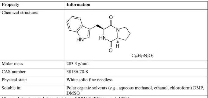
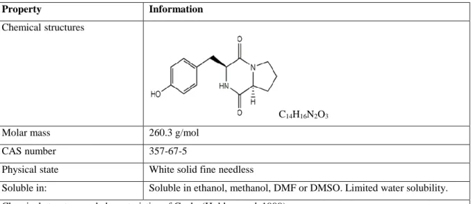
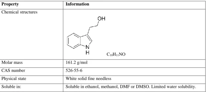
Emerging mycotoxins are fungal secondary metabolites that are neither routinely determined, nor legislatively regulated (Vaclavikova et al. 2013). Emerging Mycotoxins are usually co-produced with other well-known mycotoxins (Hussein and Brasel 2001). The most relevant and frequently occurring emerging mycotoxins are Fusarium toxins including Enniatins (ENNs), Beauvericin (BEA), Apicidin (API), Aurofusarin (AFN), Moniliformin and Fusaproliferin (Gruber-Dorninger et al. 2016; Jajić et al. 2019). ENNs and BEA belong to a group of cyclic hexadepsipeptides, AFN is a dimeric naphthoquinone, and API is a cyclic tetra peptide (Frandsen et al. 2006; Niehaus et al. 2014; Rodríguez-Carrasco et al. 2016). Furthermore, other fungal species such as Alternaria, Aspergillus and Penicillium produce some emerging mycotoxins such as Brevianamid-F (BRV-F), Emodin (EMO) and Cyclo-Pro-L-Tyr (Cyclo) (Streit et al. 2013). In addition, Acremonium which is perceived to be a heterogeneous taxon, also produce some emerging mycotoxins such as Tryptophol (TRPT) (Glenn et al. 1996). As these mycotoxins have only been discovered over the last few decades, they are to date poorly investigated (Springler et al. 2016b). Although recent sensitive analytical methods via LC-MS/MS has assisted the discovery of new fungal secondary metabolites (Malachová et al. 2014), but still the toxicology, toxicokinetics and toxicodynamics data of these metabolites are fragmentary (Taevernier et al. 2015).
Emerging mycotoxins can occur in high frequency and sometimes also in high concentrations in cereals and in other grain-based products (Rodríguez-Carrasco et al. 2016; Gruber-Dorninger et al. 2016). In addition, their presence was reported in mold of water-damaged houses in milder climate regions such as North America and Western Europe (Taevernier et al. 2016). Thus, emerging mycotoxins gain growing interest due to their rapidly increasing presence across the food chain (Lucioli et al. 2013; Fraeyman et al. 2018). Although emerging mycotoxins are concerned, maximum permitted levels may not be proposed in the immediate future. This is primarily due to the lack of data related to their occurrence, contamination level, and toxicity. In order to better assess the risk of these mycotoxins, regular surveillance is a prerequisite to understand their significance as natural contaminants in human and animal nutrition (Jestoi 2008). Among them, the ten most prevalent emerging mycotoxins in feed are presented.
1.3.1. Enniatins (ENNs)
Origin
ENNs (A, A-1, B and B-1) are cyclic hexadepsipeptides (Table 5) secondary metabolites that have been known since few decades (Ivanova et al. 2006), and produced by several Fusarium species, such as F. avenaceum, F. oxysporum, F. poae, or F. tricinctum (Gruber-Dorninger et al. 2016). So far 29 species of ENNs have been isolated and characterized, either as a single compound or mixtures of inseparable homologs (Sy-Cordero et al. 2012). The most frequently detected ENNs in food and feed are ENN A, A-1, B and B-1 (Kamyar et al. 2004; Fraeyman et al. 2017). Naturally occurring ENNs commonly consist of three d-2-hydroxycarboxylic acid residues linked alternatively to three l-N-methyl-amino acid residues (Table 5) (Uhlig et al. 2006).
Table 5: Characteristics of ENNs
Property Information
Chemical structures
Molar mass g/mol ENN-A 681.9 ENN-A1 667.9 ENN-B 639.8 ENN-B1 653.9
CAS number 2503-13-1 4530-21-6 917-13-5 19914-20-6
Physical state Colourless fine needles
Soluble in: Polar organic solvents (e.g., aqueous methanol, ethanol, chloroform) DMP, DMSO and
poor solubility in water
Chemical structure and characteristics of ENNs (ENN-A, A1, B and B1) (Uhlig et al. 2006; Escrivá et al. 2015).
Occurrence
ENNs were detected in food (63%, n= 4,251), feed (32%, n = 3,640) and unprocessed grains (76%, n = 2,647) samples collected between 2010 and 2013 in 12 EU countries (EFSA 2014). ENNs (A, A-1, B and B-1) were also detected in more than 90% of feed and feed raw materials (n=83) with maximum concentrations of 1,745μg/kg, 2,216 μg/kg, 780 μg/kg and 2,690 μg/kg respectively (Streit et al. 2013). In pig feed (n=1,141, worldwide) samples, ENNs (A, A-1, B and B-1) were found in 50%, 77%, 82% and 82% with maximum concentration of 307 μg/kg, 549 μg/kg, 1,514 μg/kg and 1,846 μg/kg respectively. ENN-B and ENN-B1 were the most
prevalent than of ENN-A and ENN-A1 as well the concentrations of ENN-B and ENN-B1 were higher than of ENN-A and ENN-A1 (Novak et al. 2019). ENNs (A, A-1, B and B-1) were also found in Egyptian feed (87%, 91%, 93% and 91%) samples (n=77) and maize (32%, 35%, 44% and 28%) samples (n=79) respectively. Concentration of ENNs was higher in feed (1.9 – 25 µg/kg) rather than maize (<1 µg/kg). Since feeds are composed of several raw materials, therefore compound feeds might be higher contaminated than maize (Abdallah et al. 2017). ENNs (A, A-1, B and B-1) were also found in Finland’s grain samples (wheat, barley, rye and oats, n=38) collected in 2001 and 2002. The incidence of positive samples and the concentrations of ENNs were quite high in both years, especially for ENN-B and B1 that were detected in all samples (maximum concentrations of 18,300 μg/kg and 5,720 μg/kg respectively). ENN-A and A1 were detected in 68 % of the samples with the highest levels of 950 μg/kg and 2,000 μg/kg respectively (Jestoi et al. 2004). ENN-B was also found in 70 % of the baby food samples at levels up to 1,100 µg/ kg and in 44 % of pasta samples at levels of up to 106 µg/kg while other authors reported contamination rates of between 50 – 90 % of wheat, maize and barley samples with total concentrations of ENNs of up to 500 mg/kg (Juan et al. 2013).
Toxicity
ENNs possess a wide range of biological properties, such as; cytotoxicity, hemolysis, permeability and skin damage and decrease of TEER. ENNs have ionophoric properties and can form complexes with cations (Kolf-Clauw et al. 2013; Taevernier 2016; Fraeyman et al. 2016; Springler et al. 2016b; Olleik et al. 2019). Cytotoxicity of ENNs (A, A1, B and B1) was assessed during 24 h incubation in both proliferating and differentiated IPEC-J2 cells. ENNs were ranked in the following order of decreasing toxicity ENN-A > > ENN-A1 > ENN-B1> > >ENN-B respectively (Fraeyman et al. 2018). In similar cell line, after 48 h incubation all ENNs (A, A1, B and B1) showed almost similar absolute IC50 values of 3.40 µM, 4.15 µM, 3.25 µM
and 3.67 µM respectively (Novak et al. 2019). In a similar time of exposure, cytotoxic effect of ENNs (0 to 100 µM) were studied in different cell types of human origin such as Caco-2, human normal lung epithelial cells (BEAS-2B), human normal epidermal keratinocytes (HEK), human liver cell line HepG2, human normal endothelial cells (HUVEC) and human gastric cell line (N87). Effect of ENNs in different cell types was dissimilar (Table 6). Among them, N87 cells were more sensitive than other cell lines (Olleik et al. 2019).
Table 6: Cytotoxicity of ENNs in different cell lines
Organ Cell Type Toxicity Range of IC50
Human intestinal
cells Caco-2
ENN-A > ENN-A1 > ENN-B1 >ENN- B
1.1 µM to 4.6 µM Human lung
epithelial cells BEAS-2B 5.7 µM to 43.7 µM
Human epidermal
keratinocytes HEK 2 µM to 54 µM
Human endothelial
cells HUVEC 2.8 µM to 17.3 µM
Human liver cells HepG2 ENN-A > ENN-B > ENN-A1 ≈ENN- B1 3 µM to 5.6 µM
Human gastric
carcinoma cell line N87 ENN-A1 > ENN-B1 > ENN-A >ENN- B 0.003 µM to 1.7 µM
Furthermore, hemolytic activity of ENNs was assessed on human erythrocytes. According to their hemolytic activity, ENNs were ranked in the following order of toxicity; A> ENN-A1> ENN-B1> ENN-B respectively (Olleik et al. 2019).
In another study performed on Caco-2 cells, ENN-B decreased cell viability from 2.5 to 10 µM up to 30% 24 h whereas at 48 and 72 h it decreased the viability from 1.25 to 10 µM up to 50% and over 90% respectively. The obtained IC50 were of 9.2 µM, 6.9 µM and 5.09 µM after 24 h,
48 h and 72 h respectively (Fernández-Blanco et al. 2016). For tumor cells (neuronal origin), B was highly toxic. In a short term treatment (8 h) and at a very low concentration, ENN-B (0.1-1 µM) had tumor promoting functions based on growth stimulation (Dornetshuber et al. 2007).
Taking into account the above results, ENNs are toxic in vitro, whereas the in vivo studies that have been carried out in rodent and mice, have shown very low or no toxicity. Oral doses of 0.5–1 g/kg body weight (b.w) per day over 6 days to mice and single oral doses of up to 50 mg/kg b.w/day in rats did not produce toxic effects (Kolf-Clauw et al. 2013). In a study of (Rodríguez-carrasco et al. 2016), 5 mg/kg b.w ENN-B administrated to the mice for 9 days did not affect body weight, food intake and behavior of animals. In another study, 20.91 mg/kg b.w/day of ENN-A was administrated in the feed to a 2-month-old female Wistar rats, during the 28 day experiment and no adverse effect were seen in organ weight and histology of duodenum (Manyes et al. 2014). In similar treatment period 465 mg/kg feed, ENN-A was assessed in rats, no significant difference in feed intake and no gross illness was observed (Juan et al. 2014).
Concerning the metabolism of ENNs, it depends on the species or compound. A toxicokinetic study was performed in one piglet, in order to investigate the simultaneous detection and quantification of ENN-A, A1, hB and B1 in animal plasma. The pig received a single oral
intra-gastric bolus of 0.05 mg/kg b.w of all these mycotoxins including BEA. Blood samples were collected before (0 min) and at 10, 20, 30 and 40 min, 1, 1.5, 2, 3, 4, 6, 8, 10 and 12 h post-administration. The authors claimed that despite of their similar chemical structure, there was a big difference in oral absorption between the different ENNs. ENN-B seemed to have the highest oral absorption, followed by ENN-B1, A1 and finally ENN-A. The maximal plasma concentrations (Cmax) for ENN-B, B1, A1 and A, were 73.4, 35.2, 11.6 and 6.8 ng/mL
respectively. The time to maximal plasma concentration (Tmax) was 20 min after bolus
administration for ENN-B1, B and A1, whereas the Tmax for ENN-A was 30 min
post-administration. The authors claimed that the elimination rate of all ENNs was fast and comparable to DON (Devreese et al. 2013). Furthermore, toxicokinetic properties and absolute oral bioavailability of ENN-B1 was evaluated in pig. Pigs were administered ENN-B1 (0.05 mg/kg b.w), either by oral gavage or by intravenous (IV) injection in the ear vein. ENN-B1 was rapidly absorbed after oral administration. The absolute oral bioavailability was 91% after 2 h. After IV administration, ENN-B1 was distributed and eliminated in accordance with oral administration (Devreese et al. 2014).
1.3.2. Beauvericin (BEA)
Origin
BEA is a cyclic hexadepsipeptide (Table 7) synthesized by several Fusarium species, including F. subglutinans, F. proliferatum, F. oxysporum, F. moniliforme, F. anthophilum and F.
beauvrecin (Fotso and Smith 2003). BEA was firstly isolated fromBeauveria bassiana (Hamill
et al. 1969) and reported for its insecticidal properties and its toxicity to Artemia salina (Randazzo et al. 1993). BEA is known to exert a broad spectrum of biological effects such as, antibiotic, anti-inflammatory activities, as well as anticancer effects in various cancer cell lines (Heilos et al. 2017).
Table 7: Characteristics of BEA
Property Information
Chemical structures
C45H57N3O9
Molar mass 784.0 g/mol
Physical state Colourless fine needles
CAS number 26048-05-5
Soluble in: Polar organic solvents (e.g., aqueous methanol, ethanol, chloroform) DMP,
DMSO and poor solubility in water
Chemical structure and characteristics of BEA (Logrieco et al. 1998; Liuzzi et al. 2017).
Occurrence
BEA was reported in food (80%, n=732), feed (79%, n=861) and unprocessed grains (46%, n=554) samples collected between 2010 and 2013 in 12 EU countries. For food, the highest mean concentrations were measured in dried fruits (29.5 µg/kg), followed by oilseeds (8.86 µg/kg) and cereal-based food for infants and young children (8 µg/kg). For feed and unprocessed grains, BEA was present in maize gluten (EFSA 2014). In a worldwide survey of pig feed, BEA was reported in 68% of total samples (n=1,141) with maximum concentration of 413 µg/kg (Novak et al. 2019). BEA was found in high prevalence (98%, n=83) with maximum concentration of 2,326 µg/kg in feed and feed raw materials collected in EU (Streit et al. 2013). Furthermore, occurrence of BEA was reported from different countries in various commodities. BEA was found 33-95% in several food commodities, such as wheat based products, rice, maize based products and barley based products, collected in Italy, Spain, Morocco and Tunisia (n=265), with maximum concentrations 844 µg/kg, 57.4 µg/kg, 73.9 µg/kg and 82 µg/kg respectively (Serrano et al. 2012). In Italian corn (n=94) and oat (n=7) samples BEA was found 27-57% in maximum concentrations 41 µg/kg (Ritieni et al. 1997; Juan et al. 2013). All the wheat samples (n=13), from Finland and all the rice samples (n=70) from morocco were contaminated by BEA with maximum concentration 3,500 µg/kg and 12,810 µg/kg respectively. Even, 33% of baby food (n=68) were contaminated by BEA with maximum concentration of 10,600 µg/kg in Morocco (Logrieco et al. 2002b; Meca et al. 2010; Sifou et al. 2011). In addition, BEA was reported in Egyptian maize (63%, n=79) and feed (88%, n=77). Concentration of BEA in feed (88 µg/kg) was higher than maize (63 µg/kg) (Abdallah et al. 2017).
Toxicity
Acute exposure to BEA is not a concern to human health (EFSA 2014). However, in human and mammalian cell lines, BEA demonstrated toxic effects as it induces apoptosis, increases cytoplasmic calcium concentration and leads to DNA fragmentation (Logrieco et al. 2002a; Luz et al. 2017). The toxicity of BEA is primarily regarded to its ionophoric properties that alters the normal physiological concentrations of cations in and out of cells, acting as an ion carrier through the plasma membrane (Hilgenfeld and Saenger 1982; Fotso and Smith 2003). It also induces lipid peroxidation and alters kinetics in heart metabolism (Ruiz et al. 2011). Cytotoxic effect of BEA was assessed on different human cell lines: Caco-2, BEAS-2B, HEK, HepG2, HUVEC and N87 for 48 h. They were ranked based on the following order of decreasing sensitivity: HUVEC> HepG2> Caco-2> HEK> BEAS-2B>N87, with IC50 of 2.4
µM> 3.4 µM> 3.9 µM> 5.4 µM> 6.3 µM> 27.5 µM respectively (Olleik et al. 2019). In addition, BEA revealed toxic effect on human fibroblast-like cell line (MRC-5) After 24 h, the IC50 was of 5.0 µM and 1.1 µM via Alamar BlueTM and BrdU assay respectively (Ivanova et
al. 2006). Furthermore, a toxicogenomic study was performed to investigate gene expression changes triggered by BEA exposure (1.5, 3 and 5 μM) during 24 h in Jurkat cells, a human lymphoblastic T cell line, through RNA-sequencing and differential gene expression analysis. The results demonstrated BEA-induced mitochondrial damage affecting the respiratory chain and pointing to apoptosis through the caspase cascade. The most significantly altered pathways genes, involved in the respiratory chain, were significantly down-regulated. These results bring greater relevance to mitochondria as a target site for BEA induced cytotoxicity in cellular models (Escrivá et al. 2018).
Nevertheless, BEA is identified toxic in vitro, but is reported to be not toxic in vivo. Toxicity and pharmacological behavior of BEA was investigated in mice during 9 days, no effect on body weight, food intake and behavior was observed at 5 mg/kg b.w (Rodríguez-carrasco et al. 2016). However, in a brain influx study performed in mice, BEA crossed the blood-brain barrier (Taevernier et al. 2016). Therefore, no conclusion could be drawn with respect to chronic exposure due to the lack of relevant in vivo toxicity data.
Metabolization of BEA was studied in pig. A toxicokinetic study was performed in one piglet, in order to investigate detection and quantification of BEA in plasma. The pig received a single oral intra-gastric bolus of 0.05 mg/kg b.w of BEA along with ENNs (ENN-A, A1, B and B1). Blood samples were drawn before (0 min) and at 10, 20, 30 and 40 min, 1, 1.5, 2, 3, 4, 6, 8, 10
and 12 h post-administration. The authors claimed that for BEA, no plasma concentration-time profile was designed as the plasma concentrations were above the limit of quantification (LOQ) level at only 2 time points, i.e. 0.51 ng/mL at 30 min and 0.82 ng/mL at 40 min post-administration. Thus, BEA revealed a lower oral bioavailability compared to ENNs (Devreese et al. 2013).
1.3.3. Aurofusarin (AFN)
Origin
AFN is a dimeric naphthoquinone belonging to the naphthoquinone group of polyketides (Table 8) (Frandsen et al. 2006). AFN is a yellow-red pigment produced by Fusarium head-blight disease caused by several species of Fusarium, such as F. graminearum, F. culmorum, and F. crookwellense (Kim et al. 2006).
Table 8: Characteristics of AFN
Property Information
Chemical structures
C30H18O12
Molar mass 570.4 g/mol
Physical state Black and brown fine needles
CAS number 88360-87-6
Physical state Dependent on the pH value of the solvent, ranging from golden yellow in
acidic solvents to red/purple in alkaline solvents
Soluble in: Polar organic solvents (e.g., aqueous methanol, ethanol, chloroform) DMP,
DMSO and poor solubility in water Chemical structure and characteristics of AFN (Tanaka and Tamura 1961).
Occurrence
AFN has been identified in feed ingredients in particular in contaminated grains and known as a new crop pollutant (Dvorska et al. 2003). AFN was found in feed raw materials 84% (n=83, Europe), finished feed 80.7% (n=1,141, worldwide), 73% (n=77, Egypt) and maize 9% (n=79, Egypt) with maximum concentration of 17,659 µg/kg, 85,360 µg/kg, 3,005 µg/kg and 1,858 µg/kg respectively (Streit et al. 2013; Abdallah et al. 2017; Novak et al. 2019). In addition, AFN was found in settled dust of Polish poultry farms (n=13), among 27 other detected chemicals, with the highest concentration of 281.44 µg/kg (Skóra et al. 2016).