HAL Id: tel-01381998
https://tel.archives-ouvertes.fr/tel-01381998
Submitted on 15 Oct 2016
HAL is a multi-disciplinary open access archive for the deposit and dissemination of sci-entific research documents, whether they are pub-lished or not. The documents may come from teaching and research institutions in France or abroad, or from public or private research centers.
L’archive ouverte pluridisciplinaire HAL, est destinée au dépôt et à la diffusion de documents scientifiques de niveau recherche, publiés ou non, émanant des établissements d’enseignement et de recherche français ou étrangers, des laboratoires publics ou privés.
activation during the innate immune response in
Drosophila
François Bonnay
To cite this version:
François Bonnay. Deciphering regulatory mecharnsms of the lMD pathway activation during the innate immune response in Drosophila. Innate immunity. Université de Strasbourg, 2014. English. �NNT : 2014STRAJ019�. �tel-01381998�
École doctorale des Sciences de la Vie et de la Santé
UPR9022 CNRS Réponse Immunitaire et Développement chez les Insectes
Thèse présentée à l’Université de Strasbourg par
François Bonnay
Soutenue publiquement le 14 octobre 2014
Pour obtenir le grade de Docteur de l’université de Strasbourg
Spécialité : Aspects moléculaires et cellulaires de la Biologie
Caractérisation des mécanismes de régulation de la voie IMD au cours
de la réponse immunitaire chez Drosophila melanogaster
Thèse dirigée par
Pr. Jean-Marc Reichhart Professeur des Universités
Institut de Biologie Moléculaire et Cellulaire, Strasbourg, France
Rapporteurs
Dr. Dominique Ferrandon Directeur de recherche CNRS
Rapporteur interne, Institut de Biologie Moléculaire et Cellulaire, Strasbourg,
Président du jury France
Dr. Christian Muchardt Directeur de recherche CNRS
Rapporteur externe Institut Pasteur, Paris, France
Dr. Amir Orian Associate Professor
I. Acknowledgments
First, I would like to address my deepest gratitude to my PhD supervisor, Prof. Jean-Marc Reichhart for incorporating me in his team and giving me all the required support to accomplish my goals. His knowledge, advices and supports have made of this PhD thesis a great experience. I am equally thankful to Dr. Nicolas Matt, my second PhD supervisor, for constantly supporting me from the beginning of Master 2. His help, his advices and his involvement in my PhD were determinant during these four past years.
I am deeply thankful to the PhD committee members, Dr. Dominique Ferrandon, Dr. Christian Muchardt and Dr. Amir Orian who accepted to read this manuscript and to come to Strasbourg to judge my work.
I would like also to thank the additional contributors of my PhD work, who contributed to the data that are presented in this manuscript: Eva Berros-Cohen, Xuan-Hung Nguyen, Eric Batsché and Miriam Yamba. I also address a big thank-you to Dr. David Gubb who helped me a lot with my English and more generally with the PhD writing.
I am indebted to Dr. Dominique Ferrandon, Prof. Jean-Luc Imler, Dr. Carine Meignin and Dr. Vincent Leclerc who also largely contributed to my present and future professional situation by advising me and supporting my applications.
More generally, I am profoundly honored to have been part of the UPR9022 community, and I thank all its members for the nice social and scientific moments I spent. This PhD thesis was the opportunity to meet great people that I hope to keep contact with. I also want to address a specific acknowledgement to Dr. Stefanie Müller and Dr. Cordula Kemp, who were the first to integrate me in the lab and teach me how to deal with flies. My last acknowledgments go to my PhD colleagues and friends Samantha Haller, Vincent Barbier, Sarah Merkling, Jelena Patrnogic, Laure Nivlet and Adrien Franchet who supported me during these four years. I wish the best to all of you.
II. Table of Contents
I. Acknowledgments ... 1
II. Table of Contents ... 2
III. Table of Figures ... 4
IV. Abbreviations ... 5
V. General Introduction ... 10
VI. Chapter 1: Innate immune responses in Drosophila melanogaster ... 13
1. NF-κB pathways in Drosophila ... 13
1.1 The IMD pathway ... 14
1.1.1 IMD pathway recognition events ... 14
(a) General features of PGRPs ... 15
(b) PGRPs involved in the activation of the IMD pathway ... 15
1.1.2 IMD pathway signaling events ... 17
(a) Establishment of the IMD-IKK signalosome ... 17
(b) Post-translational activation of Relish ... 19
(c) Relish transcriptional activity in the nucleus ... 20
(d) Positive regulators of Relish transcriptional activity ... 21
1.1.3 IMD pathway effectors ... 22
1.1.4 IMD pathway negative regulation ... 23
(a) Control of DAP-type PGN recognition ... 23
(b) Control of IMD-IKK signaling ... 24
(c) Control of Relish cleavage and stability ... 25
(d) Control of Relish activity in the nucleus ... 26
1.2 The Toll pathway ... 30
1.2.1 Toll pathway recognition events ... 31
(a) The PRR recognition pathway ... 32
(b) The danger signal recognition pathway ... 32
1.2.2 Toll pathway signaling events ... 33
1.2.3 Toll pathway effectors ... 35
1.2.4 Toll pathway negative regulation ... 36
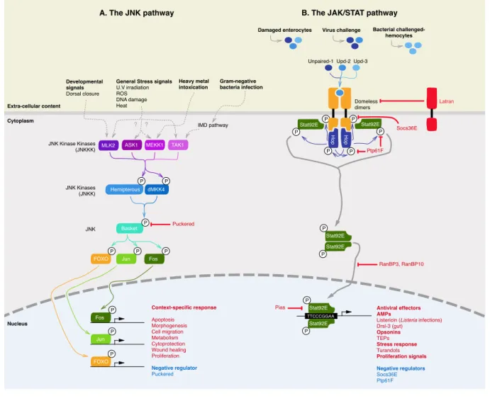
2. NF-κB-independent immune pathways in Drosophila: The JNK and JAK/STAT pathways ... 39
2.1 The JNK pathway ... 39
2.2.1 Biological relevance of the JAK/STAT pathway ... 41
2.2.2 Signaling events of the Drosophila JAK/STAT pathway ... 42
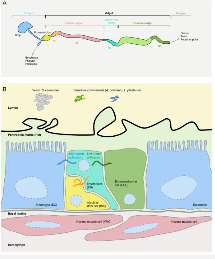
3. Local immune responses ... 45
3.1 Intestinal immune responses ... 45
3.1.1 Drosophila gut physiological properties ... 45
(a) Anatomical and functional regions of Drosophila gut ... 45
(b) Passive defense mechanisms of Drosophila gut ... 47
3.1.2 Active defense mechanisms ... 50
(a) The oxidative burst ... 50
(b) Local production of anti-microbial peptides (AMPs) ... 54
(c) Maintenance of gut homeostasis ... 55
3.2 Other local immune responses: trachea and male genital plates ... 59
4. Systemic immune responses ... 61
4.1 Cellular immune responses ... 61
4.1.1 Drosophila hematopoiesis ... 61
4.1.2 Phagocytosis ... 63
4.1.3 Encapsulation ... 65
4.1.4 Melanisation ... 65
4.1.5 Coagulation ... 66
4.2 Humoral immune response ... 67
5. Intrinsic immune response against viruses ... 68
6. Objectives and aims of the PhD work ... 69
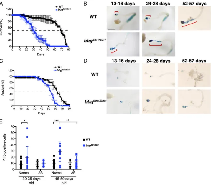
VII. Chapter 2: big-bang gene modulates gut immune response ... 71
1. Scientific context of the study ... 71
1.1 The big-bang gene ... 71
1.2 Midgut immune responses and homeostasis ... 72
2. Manuscript ... 74
VIII. Chapter 3: Akirin specifies NF-κB selectivity of Drosophila innate immune response via chromatin remodeling ... 75
1. Scientific context of the study ... 75
1.1 The akirin gene family ... 75
1.2 Known functions of Akirins in the immune responses of metazoa ... 75
1.3 Known functions of Akirins in the development of metazoa ... 77
1.4 What are the molecular functions of Akirins? ... 78
1.5 SWI/SNF chromatin remodeling ... 78
1.7 SWI/SNF complexes in mammals ... 82 2. Manuscript ... 84 IX. References ... 85
III. Table of Figures
Figure 1 The life cycle of Drosophila melanogaster. ... 12! Figure 2 The IMD pathway of Drosophila melanogaster. ... 29! Figure 3 The Toll pathway of Drosophila melanogaster in immunity. ... 39! Figure 4 The Jun N-terminal kinase (JNK) and JAK/STAT pathways in Drosophila
melanogaster. ... 44!
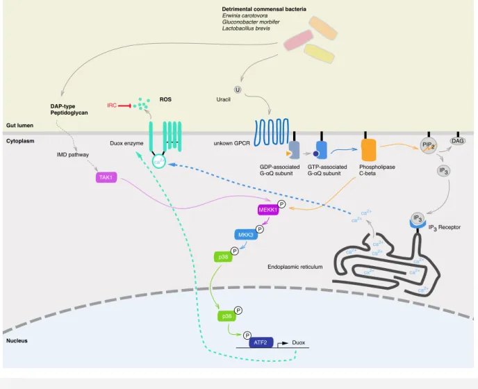
Figure 5 Histogical and cellular organization of Drosophila melanogaster midgut. ... 49! Figure 6 ROS synthesis through the Duox activation and expression pathways in
Drosophila melanogaster ... 53!
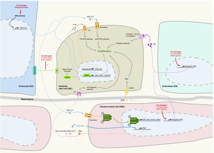
Figure 7 Signaling pathways governing intestinal stem cells (ISC) proliferation in the
IV. Abbreviations
20E: 20-hydroxyecdysone AA: amino acidAD: Adherens junction
AgAkirin: Anopheles gambiae Akirin
AMP: Anti-microbial peptide ANT-C: Antennapedia complex AP1: Activator protein 1
APC: Adenomatosis polyposis coli ARID: AT-rich interaction domain ATF2: Activating transcription factor 2 ATP: Adenosine triphosphate
Att: Attacin
BAFs: Brahma-associated factors
BAP complex: OSA-associated
Brahma complex
Bap170: Brahma-associated protein 170 kDa
Bap55: Brahma-associated protein
55kDa
Bap60: Brahma-associated protein
60kDa
Bbg: Big-bang
Bcl3: B-cell lymphoma 3 BCR: B-cell receptor
Bfl-1: Bcl2-related gene expressed in fetal liver 1
Brg-1: Brahma-related gene 1 Brm: Brahma
BX-C: Bithorax complex
C106: Spätzle 106 amino acids C-terminal fragment
C3PO: Component 3 promoter of RISC
Caspase: Cysteine-dependent
aspartate-directed protease CD: Crohn’s disease
CDRE: Caudal protein DNA
recognition element
CeAkirin: Caenorhabditis elegans
Akirin
Cec: Cecropin
Chd: Chromodomain-helicase DNA binding
ChIP: Chromatin Immunoprecipitation CK1α: Casein kinase 1α
CrPV: Cricket paralysis virus CYLD: Cylindromatosis DAG: Dyacylglycerol
DAMP: damage-associated molecular pattern
DAP-type: meso-diaminopymelic-type DAV: Drosophila A virus
DCE: Dopachrome conversion enzyme DCHS: Dachsous
Dcr-2: Dicer-2
DCV: Drosophila C virus Dcy: Drosocrystalin DD: Death domain
DDC: DOPA decarboxylase enzyme DED: Death effector domain
DFV: Drosophila F virus
DIAP2: Drosophila inhibitor of
DIF: Dorsal-related immunity factor Dilp: Drosophila insulin-like peptide
DmAkirin: Drosophila melanogaster
Akirin
DMAP1: DNA methyl transferase 1 Dnr-1: Defense repressor 1
DOPA: 3,4-dihydroxyphenylalanine Dpt: Diptericin
DPV: Drosophila P virus
Dredd: Death related ced-3/Nedd2-like protein
Drs: Drosomycin Drsl: Drosomycin-like Dsh: Dishevelled
Dsp1: Dorsal-switch protein 1
dSR-Cl: Drosophila scavenger receptor Cl
dsRNA: double-stranded RNA DSS: Dextran sodium sulfate Duox: Dual oxidase
DXV: Drosophila X virus EB: Enteroblast
EC: Enterocyte
EcR: Ecdysone receptor EEC: entero-endocrine cell EGF: Epidermal growth factor EGF: Epidermal growth factor
EGFR: Epidermal growth factor
receptor
ER: Endoplasmic reticulum
ERK: Extracellular regulated kinase ESC: Embryonic stem cell
ET: Eye transformer
FADD: FAS associated death domain
Faf: Fat facets
FHV: Flock-house virus FOXO: Forkhead box O FT: Fat
Fz: Frizzled
G707: Gluconobacter sp. strain EW707 Gcm: Glial cell missing
GNBP: Gram-negative bacteria binding protein
GPCR: G protein-coupled receptor Gprk2: G protein-coupled receptor kinase 2
Grass: Gram-positive specific serine protease
GSK3: Glycogen synthase kinase 3 GTP: Guanosine triphosphate Gαq: G protein α q sub-unit H3K4: Histone 3 Lysine residue 4 H3K4ac: H3K4 acetylation
H3K4me: H3K4 methylation H3K4me3: H3K4 tri-methylation HAT: Histone acetyl transferase HDAC: Histone deacetylase HMG: High mobility group HPO: Hippo
hTRIF: human TIR domain containing adapter inducing interferon-β
IAP: Inhibitor of apoptosis
IBD: Inflammatory bowel disease IBM: IAP2 binding motif
IFN: Interferon Ig: Immunoglobulin
IIV6: Invertebrate iridescent virus 6 IKK: Inhibitor of NF-κB Kinase
IL: Interleukin
IMD: Immune deficiency Ino80: Inositol auxotroph 80 InR: Insulin receptor
IP3: Inositol 1,4,5-triphosphate
IRC: Immune-regulated catalase Ird5: Immune-response deficient 5
IsAkirin: Ixodes scapularis Akirin
ISC: Intestinal stem cell Iswi: Immitation switch IκB: Inhibtor of NF-κB
JAK/STAT: Janus Kinase / Signal
Transducers and Activators of
Transcription
JNK: Jun N-terminal kinase KD: Knock-down
Key: Kenny KO: Knock-out
LPS: Lipopolysaccharide LRR: Leucin-Rich-Repeat
Lys-PGN: Lys-type peptidoglycan Mad: Mothers against Dpp
MAMP: Microbial-associated molecular pattern
MAP: Mitogen associated protein MAPK: MAP kinase
MAPKK: MAPK kinase MAPKKK: MAPKK kinase
MATS: Mob as tumor suppressor MEKK1: MEK kinase 1
MHC: Major histocompatibility complex ModSP: Modular serine protease MP1/2: Melanization protease 1/2 MPDZ: Multiple PDZ domain protein
Mya: Million years ago
Myd88: Myeloid differentiation primary response gene 88
NADPH: Nicotinamide adenine
dinucleotide phosphate
NF-κB: Nuclear factor kappa-light-chain-enhancer of activated B cells NimC1: Nimrod C1
NLS: Nuclear localization signal Nt: nucleotide
PBAP complex: Polybromo-associated Brahma complex
Pdk1: Phosphatidylinositol-dependent kinase 1
PDZ: PSD-95, Discs-large, ZO-1
PEST: Proline, Glutamate, Serine, Threonine-rich
PGN: Peptidoglycan
PGRP: peptidoglycan-recognition
protein
Pias : Protein inhibitor of activated STAT
PIP2 : Phosphatidylinositol
4,5-biphosphate
Pirk: Poor immune response upon knock-in
PLC-β: Phospholipase C-β PM: Peritrophic matrix
PMA: Phorbol 12-myristate 13 acetate PO: Phenoloxydase
Posh: Plenty of SH3s
PP2A: Protein phosphatase 2A pPA: PPO activating enzyme PPO: Prophenoloxydase
PRR: Pattern-recognition receptor Psh: Persephone
pSJ: pleated septate junction
PTEN: Phosphatase and tensin
homolog
Ptp61F: Protein tyrosine phosphatase 61F
Pvf2: PDGF- and VEGF-related factor 2
Pvr: Platelet-derived Growth Factor (PDGF)-Vascular Endothelial Growth Factor (VEGF) Receptor
RAG: Recombination-activating gene RanBP : Ras-like guanine nucleotide-binding protein
RHD: Rel-homology domain
RING: Really interesting new gene RIP: Receptor interacting protein RISC: RNA-induced silencing complex RNAi: RNA-interference
RNAse: Ribonuclease
ROS: Reactive Oxygen Species SANT: Swi3, Ada2, N-CoR, TFIIIB SAPK: Stress-activated protein kinase pathway
SAV: Salvador
Sayp: Supporter of activation of Yellow protein
SC: Synaptonemal complex Scr: Sex-comb reduced Sd: Scalloped
Serpin: Serine protease inhibitor SIGMAV: Sigma virus
SINV: Sindbis virus
siRNA: short-interfering RNA SJ: Septate junction
Snr1: SNF5-related 1
Soc36E: Suppressor of cytokine
signaling 36E SP: Sex peptide
SPE: Spätzle processing enzyme Spn: Serpin, Serine-protease inhibitor Spz: Spätzle
SRR: Serin-rich region
sSJ: Smooth septate junction ssRNA: single-stranded RNA
SUMO: Small Ubiquitin-like Modifier SWI/SNF: Mating-type switching / Sucrose non-fermentable
TAB2: TAK1-associated binding
protein 2
TAK1: TGF-β-activated kinase 1 Tcf: T-cell factor
TCR: T-cell receptor TCT: Tracheal cytotoxin
TEP: Thioester-containing protein TGF-β; Transforming growth factor beta
Tip60: TAT-interactive protein 60kDa TIR: Toll/interleukin-1 receptor
TJ: Tight junction TLR: Toll-like receptor TNF: Tumor necrosis factor
TNFR: Tumor-necrosis factor receptor Tor: Target of rapamycin
Tot: Turandot
trxG: Trithorax group
TSS: Transcription start site UC: Ulcerative colitis
Uev1a: Ubiquitin-conjugating enzyme variant 1a
Upd: Unpaired Usp: Ultraspiracle
V,D,J: Variable, Joining, Diversity VLR: Variable lymphocyte receptor VMC: Visceral muscle cell
vsiRNA: viral siRNA
VSV: Vesicular stomatitis virus Wbp2: WW-binding protein 2 Wg: Wingless
WTS: Warts Yki: Yorkie
Zfh1: Zinc-finger homeodomain 1 ZO1: Zonulla occludens 1
V. General Introduction
During evolution, metazoans have established a powerful immune system to survive pathogenic invading microorganisms. There are two main types of defense systems: innate and adaptive.
The innate immune system predates the adaptive response and consists of a package of defense mechanisms that has been conserved for more than a billion years within the animal kingdom. The innate immune system involves a wide variety of cells, effectors and molecular pathways that give a robust and immediate response to immune challenge. An active innate immune mechanism requires three categories of molecules: i) Sensors, able to discriminate and detect microbial pattern or danger signal and to engage a downstream signaling pathway. ii) Adaptors, constituting the molecular pathways driving the sensing signal to the production of the effectors. iii) The induced effector molecules, which can directly (e.g. Anti-microbial Peptides (AMPs), Reactive Oxygen Species (ROS)) or indirectly (e.g. Cytokines, Fever) counteract microbial challenges.
The adaptive immune system appeared more recently on an evolutionary scale, around 650 Million years ago (Mya), among the ancestors of jawless fishes (Kasahara and Sutoh, 2014). This adaptive system is based on antigen-specific recognition and maintains a memory of the response. This last property enabled the development of vaccines, which represents, together with the discovery of antibiotics, one of the major achievements of contemporary bio-medical research. As far as is known, two main branches of the adaptive immune system have diverged from these ancestral vertebrates, based on T-Cell and B-Cell receptors (TCRs and BCRs) in gnathostomes or based on variable lymphocyte receptors (VLRs) in jawless vertebrates (e.g. lampreys, hagfishes). In jawed vertebrates, TCRs and BCRs are expressed clonally on lymphocytes and recognize a wide variety of antigens (Tonegawa, 1983). To possess such plasticity in the recognition motif, vertebrate genes encoding these receptors somatically recombine the Variable (V), Joining (J), or V, Diversity (D) and J genes fragments through double-stranded DNA breaks induced by the recombination-activating gene (RAG) nuclease (Schatz and Swanson, 2011). A third component of this adaptive immune system, the Major
Histocompatibility Complex (MHC) molecules, is required for antigen recognition by the αβ subset of TCRs (Klein and Sato, 2000). In jawless vertebrates, the diversity of antigen recognition is produced by the assembling of variable Leucin-Rich-Repeat (LRR) modules encoding the VLRs in lymphocyte lineages (Nagawa et al., 2007). Importantly, the activation of the adaptive immune system strongly relies on concomitant innate immune responses (Fearon and Locksley, 1996).
The scientific context of my PhD was the exploration of innate immune mechanisms and I will therefore focus the rest of the manuscript on this aspect. In humans the innate immune system is required to defend against microbial challenges. When abnormally regulated however, innate immune responses contribute to a range of pathologies including autoimmune diseases, chronic inflammation and cancer (Maeda and Omata, 2008). Chronic inflammation-related pathologies such as atherosclerosis, type II diabetes or inflammatory-bowel diseases (IBDs) are difficult to cure with currently available anti-inflammatory therapeutic molecules and have become a major health problem (Tabas and Glass, 2013). The understanding in fine-tuning mechanism as well as deciphering the innate immune pathways cannot be dissociated from the unraveling of the next generation of therapeutic molecules.
Drosophila melanogaster is a small fly that has been widely used during the
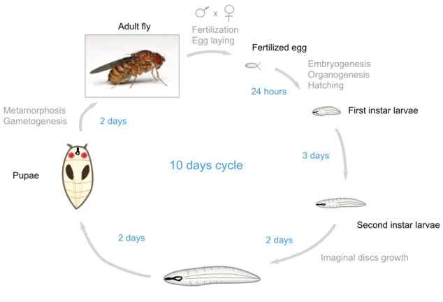
past hundred years to investigate complex biological questions, notably in genetics and developmental biology. The advanced genetic tools and the short generation time of Drosophila (8-10 days) (Figure 1) contributed to its success as a model organism. Importantly, Drosophila and humans share many genes and molecular pathways with similar functions (Rubin, 2000). Drosophila is well suited for deciphering the fundamental mechanisms underlying the innate immune response as unlike in vertebrates, the defense mechanisms of invertebrates rely entirely on innate immune responses. Although Drosophila and humans diverged more than 800 Mya in evolution, they share many molecular pathways underlying the activation of their innate immune systems (Hoffmann and Reichhart, 2002). Massive efforts during the past twenty years to describe the Drosophila innate immune system has largely contributed to the characterization of mammalian Toll-like Receptors (TLRs) and
NF-κB pathways (Leulier and Lemaitre, 2008) and demonstrated the relevance of this model to study innate immunity.
Adult fly Fertilization
Egg laying
Embryogenesis Organogenesis Hatching
Imaginal discs growth Metamorphosis Gametogenesis 24 hours 3 days 2 days 2 days 2 days 10 days cycle Fertilized egg
First instar larvae
Second instar larvae
Third instar larvae Pupae
x
Figure 1 The life cycle of Drosophila melanogaster.
Additionally, work on Drosophila innate immunity may valuably contribute to understanding the biology of other arthropods human diseases vectors. Such vectors include other Diptera such as the sand fly (Phlebotominae, vector of Leishmaniasis), the buffalo gnat (Simuliidae, vector of Onchocerciasis), the Anopheles and Aedes mosquitoes (vector of malaria and filariasis, dengue fever, yellow fever and chicungunya). These pest species represent a major and growing threat for human health.
The aim of my PhD research was to improve our understanding of the innate immune response activation using Drosophila melanogaster as a model system. To fulfill this goal, I characterized the molecular function of two genes, big-bang (bbg)
and akirin, respectively implicated in the intestinal and systemic immune system of
Drosophila. The following introduction sections present a broad overview of the
known mechanisms of Drosophila innate immune responses, with an emphasis on NF-κB pathways biology.
VI. Chapter 1: Innate immune responses in Drosophila
melanogaster
In the wild, Drosophila lives in a microorganisms-rich environment including decaying fruits. As a consequence, this organism is constantly exposed to microbial threats during nutrition and had to develop a powerful innate immune system. The first layer of Drosophila innate immune response is located at the putative entry sites of natural microbial infections: the gut, the trachea and the genital plates. To prevent a potential invasion in its internal cavity (the hemocoele), Drosophila has developed a set of defense mechanisms specific to these tissues, so-called the local immune responses, described thereafter (3.). This local immune system is sufficient to contain most microorganisms, but some pathogenic species such as Pseudomonas
aeruginosa are able to cross the epithelial barriers and spread in the hemocoele
(Limmer et al., 2011a). When such entomophagous pathogens invade the internal cavity of flies, or following septic injury, a second layer of defense mechanisms is activated in the hemocoele: the systemic immune response (4.).
For clarity, I will first describe the main molecular immune pathways of
Drosophila that influence both local and systemic immune responses. These include
the κB-dependent pathways Immune Deficiency (IMD) and Toll (1.) and two NF-κB-independent additional molecular pathways: the Jun N-terminal Kinase (JNK) and the Janus Kinase / Signal Transducers and Activators of Transcription (JAK/STAT) pathways (2.). Finally, I will describe the intrinsic defense mechanisms deployed by
Drosophila to fight viral infections (5.).
In Drosophila, the Toll and the Immune deficiency (IMD) pathways play a fundamental role in the defense against invasive microbes by triggering the massive release of anti-microbial peptides. These pathways are able to recognize, discriminate and fight three main pathogen families of flies: Gram-negative bacteria, Gram-positive bacteria and fungi. So far, the functions of these pathways have been mostly characterized in three main immune tissues: i) the fat-body, a pseudo-epithelial tissue required for lipid storage with functional equivalence to the mammalian liver, but also the most potent organ of Drosophila systemic immune responses ii) the hemocytes, specialized phagocytic cells and iii) the digestive tract. This section of the introduction describes the current knowledge of the Toll and IMD κB pathways without tissue restriction. Additional details about specific local NF-κB pathways activations are provided in the section 3.
1.1 The IMD pathway
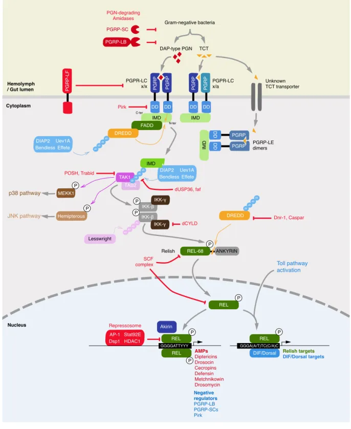
The IMD pathway controls the expression of a set of anti-microbial peptides, one of the stronger arms of Drosophila immune effectors. The absence of a functional IMD pathway activation leads to a high susceptibility of flies to Gram-negative bacterial infections, but not to Gram-positive bacterial or fungal infections (Lemaitre et al., 1995a). Conversely, when over-activated, the IMD pathway is a source of pathologies in flies (Paredes et al., 2011a). This section describes the known mechanisms of IMD pathway in flies as well as the numerous regulatory mechanisms blocking this activation (Figure 2). Note that a large portion of the proteins involved in IMD pathway signaling have a close ortholog in the mammalian Tumor-Necrosis Factor Receptor (TNFR) pathway (Hoffmann, 2003), one of the primary pathway involved in inflammation (Locksley et al., 2001). This high degree of conservation validates the relevance of studying of Drosophila IMD pathway for bio-medical research.
1.1.1 IMD pathway recognition events
The IMD pathway is initiated through the recognition of meso-diaminopymelic-type (DAP-meso-diaminopymelic-type) peptidoglycan. This microbial-associated molecular pattern (MAMP)
is contained in Gram-negative bacteria and some Gram-positive bacilli. Two pattern-recognition receptors (PRR), members of the peptidoglycan-pattern-recognition proteins (PGRPs) family are involved in such recognition: PGRP-LC, PGRP-LE (Neyen et al., 2012).
(a) General features of PGRPs
PGRP family of receptors is conserved from invertebrates to mammals and is composed in Drosophila of 13 genes encoding at least 17 independent PGRPs isoforms through alternative splicing (Werner et al., 2000). PGRP receptors are classified into small-sized (182 to 203 amino-acids (AA)) PGRP-S and long-sized (215 to 520 AA) PGRP-L receptors. All PGRPs proteins have a PGRP domain, closely related to the T7 bacteriophage type II amidases secreted enzymes involved in PGN degradation (Kang et al., 1998).
In Drosophila, six members of PGRP family possess a PGRP domain bearing a functional PGNdegrading amidase activity: PGRPSB1, SB2, SC1A, SC1B, -SC2 and –LB. The amidase activity of these receptors gives them roles in the negative regulation of immune responses via the scavenging of immune-potent PGN (Bischoff et al., 2006; Guo et al., 2014; Paredes et al., 2011a; Zaidman-Rémy et al., 2011). By contrast, the seven other Drosophila PGRPs (PGRPSA, SD, LA, LC, -LD, -LE, and –LF) do not have an amidase activity and are involved either in sensing and signal transduction to immune pathways (PGRP-SA, -SD, -LA, -LC, -LE), or in the negative regulation of immune responses (PGRP-LE, -LF) (Bischoff et al., 2004; Choe et al., 2005a; Gendrin et al., 2013; Kaneko et al., 2006; Maillet et al., 2008; Michel et al., 2001). All PGRP-Ss have a secretion signal peptide in their N-terminal part and are therefore exclusively located outside of the cell. Finally, PGRP-Ls are either trans-membrane (PGRP-LA, -LC, -LD, -LF), intracellular (PGRP-LE) or secreted (PGRP-LE, -LB) proteins (Werner et al., 2000).
Among PGRP family members, PGRP-LC is the main contributor for IMD signaling in the systemic immune system. PGRP-LC encodes three isoforms through alternative splicing: PGRP-LCa, PGRP-LCx and PGRP-LCy. These isoforms differ in their PGN-recognition domain (Neyen et al., 2012). PGRP-LCy lacks a functional PGN-recognition domain and may therefore act as a negative regulator of other PGRP-LC isoforms. By contrast, the PGRP-LCx isoform is necessary and sufficient to respond to Gram-negative and Gram-positive bacterial challenge as the LCx homodimer can recognize DAP-type PGN multimers. By contrast, PGRP-LCx/PGRP-LCa heterodimers recognize the monomeric PGN known as tracheal cytotoxin (TCT).
Unlike PGRP-LC, PGRP-LE recognizes only TCT. Moreover, PGRP-LE is crucial for the local activation of IMD in the midgut while it is dispensable for immune activation in the fat-body (Bosco-Drayon et al., 2012a). Thus, pattern-recognition receptors are expressed in region-specific patterns along the length of the fly intestine. On one hand, the trans-membrane PGRP-LC receptor plays a predominant role in the foregut and the hindgut. On the other hand the intracellular PGRP-LE’s sensing function is required in the midgut (Bosco-Drayon et al., 2012b; Neyen et al., 2012). Interestingly, as intracellular PGRP-LE is the only sensor in the midgut, it suggests that a yet-unidentified trans-membrane TCT transporter is involved in such recognition. The indirectness of PGRP-LE-dependent midgut recognition of bacteria is thought to prevent undesired and potentially harmful over-activation of the IMD pathway at this very location, where the permeability towards external components is greater than the other sections of the gut (further detailed in 3.1.1.).
Intriguingly, PGRP-LC is required for the IMD pathway activation in the foregut proventriculus, while PGRP-LE selectively promotes the expression of negative regulators of the IMD pathway (Bosco-Drayon et al., 2012b). Finally, a recent study, indicates that the trans-membrane PGRP-LA protein activates the IMD pathway activation in the gut. The mode of action of PGRP-LA is unclear as this protein has neither a predicted PGN-binding domain nor amidase enzymatic activity (Gendrin et al., 2013).
1.1.2 IMD pathway signaling events
(a) Establishment of the IMD-IKK signalosome
DAP-type PGN is recognized by PGRP-LC and -LE proteins at their C-terminal domains (Chang et al., 2006; Lim et al., 2006). Following this recognition, PGRP-LC or –LE oligomerize and transduce the activation signal through their N-terminal domain (Choe et al., 2005b). The signal transduction is mediated through a specific AA sequence named the “core motif” in the N-terminal domain. The core motif is conserved between PGRP-LC and –LE (Kaneko et al., 2006) and shares a strong homology with the human Toll/interleukin-1 receptor (TIR)-domain-containing adapter-inducing interferon-β (hTRIF) protein. Interestingly, hTRIF is also involved in Toll-like Receptor pathway at the level of signal transduction by the pattern-recognition receptors (Meylan et al., 2004). The activation of PGRP-LC and -LE allows the recruitment of the adaptor molecules Immune Deficiency (IMD) and FAS associated Death domain (FADD) plus Death related ced-3/Nedd2-like protein (Dredd) (Leulier et al., 2000, 2002; Naitza et al., 2002).
To establish the formation of a signaling complex, the first interaction occurs between PGRP-LC or –LE and IMD. IMD possesses a death-domain (DD), a protein-protein interaction domain homologous to the mammalian receptor interacting proteins (RIP) (Georgel et al., 2001). PGRP-LC and –LE’s interaction with IMD requires the core motif but is not direct, suggesting the involvement of a third unknown molecule involved in this process (Kaneko and Silverman, 2005). Subsequently, FADD is recruited onto IMD. FADD contains a DD and another protein-protein interaction domain, Death Effector Domain (DED). Dredd, is further recruited into this complex and also carried a DED domain through which it interacts with FADD.
Dredd, is a cysteine-dependent aspartate-directed protease (caspase) ortholog to the mammalian Caspase-8. Besides its role in the IMD pathway activation, Dredd acts as an effector of Reaper, Grid and Hid-mediated apoptosis in
E3 ubiquitin ligase Drosophila inhibitor of apoptosis 2 (DIAP2) and the E2 ubiquitin conjugating Ubiquitin-conjugating enzyme variant 1A (Uev1a) together with Bendless and Effete activates Dredd by K63-linked poly-ubiquitinylation (Meinander et al., 2012). Once poly-ubiquitinylated, Dredd is able to cleave IMD at aspartic residue 30, hence truncating the 30 N-terminal amino acids. The cleaved N-terminal of IMD exposes an IAP2 binding motif (IBM) that allows the recruitment of the tetrameric DIAP2, Uev1a, Bendless and Effete complex (Zhou et al., 2005). This complex will add K63-linked ubiquitin chains on cleaved IMD, which will serve as a scaffold to recruit the MAP kinase kinase kinase (MAPKKK) Transforming growth factor beta (TGF-β)-activated kinase 1 (TAK1) and TAK1-associated binding protein 2 (TAB2) (Kleino et al., 2005).
The resultant heptameric protein complex can activate i) the MAPK p38 pathway to sustain ROS production possibly by phosphorylating MEKK1 (further detailed in 3.1.2(a)) (Ha et al., 2009a), ii) the Jun N-terminal kinase (JNK) pathway to promote stress response and wound healing by phosphorylating the JNK kinase (JNKK) Hemipterous (further detailed in 2.1.) (Silverman et al., 2003) and iii) the Inhibitor of NF-κB Kinase (IKK) complex (Silverman et al., 2003; Vidal et al., 2001). IKK complex activation by TAK1 and TAB2 likely involves a phosphorylation event on the IKK complex, as it is described in mammals (Wang et al., 2001), but this event was not described so far in Drosophila.
Drosophila IKK complex contains the catalytic subunit IKKβ (also named
immune-response deficient 5 (Ird5)) and the regulatory subunit IKKγ (also named Kenny (Key)). Together, Ird5 and Kenny mediate the phosphorylation of the NF-κB factor Relish, the final player of the IMD pathway and an ortholog of the mammalian p100 and p105 NF-κB factors. This step is mandatory for Relish activation (Ertürk-Hasdemir et al., 2009). Interestingly, a recent study demonstrated that Ird5 kinases require Small Ubiquitin-like Modifier (SUMO) ligation on their K152 residue to be
functional in the IMD pathway activation (Fukuyama et al., 2013). This study also showed that Lesswright (also named Ubc9), a putative SUMO-conjugating enzyme is required in the SUMOylation of Ird5.
(b) Post-translational activation of Relish
Relish is a 110kDa protein with functionally distinct N-terminal and C-terminal portions (Dushay et al., 1996). Following proteolytic cleavage of its C-terminal IκB-like domain, the N-terminal domain of Relish can translocate from the cytoplasm to the nucleus, where it acts as a NF-κB transcription factor. On one hand, the N-terminal portion of Relish (Rel-68) contains a Rel-homology domain (RHD), responsible for the transcription factor activity of the protein, two serine-rich regions (SRR) and a nuclear localization signal (NLS). On the other hand, the C-terminal portion of Relish (Rel-49) contains an IκB-like domain containing multiple Ankyrin repeats, responsible for Relish cytoplasmic sequestration by hindering NLS accessibility and a PEST (Proline, Glutamate, Serine, Threonine-rich) domain. Of note, functional study of Relish domains indicated that the SRR and PEST domains were negative regulators of Relish activation that prevent the full-length protein from entering the nucleus. In particular, the removal of the SRR between S29 and S45
converts Relish to a constitutively active form, RelishΔS29-S45 (Stoven et al., 2003).
When the IMD pathway is activated, Relish N-terminal (Rel-68) and C-terminal (Rel-49) portions are separated through a Dredd-mediated cleavage. This cleavage occurs at residue D545 following the recognition of the L542Q543H544D545G546 caspase
cleavage motif (Kim et al., 2014; Stoven et al., 2003). While Rel-68 is immediately imported to the nuclear compartment to act as a transcription factor, Rel-49 is stably maintained in the cytoplasm, with no known function (Stoven et al., 2003). IKK
complex-mediated phosphorylations occur on serine residues S528 and S529 precisely
at the very end of the N-terminal portion. These phosphorylations are required for Relish-mediated RNA polymerase-II recruitment and subsequent gene activation. Nonetheless, these phosphorylations are dispensable for Dredd-mediated Relish cleavage and Relish subsequent nuclear translocation, although activated IKK complex participates in Relish cleavage in a non-catalytic way (Ertürk-Hasdemir et al., 2009; Stoven et al., 2003). Nuclear Rel-68 binds to its cognate cis-elements, named κB response elements (κB-REs). Relish κB-REs are contained in the promoter of hundreds of genes, including anti-microbial peptide-coding genes such as diptericin, attacin and cecropin described further (1.1.3.) (Hetru and Hoffmann, 2009).
(c) Relish transcriptional activity in the nucleus
NF-κB factors work as dimers to recognize a κB-REs composed of the consensus sequence 5’-GGGRNWYYCC-3’ (R: purine (G or A); N: any nucleotide; W: A or T; Y: pyrimidine derivative (C or T)) (Gilmore, 2006). During NF-κB trans-activation events, the RHD domain of each NF-κB monomer mediates base-specific contacts through the DNA major groove to one half site wherein the flanking (G)GG/(C)CC sequences are contacted by conserved residues among NF-κB family members. By contrast, the inner more variable sequence (RNWY) is recognized by more specific regions of each NF-κB member. Work on mammalian NF-κB factors demonstrated that the variable central nucleotide (N) plays a crucial role in determining the binding specificity of different NF-κB-dimers as well as the outcome of such binding (Wang et al., 2012). For example, dimers of the NF-κB factor that initiates the mammalian inflammatory response, RelA preferentially bind central (A/T)-containing κB-REs to activate transcription. By contrast, when bound to a central (A/T)-containing κB-REs, the p52:B-cell lymphoma 3 (Bcl3) atypical NF-κB dimers recruit the histone deacetylase HDAC3 to repress transcription. When bound to a central (C/G)-containing κB-REs however, these same p52:Bcl3 dimers recruit instead the histone acetyl transferase (HAT) TAT-interactive protein 60kDa (Tip60) to activate transcription. This binding specificity makes sense, as p52:Bcl3 dimers have repressive functions on inflammation and as repressed (A/T)-containing κB-RE are found in pro-inflammatory genes (Interleukin-23 (IL-23), IL-6, IL-8) whereas promoted (C/G)-containing κB RE are found in anti-inflammatory cytokines (IL-10) (Wang et al., 2012).
The Drosophila genome encodes three NF-κB factors: Relish, Dorsal and Dorsal-related immunity factor (DIF), Dorsal and DIF being primarily involved in the second NF-κB pathway, the Toll pathway mostly directed against Gram-positive bacteria and fungi (further detailed in 1.2.). Upon IMD pathway activation, Relish forms homo-dimers that induce the expression of IMD pathway target genes. Relish homo-dimers recognize preferentially a sequence of four Gs followed by a three nucleotide A/T-rich stretch and three pyrimidine bases (GGGGATTYYY). Upon Toll
pathway activation, DIF homo-dimers preferentially bind a sequence of three Gs followed by four to five A/T-rich nucleotides (GGGAAA(A/T/G)YCC). Additionally, perfect palindromic GGGAATTCCC and GGGGAAAACCCC sequences are efficiently bound by both Relish and DIF homo-dimers (Busse et al., 2007). Moreover, a study demonstrated that, upon the activation of both Toll and IMD pathways, Relish can form hetero-dimers with DIF or Dorsal and activate both Toll and IMD pathways target genes (Tanji et al., 2010a). Another study identified the response element of such heterodimers as GGGA(A/T)TC(C/A)C (Senger et al., 2004).
(d) Positive regulators of Relish transcriptional activity
During the immune response, several transcription factors or other nuclear proteins may act together with or in parallel of Relish to sustain the transcriptional activation of Relish targets. These transcriptional “helpers” are described below.
First, the GATA transcription factor family, which binds GATA sequences, was shown to positively influence Relish-targeted transcription in tissue-specific contexts during larval stages (Petersen et al., 1999; Senger et al., 2006). This family of factors contains five members in Drosophila: Pannier, Serpent, Grain, dGATAd and dGATAe. GATA motifs, (A/T)GATA(A/G) are present in proximity to κB RE in a large number of insect immune-related genes (Kadalayil, 1997). However, only Serpent has been shown to be required for the expression of the cec-A1 gene, one of the target of Relish, in the larval but not the adult fat-body (Petersen et al., 1999) while dGATAe was shown to participate in Relish-dependent transcription in the larval midgut (Senger et al., 2006).
Seconds, the steroid hormone 20-hydroxyecdysone (also named 20E) produced during Drosophila metamorphosis at larval and pupal stages is a potent positive regulator of both IMD and Toll-pathway immune responses (Dimarcq et al., 1997; Flatt et al., 2008; Meister and Richards, 1996; Zhang and Palli, 2009). 20E first binds to heterodimers of the Ecdysone Receptor (EcR) and Ultraspiracle (Usp) nuclear receptors (Yao et al., 1993). This signaling complex induces the expression
of multiple target genes, many of which are themselves transcription factors themselves, creating a complex cascade of signaling events (Thummel, 1996). A recent study analyzed 20E-provoked immune up-regulation in S2 cells and identified four transcription factors required in this process: EcR, Broad, Serpent and Pannier (Rus et al., 2013). This work also demonstrated that 20E was able to induce the production of PGRP-LC in an immune-stimulation-independent manner to further sustain all IMD signaling outputs.
Finally, a novel player of Relish-mediated transcription, the nuclear protein Akirin, has been shown to be required in the transcription of attacin-A and
diptericin-A in Drosophila (Goto et al., 2008). Interestingly, this gene is well conserved among
animal species, from mosquitoes to vertebrates, in which the gene was duplicated (akirin-1 and akirin-2). In mice, Akirin-2, the closest homolog of Drosophila Akirin was shown to be required for a key NF-κB targets transcription upon immune challenges (Goto et al., 2008). However, molecular mechanisms of Akirin’s mode of action towards NF-κB-dependent transcription had not been described. In this context, my PhD work on Drosophila Akirin (Bonnay et al., 2014) and a parallel study performed in mice by our collaborators (Tartey et al., 2014) aimed at better understanding Akirins’ mode of action during NF-κB-activated immune responses.
1.1.3 IMD pathway effectors
The best-characterized induced effectors of the IMD pathway are anti-microbial peptides (AMPs). These small secreted peptides (mostly less than 10kDa, with the exception of Attacins) play a central role in the defense of procaryotes, vertebrates, plants and other invertebrates against micro-organisms (Toke, 2005).
Drosophila melanogaster has seven AMP families: Diptericins (Dpt-A and -B),
Attacins (Att-A, -B, -C and -D), Cecropins (Cec-A1, -A2, -B, -C and Andropin), Drosomycins (Drosomycin (Drs), Drs-like (Drsl) -1, -2, -3, -4, -5 and -6), Metchnikowin, Defensin and Drosocin. IMD pathway activation induces transcription of all these AMP families.
The AMP families can be subdivided into three groups based on their specific microbicidal activity: i) Drosomycins and Metchnikowins have fungicidal activities, ii) Defensin is effective against Gram-positive bacteria and iii) Attacins, Cecropins, Diptericins and Defensin fight Gram-negative bacteria (Bulet, 1999). All of these peptides have a positive net charge at physiological pH and mostly bear amphiphilic α-helices or hairpin-like β-sheets in their structure. The predicted activity of AMPs is to perforate microbial cell walls, although their precise mode of action remains to be investigated (Bulet, 1999). In addition to AMPs, the IMD pathway induces a few hundred of other molecules via Relish transcriptional activity (Levy et al., 2004). These genes encode proteins with diverse immune functions such as microbial-recognition, phagocytosis, melanization, production of reactive oxygen species or iron sequestration (Ferrandon et al., 2007).
1.1.4 IMD pathway negative regulation
When inappropriately regulated, the IMD pathway is associated with pathologies in flies. For example, in the brain, uncontrolled activation of IMD leads to brain damage and neurodegeneration that are directly linked with the production of AMPs (Cao et al., 2013). Upregulated IMD pathway activations in the gut, which is in constant contact with microorganisms can lead to a premature death (Guo et al., 2014; Lhocine et al., 2008; Maillet et al., 2008; Paredes et al., 2011b). To prevent inappropriate microbial activation, flies have developed a battery of negative regulators that fine-tune the IMD pathway. Inhibitory proteins have been identified at almost all the key steps of IMD pathway activation: (a) DAP-type PGN recognition, (b) IMD-IKK signaling platform, (c) Relish cleavage and (d) Relish activity in the nucleus.
(a) Control of DAP-type PGN recognition
Four Peptidoglycan Recognition Proteins inhibit the initiation of the pathway directly at the level of DAP-type PGN recognition: PGRP-LB, -LE, -LF and -SC. The PGRPs with functional amidase activity (PGRP-LB and PGRP-SCs) probably scavenge available bacterial PGN. The resultant lowering in PGN activity will reduce
PGN binding by the non-catalytic PGRP-LC and -LE PRRs, which would downregulate the IMD pathway (Guo et al., 2014; Paredes et al., 2011a; Zaidman-Rémy et al., 2006, 2011). Secondly, LF acts a competitive inhibitor of PGRP-LC dimerization. PGRP-LF is unable to bind PGN itself and also lacks an intracellular signaling domain but PGRP-LF association with PGRP-LC blocks formation of the active PGRP-LC homodimer, which is required for IMD signaling (Basbous et al., 2011; Maillet et al., 2008). In the proventriculus of the Drosophila foregut, PGRP-LE acts as a negative regulator by promoting the expression of PGRP-LB, PIRK (further detailed below) and PGRP-SC1 (Neyen et al., 2012).
An additional negative regulator acting at the level of PGRP proteins, Poor immune response upon knock-in (Pirk) protein has been identified. Pirk interacts with PGRP-LC to change its sub-cellular localization from the cytoplasmic membrane to perinuclear structures, therefore preventing PGN recognition (Lhocine et al., 2008). With the exception of PGRP-LF, all these negative regulators of IMD pathway are induced upon IMD pathway activation and therefore work as negative feedback loops of the IMD pathway activation. Finally, the Toll 8 member of Toll receptors (also called Tollo) constitutively down-regulates IMD pathway activation in the larval tracheal epithelium (Akhouayri et al., 2011a). Tollo binds the ligand Spätzle2 (also known as Neutrophin 1 or DNT1) and Ectoderm-expressed 4 (Ect4), a putative Toll/interleukin-1 receptor (TIR)-domain adaptor to mediate a negative regulation of IMD signaling at the level of PGRP-LC and IMD.
(b) Control of IMD-IKK signaling
The ubiquitination of IMD is a crucial step in the activation of the pathway, and this step is the target of multiple ubiquitinating and de-ubiquitinating enzymes. First, the ubiquitin-specific protease dUSP36 (also called Scrawny) degrades the K63-linked ubiquitin chain of IMD required for signaling, while promoting the formation of K49-linked ubiquitin chains, which target IMD for proteasome degradation. As a consequence, Scrawny blocks IMD signaling and provokes the degradation of IMD by the proteasome (Thevenon et al., 2009). Another ubiquitin-specific protease, fat
facets (faf) was also demonstrated to have a negative impact on IMD pathway, probably by modulating IMD ubiquitination and/or stability state (Yagi et al., 2013).
Third, an E3 ubiquitin ligase, Plenty of SH3s (POSH) poly-ubiquitinates TAK1, targeting TAK1 for proteasomal degradation, and therefore diminishing the activation of the IKK complex (Tsuda et al., 2005). Additionally, a recent study demonstrated that TAK1 K63-poly-ubiquitinylation was required for IMD pathway signaling and that this step was targeted by a regulatory mechanism involving the ubiquitin protease Trabid (Fernando et al., 2014). The absence of Trabid constitutively activates the IMD pathway, leading in particular to intestinal damages. However, the (Fernando et al., 2014) study does not document how TAK1 activation poly-ubiquitinylation initially occurs. Finally, the Drosophila homolog of Cylindromatosis (CYLD), a known de-ubiquiting enzyme down-regulating the Tumor Necrosis Factor (TNF) Receptor pathway in mammals (Trompouki et al., 2003), dCYLD has also been shown to down-regulate the IMD pathway by interacting with the IKKγ Kenny protein. Although the molecular event establishing this negative regulation has not been identified, these results suggests that Kenny would require ubiquitination for signaling (Tsichritzis et al., 2007).
(c) Control of Relish cleavage and stability
Two proteins, Defense repressor 1 (Dnr-1) and Caspar have been shown to interfere with Dredd-mediated Relish cleavage. Dnr-1 was first shown to act as a negative regulator of the IMD-dependent Diptericin-LacZ transgene in Drosophila S2 cells (Foley and O’Farrell, 2004). This study also demonstrated that Dnr-1 was stabilized upon IMD pathway activation, further establishing this protein as a bona
fide retro-controlling protein. A more recent study demonstrated that Dnr-1 blocks
IMD pathway activation by interacting with through the C-terminal RING domain of Dredd. The RING (Really Interesting New Gene) domain, usually found on inhibitor of apoptosis (IAP) caspase inhibitors (Guntermann et al., 2009; Vaux and Silke, 2005). According to the Guntermann study, Dnr-1 is probably involved in Dredd proteasomal degradation since IAP family members inhibit their targeted caspase by poly-ubiquitination and proteasome addressing (Guntermann et al., 2009).
Relish cleavage by Dredd can also be inhibited by Caspar, a multiple ubiquitin-related domain protein (Kim et al., 2006a). Although the molecular mechanism of such inhibition has not been investigated in Drosophila, its closest human homolog, hFAF1 has been shown to activate the ubiquitin-proteasome pathway (Song et al., 2005). Since Caspar is genetically required at the level of Relish cleavage, it is tempting to speculate that Caspar would target Dredd for proteasomal degradation (Kim et al., 2006a). The IMD pathway activation can also be fine-tuned by the regulation of Relish protein pool. In particular, Relish stability is directly affected by the E3 ubiquitin ligase Skpa, dCullin, F-box (SCF) complex, promoting Relish proteasomal degradation (Khush et al., 2002).
(d) Control of Relish activity in the nucleus
Once cleaved and imported in the nucleus, the activated Rel-68 may encounter an additional layer of inhibition from specific nuclear factors before being able to trans-activate its cognate target genes. Five transcription factors in particular, Activator protein 1 (AP1), Signal Transducer and Activator of Transcription (STAT) 92E, Dorsal-switch protein 1 (Dsp1), Zinc-finger homeodomain 1 (Zfh1) and Caudal are able to block the activation of the IMD pathway at the level of Relish (Kim et al., 2007, 2005; Myllymäki and Rämet, 2013; Ryu et al., 2008). AP1 (also called Jun-related Antigen, Jra or Jun) is a transcription factor activated by the JNK signaling pathway (further detailed in 2.) while STAT92E is at the top of the activation of the Janus Kinase (JAK)/STAT pathway (further detailed in 2.). Alternatively to their role in trans-activating their own target genes transcription, these two transcription factors can form a repressosome complex with the High mobility group (HMG) protein Dsp1 and the histone deacetylase 1 (HDAC1) to down-regulate Relish-dependent transcription (Kim et al., 2007). Dsp1 works as the nucleating factor linking all the members of this complex and specifies its binding to Relish-target promoters, likely in the close proximity of Relish κB Response elements, as suggested by the binding specificity of the mammalian homolog of Dsp1, HMGB1 (Goodbourn et al., 1986). Consequently, Relish is displaced from its response-element and is no longer able to induce transcription. This displacement seems to occur in wild-type flies rapidly after
immune challenge (15min) and is more pronounced at later time points (8h), suggesting that this mechanism is required for a proper termination of the IMD pathway immune response. Intriguingly, the removal of a member of this repressosome complex increases Relish-target AMPs production in response to a Gram-negative bacterial septic injury but decreases flies’ survival to such an infection in a Relish-dependent manner, pointing out the harmfulness of unresolved immune responses in Drosophila (Kim et al., 2007).
Another transcription factor, Zinc-finger homeodomain 1 (Zfh1) also functions as a negative regulator of Relish target genes expression in S2 cells and in the adult fat body. However, Zfh1 only represses a subset of Relish-target genes in vivo, as its absence leads to an up-regulation of attacin-A and cecropin-B, but did not change the expression of diptericin-B, attacin-B and attacin-D transcription upon immune stimulation. Zfh1 contains multiple Zinc finger domains and one homeobox domain allowing this protein to interact both with DNA and other transcription factors or inhibitors. Nonetheless, the mechanism by which Zfh1 mediates its repression remains unknown and could be indirect as no interaction with Relish was detected and no putative binding site of this factor was found on targeted promoters (Myllymäki and Rämet, 2013).
Finally, in the Drosophila gut, another homeobox transcription factor, Caudal, was found to play a crucial role in dampening a subset of Relish-target genes, specifically AMPs, in the proventriculus and the posterior part of the midgut. Importantly, caudal deficient flies’ gut were shown to over-express AMPs, displayed an elevated number of apoptotic epithelial cells and carried an altered microbiota favoring the proliferation of a pathogenic commensal, Gluconobacter sp. strain EW707 (G707). As a consequence, conditional KD of caudal in the gut was sufficient to decrease the lifespan of flies in a microbiota-dependent manner, as antibiotics treatment partially rescued this phenotype (Ryu et al., 2008). Caudal is predicted to bind to Caudal-protein DNA recognition elements (CDRE) that are found in AMP promoters (Ryu et al., 2004). Molecular mechanisms by which Caudal would repress Relish transcription however, have not been described. Note that Caudal can also act as an activator to express the basal level of expression of AMP genes such as
cecropin and drosomycin in specific Drosophila tissues such as S2 cells, the trachea,
ANKYRIN REL-68 DD IMD FADD TAK1 MEKK1 p38 pathway JNK pathway Toll pathway activation IKK-β IKK-γ IKK-γ IKK-β TAB2 DREDD DIAP2 Bendless Effete Uev1A AP-1 Dsp1 HDAC1 Stat92E DIAP2 Bendless Effete Uev1A IMD IMD IMD DD PG R P PG R P DD DD PG R P PG R P PGRP-LB PG R P-L F REL REL REL Repressosome Relish Nucleus Hemolymph / Gut lumen Cytoplasm Gram-negative bacteria PGN-degrading Amidases DAP-type PGN PGPR-LC x/x Pirk POSH, Trabid dCYLD SCF complex Dnr-1, Caspar dUSP36, faf Lesswright PGRP-SC PGPR-LC x/a PGRP-LE dimers Unknown TCT transporter TCT P P P P P P Hemipterous P P Akirin Negative regulators PGRP-LB PGRP-SCs Pirk AMPs Diptericins Drosocin Cecropins Defensin Metchnikowin Drosomycin DD DD PGRP PGRP GGGGATTYYY DIF/Dorsal P REL Relish targets DIF/Dorsal targets GGGA(A/T)TC(C/A)C ub ub ub ub DREDD ub ub ub ub ub su su su su ub ub ub C-ter N-ter
Figure 2 The IMD pathway of Drosophila melanogaster.
IMD is specifically activated through the recognition of Gram-negative bacteria-derived meso-diaminopymelic-type (DAP-type) peptidoglycan (PGN) and tracheal cytotoxin (TCT) by the Peptidoglycan recognition (PGRP) domain of Peptidoglycan recognition protein -LC and -LE (PGRP-LC, -LE). PGRP-LC isoforms x homodimerize to recognize DAP-type PGN, while PGRP-LC isoform x
and a heterodimerize to recognize TCT. PGRP-LE dimers recognize only TCT. PGRP-LC and -LE death-domains recruit Immune deficiency (IMD), FAS associated Death domain (FADD) and Death related ced-3/Nedd2-like protein (Dredd). An ubiquitin-ligase complex formed by the E3 ubiquitin ligase Drosophila inhibitor of apoptosis 2 (DIAP2) and the E2 ubiquitin conjugating Ubiquitin-conjugating enzyme variant 1A (Uev1a), Bendless and Effete activates Dredd by K63-linked poly-ubiquitinylation. Activated Dredd cleaves IMD N-terminal domain. Cleaved IMD is further K63-polybuquitinylated by DIAP2-Uev1a-Bendless-Effete complex and recruit Transforming growth factor beta (TGF-β)-activated kinase 1 (TAK1) and TAK1-associated binding protein 2 (TAB2). Consequently, TAK1 is able to activate the p38 pathway by phosphorylating MEKK1, the Jun N-terminal Kinase (JNK) pathway by phosphorylating Hemipterous, or the Inhibitor of NF-κB (IκB) Kinase (IKK) complex formed of IKKβ and IKKγ subunits. Phosphorylated IKKβ is sumoylated by Lesswright and consequently phosphorylates the N-terminal portion of the NF-κB factor Relish to enable its transcriptional activity. Relish is separated from its IκB-like C-terminal ankyrin repeats region by Dredd through proteolytic cleavage.
The NLS-containing N-terminal portion of Relish (Rel-68) is then imported to the nucleus while the IκB-like C-terminal portion (Rel-49) remains in the cytoplasm. Phosphorylated Rel-68 homodimerize or heterodimerize with Dorsal-related immunity factor (DIF) or Dorsal if both Toll and IMD pathway are activated. Rel68 homodimers bind their cognate κB Response element, the consensus sequence 5’-GGGGATTYYY-3’ (Y: C or T) and activate IMD-pathway target genes with the help of the nuclear protein Akirin. Relish-target genes include antimicrobial-peptides (AMPs) and negative regulators retro-controlling the activation of the pathway. Rel68/DIF or Rel68/Dorsal bind to the κB Response element 5’-GGGA(A/T)TC(C/A)C-3’ and are able to activate both IMD and Toll pathways target genes. Negative regulators, highlighted in red, act at almost every step of the pathway activation and are described more in detail in the main text. Tissue-specific negative regulators of the IMD pathway were not included in the scheme but detailed in the main text.
1.2 The Toll pathway
The Toll pathway was the first characterized NF-κB pathway in Drosophila. Its discovery was initiated by genetic screens to identify genes involved in early embryonic development. These screens, conducted by Christiane Nüsllein-Volhard and Eric Wieschaus, identified 15 genes controlling embryonic segmentation (Nüsslein-Volhard and Wieschaus, 1980). This work constituted the basis for the discovery of dorso-ventral patterning genes, including most of the known members of the Toll pathway (Belvin and Anderson, 1996).
Besides its role in the establishment of dorso-ventral axis formation during embryogenesis, the Toll pathway plays a crucial role in Drosophila immunity against Gram-positive bacteria and fungi. Flies deficient in this pathway succumb more rapidly to Gram-positive bacterial and fungal infections (Lemaitre et al., 1996). Of note, unlike the IMD pathway, the known Toll pathway spectrum of action goes wider than just AMP production. Indeed, the Toll pathway was shown to play an important role in the cellular immune response (hemocyte differentiation and proliferation; melanization) in larvae, which provides a defense line against both unicellular microorganisms and pluricellular parasites (Bettencourt et al., 2004a; Lemaitre et al., 1995b; Qiu et al., 1998; Sorrentino et al., 2004; Zettervall et al., 2004). The terminal signaling molecules of this pathway are two closely related NF-κB members (more than 45% of identity) to mammalian c-Rel, Rel-A and Rel-B NF-κB factors (Hetru and Hoffmann, 2009): DIF and Dorsal (Ip et al., 1993; Lemaitre et al., 1995b; Rutschmann et al., 2000).
Importantly, the discovery of an immune function for the Toll receptor, (Lemaitre et al., 1996) has strongly influenced and accelerated the characterization of Toll-like Receptors (TLR), one of the most potent family of pattern-recognition receptors in mammals. Hereafter are described the molecular events leading to
Drosophila Toll pathway activation and its negative regulation (Figure 3).
1.2.1 Toll pathway recognition events
The Toll pathway is able to sense fungi, Gram-positive bacteria and some Gram-negative bacteria through two categories of recognition mechanisms: the recognition of microbe-associated molecular patterns (MAMPs) by PRRs (so-called PRR pathway) and the recognition of so-called “danger-signal”. This last term was introduced by Polly Matzinger to define deleterious molecules from self or non-self produced in the case of infection or sterile damage (Matzinger, 1994). During PRR pathway activation, a set of pattern recognition receptors recognizes Lys-type peptidoglycan (Lys-PGN) from Gram-positive bacteria and β-glucans from fungi (a). Alternatively, danger signals, in this case, proteases produced by fungi, Gram-positive bacteria and possibly some Gram-negative bacteria, are sensed by a proteolytically activable protease engaging the “danger-signal” pathway (b).
(a) The PRR recognition pathway
In contrast to the IMD pathway, the Toll pathway PRRs are secreted proteins circulating in the hemolymph. These PRRs belong to the Peptidoglycan recognition protein (PGRP, previously described in 1.1.1a) and Gram-negative bacteria binding proteins (GNBP) families. GNBP proteins are characterized by their N-terminal β-glucan-binding protein domain and a C-terminal enzymatic β-glucanase domain (Ochiai and Ashida, 2000). Of note, this family of PRR is conserved in most invertebrates but has not been identified in vertebrates so far.
Gram-positive bacterial Lys-type PGN is recognized by GNBP1, PGRP-SA and PGRP-SD (Gobert et al., 2003; Michel et al., 2001; Pili-Floury et al., 2004) while fungal β-glucans are recognized specifically by GNBP3 (Gottar et al., 2006). During Lys-type PGN recognition events, PGRP-SA and GNBP1 are physically bound in a complex. Upon the formation of this complex, GNBP1 β-glucanase domain hydrolyzes the Lys-type PGN, resulting in the formation of glycan reducing ends further recognized by PGRP-SA (Wang et al., 2006). A contradictory study showed that GNBP1 did not have such enzymatic activity but instead acted as a linker between PGRP-SA and the downstream signaling component ModSP (Buchon et al., 2009a). Alternatively, PGRP-SD was also shown to recognize Lys-type PGN from Gram-positive bacteria (Bischoff et al., 2004). Interestingly, a structural study suggested that PGRP-SD can recognize DAP-type PGN, further implying that Toll pathway may also be able to recognize Gram-negative bacteria through its PRR recognition pathway (Leone et al., 2008).
(b) The danger signal recognition pathway
In addition to the PRR pathway, bacteria and fungi can be sensed through the activation of the Serine-Protease Persephone (Psh) (El Chamy et al., 2008; Gottar et al., 2006). Psh is first produced as an inactive zymogen that requires activation by exogenous protease cleavage to give a catalytically active Serine protease. Identified proteases provoking Psh cleavage are the cuticle-degrading PR1 subtilisin-like
proteases released by the entomopathogenic fungi Beauveria bassiana (B. bassiana) and Metarhizium anisopliae (M. anisopliae) (Gottar et al., 2006) and proteases from
Bacillus subtilis Gram-positive bacterium and Aspergillus oryzea fungi (El Chamy et
al., 2008). Of note, secreted proteases from pathogenic Gram-negative bacteria such as Pseudomonas aeruginosa may potentially also be recognized by this mechanism, as the Toll pathway is induced and required for survival following P. aeruginosa infection (Lau et al., 2003; Limmer et al., 2011a). Finally, a recent study reports that Psh-dependent Toll pathway activation would play a role in the recognition of endogenous damage-associated molecular patterns (DAMPs) from non-apoptotic cell death, in a model of apoptosis-deficient flies (Ming et al., 2014).
1.2.2 Toll pathway signaling events
Following microbial recognition, Toll pathway signaling is initiated through an extracellular proteolytic signaling cascade leading to the activation of the transmembrane Toll receptor, which is the starting point of the intracellular pathway. Upon recognition Lys-type PGN or β-glucans by the PGRP-SA-GNBP1, PGRP-SD or GNBP3 receptors, a proteolytic cascade is initiated. The cascade includes Modular serine Protease (ModSP) and Gram-positive Specific Serine Protease (Grass) and ends with Spätzle-processing enzyme (SPE) (Buchon et al., 2009a; El Chamy et al., 2008; Kellenberger et al., 2011). Alternatively, following microbial protease cleavage, Psh may directly process and activate SPE (El Chamy et al., 2008; Gottar et al., 2006). Once activated, SPE processes the Pro-spätzle ligand to its active Toll-binding form Spätzle (Spz). Spz is a member of the cysteine knot family of growth factor cytokines. Of note, in dorso-ventral patterning, Spz is processed through a different Serine-protease cascade composed of Nudel, Gastrulation Defective, Snake and Easter (Chasan et al., 1992; Hong and Hashimoto, 1995).
Pro-spätzle circulates as an inactive dimeric precursor that is unable to bind its cognate receptor, the Toll receptor (Hu et al., 2004). Toll receptors are trans-membrane proteins composed of a composite Leucine-rich repeat (LRR)-containing extracellular ectodomain, a single-span transmembrane region and an intra-cellular signaling domain referred as to Toll / Interleukine-1 Receptor (TIR)-domain (Imler