HAL Id: tel-02414632
https://tel.archives-ouvertes.fr/tel-02414632v2
Submitted on 7 Jul 2020
HAL is a multi-disciplinary open access
archive for the deposit and dissemination of sci-entific research documents, whether they are pub-lished or not. The documents may come from teaching and research institutions in France or abroad, or from public or private research centers.
L’archive ouverte pluridisciplinaire HAL, est destinée au dépôt et à la diffusion de documents scientifiques de niveau recherche, publiés ou non, émanant des établissements d’enseignement et de recherche français ou étrangers, des laboratoires publics ou privés.
Generic and specific computational principles for visual
anticipation of motion trajectories
Selma Souihel
To cite this version:
Selma Souihel. Generic and specific computational principles for visual anticipation of motion tra-jectories. Bioinformatics [q-bio.QM]. COMUE Université Côte d’Azur (2015 - 2019), 2019. English. �NNT : 2019AZUR4114�. �tel-02414632v2�
Principes computationnels
génériques et spécifiques à
l’anticipation visuelle du mouvement
Selma Souihel
INRIA Sophia Antipolis
Présentée en vue de l’obtention du grade de docteur en
Informatique
d’Université Côte d’Azur Dirigée par : Bruno Cessac Soutenue le : 18/12/2019
Devant le jury, composé de :
Laurent Perrinet, Dr., Aix Marseille Université
Matthias Hennig, Dr., University of Edinburgh
Michael J. Berry II, Dr., Princeton University
Frédéric Chavane, Dr., Aix Marseille Université
Stephanie Palmer, Dr., University of Chicago
Olivier Marre, Dr., Institut de la Vision Benoit Miramond, Dr., Université Côte d’Azur
Principes computationnels génériques et spécifiques à
l’anticipation visuelle du mouvement.
Jury :
Président du jury
Benoît Miramond, Professeur, Université Côte d’Azur
Rapporteurs
Laurent Perrinet, Chercheur, Université Aix Marseille Matthias Hennig, Reader, University of Edinburgh
Michael J. Berry, Associate Professor, Princeton University
Examinateurs
Bruno Cessac, Directeur de Recherche, Inria Sophia Antipolis
Frédéric Chavane, Directeur de Recherche, Université Aix Marseille Université
Invités
Stephanie Palmer, Assistant Professor, University of Chicago Olivier Marre, Chercheur, Institut de la Vision
French abstract
La vision commence dans la rétine, où la lumière est convertie en signaux élec-triques par les photorécepteurs. Les signaux sont envoyés aux cellules bipolaires puis aux cellules ganglionnaires, responsables de la formation des trains de potentiels d’action. L’information visuelle est ensuite transmise au thalamus par le nerf optique, qui la relaie au cortex visuel. La phototransduction seule nécessite du temps, jusqu’à 150 ms, auxquelles s’ajoutent les délais introduits par les transmissions synaptiques en-tre les trois unités . Cela monen-tre la nécessité d’avoir des mécanismes compensatoires pour réduire les délais de traitement. Ces mécanismes sont connus sous le terme d’anticipation.
L’anticipation se produit d’abord au niveau de la rétine et se poursuit ensuite dans le cortex visuel primaire. Dans la rétine, elle se caractérise soit par un décalage du pic de réponse des cellules ganglionnaires, soit par une onde d’activation à courte portée. Dans le cortex, elle se caractérise par une onde d’activation à plus grande portée.
La première contribution de cette thèse est le développement d’un modèle d’anticipation dans la rétine, avec trois types de cellules ganglionnaires : les cellules Fast OFF avec contrôle de gain, les cellules sélectives à la direction avec connectivité via les synapses électriques et les cellules sensibles au mouvement différentiel cou-plées via les cellules amacrines. La deuxième contribution consiste à utiliser notre modèle comme entrée d’un modèle cortical capable de reproduire l’anticipation telle qu’observée dans d’imagerie optique. Nous avons étudié en particulier les phénomènes non linéaires impliqués dans l’anticipation, ainsi que la connectivité, tant au niveau de la rétine que du cortex visuel primaire.
Le modèle intégré rétine-cortex nous a permis d’étudier les effets de l’anticipation sur des stimuli en deux dimensions, et mettre en avant l’aspect collaboratif des méch-anismes d’anticipation dans la rétine et dans le cortex.
English abstract
Vision is initiated in the retina, where light is converted into electrical signals by photoreceptors, sent to bipolar cells then ganglion cells, generating spike trains. Visual information is then transmitted to the thalamus via the optic nerve which in turn transmits it to the visual cortex. The retinal processing alone takes time, up to 150 ms, not to mention the time lags introduced by synaptic transmissions between the three processing units. This shows that the existence of compensatory mechanisms to reduce processing delays is absolutely essential. These compensatory mechanisms are known as anticipation.
Anticipation first occurs at the level of the retina and is further carried out by the primary visual cortex. In its first occurrence, anticipation is either characterized by a shift in the the peak response, or a short range wave of activation. In the second case, it is characterized by a wider range wave of activation.
The first contribution of this thesis is the development of a generalized 2D model of the retina, mimicking three types of ganglion cells : Fast OFF cells with gain control, direction selective cells with gap junction connectivity, and differential motion cells connected through an upstream amacrine circuit, able of anticipating different kind of moving stimuli. The second contribution is to use our retina model as an input to a mean field cortical model to reproduce motion anticipation as observed in voltage sensitive dye imaging recordings. Throughout our work, we will study the effect of non linear phenomena involved in anticipation, as well as connectivity, both at the level of the retina and the primary visual cortex.
The integrated retinocortex model allowed us to study the effects of anticipation on two-dimensional stimuli, and to highlight the collaborative aspect of anticipation mechanisms in the retina and the cortex.
Acknowledgment
I would first like to deeply thank my advisor, Bruno Cessac, who gave me the opportunity to pursue this PhD. Throughout these three years, he has been of tremen-dous support, both at the professional and the personal level. I’m proud of being able to consider him as my mentor. I would also like to thank Frédéric Chavane and Olivier Marre, who highly contributed to define the "Trajectory" of my thesis. I am very grate-ful to Frédéric and Olivier along with Benoit Miramond, Laurent Perrinet, Michael J. Berry, Matthias Hennig and Stephanie Palmer, for accepting to examine this thesis.
Many thanks to all the Inria Biovision team past and current members : Pierre, Marie-Cécile, Teva, Dora, Marco, Josselin, Iliann, Hui Yin, Simone, Iganacio, who have always made sure the work atmosphere is pleasant. Special thanks to Jenny, my office-mate and friend, who was supportive along the way ... Warm thanks to all my Inria friends and colleagues.
On a more personal note, I would like to thank my dear friend Clément, for his almost perpetual good mood, the morrocan squad, Ayoub, Ichrak, Zineb and Simo, who were home far from home, and the music band past and current members, mainly Nathalie, Marco and Ben, for the bless I have been having playing with them.
Enfin, je dédie cette thèse à mes parents, dont le soutien et l’encouragement m’ont toujours poussée à donner le meilleur de moi-même. J’espère vous rendre fiers. Je la dédie également à mes frères et soeur, Yassine, Siham et Simo, et ma nièce la petite Nour, en espérant qu’elle en fera une un jour :)
Funding body : ANR project "Trajectory"
This thesis has been entirely funded by the ANR project "Trajectory" (ANR-15-CE37-0011). Coordinator of the project : Dr. Frédéric Chavane (Aix-Marseille Univer-sité Institut de Neurosciences de la Timone)
Partners:
• AMU INT : Aix Marseille Université Institut de Neurosciences de la Timone • INSERM IDV : INSERM Institut de la Vision
• INRIA : Institut National de Recherche en Informatique et en Automatique • USM UV : Universidad Santa Maria & Universidad de Valparaiso
Contents
Résumé Abstract
French introduction 2
English introduction 6
What is the thesis about ? 10
1 Introduction to the visual system 12
1.1 The retina . . . 13
1.1.1 General organization of the retina . . . 13
1.1.2 Motion processing in the retina . . . 16
1.2 The visual cortex . . . 22
1.2.1 General organisation of the visual cortex . . . 22
1.2.2 Motion processing in the visual cortex . . . 26
2 Anticipation in the retina and the primary visual cortex 29 2.1 The flash lag effect . . . 30
2.2 Anticipation in the retina . . . 31
2.2.1 Experimental evidence . . . 32
2.2.2 Anticipation models . . . 34
2.3 Anticipation in the primary visual cortex . . . 36
2.3.1 Experimental evidence . . . 37
3 Developing a 2D retina model 41 3.1 Retina organization . . . 42
3.2.1 Stimulus integration . . . 42
3.2.2 Bipolar cells voltage . . . 47
3.2.3 Gain control . . . 48
3.3 Amacrine cells layer . . . 49
3.3.1 Bipolar to amacrine cells connections . . . 49
3.3.2 Dynamics . . . 50
3.3.3 Probabilistic model of amacrine connectivity . . . 51
3.4 Ganglion cells . . . 58
3.4.1 Bipolar cells pooling . . . 58
3.4.2 Ganglion cells response . . . 58
3.4.3 Fast OFF cells . . . 59
3.4.4 Direction selective ganglion cells and gap junctions connectivity 62 4 Mathematical results 66 4.1 Anticipation time in the gain control model . . . 66
4.1.1 Peak times in the ganglion cell activity . . . 68
4.1.2 Anticipation time in a simple example . . . 68
4.1.3 Peak time without bipolar gain control and without lateral con-nectivity . . . 70
4.1.4 Peak time with bipolar gain control and without lateral connec-tivity . . . 71
4.2 The role of gap junctions . . . 72
4.2.1 Propagating wave of activity . . . 72
4.2.2 Effect of gain control after gap junctions . . . 73
4.2.3 Effect of gain control before gap junctions . . . 75
4.3 Differential motion sensitive ganglion cells : Amacrine connectivity . . . 76
4.3.1 General mechanisms . . . 76
4.3.2 Nearest neighbours interactions . . . 87
5 Simulations results of the retina model 93 5.1 Anticipation variability with bar’s characteristics . . . 93
5.2 Amacrine connectivity . . . 94
5.2.1 Anticipation variability : Laplacian connectivity . . . 94
5.3 Gap junction connectivity . . . 109
5.3.1 The model equations . . . 109
5.3.2 Anticipation variability . . . 109
5.4 Response to 2D stimuli . . . 114
5.4.1 Flash lag effect . . . 114
5.4.2 Gain control accounts for angular anticipation . . . 115
5.4.3 Retina response to a parabolic trajectory . . . 116
6 Primary visual cortex model 119 6.1 General introduction to the mean field model . . . 119
6.1.1 Mean field equations . . . 121
6.1.2 Transfer function . . . 122
6.1.3 Application to the Adaptive Exponential IF model . . . 124
6.1.4 Application to the Hodgkin Huxley model . . . 124
6.2 Cortical representation of apparent motion . . . 130
6.2.1 Experimental and modeling results . . . 130
6.2.2 Reproducing apparent motion results . . . 131
6.3 Studying anticipation in V1 . . . 133
6.3.1 Experimental measures (Courtesy of F. Chavane) . . . 133
6.3.2 Studying anticipation with the mean field cortical model . . . 134
6.3.3 Understanding the origin of the "shoulder" effect . . . 138
6.3.4 Studying the latency and time to peak . . . 140
6.3.5 Concluding remarks . . . 144
7 Results of the retino-cortical model 145 7.1 Connecting the retina and the cortex models . . . 145
7.2 Latency and time to peak using a retinal drive . . . 146
7.2.1 LN retina drive . . . 146
7.2.2 Gain control retina drive . . . 148
7.2.3 Amacrine connectivity retina drive . . . 149
7.2.4 Gap junction connectivity retina drive . . . 151
7.2.5 Removing edge effects . . . 153
7.2.6 Synthetic view of the spatio-temporal . . . 158
8 Perspectives 163
8.1 On spike train correlations . . . 163
8.1.1 Pairwise correlations algorithm . . . 163
8.1.2 Correlations results on experimental recordings . . . 171
8.2 Macular : a simulation platform of the retina and V1 . . . 176
8.2.1 General presentation . . . 176
8.2.2 Development process . . . 176
English conclusion 181 French conclusion 184 9 Appendix 187 9.1 Parameters of the retina model . . . 187
9.2 Parameters of the cortical model . . . 188
9.3 Anticipation time . . . 189
9.3.1 Kernel form . . . 189
9.3.2 General equation for the anticipation time . . . 189
9.3.3 Moving bar . . . 190
9.3.4 Time of the response peak without gain control . . . 194
Publications 202
French introduction
Le système visuel est une des machines d’encodage de l’information les plus évoluées qui existent, de part son efficacité, à la fois en termes de traitement et de consommation d’énergie. La vision commence au niveau de la rétine, récepteur qui est improprement assimilé à une caméra, mais qui est en réalité capable d’effectuer des opérations bien plus élaborées, notamment d’extraction et de compression des carac-téristiques utiles de la scène visuelle, dans le but de coder l’information avec le plus d’efficience. Contraste, orientation, mouvement, sont autant d’éléments que la rétine est capable d’extraire. L’information ainsi codée par la rétine est ensuite transmise au thalamus, relais central du système visuel, qui la transmet à son tour au cortex visuel, la partie du cerveau spécialisé dans le décodage et le traitement de l’information vi-suelle. Le traitement visuel requiert du temps cependant: une barre en mouvement projetée sur la rétine génère une activité qui arrivera au moins 200 ms plus tard au cortex, le temps pour une voiture roulant à 50 km/h de parcourir environ 3m. Il va sans dire que de tels délais de traitement peuvent avoir de graves conséquences sur la sécurité des personnes, notamment leur capacité à éviter des obstacles en mouve-ment. Plus généralement, les délais introduits par le système visuel peuvent impacter la survie des espèces qui ont besoin d’une représentation exacte des mouvements ayant lieu dans le monde les entourant, pour avoir la capacité de fuir d’éventuels préda-teurs ou de pourchasser leur proies. Comment les êtres vivants font-ils donc pour compenser ces retards? Une des pistes de réponse consiste à considérer l’anticipation et l’extrapolation du mouvement par la rétine puis par le cortex visuel primaire, V1. C’est l’objet de cette thèse, effectuée dans le cadre du projet ANR «Trajectory», en col-laboration avec F. Chavane et S. Chemla de l’Institut des Neurosciences de la Timone à Marseille, Olivier Marre de l’Institut De la Vision à Paris, et Alain Destexhe et Matteo Di Volo de l’Institut des Neurosciences de Paris Saclay.
Dans un premier temps, nous nous sommes intéressés aux différents mécanismes pouvant expliquer l’anticipation au niveau de la rétine. Nous choisissons dans la suite de modéliser la sortie rétinienne en termes de fréquences de décharge, nombre moyen d’impulsions électriques générés par les cellules ganglionnaires. Dans la rétine, nous faisons la distinction entre deux types d’anticipation: le premier est marqué par un maximum de fréquence de décharge en réponse à un objet en mouvement arrivant en avance par rapport au pic obtenu lorsque le même objet est seulement "flashé". Le second type d’anticipation consiste, quant à lui, en une montée de l’activité d’une cellule ganglionnaire avant que l’objet n’arrive dans son champ récepteur. Berry et al. [1] ont montré que le premier type d’anticipation peut être modélisé par un mod-èle linéaire-non linéaire, implémentant un mécanisme de contrôle de gain au niveau des cellules ganglionnaires, et qui a pour effet d’avancer le pic de réponse des cel-lules. Ce modèle a été repris par Chen et al. [2], qui ont implémenté le même mécan-isme au niveau des cellules bipolaires, pour reproduire deux effet supplémentaires: la réponse à l’apparition d’une barre, ainsi qu’à la réponse au mouvement après un temps d’immobilité. Le modèle original étant unidimensionel, nous avons dans un premier temps étendu et implémenté le modèle à contrôle de gain en 2 dimensions, et étudié comment le temps d’anticipation et le maximum de fréquence de décharge dépendaient des caractéristiques du stimulus: contraste, taille et vitesse de la barre. Notre modèle implémente également la sensibilité à l’orientation, en adaptant une méthode de vision par ordinateur au filtrage spatio-temporel anisotrope du stimulus. Les modèles à contrôle de gain reproduisent des effets locaux d’anticipation, qui trou-vent leurs origine biophysique dans l’adaptation, expliquée par l’inactivation de canaux ioniques. Néanmoins, l’anticipation peut également être étudiée du point de vue col-lectif, en tenant compte des interactions entre les différentes cellules. Il est dans ce cas nécessaire de comprendre comment les cellules rétiniennes sont connectées et commu-niquent entre elles. Des études ont mis en évidence le couplage des cellules ganglion-naires sensibles à la direction à travers les synapses électriques. Nous avons intégré ce couplage, basé sur le sur le modèle développé par Trendholm et al. [3], dans notre modèle et étudié en quoi il améliore l’anticipation. Puis nous nous sommes intéressés à un circuit rétinien capable d’expliquer le mouvement différentiel: un objet ayant un mouvement différent de celui de l’arrière-plan induit une activité plus saillante. Ce circuit introduit une connectivité via les cellules amacrines agissant en amont des cellules ganglionnaires. La mise en équations de ce circuit a permis de comprendre
le rôle des temps caractéristiques et des poids synaptiques dans le comportement du système couplé, et de déterminer le régime de paramètres dans lequel cette tivité latérale permet d’améliorer l’anticipation. Enfin, dans les deux cas de connec-tivité, nous avons mis en évidence l’existence d’une onde de propagation, à même d’extrapoler différentes formes de trajectoires.
Afin d’ancrer notre modèle de connectivité dans la réalité biologique, nous avons développé un modèle probabiliste de connectivité entre les cellules amacrines et bipo-laires, s’inspirant de la forme de l’arbre dendritique des cellules amacrines. Nous ex-plorons dans ce cas le changement des propriétés d’anticipation des cellules ganglion-naires couplées par les cellules amacrines.
Le modèle 2D, implémentant le contrôle de gain, la sélectivité à l’orientation et les deux formes de connectivité, nous permet d’étudier l’anticipation dans le cas de stimuli plus complexes que le stimulus classique : une barre en translation rectiligne uniforme. Pour cela, nous concevons des stimuli avec des trajectoires curvilignes, des mouve-ments accélérés, et des objets plus complexes qu’une simple barre. Loin de faire une étude exhaustive de l’anticipation dans le cas de ces stimuli complexes, nous extrayons des propriétés qualitatives permettant d’évaluer l’anticipation rétinienne et ces effets.
Dans la deuxième partie de cette thèse, nous avons examiné les mécanismes d’anticipation dans le cortex visuel primaire. Des études expérimentales se basant sur l’imagerie optique ont mis en évidence un mécanisme d’anticipation dans le cortex. Ce mécanisme peut être expliqué par la propagation de l’activité via la connectivité latérale, qui a pour effet d’augmenter l’activité des colonnes corticales avant l’arrivée de l’objet dans leurs champs récepteurs.
Comment la répartition des tâches se fait-elle donc entre la rétine et le cortex et en quoi l’anticipation rétinienne impacte-t-elle l’anticipation corticale?Pour répondre à cette question, nous partons d’un modèle de champs moyen du cortex primaire, développé par M. Di Volo et A. Destexhe de l’Institut des Neurosciences de Paris Saclay, avec lesquels nous avons collaboré. Ce modèle a initialement été proposé pour reproduire les résultats de l’imagerie optique concernant le stimulus du mouvement apparent, en-tre deux tâches gaussiennes spatialement distants, et apparaissant de façon successive. Nous avons étendu la validité de ce modèle en montrant qu’il permet également de reproduire partiellement l’anticipation corticale, en termes de latence et de temps au pic, en accord avec les résultats obtenus expérimentalement.
Nous avons ensuite connecté le modèle de rétine que nous avons développé au mod-èle cortical, en omettant le thalamus. Le but n’était pas d’avoir un modmod-èle exhaustif du système visuel, mais bien de considérer une entrée rétinienne réaliste au cortex, en contraste avec les modèles communément développés qui prennent généralement des entrées qui ne sont pas biologiquement réalistes (activité constante, bruit blanc). C’est à notre connaissance le premier modèle de ce type, intégrant des mécanismes sophistiqués à la fois au niveau de la rétine et du cortex. Pour connecter le modèle rétinien au modèle cortical, nous avons utilisé des conversions spatiales et des densité cellulaires spécifiques aux primates. Le modèle intégré rétine-cortex nous a permis d’évaluer l’effet de l’anticipation dans la rétine sur l’anticipation corticale notamment en termes de latence et de temps au pic, et de mettre en évidence l’aspect collaboratif de ces deux effets.
Le modèle 2D intégré rétine-cortex nous permet également d’étudier l’anticipation de stimuli plus complexes, et d’émettre des hypothèses quant à la coopération entre la rétine et le cortex dans l’anticipation des trajectoires.
Enfin, dans un travail en cours, nous avons développé une méthode de calcul des corrélations entre les trains de spikes dans le cas non stationnaire, et l’avons appliqué à des données expérimentales de rétine de salamandre et de souris (avec la permission d’Olivier Marre). Nous avons mis en évidence une variation des corrélations au cours du mouvement, liées aux interactions entre les cellules. Ces interactions suggèrent que le mouvement d’un objet peut être extrapolé, non seulement par les fréquences de décharge, mais aussi par les corrélations des spikes qui reflètent les corrélations spatio-temporelles dans la trajectoire d’un objet en mouvement.
Nos résultats peuvent à l’avenir être utilisé pour motiver des travaux théoriques et expérimentaux qui auront pour but de mieux comprendre la prédiction au sens large, à la fois au niveau de la rétine et du cortex.
English introduction
The visual system is an incredibly efficient information encoding machinery, both in terms of data processing and energy consumption. Vision begins in the retina, a re-ceptor that is often and improperly thought of as a camera. However, it is actually able to perform much more elaborate operations than a camera, such as extracting and com-pressing useful features of the visual scene, in order to code information as efficiently as possible. Contrast, orientation, movement, these are all elements the retina is able to infer. The information encoded by the retina is then transmitted to the thalamus, the central relay of the visual system, which in turn transmits it to the visual cortex, the part of the brain specialized in decoding and processing visual information.
Visual processing requires time : a moving bar projected on the retina generates an activity that will reach the cortex at least 200 ms later; a car traveling at 50 km/h can cross about 3m during this time. It is evident that such processing times can have se-rious consequences for the safety of people, including their ability to avoid moving obstacles. More generally, the delays introduced by the visual system can impact the survival of species that need an accurate representation of the movements taking place in the surrounding world, in order to have the ability to escape predators or to hunt their prey. So how do living beings compensate for these delays? One of the possible responses consists in considering the anticipation and extrapolation of movement oc-curring at the level of the retina and the primary visual cortex, V1. This is the purpose of this thesis, carried out as part of the ANR (French Research Agency) project “Trajec-tory”, in collaboration with F. Chavane and S. Chemla from Institut des Neurosciences de la Timone Marseille, Olivier Marre from Institut De la Vision in Paris and Alain Destexhe and Matteo di Volo from Institut des Neurosciences Paris Saclay.
First, we focused on the different mechanisms that can explain anticipation at the retina level. We have modeled the retina output in terms of firing rate, the average
number of spikes emitted by a ganglion cell. In the retina, we distinguish between two types of anticipation: the first is denoted by a peak firing rate response to a moving object occurring before the peak response to the same object when flashed. The second type of anticipation consists in a rise in the cell’s activity before the object enters in its receptive field. Berry et al. [1] have shown that the first type of anticipation can be modeled by a linear-non-linear model, implementing a gain control mechanism at the level of ganglion cells, and which has the effect of advancing the peak response of the cells. This model was later used by Chen et al. [2] who implemented the same mech-anism at the level of bipolar cells to reproduce two additional effects: the response to the appearance of the bar, and to its motion onset. The original model being one-demensional, we first extended the gain control model in 2 dimensions, and studied how anticipation time and maximum firing rate depend on the characteristics of the stimulus: contrast, bar size and speed. Our model also implements orientation selec-tivity, by adapting a computer vision method to anisotropic spatial-temporal filtering of the stimulus.
In the feed-forward pathways, gain control models reproduce local anticipatory ef-fects, which can be biophysically explained by adaptation, which in turns is explained by the inactivation of ion channels. Nevertheless, anticipation can also be studied from the point of view of the population, taking into account the interactions between the different cells. In this case, it is necessary to understand how retinal cells are connected and communicate with each other. Studies have shown a class of ganglion cells, selec-tive to directions, is coupled through gap junctions. We have integrated this coupling into our model and studied how it improves anticipation. Then, we focused on a reti-nal circuit capable of explaining differential motion: an object with a different motion from its background induces more salient activity. This circuit introduces a connec-tivity pathway involving amacrine cells, acting upstream of ganglion cells. In order for our connectivity model to be biologically plausible, we developed a probabilistic connectivity model between amacrine and bipolar cells, inspired by the shape of the synaptic arbor of amacrine cells. The study of this circuit equations has made it pos-sible to highlight and understand the role of characteristic times and synaptic weights in the behavior of the coupled system, and to determine the parameters regime in which amacrine connectivity outperform gain control, in terms of anticipation. We also explored the change in the anticipation properties of the system, when using the probabilistic connectivity. Finally, in both connectivity pathways, we have highlighted
the existence of a propagation wave, able to extrapolate different trajectories.
The coupled 2D model allows us to study anticipation in the case of stimuli that are more complex than a bar in smooth motion. To do this, we design stimuli with curved trajectories, accelerated movements, and more complex objects. Far from making an exhaustive study of anticipation in these complex stimuli, we extract qualitative prop-erties to evaluate anticipation and its effects.
In the second part of this thesis, we examined anticipation mechanisms in the pri-mary visual cortex. Experimental studies conducted by the laboratory of F. Chavane and based on optical imaging have shown an anticipation mechanism in V1 that can be explained by the propagation of activity via lateral connectivity. This propagation has the effect of increasing the activity of cortical columns before the bar reaches their receptive field.
How is the division of labor between the retina and the cortex organized and how does retinal anticipation impact cortical anticipation? To answer this question, we have used a mean field model of the primary cortex, developed by M. Di Volo and A. Destexhe from Institut des Neurosciences de Paris Saclay, with whom we have collaborated. Their model has initially been proposed to reproduce the results of optical imaging in the case of the apparent motion stimulus : a motion illusion occurring between two spatially distant Gaussian spots, which appear successively. We have extended the validity of this model by showing that it is partially able to reproduce cortical antici-pation, in terms of latency and time to peak, in accordance with the results obtained experimentally.
We then connected the retina model we developed in the first part of the thesis to the cortical model, omitting the thalamus. The goal here is not to have an exhaustive model of the visual system, but to consider a realistic retinal entry to the cortex, in con-trast to commonly developed cortical models which generally take non biologically realistic entries. To the best of our knowledge, this is the first model of its kind, in-tegrating advanced mechanisms in both the retina and the cortex. In order to connect the retina model to the cortical one, we used spatial and density conversions specific to primates. The integrated retino-cortical model allowed us to evaluate the effect of reti-nal anticipation on cortical anticipation, particularly in terms of latency and peak time, and to highlight their collaborative aspect. The integrated 2D retino-cortical model al-lows us to study the anticipation of more complex stimuli, and to make hypotheses
about the cooperation between the retina and the cortex in the anticipation of trajecto-ries. In particular, we explored the changes in the cortical wave of activation proper-ties, when V1 is driven by a retina output implementing the anticipation mechanisms above-mentioned.
Finally, in an ongoing work, we developed a method for calculating correlations between spike trains in the non-stationary case, and applied it to experimental data from salamander and mice retina, then to Poissonian spike trains generated from our retina model. We found a variation in correlations during movement, related to inter-actions between cells. These interinter-actions suggest that the motion of an object can be extrapolated, not only through firing rates, but also by the spike trains correlations that reflect the spatio-temporal correlations in its trajectory.
Our results can be used in the future to motivate theoretical and experimental work aiming at a better understanding of prediction in a broader sense, both in the retina and in the visual cortex.
What is the thesis about ?
This thesis is first and foremost a computer science thesis, based on a computa-tional development, implementation and study of an integrated retino-cortical model.
We will show throughout this thesis that the retina is a complex neural entity, which does not only transform incoming light into a simple neural code, but also per-forms complex processing tasks. We will also emphasize the role that lateral connec-tivity, both at the level of the retina and the primary visual cortex, plays in motion anticipation.
Chapters description
In the introductory chapters (I and II), we will review the characteristics of retinal encoding and feature extraction capacity, as well as cortical processing, mainly in terms of motion detection. We will then emphasize the anticipatory mechanisms occurring at the level of the retina and the primary visual cortex, and review some models that have been used to account for these effects.
In the modeling chapters (III and VI), we introduced the 2D phenomenological model we have developed for retina anticipation, implementing gain control and lateral con-nectivity through gap junctions and an amacrine cell circuit. The model also accounts for orientation selectivity, through a method inspired from computer vision. We will then introduce the mean field model we have used to reproduce cortical anticipation. The novelty of our work lies in the use of the retina model output as an input to the cortical model.
In the results chapters (IV, V and VII), we will discuss the mathematical and simulation results of motion anticipation, and emphasize the role that lateral connectivity both in the retina and V1 is likely playing. In particular, we will show how lateral connectivity
can improve anticipation in the retina. We will also emphasize the existence of a prop-agating wave with different characteristics, both in the retina and in the cortex, as an outcome of this connectivity.
Finally, in the last chapter, we will introduce a method for correlation evaluation, and test it on recordings of ganglion cells . We will also present the simulation platform that is currently being developed within the Inria team Biovision, and discuss the per-spective of implementing our work in the software.
Chapter 1
Introduction to the visual system
Handling the conversion of light into visual concepts is a complex problem that only a fully developed biological system can handle [4]. Vision tasks involve recog-nizing objects, discriminating them from the backgrounds, interpreting spatial cues such as orientation and contrast ... These tasks become all the more difficult to attend to when dealing with moving objects. The neural computations underlying the pro-cessing of moving visual scenes are still not fully understood. Beyond the utility of expanding the global knowledge of biological vision, a better understanding of the involved mechanisms would also allow bio-inspired vision systems to optimize their visual information processing and improve their performance.
Vision starts when light passes through the cornea and the lens, and is then focused on the retina, the first milestone of neural visual processing. As in a camera, the retinal representation of the visual scene is reversed. The retina transforms incoming photons into neural signals and transmits them to several brain regions, via the optic nerve. The well functioning of the retina is therefore fundamental for seeing, and the loss of information at this level could have dramatic consequences on vision. This explains the growing interest to study how the retina encodes visual information, and under-stand the specificities of its underlying circuitry. [4]
The output cells of the retina are retinal ganglion cells, whose axons gather to form the the fibers of the optic nerve. Ganglion cells encode and extract relevant features of the visual scene, and send them in parallel streams to the thalamus through the optic nerve, in the form of spike trains. The thalamus relays the visual information to the visual cortex, ultimately enabling us to see.
Figure 1.1: General representation of the visual pathway. Vision starts at the level of the eye, where the retina converts the incoming light to an electrical neural signal. This signal is then transmitted to the LGN via the optic nerve which in turn sends it to the primary visual cortex. [5] .
1.1
The retina
1.1.1
General organization of the retina
PhotoreceptorsLight is first absorbed by the photoreceptors, located in the outer nuclear layer. These cells that have the ability to perform phototransduction, i.e. the conversion of light into electrical signal. Photoreceptors are divided into two types, rods and cones. Rods are more sensitive to faint light, which makes them responsible for our night vision. They are mostly located in the periphery of the retina. Cones on the other hand are concen-trated in the central part of the retina, also known as the fovea, and are at the origin of color vision. They can generally be differentiated into three subtypes, responding to either red, green or blue light. Their combination allows us to perceive colors. Most mammals and rodent retinas have however a preponderance of rods[6].
Photoreceptors hyperpolarize with light and depolarize in darkness, releasing gluta-mate, an excitatory neurotransmitter. Their signals is then transmitted to downstream cells, horizontal and bipolar cells.
In the second cell layer, known as the inner nuclear layer, are located horizontal, bipolar and amacrine cells.
Horizontal cells
Horizontal cells have AMPA and glutamate receptors, and consequently hyperpolarize to light. Theses cells influence bipolar cells, but it is still unclear whether they do it directly or through a feedback mechanism, via photoreceptors. As they release GABA, an inhibitory neurotransmitter, horizontal cells are accountable for the antagonistic center surround receptive field of bipolar cells.
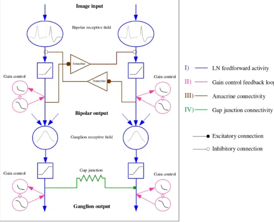
Bipolar cells
Bipolar cells receive signals from the photoreceptors along with inhibitory feedback from horizontal cells. They then transmit the visual information to ganglion cells, which is also modulated by the activity of amacrine cells. Their intermediate position makes them essential to understand how signals are transmitted from the photore-ceptors to ganglion cells, but it also makes it more challenging to record their neural activity. As a consequence, little is known about the response properties of bipolar cells, whose activity is often modeled as a linear filter applied to the stimulus, fol-lowed by a non-linearity. Bipolar cells don’t uniformly respond to the glutamate re-lease of photoreceptors. According to the type of receptor they express, they are either hyperpolarized or deporalized by light, which gives rise to two major classes of bipo-lar cells, ON-center and OFF-center bipobipo-lar cells. Bipobipo-lar cells also receive GABAergic inhibitory input from horizontal cells, shaping their center-surround receptive field. [7]
Amacrine cells
Amacrine cells are the most diverse retinal cell class, they however remain very in-sufficiently understood. This cell family consists of two major types, small-field and wide-field cells, based on the size of their dendritic arbor. They are involved in both feed-forward and lateral connectivity pathways [8].
Amacrine cells are activated through glutamatergic input from bipolar cells. They con-versely release two types of inhibitory neurotransmitters : GABA and glycine, onto bipolar cells and ganglion cells, and can also provide a feedback to other amacrine cells. They are also connected to bipolar cells and ganglion cells via gap junctions.
Ganglion cells
Ganglion cells, whose axons form the fibers of the optic nerve, are the output layer of the retina. They integrate the visual information processed by the upstream layers and transmits it to the brain, in the form of action potentials. They receive excitatory glu-tamatergic input from bipolar cells, and inhibitory GABAergic and glycinergic inputs from amacrine cells. Ganglion cells have a wide diversity of dentritic field morpholo-gies, yielding different sizes of receptive fields. These receptive field are mainly char-acterized by the receptive field’s configuration of upstream bipolar cells. According to their retinal location, ganglion cells will either communicate with a small number of photoreceptors (as few as five in the fovea), or a large number (up to many thousands in the periphery)[6] [9]. Ganglion cells play an essential role in objects shape and mo-tion detecmo-tion.
A small number of ganglion cells is photosensitive, and contribute to circadian rhythms and light reflex. These cells contain their own photopigment, melanopsin, which makes them sensitive to light even when photoreceptors are not responding.
Figure 1.2: Simple retina representation. The photoreceptors transform the light into an electrical signal transmitted to the bipolar cells. These neuronal signals are modified by horizontal cells which also ensure the lateral connectivity of photoreceptors and bipolar cells. Bipolar cells then forward the signal to ganglion cells, taking into account the lateral interactions afforded by amacrine cells. [5] .
1.1.2
Motion processing in the retina
While color vision and binocular vision are not shared between all the species, motion detection is an essential visual capability common to all of them. As a conse-quence, studying visual motion processing has been one of the fundamental challenges facing systems neuroscience. Studies have revealed that sensitivity to motion starts al-ready at the level of the retina, allowing different species to navigate the world, and detect the possible presence of mates, predators or preys. We will review in this sec-tion some aspects of the understanding of mosec-tion detecsec-tion by the retina, giving as examples direction selectivity, speed tuning and lag normalization.
1.1.2.1 Direction selectivity
There exists a wide range of theoretical and biological approches to studying reti-nal processing of motion. Moving stimuli are generally considered as a spatiotemporal pattern of light intensity projected on the retina, from which retina extracts relevant in-formation, such as the direction of image motion. Detecting motion requires neural networks able to process in a non-linear fashion moving stimuli, asymmetrically in time. [10] [11] [12] [13]
Direction sensitivity is the first mechanism involved in motion sensitivity, achieved through the interaction of On and Off pathways [14] (See Fig. 1.3).
Direction selective ganglion cells (DSGCs) are triggered differentially by the di-rection of a visual stimulus, responding more strongly when the motion of the object corresponds to their preferred direction. This feature is not dependent on the stimulus shape, size or color. There exists three types of DS cells in the retina : ON/OFF DSGCs, which respond both to the leading and the trailing edge of the stimulus, ON DSGCs, which only respond to the leading edge, and OFF DSGCs, which only respond to the trailing edge.
ON/OFF DCGCs are divided into 4 major types, according to their prefered di-rection : ventral, dorsal, nasal or temporal. Cells of different types differ in their den-dritic configuration and synaptic projections. For instance, nasal cells have a dentritic arbor with an asymmetry toward the nasal direction.
From a mechanistic point of view, DSGCs receive their inputs from bipolar and star-bust amacrine cells, responding to their directional preference with a large excitatory postsynaptic potential followed by a small inhibition. Conversely, when stimulated by
Figure 1.3: Schematic of the mammalian retina wiring, highlighting the rod and cone pathways. Left, ON pathways. Right, OFF pathways. The cone circuitry is illustrated using two cone photoreceptors : on the left hand side, the photoreceptor is connected to the ON ganglion cell through an ON cone bipolar cell. On the right hand side, the cone photoreceptor is connected to the OFF ganglion cell via an OFF cone bipolar cell. The rod pathways are more diverse : they can be either direct (OFF3); connected through a rod bipolar, an amcrine cell, and an ON or an OFF cone bipolar cell (ON1, OFF1); or connected through a cone photoreceptor and an ON or an OFF cone bipolar cell (ON2, OFF2).[14]. .
an object moving in their null direction (i.e the opposite of their preferred direction), they respond with a small excitatory postsynaptic potential followed by a large inhibi-tion. Starbust amacrine cells (SACs), whose dendrites emerge radially from the soma, have been shown to express important direction selectivity properties. Optical calcium imaging has specifically revealed that SACs respond strongly to centrifugal motion, while they are inhibited by the centripetal motion. More generally, when SACs were silenced with toxins, direction selectivity was strongly undermined.
SACs possess two types of neurotransmitters, acetylcholine, allowing excitation and GABA, allowing inhibition. Studies have shown an uneven repartition of gabaergic and cholinergic SACs synapses onto the dendritic arbor of DSGCs, providing direction sensitivity. (See Fig. 1.4)
As stated before, direction selectivity could also be an intrinsic feature of DSGCs, due to the asymmetry of their dendritic field. Trenholm [3] showed the existence of DS-GCs that conserve their direction selectivity even in the presence of cholinergic and GABAergic inhibitors. This property is further enhanced by the presence of gap junc-tions between cells that are tuned to the same direction, increasing the response of
Figure 1.4: A) Distribution of the excitatory (cholinergic) synapses and inhibitory (gabaergic) synapses along the motion axis. On the null side of the dendritic arbor of SACs, more inhibitory synapses are present, reducing as a consequence the activity of DSGCs along the non preferred axis. Conversely, on the preferred side of the den-dritic tree, more cholinergic and glutamatergic synapses are present, facilitating the emergence of action potentials at the level of DSGCs. B) Details of the neurotransmitter exchanges between SACs, bipolar cells and DSGCs involved in the direction selectivity pathway. Glutamate is represented in green, Acetylcholine in red and GABA in blue [15]. .
downstream cells (cells that are located further in the connectivity graph) to an object moving in their preferred direction.
1.1.2.2 Speed processing
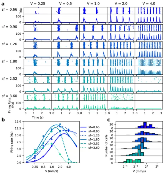
Speed tuning has been recently identified as a property inherent to a certain class of ganglion cells. Ravello et al. [16] have studied speed selectivity of RGCs using mo-tion cloud stimuli, artificial textures which conserve some of the properties of natural images, mainly in terms of frequency bandwidths. These stimuli are characterized by a spatial frequency, a spatial frequency bandwidth (equal to 0 for regular gratings) and a temporal frequency, resulting in a broader and less binary distribution of contrast, where low contrasts are more frequent.
Figure 1.5: RGC response to grating stimuli with different spatial and temporal fre-quencies. (a) Example response (raster plot and firing rate) of a single cell at each com-bination of spatial frequency and speed. (b) Fitting of response curves with skewed Gaussians. (c) Distribution of speed responsiveness across cells show a preferred speed that decreases with the spatial frequency. [16] .
high density of RGCs. In particular, they showed that the speed selectivity property is not inherent to a single type of RGCs, but rather shared between different classes. They first recorded the RGC response to gratings with different spatial and temporal frequencies. There is a trade-off between spatial and temporal frequencies, the higher the spatial frequency, the more the response curve shift towards lower speeds. High firing rate frequencies are reached for a combination of intermediate values of spatial frequency and speed. (See Fig. 1.5)
When presenting the rodent with motion clouds, the response curves show a narrow-ing effect around the preferred speed, i.e the response to low and high speeds is much weaker for motion clouds than simple gratings. The higher the complexity of the mo-tion cloud, the larger is its spatial frequency bandwidth and the narrower is the speed response curve around the preferred speed. This shows a finer speed tuning of RGCs in naturalistic conditions.
1.1.2.3 Lag normalization
Direction selective ganglion cells (DSGCs) in the mouse retina are selective to ob-jects moving in the cardinal directions. There exists a class of DSGCs coupled through gap junctions, eliciting spiking 106 +- 16 µm before the stimulus reaches their dendritic field. Trenhlom & al. [3] have emphasized the role of coupling in these cells response : uncoupled cells begin responding when a bar effectively enters their receptive field, i.e, their dentritic field extension, whereas coupled cells start responding before the bar reaches their dendritic field. The coupling considered here is towards the side from which the stimulus is approaching.
This response property accounts for lag normalization : coupled cells far from onset of the bar start responding when the bar reaches a constant distance from their soma, whatever its velocity, while uncoupled cells detect the bar at a position which is further shifted as the bar velocity increases.
Figure 1.6: Lag normalization in an electrically coupled network of DSGCs. a) Schematic of the model : the nth ganglion cell receive a total current which is the sum of the bipolar input and the weighted pre-junctional input from the previous ganglion cell. The strength of coupling is the only free parameter in their model, and is tuned to best tune the data. b) Simulation results of the first and the sixth cell of the network, for different stimulus speeds. Lag normalization is developed by cells far from the start of motion. [3]
Partial conclusion
In this first section, we reviewed the physiological organization of the retina, and gave examples of motion processing mechanisms and pathways, showing that the retina is not a mere camera, but is rather able to perform complex computations. In particular, we reviewed the role of connectivity (amacrine cells connectivity as well as gap junction coupling) at the level of ganglion cells, in motion and speed processing.
1.2
The visual cortex
1.2.1
General organisation of the visual cortex
The visual cortex is the part of the brain specialized in the processing of visual
in-formation. It receives the sensory inputs from the Lateral Geniculate Nucleus1located
at the level of the thalamus.
When cells in the visual cortex are stimulated within their receptive field, in a given way, they emit action potentials. This defines their neuronal tuning, i.e the stimu-lus properties to which they are sensitive. This tuning becomes all the more complex when going towards higher visual areas. For instance, if the receptive fields of cells in V1 correspond to simple stimulus feature such as orientation, some cells in the inferior temporal cortex will only fire if a definite object appears in their receptive field [17]. There exists two primary visual streams, receiving inputs from the primary visual cor-tex, the dorsal and the ventral stream. The dorsal pathway starts with V1, goes through V2, the dorsomedial area, the medial temporal area and the posterior parietal cortex. It is mainly involved in the processing of motion, and the coding of location. The ventral stream begins as well with V1, goes across V2, V4 and the temporal cortex. It is mainly associated with the shapes and objects recognition, and is also involved in long-term memory. (See Fig. 1.7)
Figure 1.7: Organization of the visual cortex into two parallel streams.[18]
1The Lateral Geniculate Nucleus (LGN) is a central relay in the visual pathway. It transmits to the
primary visual cortex the visual information received from the retina, via the optic nerve. The LGN lies in both the left and right brain hemispheres. It contains layers of parvocellular (small sized cells receiving their inputs from midget ganglion cells) and magnocellular cells (larger cells receiving their inputs from parasol ganglion cells).
1.2.1.1 The primary visual cortex
The primary visual cortex is the most commonly studied area in the visual cortex. Each brain hemisphere has a cortical area V1 receiving the visual signal from the con-tralateral eye. It is essentially involved in the processing of static and moving stimuli, and is specialized in pattern recognition. It is able to differentiate different features such as orientations, colors, spatial frequencies, motion . . . V1 neurons can thus be viewed as a set of specialized spatio-temporal filters able to selectively respond to each of these features. Neurons with similar response properties are arranged in columns, which are in turn assembled in larger structures called modules. Each module is able to decode and analyze a small area of the visual field.
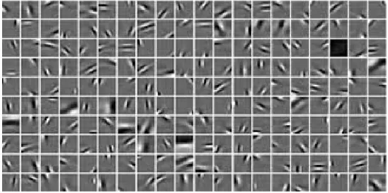
V1 is thus believed to implement a filter bank of 2D "Gabor filters" (See Fig. 1.8), which reproduces the cortical cells responses to impulse stimuli. [19] [20] [21]
Figure 1.8: Image structures can be captured using a bank of oriented basis functions. The set of basis functions has been obtained with a sparse coding algorithm. [21]
From an anatomical point of view, the multi-level processing of the primary vi-sual cortex is based on the feed-forward pathway, i.e the activity coming from the retina via the LGN, and extensive lateral connections between the cortical columns. The direct feed-forward activity alongside the contribution of lateral connectivity are then projected onto V2, V3, V5. V1 also receives feedback projections from V2, V3, V4 and V5.
Neurons in V1 are organized according to a retinotopic map. Indeed, the spatial posi-tions of RGCs within the retina is conserved by their neuronal projecposi-tions in the LGN, and the same topology is also preserved at the level of V1. When measuring the loca-tions of receptive fields along V1, one can see that adjacent RF centers from posterior
to anterior correspond systematically to ganglion cells located from the fovea towards the periphery.
As stated earlier, orientation and direction selectivity are strongly present at the level of V1. The vast majority of V1 neurons are orientation selective to a certain extent, but only approximately 30% of them show strong direction selectivity.
Cells in V1 have also been shown to be either monocular, i.e solely responding more strongly to one eye, or binocular, i.e cells responding maximally to stimulation in both eyes or with a weak bias to a given eye excitation. Their proportion vary according to the area of V1 from which the cells are recorded. Binocular cells are involved in binoc-ular disparity : the difference in the objects location between the left and right eyes, used for depth perception.
Hubel and Wiesel [22], who won the Nobel prize for their studies of the physiolog-ical and functional organization of neurons in V1 (orientation and direction selectiv-ity, binocularity . . . ) , emphasized the existence of three types of neurons : simple cells, complex cells, and hypercomplex cells. Each of these different types can be dis-criminated based on how they respond to visual stimuli. Simple cells respond best to elongated edges. They are orientation selective, and can be monocular or binocu-lar. Their receptive fields has distinct ON and OFF subregions. Complex cells are also orientation selective, but are mostly all binocular. Their receptive fields are homoge-neous, responding to either ON or OFF stimuli. Finally, hypercomplex cells are similar to complex cells, with the difference that they have an end-stopping property : they maximally respond to a cell with a given length, and their response starts decrease for higher lengths, while complex cells response increases with the bar length before reaching a plateau. (See Fig. 1.9)
1.2.1.2 Higher order areas
Higher order areas in the visual cortex are responsible for the refinement of the visual information pre-processed by V1. V2 improves the ability to discriminate lines and edges, and enhance color interpretation. For instance, V2 is responsible for color constancy, which accounts for a constant color perception, regardless of illumination levels.
V3 is specialized in form processing but is poorly sensitive to color. Most of V3 cells are orientation selective, and the activity of some is modulated by motion and depth.
Figure 1.9: Complex cells response increases with the size of the bar before reaching a constant value, while hypercomplex cell response is maximum for an intermediate size [23].
They are also highly sensitive to contrast.
V4 is mainly responsible for color processing. Like V2, V4 is also selective to orienta-tion and spatial frequency, but is however tuned to more complex object features such as geometric shapes. Recent studies have emphasized the existence of long-term
plas-ticity 2at the level of V4, sensitivity to salient stimuli, and the ability of its receptive
fields to change over time, depending on attention levels.
Finally, V5, also known as the medial temporal area, is involved in motion processing. Cells in V5 are largely tuned to stimuli speed and direction. More details on the role of V5 and motion processing will be introduced in the next section.
An important feature at the level of higher order visual areas is that they are strongly influenced by past experience based expectations. Though these expectations can lead to misinterpretations or misperception of the external world, they enable the brain to process visual information very quickly [24].
1.2.1.3 The retinotopic representation
Cells in V1 form together a topographic map of the retina input, known as the retintopic representation. Each half of the visual field is displayed as a map on the contralateral hemisphere, while the fovea is mirrored on the occipital pole. The fovea refers to the center part of the visual representation, where cells have the highest
reso-2Synaptic plasticity is the property that connections between neurons, called synapses, have to
change their strength according to their activity. It encompasses the multiple mechanisms involved in modifying synaptic transmissions over time. Long-term plasticity refers to phenomena that occur over a longer time range, lasting minutes or more.
lution, i.e the smallest receptive fields. The mapping and identification of visual areas has been achieved using magnetic resonance imaging (MRI). It has been mainly used to investigate changes in cortical activity patterns when subjects are presented with flashed checkerboard stimuli, with a changing position.
While the primary visual cortex is only split into to hemispheres, the retinotopic repre-sentation of V2 and V3 is split into 4 quadrants. Nowadays, more than 20 visual areas have been characterized, showing different levels of retinotopic representation.
Figure 1.10: The Retinotopy paradigm. In order to measure retinotopic maps in the cortex, two stimuli are used, a ring expanding stimulus and a rotating wedge. Data has been recorded on the left hemisphere. The figure shows how the eccentricity and the polar angle are mapped into the retinotopic map of the visual cortex. [25]
1.2.2
Motion processing in the visual cortex
Motion perception is one of the most important tasks that the visual cortex at-tends to, analyzing and processing the visual information by a large number of inter-connected neurons and areas. The complexity of cells’ response increases from lower to higher cortical areas, and this is also applicable to motion processing. The latter starts at the level of V1 where there is a large proportion of direction selective cells. Mo-tion processing is further pursued at the level of the medial temporal area (MT or V5), where cells are selective to both direction and speed. The first motion detector model has been proposed by Hassenstein and Reichardt [26](See Fig. 1.11). The model relies on changes in contrast of two spatially distant locations, inside the receptive field of a motion sensitive neuron. The neuron will only produce a response if contrast changes
are temporally delayed, and is thus not only selective to direction, but also to motion velocity. Several models have been developed based on Reichardt detectors, but they all share the common feature of integrating spatio-temporal variations of the contrast, constituting the motion sensor’s receptive field. Consequently, to perceive motion as coherent and uninterrupted, an additional integration over motion detectors is hypoth-esized to take place. This integration usually takes the form of a pooling mechanism, over visual space and time.
Figure 1.11: The motion detector model is composed of two subunits. The input re-ceived at the level of one unit is sent to the opposite unit, to be multiplied with the op-posite unit’s input with a time delay. The output of the two units are then substracted to form the output of the motion detector. Adapted from [27].
Studies [28] [29] have also shown the existence of a bias for centrifugal and cen-tripetal motions. From a functional point of view, this could be due to the prevalence of radial motion in self-locomotion situations. When walking forward for instance, the visual scene moves centrifugally on the retina, and the opposite occurs when moving backward.
Predictive coding The brain can be seen, from a probabilistic point of view, as an engine that computes the probabilities of the most likely causes to a given neuronal activity, assuming that the computations would converge to a single optimal solution.
In order to understand how this solution is found, many studies have investigated pre-dictive aspects of neuronal processing, directing the reconstruction of possible stimuli towards the most likely scenarios [30] [31].
The term "predictive coding" refers to the role played by predictions in the shaping of neural signals, decreasing neural activity when the stimulus does not contradict the prediction, and giving rise to a strong response when the prediction is inaccurate. There is a class of neurons, called object detectors, that respond only when the move-ment of an object does not correspond to the predicted trajectory. This type of neurons will have a particularly high response when an object that has not been predicted ap-pears in the visual field. Conversely, these neurons are very strongly inhibited when the movement of the object corresponds to the prediction.
Partial conclusion
In this section, we reviewed the physiological organization of the cortex, with an emphasize on the primary visual cortex. We presented an example of motion pro-cessing at the level of the cortex, and introduced the notion of predictive coding. This notion is highly linked to anticipation. In the following, we will not focus on the prob-abilistic aspect of predictive coding in the retina and the cortex, but rather on its mech-anistic aspects.
Chapter 2
Anticipation in the retina and the
primary visual cortex
Estimation of a moving object precise position is a non trivial problem that the brain has to attend to, taking into account the time lags that are involved in neuronal computations. Let’s consider a human subject, interacting with a moving object, that he has to catch for instance. How does he compensate for the delays introduced by his nervous system ? Some studies suggest that the origin of compensation is motor : the subject performs a muscular action in response to a predicted cause [32] [33] [34]. This would explain why practice, mental or physical, not only improves the timing of actions, but also the velocity at which they are realized [35] [36].
Other studies show that motion anticipation is in fact sensory : the visual system is able to extrapolate the trajectory of a moving object, delivering an anticipated repre-sentation of the object’s position, when its motion is deterministic. [1] [37] [38]. It has been shown that such anticipatory mechanisms exist, first at the level of the retina, and are further carried out by the primary visual cortex.
It is likely that anticipation is ultimately due to the cooperation of both compensation mechanisms [39]. In that sense, Nijhawan et al. suggest that "compensation mech-anisms [...] belong to a general principle of how the brain carries out computation efficiently in the spatio-temporal domain".
In this chapter, we will focus on motion anticipation in the retina and primary visual cortex. We will first present the psychophysical experiment known as the flash lag effect that highlights anticipation from a perceptual point of view. We will then
ex-plore the experimental findings concerning anticipation in the retina and visual cortex. Finally, we will present, in each of the sub-parts, the models that have been developed to reproduce anticipatory effects.
2.1
The flash lag effect
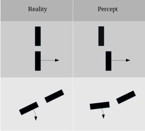
The flash lag effect is an optical illusion where a bar moving along a smooth tra-jectory and a flashed bar are presented to the subject, and are perceived with a spatial displacement, while they are actually aligned. A variation of this illusion consists of a bar moving in rotation, a bar flashed in angular alignment, giving rise to a perceived angular discrepancy. (See Fig. 2.1)
Neuroscience has explored many explanations for this illusion, including motion ex-trapolation. The visual system being predictive, it processes differently a bar in smooth motion, whose motion can be extrapolated, and a flashed bar which cannot be pre-dicted by the system.
A second explanation is that the visual system, rather than extrapolating trajectories, simply processes moving objects with a smaller latency than flashed objects [40]. In the first conception, the actual position of the moving object is anticipated, while in the second, both the moving and the flashed objects elicit delayed responses, with a delay that is reduced in the case of motion.
A third explanation suggests that the flash lag effect is due to postdiction, in other words, the perception of the flash is conditioned by events happening after its appear-ance [41] [42]. This hypothesis is inspired by the the color phi illusion, where two dots of different colors appearing at two discrete yet close positions, with a small latency, will be perceived as a single moving dot which color has changed.
In the following, we will restrain our literature review to the first hypothesis, i.e motion anticipation and trajectory extrapolation. These two phenomena have been shown to occur at the level of the retina and the primary visual cortex for different species : small animals such as the salamander and the rabbit in the case of retinal anticipation, and bigger animals such as cats and monkeys in the case of cortical antic-ipation.
Figure 2.1: Representation of the flash lag effect. Arrows denote moving bars, versus the bar flashed in alignment. In the case of translation, the flashed bar is perceived as lagging behind the moving bar, while in the case of rotation, it is perceived with an angular displacement.
2.2
Anticipation in the retina
When an object moves across the visual field, its motion elicits a series of neu-ronal activities, at the level of the retina, transmitted to the LGN, that will eventually be decoded at the level of the visual cortex. All this encoding and decoding processes take time, and emphasize the necessity of having mechanisms which compensate for the generated delays.
The general consensus nowadays among retina experts, it to consider that the retina performs general features extraction, rather than just being a mere transmitter, for the visual cortex to be able to process stimuli with more efficiency. The complexity of retinal processing is achieved through a variety of cells mechanisms [6] and different connectivity pathways [43], [44], [45]. One of the most interesting features that the retina attends to is motion anticipation of moving objects. We will first present experi-mental evidence showing motion anticipation of a moving bar at the level of the retina, and we will then introduce the models that have been developed to account for it.
2.2.1
Experimental evidence
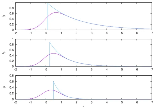
At the level of the retina, an object moving along a trajectory generates an activity in advance to its future position. Berry et al. [1] have first shown that local gain control mechanisms occurring at the level of bipolar and ganglion cells can explain the local anticipation of a moving bar. These mechanisms explain the change in the shape of response observed in experimental data, bringing the cells to their peak activity state earlier than when they respond to a flashed bar, without modifying the time at which the activity starts increasing, with respect to the size of the receptive field.
Figure 2.2: Ganglion cells firing rates in response to flashed and moving bars. a,c,e Recording of Fast OFF salamander ganglion cells. b,d,f, recordings of brisk-sustained OFF rabbit ganglion cells. The first row shows the response to a bar flashed for 15ms.
The second and third rows show the response to a bar moving at 0.44mms−1, in
oppo-site directions. Error bars are obtained from the repeated presentations of the stimulus. The bar is the three configurations is 90% contrast, and 133 µm width. [1]
Berry et al. recorded the responses of OFF-type ganglion cells in salamander and rabbit retinas, in response to two stimuli : a bar flashed in the centre of the individ-ual cells receptive field, and bar moving at a constant speed across the receptive field. Fig. 2.2 shows that the flashed bar generates a narrow response with a peak occurring
after the flash, while the moving bar elicits a wider response, with a peak firing rate that seemingly occurs ahead of the center of the bar, near its leading edge. This effect doesn’t depend on the direction in which the bar moves, showing that this anticipatory effect is not due to direction selectivity.
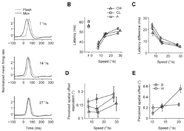
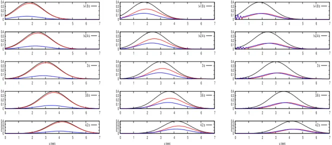
Another study by Johnston et al. [46] emphasized the role of inhibition at the level of the retinal connectome in the existence of anticipatory mechanisms. Fig. 2.3 shows, using an experimental setting similar to the one used by Berry et al., the existence of an-ticipation, this time in the goldfish retina. Both studies show that motion anticipation is velocity dependent (Fig. 2.4). However, this velocity tuning varies across species, and according to the ganglion cell type. Similarly, the anticipation ability degrades at low contrasts.
Figure 2.3: A) Goldfish ganglion cells firing rate when responding to a bar flashed for 100 ms. B) Ganglion cells firing rate when responding to a bar moving at 500µm/s. The bar is 100% contrast and 160µm width. [46]
Johnston et al. have demonstrated that motion anticipation arises from the gen-eral properties of the retina connectome, namely the excess of inhibitory connections compared to excitatory ones. In particular, they show that bipolar gain control mecha-nisms are not responsible for motion anticipation. They emphasize instead, through pharmacological disruptive tests, the role played by feedforward inhibition, which ganglion cells receive from amacrine cells. This inhibition could be at the origin of