HAL Id: tel-02294276
https://tel.archives-ouvertes.fr/tel-02294276
Submitted on 23 Sep 2019HAL is a multi-disciplinary open access archive for the deposit and dissemination of sci-entific research documents, whether they are pub-lished or not. The documents may come from teaching and research institutions in France or abroad, or from public or private research centers.
L’archive ouverte pluridisciplinaire HAL, est destinée au dépôt et à la diffusion de documents scientifiques de niveau recherche, publiés ou non, émanant des établissements d’enseignement et de recherche français ou étrangers, des laboratoires publics ou privés.
Characterization of the mitochondrial translation
apparatus of Arabidopsis thaliana
Florent Waltz
To cite this version:
Florent Waltz. Characterization of the mitochondrial translation apparatus of Arabidopsis thaliana. Vegetal Biology. Université de Strasbourg, 2018. English. �NNT : 2018STRAJ087�. �tel-02294276�
UNIVERSITÉ DE STRASBOURG
ÉCOLE DOCTORALE DES SCIENCES DE LA VIE ET DE LA SANTÉ
Institut de biologie moléculaire des plantes – CNRS – UPR2357
THÈSE DE DOCTORAT
présentée par :Florent WALTZ
soutenue le : 6 Décembre 2018
pour obtenir le grade de :
Docteur de l’université de Strasbourg
Discipline : Sciences de la Vie et de la Santé
Spécialité : Aspects Moléculaires et Cellulaires de la Biologie
Characterization of the mitochondrial translation
apparatus of Arabidopsis thaliana
THÈSE dirigée par :
M. GIEGE Philippe Docteur, université de Strasbourg
RAPPORTEURS :
Mme. LURIN Claire Docteur, université de Paris-Saclay
M. LIGHTOWLERS Robert Professeur, université de Newcastle
AUTRES MEMBRES DU JURY :
Mme. DUCHENE Anne-Marie Professeur, université de Strasbourg Examinatrice interne
M. MIREAU Hakim Docteur, université de Paris-Saclay Membre invité
Acknowledgments/Remerciements
First, I would like to thank the members of the Jury, Pr. Anne-Marie Duchêne, Dr. Claire Lurin and Pr. Robert Lightowlers for agreeing to evaluate my work. As invited members, I would also like to thank Dr. Yaser Hashem and Dr. Hakim Mireau.
Un grand merci à Philippe pour m’avoir accueilli au sein de son équipe. Merci à lui pour sa patience, son implication et ses excellents conseils tout au long de ma thèse qui m’ont permis d’acquérir de nombreuses connaissances scientifiques et techniques. Merci de m’avoir fait confiance en me permettant de travailler sur ce super projet, même quand ça ne marchait pas. Merci également pour la liberté que j’ai pu avoir sur le projet et de m’avoir donné la possibilité de participer à de nombreux congrès !
Merci à Anne-Marie, Laurence, Gag, Hélène, Thalia, José, André et Heike d’avoir bien voulu partager leurs connaissances au cours de nos séances de questions lors des entrainements de Master, et plus tard pendant ma thèse. J’ai aussi appris beaucoup grâce à eux. Merci particulièrement à Laurence pour sa confiance lors des RNA Salons.
Merci à Hakim et son équipe pour m’avoir accueilli pendant deux semaines à l’INRA de Versailles. Merci à Trung (Master Trung) pour sa patience durant mon apprentissage du ribosome-profiling. Merci à Yaser pour le temps passé à l’IGBMC sur le microscope, malgré les galères, et sans qui la partie structurale du projet n’aurait pas été possible. Merci également à Jailson pour ses conseils pour les purifs de ribosomes.
Mes remerciements s’adressent bien évidemment aux membres de l’équipe, présents ou passés, qui ont fait de ces 3 ans une expérience enrichissante, merci pour leur bonne humeur, leur soutien et leurs précieux conseils. Anthony qui m’a appris la majorité des techniques de bases, Cédric qui est maintenant parti et Matthieu qui reprend la suite, Géraldine pour sa bienveillance et ses conseils, Ayoub pour son caractère jovial et Kamel qui a maintenant sa propre équipe. Je tiens aussi à remercier particulièrement Mathilde qui est arrivée au moment où j’ai débuté ma thèse, et avec qui j’ai toujours apprécié discuter et qui m’a été d’une grande aide avec les manips. Merci tout particulièrement à Pierre également pour les bons moments passés au labo ou en dehors, ainsi que pour son aide.
Je tiens également à remercier les stagiaires qui sont passés par le labo, notamment Herrade qui m’a suivie à la trace dans mon parcours académique et avec qui j’ai également pu passer d’excellents moments en dehors du travail (du coup aussi merci à Thibaud pour supporter Herrade au quotidien).
Egalement merci à mes camarades de Master, qu’ils aient continué à l’IBMP ou non, Nicolas, Deborah, Yannick, ainsi que les autres étudiants en thèse pour leur bonne humeur (presque) toujours au rendez-vous, Hélène, Marlène, Marco, Magdalène, Arnaud, Guillaume, Stéphanie, Marion, Thibaud, Adrien, etc …
Merci à Joern et mes collègues moniteurs avec qui j’ai pu durant ces trois dernières années découvrir l’enseignement, qui fut aussi une expérience à part entière.
Merci à Johana, Lauriane et Philippe de la plateforme protéomique pour avoir toujours été à l’écoute et avoir été aussi réactifs, même lorsque je leur demandais de passer des séries de 12 échantillons en 3 jours. Sans eux rien de ce que j’ai fait n’aurait été possible.
Merci à Todd et Stefano pour les conseils durant ma mi-thèse. Thank you Misha for your advices and for allowing me to use your gradient collector.
Merci à ma famille et particulièrement à mes parents qui ont su me montrer le chemin et éveiller ma passion pour les sciences. Merci pour m’avoir toujours soutenu depuis le début même si je sais qu’il est parfois difficile de saisir les tenants et les aboutissants de ce que je fais.
Merci à Caroline avec qui j’ai partagé ses trois dernières années. Les longues heures au 306 pendant le stage à discuter, les voyages autour du monde (jusqu’en Chine), les concerts, etc. Merci pour le soutien que tu m’as apporté dans les bons et les mauvais moments. J’ai pu partager / faire / voir / visiter / découvrir tellement de choses que je n’avais pas faites avant et je t’en suis reconnaissant. Je te fais entièrement confiance pour ta thèse, tu en es plus que capable, n’en doute pas. Merci moumou.
Je tiens aussi à remercier mes bons amis Nicolas et Nathan qui m’ont aussi soutenus à leur manière durant toute ma scolarité (c’est-à-dire pratiquement depuis la maternelle).
Plus généralement, merci à tout le personnel de l’IBMP, les responsables des différentes plateformes et les membres des différentes équipes que j’ai pu croiser au détour d’un couloir.
Abbreviations
aa : amino acid
ADP : Adenosine diphosphate AOX : Alternative Oxydase Asn : Asparagine
Asp : Aspartate
ATP : Adenosine triphosphate
AtPRORP : Arabidopsis thaliana PRORP BN-PAGE : Blue Native PAGE
Ca2+ : Calcium
cDNA : complementary DNA co-IP : Co-immuno-precipitation CP : Central protuberance
cryo-EM : Cryogenic electron microscopy C-ter : Carboxyl-terminus
DNA : Deoxyribonucleic acid ETC : Electron Transport Chain
FAD: Flavin adenine dinucleotide
Fe/S : Iron-sulphur
FECA : First Eukaryotic Common Ancestor GDP : Guanosine diphosphate
GFP : Green Fluorescent Protein GTP : Guanosine triphosphate
IMM : Internal mitochondrial membrane IMS : Inter-membrane space
kb : Kilobase kDa : KiloDalton
kDNA : Kinetoplastid DNA
LB medium : Lysogeny broth medium LC-MS/MS : Liquid chromatography–mass
spectrometry
LECA : Last Eukaryotic Common Ancestor LSU : Large Subunit
MAM : Mitochondrial associated
membranes
MCS : Membrane contact sites Met : Methionine
MICOS : Mitochondrial contact site and
cristae organizing system
mRNA : messenger RNA MS : Mass spectrometry
MS medium : Murashige and Skoog medium mtDNA : mitochondrial DNA
MTS : mitochondrial targeting sequence NAD : Nicotinamide adenine dinucleotide nDM : n-Dodecyl β-D-maltoside
N-ter : amino-terminus O2 : Dioxygen
OD : Optical density
OMM : Outer mitochondrial membrane OXPHOS : Oxidative phosphorylation PAGE : Polyacrylamide gel electrophoresis PCD : Programmed cell death
PCR : Polymerase chain reaction
PfPRORP : Plasmodium falciparum PRORP Pi : Inorganic phosphate
PPR : Pentatricopeptide repeat PRORP : Protein Only RNase P PTC : Peptidyl transferase center PVDF : Polyvinylidene fluoride
RcPRORP : Romanomermis culicivorax
PRORP
RNA : Ribonucleic acid rPPR : ribosomal PPR rRNA : ribosomal RNA RT : Reverse transcription S : Svedberg
SD sequence : Shine-Dalgarno sequence SDS : Sodium dodecyl sulfate
Ser : Serine
ssRNA : Single stranded RNA SSU : Small subunit
TCA cycle : Tricarboxcylic acid cycle T-DNA : Transfer DNA
TIM : Translocase of the inner membrane TOM : Translocase of the outer membrane TPR : Tetratricopeptide repeat
tRNA : Transfert RNA
UTR : Untranslated Transcribed Region VDAC : Voltage-dependent anion channels WT : Wild-type
List of figures and tables
List of figures
Figure 1: The two-domains tree of life
Figure 2: The endosymbiosis in eukaryote evolution
Figure 3: Energy availability in prokaryotes versus eukaryotes Figure 4: The overall mitochondria organization
Figure 5: The TCA cycle
Figure 6: The mitochondrial ETC – the oxydative phosphorylation Figure 7: Supercomplexes organization
Figure 8: Mitochondrial genomes sizes and contents Figure 9: RNA editing in plant mitochondria
Figure 10: Mitochondrial introns
Figure 11: PPR proteins structure and binding mechanism Figure 12: The different classes of PPR proteins
Figure 13: PPR repartition across eukaryotes
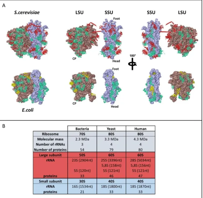
Figure 14: Eukaryotic and prokaryotic ribosomes structure and composition Figure 15: The processus of translation in eukaryotes and prokaryotes
Figure 16: The mitochondrial ribosomes are bound to the inner membrane of mitochondria Figure 17: Mitoribosomes structures
Figure 18: The mRNA recruitment to mitoribosomes
Figure 19: Mitochondrial ribosomes purification protocols used during this study
Figure 20: Occurrence of mitoribosomes and cytoribosomes in crude ribosome fractions Figure 21: Mitochondria lysis optimization
Figure 22: Attempts to resolve mitoribosome subunits using cauliflower mitochondria Figure 23: The 336/336L double mutant and construction of the 336-HA (rPPR1-HA) line Figure 24: BN Page analyses
Figure 25: Mitoribosome co-immunoprecipitation using 336HA Figure 26: Mitochondrial rpl15-HA IP
Figure 27: PfPRORP Figure 28: RcPRORP
Figure 29: MNU2 expression and purification
Figure 30: Arabidopsis mitochondrial central protuberance (CP) proteins compared with E. coli CP structure
Figure 31: mS83, “adenylyl cyclases” proteins Figure 32: mS84, an “IF2-like” protein
List of tables
Table 1: The mitochondrial genomes composition Table 2: Number of PPR genes in different organisms Table 3: Mitoribosomes composition
Table 4: Purification optimization using different salt concentrations Table 5: Compared protein compositions of charaterized mitoribosomes Table 6: List of primers
i
Table of content
INTRODUCTION
PREAMBLE: MITOCHONDRIA AS THE KEY TO EUKARYOTE ORIGIN AND EXPANSION ... 1
THE COMPARTMENTS OF GENE EXPRESSION IN PLANTS (VIRIDIPLANTAE) ... 6
MITOCHONDRIA IN PLANTS AND OTHER EUKARYOTES ... 7
OVERALL STRUCTURE OF MITOCHONDRIA: ... 7
METABOLIC FUNCTIONS SUPPORTED BY MITOCHONDRIA: ... 8
Iron-sulfur cluster synthesis ... 9
Tricarboxylic acid (TCA) cycle – Krebs cycle... 9
Oxydative phosphorylation and the respiratory chain ... 10
MITOCHONDRIAL GENOMES: ... 12
Sizes and gene contents ... 12
Organization and structures ... 13
Why is the genome retained? ... 14
MITOCHONDRIAL GENE EXPRESSION: ... 15
Transcription ... 16
Maturation and degradation ... 17
Definition of transcript ends ... 17
RNA editing ... 18
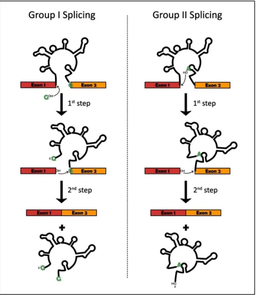
Splicing ... 19
PPR proteins ... 20
Classes of PPRs ... 20
Structure and mode of action ... 21
Repartition across eukaryotes... 22
PROTEIN SYNTHESIS ... 23
GENERALITIES ... 23
THE MECHANISM OF TRANSLATION ... 24
THE MITOCHONDRIAL RIBOSOMES: ... 25
Adaptation to a membrane-bound system ... 26
Mitoribosomes compositions ... 26
Mitoribosomes structures ... 28
Characteristic structural features of the mitoribosomes ... 29
mRNAs recruitment to the mitoribosomes ... 31
ii
Initiation ... 32
Yeast translational activators ... 32
Regulators of translation in other eukaryotes ... 33
Elongation and termination ... 34
AIMS OF THIS STUDY ... 36
RESULTS OPTIMIZATION OF ARABIDOPSIS MITORIBOSOMES PURIFICATION ... 37
ALTERNATIVE PROTOCOL TO PURIFY PLANT MITORIBOSOMES ... 39
SEPARATION OF THE SMALL AND LARGE MITORIBOSOME SUBUNITS ... 40
IMMUNO-PURIFICATION OF MITORIBOSOMES USING RPPR1 (PPR336) AS A BAIT ... 41
CHARACTERIZATION OF THE 336/336L LINE ... 41
CREATION OF 336HA PLANTS ... 41
BN-PAGE ANALYSES ... 42
MITORIBOSOME CO-IMMUNOPRECIPITATION ... 42
PUBLICATION:SMALL IS BIG IN ARABIDOPSIS MITOCHONDRIAL RIBOSOME ... 44
MITOCHONDRIAL R-PROTEINS MUTANTS ... 71
PRORP PROTEINS IN EVOLUTION ... 72
APICOPLASTIC PLASMODIUM FALCIPARUM PRORP(PFPRORP) ... 73
MITOCHONDRIAL ROMANOMERMIS CULICIVORAX PRORP(RCPRORP) ... 74
CHARACTERIZATION OF A NOVEL MITOCHONDRIAL NUCLEASE: MNU2 ... 75
PUBLICATION:DETERMINATION OF PROTEIN-ONLY RNASE P INTERACTOME IN ARABIDOPSIS MITOCHONDRIA IDENTIFIES A COMPLEX BETWEEN PRORP1 AND ANOTHER NYN DOMAIN NUCLEASE ... 77
DISCUSSION COMPOSITION OF THE PLANT MITORIBOSOME COMPOSITION COMPARED WITH OTHER MITORIBOSOMES ... 101
SPECIFICITIES OF ARABIDOPSIS MITORIBOSOME RRNAS ... 101
SPECIFICITIES OF ARABIDOPSIS MITORIBOSOME PROTEIN COMPOSITION ... 101
What about the plant specific r-proteins? ... 102
To which degree is the plant mitoribosome bound to membrane? ... 104
FUNCTIONS OF THE PPR336/RPPR1 AND 336L PROTEINS ... 105
PERSPECTIVES OF THIS WORK ... 107
iii
MATERIALS AND METHODS
MATERIALS ... 110 PLANT LINES ... 110 PPR mutants ... 110 Non-PPR mutants ... 111 CELL LINE ... 111 BACTERIAL STRAINS ... 112 Escherichia coli ... 112 Agrobacterium tumefaciens... 112 PLASMIDS ... 113
GATEWAY cloning vectors ... 113
Restriction cloning vectors ... 113
PRIMERS / OLIGONUCLEOTIDES ... 114
ANTIBODIES... 114
Primary ... 114
Secondary ... 114
METHODS ... 115
NUCLEIC ACIDS ANALYSES ... 115
RESTRICTION CLONING ... 115
GATEWAY CLONING ... 115
HEAT SHOCK BACTERIAL TRANSFORMATION ... 115
DNA AMPLIFICATION BY PCR ... 116
AGAROSE GEL ELECTROPHORESIS ... 116
PHENOL/CHLOROFORM NUCLEIC ACIDS EXTRACTION ... 116
ETHANOL PRECIPITATION ... 117
PLASMID PURIFICATION : ... 117
QUANTIFICATION : ... 117
SANGER DNA SEQUENCING : ... 117
RAPID PLANT DNA EXTRACTION FOR GENOTYPING ... 117
PLANT TOTAL RNA EXTRACTION ... 118
RT-QPCR ... 118
cDNA synthesis ... 118
qPCR ... 119
iv
PROTEINS ANALYSIS ... 120
TOTAL PROTEIN EXTRACTION : ... 120
Crude ... 120
From TRIzol extraction ... 120
PROTEIN QUANTIFICATION ... 120 Bradford ... 120 Nanodrop ... 120 PROTEIN EXPRESSION ... 121 Induction test ... 121 Protein purification ... 121 PROTEIN CO-IMMUNOPRECIPITATION ... 122
SODIUM DODECYL SULFATE–POLYACRYLAMIDE GEL ELECTROPHORESIS (SDS-PAGE) ... 122
PROTEIN TRANSFER TO A PVDF MEMBRANE UNDER LIQUID CONDITION ... 123
IMMUNODETECTION OF PROTEINS (WESTERN BLOT) ... 123
LC-MS/MS ANALYSIS ... 123
MITOCHONDRIAL COMPLEXES ANALYSIS BY BLUE NATIVE PAGE(BN-PAGE) ... 124
IN-GEL COMPLEX I ACTIVITY TEST: ... 125
PLANTS ... 125
AGRO-TRANSFORMATION BY FLORALDIP ... 125
TRANSIENT PROTEIN EXPRESSION IN NICOTIANA BENTHAMIANA LEAVES ... 126
SEEDS STERILIZATION ... 126
ARABIDOPSIS CELL CULTURE ... 126
ARABIDOPSIS MITOCHONDRIA PURIFICATION ... 126
RIBOSOMES PURIFICATION FROM MITOCHONDRIA ... 128
MICROSCOPY ... 128
CONFOCAL MICROSCOPY IMAGING ... 128
ASSESSMENT OF RIBOSOME SAMPLES BY TRANSMISSION ELECTRON MICROSCOPY ... 128
DATA COLLECTION USING CRYO-ELECTRON MICROSCOPY ... 129
BIOINFORMATIC ANALYSES ... 129 BIBLIOGRAPHY………..………135 RESUME EN FRANCAIS INTRODUCTION ... 146 RESULTATS ... 148 CONCLUSIONS ... 150
1
Introduction
This thesis is about translation in plant mitochondria. Before addressing the topics directly linked to my work, I wish to give some insights about the crucial role that the acquisition of mitochondria has played for the evolution of life on Earth. This is presented as a preamble to my thesis introduction.
Preamble: Mitochondria as the key to eukaryote origin and expansion
Life arose relatively early in the history of Earth. The first traces of life can be dated back to 3.8 billion years ago, a little more than 500 million years after Earth formation. Before the emergence of cells, a “pre-biotic” period saw the development of the molecular components required for life. A widespread theory proposes that during this pre-biotic time the first complex molecule that arose was RNA. In this hypothetical stage of Earth evolution, called “RNA world”, self-replicating RNA molecules proliferated before the evolution of DNA and proteins (Bernhardt, 2012). This theory is supported by the fact that several core components of the cell, like the ribosome – the universal cellular machine that synthesizes proteins – are composed mainly of RNA, which may constitute relics of this ancient world.During approximately 2 billion years, the Earth was populated by simple unicellular organisms, “complex” life only appeared much later. Morphologically complex living organisms – plants, animals, fungi – and single-celled “protists” (such as Trypanosoma brucei or Plasmodium
falciparum), descend from one singular ancestor LECA – Last Eukaryotic Common Ancestor –
estimated to have lived ~1.6–2 billion years ago. Therefore, they form in phylogenetics what we call a monophyletic group. This group, called Eukarya (literally “true nucleus”), or more commonly Eukaryotes, was named after their major morphological feature which is the presence of a membrane-enclosed nucleus containing the cell's genetic information. The other super-group of organisms, the first ones that have appeared on Earth, are called Prokaryotes (regrouping Bacteria and Archaea). They were named in opposition to the Eukaryotes, as they do not possess a nucleus (Sapp, 2005; Zimmer, 2009). Even though eukaryotes were originally classified according to the presence or not of a nucleus, many more cellular and molecular traits define if a given organism belongs to Eukaryotes. Among those characteristics, the nucleus should harbor nuclear pores – inside the nucleus, nuclear lamina is found along with linear chromosomes with telomeres, facilitating sexual reproduction. Complex regulatory mechanisms, including chromatin (histones …), an RNAi system and small non-coding RNAs, orchestrate gene expression at different levels.
Figure 1: The two-domains tree of life
Schematic representation of the “tree of life”. In the 70’s all cellular life was divided into the three major evolutionary lines: Eukarya (or eukaryotes), Bacteria and Archaea. But it was later discovered that eukaryotes emerged from Archaea, hence the two-domains tree of life here represented. In the early 2010’s eukaryotes were found to branch within, or as sister to, the Thaumarchaeota, Aigarchaeota, Crenarchaeota and Korarchaeota (TACK) superphylum. But more recent phylogenomic analyses now suggest that eukaryotes originated from within the Asgard archaea phylum or that they represented a sister group to them (Eme et al., 2017).
The red and green arrows represent the two major endosymbiosis events, in red between an α-proteobacteria and the ancestor of eukaryotes and in green a cyanobacteria and the ancestors of Archaeplastida.
2 Transcription is uncoupled from translation and involves extensive RNA processing (including intron splicing, capping and polyadenylation). Translation itself is much more complex, putting into action eukaryote-specific ribosomes and numerous additional translation factors. Also, an elaborate protein regulation and recycling system composed of the proteasome and an ubiquitin signaling systems are always found. The cellular eukaryotic environment is highly compartmentalized by the presence of a sophisticated endomembrane systems composed of the endoplasmic reticulum, the Golgi apparatus, endosomes, lysosomes and peroxisomes. A complex actin-based and tubulin-based cytoskeleton and associated molecular motor proteins enabled intracellular trafficking, cell motility and a complex cell cycle, including meiosis, allowing sexual reproduction. They are also able to synthesize a wide range of eukaryote-specific lipids and phospholipids (eg. sterols and sphingolipids) (Eme et al., 2017). And of course, eukaryotes are host of aerobic or facultatively aerobic, mitochondria that descended from a once free-living alpha-proteobacterium (Andersson et al., 1998).
The origin of eukaryotes has always been, and is still, subject to strong debates. The pioneering work of Carl Woese and colleagues revealed that all cellular life could be divided into the three major evolutionary lines: Eukarya (or eukaryotes), Bacteria and Archaea. Life was therefore represented as a three-domains tree of life, each domain represented as a monophyletic group, Archaea and Eukarya sharing a unique common ancestor. But already in the early 80’s molecular phylogenetic analyses showed that eukaryotes and archaea were sister groups. These results were reinforced by the multiple metagenomic approaches which allowed the sequencing and discovery of new archaeal organisms, hence permitting a better comprehension of Archaea. Most experts tend now to class Eukaryotes as a sister group of the archaeal clade Asgard, named after the realm of the gods in Scandinavian mythology, as several eukaryote-specific features never found before in any prokaryotes (specific ribosomal proteins, cytoskeleton component, ubiquitin system, trafficking machinery,…), were found in this group of organisms, favoring of a two-domains tree of life rather than the original three-domains one (Fig 1) (Zaremba-Niedzwiedzka et al., 2017).
Even though it now appears that Eukaryotes emerged from Archaea, they are by nature chimeric and symbiotic organisms. Chimeric first because when you look into eukaryote genomes, a large proportion of the genes can be traced back to prokaryote (eg. about 36% in yeast (Cotton and McInerney, 2010)). Among those prokaryotic-like genes, most of them can be traced back to bacteria (55–70%) whereas a smaller proportion is related to Archaea (20–35%). In term of
Figure 2: The endosymbiosis in eukaryote evolution
Schematic representation of the two major endosymbiosis events that led to the eukaryotes that we know today. The first major one happened during the event of eukaryogenesis (the series of events that drove the evolution of FECA to LECA). The α-proteobacteria, that later became the mitochondrion, was acquired by an “early eukaryote”, but the exact course of events is not fully understood. Later the engulfment of a cyanobacteria by an eukaryote led to the apparition of the “green-phylum” Archaeplastida.
3 functions, genes with a bacterial ancestry are overwhelmingly linked to metabolic processes, whereas archaeal genes tend to be involved in information processing – DNA replication, transcription and translation machineries – essential functions that comfort the archaeal origin of eukaryotes. This huge proportion of bacterial genes reveals the chimeric nature of eukaryotes. In the 60’s Lynn Margulis already proposed (Sagan, 1967) that eukaryotes were not the product of standard natural selection, but the result of a serial of endosymbiosis events, therefore called the “serial endosymbiosis theory”. This concept was based in part on early hypotheses, for instance from the 19th century French botanist Andreas Schimper (born in Strasbourg in 1856). On a
footnote to his 1883 work, he made the observation that the chloroplasts that are found in photosynthetic organisms shared many characteristics with cyanobacteria and thus proposed that the combination of two separate organisms may have given rise to modern photosynthetic organisms (Schimper, 1883). The Margulis theory stipulates that a number of bacteria cooperated together so closely that some cells got inside others, thus leading to the formation of the nucleus and all the complex compartmentalization of eukaryotes, predicting both the chimeric and symbiotic nature of eukaryotes. This theory was later proven wrong, but not entirely as the events of endosymbiosis took place twice during eukaryote evolution (Fig 2) (Lane, 2017). Once during early eukaryote evolution, before LECA, thus more than ~2 billion years ago, leading to what we now know as the mitochondria, which is conserved across (almost) all eukaryotes. Organisms that would have diverged before the acquisition of mitochondria (or amitochondriate eukaryotes) were investigated but never found. Such organisms, named Archezoa, were proposed by Thomas
Cavalier-Smith (Cavalier-Smith, 1989). Organisms like Giardia intestinalis and Trachipleistophora
hominis were thought to be “archezoan”, however it was later proven that these organisms
possess relics of mitochondria and had thus lost the latter secondarily, not being evolutionary intermediates. The kingdom Archezoa has therefore been abandoned (Poole and Penny, 2007). The second endosymbiosis event occurred later, about 1.5 billion years ago, with the acquisition of the chloroplast, and contributed to the apparition of the “green phylum” (or Viridiplantae which is made up of the green algae and the land plants) of eukaryotes (Dorrell and Howe, 2012). Those two components of eukaryotic cells, called organelles, are primarily energy producing compartments.
These compartments are both able to produce energy due to the presence in their membrane system of an electron transport chain (ETC). It relies on a series of protein complexes that transfer electrons from electron donors to electron acceptors via redox reactions, and couples this electron transfer with the transfer of protons across a membrane. The flow of protons across
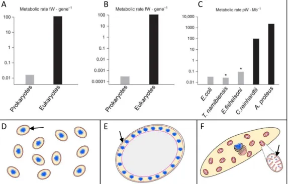
Figure 3: Energy availability in prokaryotes versus eukaryotes
In A the mean energy per gene in prokaryotes versus eukaryotes equalized for genome size are represented. The same is represented in B but equalized for genome size and cell volume. In C the power available per haploid genome (energy per gene X number of genes in one haploid genome) is represented for different prokaryotic and eukaryotic organisms. Note the log scale is each case, eukaryotes have 4-5 orders of magnitude more energy available. Both T.namibiensis and E.fishelsoni are giant bacteria, but even though they are able to reach such proportions their internal volume is metabolically quite inert. To sustain this giant life style they require multiple copies of their genomes, in the case of E.fishelsoni 200,000 copies of its 3.8 Mb genome, in order to produce enough energy. In the end, even if large bacterium do exist they are not able to sustain “true” cellular complexity, as the totality of their energetic resources have to be mobilized to sustain this life style.
D, E and F are the organisms discussed above. D Schematic representations of a medium
sized prokaryote (E.coli), E a very large prokaryote with its inert vacuole in grey
(T.namibiensis) and F a medium-sized eukaryote and its multiple mitochondria allowing large energy availability (A.proteus). Bioenergetic membranes across which
chemiosmotic potential is generated are drawn in red and indicated with a black arrow and DNA is indicated in blue.
Derived from (Lane, 2011; Lane and Martin, 2010)
A B C
4 the membrane creates a electrochemical gradient that ultimately drives the ATP synthase and permits, every ten protons passing through it, the full rotation of the ATP synthase’s head allowing 3 ADP molecules to be turned into 3 ATP molecules, which will be used in all metabolic processes (Abrahams et al., 1994). The oxidative phosphorylation, which is the name of this process in mitochondria, was first hypothesized in the early 60s by Peter Mitchell, called the “Chemiosmotic theory” (Mitchell, 1961), which won him the 1978 Chemistry Nobel Prize. In chloroplast, a somewhat similar phenomenon happens, the photophosphorylation, but this process is light dependent. It highly resembles oxidative phosphorylation, the only evolutive innovation compared to the latter is the addition of the chlorophyll pigments to use the energy of the sun to
create a high-energy electron that splits water, releasing O2 and protons. Electrons then move
spontaneously from donor to acceptor through the electron transport chain.
The acquisition of mitochondria during the eukaryogenesis, and the fact that is was retained since then, in one form or another (eg. hydrogenosomes or mitosomes) – at the exception of a Monocercomonoid parasitic protist of that entirely lost mitochondria (Karnkowska et al., 2016) – prove the original symbiotic nature of eukaryotes. This cooperative way of functioning is for sure not as symbiotic as it was originally thought to be, with the original host more-or-less “enslaving” the original endosymbiont. Indeed mitochondria is now non-autonomous as the large majority of genes originally encoded in the endosymbiont were either lost or transferred to the nucleus of the host cell. From the archaeal ancestor of eukaryotes, FECA (First Eukaryotic Common Ancestor) to LECA, many other evolutive events happened, aside the acquisition of mitochondria. The exact nature of those events, and their chronology, is still debated. In the case of mitochondria, most competing scenarios can be roughly grouped into either mito-early, which considers the driving force of eukaryogenesis to be mitochondrial endosymbiosis into a simple host, or mito-late, which postulates that an already somewhat complex cell predated the original endosymbiont through phagocytosis. Along that, which of standard natural selection, horizontal gene transfer, or endosymbiosis/phagocytosis contributed the most to what LECA was, is still an unsolved question. Many eukaryote-specific traits for sure arose from standard natural selection. As predicted by the evolutionary theory, complex traits arise via a series of small steps (Darwin, 1859). Nevertheless, the acquisition of mitochondria gave a huge boost to the evolution by simply releasing the energetical constraint of these cells. Indeed, when comparing the energy available per gene between a prokaryote and a simple eukaryote, a eukaryotic nuclear gene governs nearly 5,000 times more energy flux than a prokaryotic gene (Fig 3) (Lane, 2011; Lane and Martin, 2010). Indeed, prokaryotes can generate energy only by pumping
5 protons across their membrane, thus being limited by their size. On the other hand, eukaryotes pack hundreds of mitochondria into a single cell allowing much more energy production. With that much power available, eukaryotes were able to develop a large number of new functions, many listed before, but most importantly, it permitted to release the structural constraint of cell size and genome size. Indeed eukaryotic cells are usually much larger (around 10-100 times), they have much larger genomes (around 40-600 times) and more genes (around 5 times), along with the cell structure itself being much more complex compared to prokaryotes. Overcoming this energetical barrier contributed much likely to the rapid radiation of eukaryote into the five super-groups of organisms that we know today (Adl et al., 2012). To date, the bacteria Rickettsia prowazekii is the more closely related organism to mitochondria in term of genetic structure (Andersson et al., 1998), but the original endosymbiont was most likely a part of a sister group of α-proteobacteria (Martijn et al., 2018). On the opposite, the jakobid Reclinomonas americana, and jakobids in general, possess the mitochondrial genome which most closely resembles the ancestral proto-mitochondrial genome, due to their high gene content (Lang et al., 1997).
Altogether, endosymbiosis is a process that had a profound impact on all extant eukaryotes. The acquisition of mitochondria marks a crucial step in eukaryotes emergence and is one of the clearest examples of an evolutionary transition. Beside the evolutive questions, mitochondria is also a crucial subject of study for human health as their dysfunction has been associated with an increasingly large number of inherited disorders and is implicated in common diseases, such as neurodegenerative disorders, cardiomyopathies, metabolic syndrome, cancer, and obesity (Nunnari and Suomalainen, 2012; De Silva et al., 2015). In plants, this organelle has also attracted considerable attention since they specify a widely expanded trait leading to an inability of plants to produce functional pollen, called “cytoplasmic male sterility”. Plant mitochondria are thus of huge agronomical interest (Chen and Liu, 2014; Horn et al., 2014).
6
The compartments of gene expression in plants (Viridiplantae)
The term "plant" refers to a group of eukaryotic organisms possessing the following traits: multicellularity, presence of cell walls composed of cellulose and the ability to carry out photosynthesis with primary chloroplasts, using light, water and carbon dioxide to synthesize nutriments, making them autotrophic organisms. Viridiplantae (literally "green plants") encompass a group of eukaryotic organisms made up of the green algae, which are primarily aquatic, and the land plants (embryophytes), which emerged within them. Embryophytes include the vascular plants, such as ferns, conifers and flowering plants.
In plants, three distinct cell compartments carry out genetic expression: the nucleus, mitochondria and chloroplasts. In these compartments genetic expression is differently fulfilled, and is tightly orchestrated through inter-organellar crosstalk, mitochondria and chloroplasts being completely dependent of the nucleus. As described above, mitochondria and chloroplasts were acquired through two consecutive endosymbiosis events. The first event happened about 1.5–2 billion years ago, which resulted in the acquisition of mitochondria. It appears that the original bacterium which later became mitochondria was an α-proteobacterium (Gray et al., 1999) but these results are now being reconsidered (Martijn et al., 2018). A secondary endosymbiosis event occurred between an already fully-fledged eukaryote and a cyanobacteria over 1 billion years ago (Dorrell and Howe, 2012), which ultimately lead to the formation of plastids, and gave rise to the “green-phylum”.
During evolution, most of the genes that were initially encoded in the original endosymbionts were either transferred to the nucleus of the host cell, or between endosymbionts (Hao et al., 2010), or lost (Brown, 2003). The genome size of around 4,500 genes in the original endosymbionts decreased to 3–67 genes in mitochondria and 23–200 genes in chloroplasts, as a result these are now semi-autonomous. For example, in Arabidopsis thaliana, 18% of the nuclear genes appear to be derived from cyanobacteria (Martin et al., 2002). Genetic expression in the nucleus and in the cytosol of plants is similar to what can be found in other eukaryotes. In chloroplasts and mitochondria, almost 2 billion years of evolution led to the apparition of specific gene expression mechanisms combining bacterial-like traits with novel features that evolved in the host cell. The most striking differences are observed between mitochondria from different big groups of eukaryotes. Indeed, as the evolutive radiation into the five big groups of eukaryotes (Adl et al., 2012) seems to have occurred quickly after the acquisition of mitochondria, each of them developed specific features.
A B
Figure 4: The overall mitochondria organization
A Electron micrograph of an animal mitochondria, the different main components are
indicated. B Schematic representation of the different components of mitochondria (described in the main text).
7
Mitochondria in plants and other eukaryotes
Overall structure of mitochondria:
Mitochondria are composed of two different membranes: the outer and inner membranes. Those two membranes of distinct natures delimit two biochemically different compartments; the inter membrane space (IMS) and the matrix. The outer membrane is rather permeable thanks to the presence of β-barrel shaped porins, called VDAC for voltage-dependent ion channel (Hodge and Colombini, 1997; Mihara and Sato, 1985). Those VDAC proteins play a key role in regulating metabolic and energetic flux by allowing the transport of several metabolites such as ATP, ADP, pyruvate, malate, … Besides VDAC, the outer membrane also contains so called Translocase of the Outer Membrane, or TOM complexes (Ahting et al., 1999; Dekker et al., 1998). TOMs are involved in the entry of larger molecules in mitochondria, mainly proteins, which are recognized if a signalling sequence at their N-terminus, named MTS, is present which then actively triggers the import (Emanuelsson et al., 2007; Omura, 1998). With the relocation of the majority of the original endosymbiont’s genes in the nucleus, most of the mitochondrial proteome has to be first translated by cytosolic ribosomes (often being associated to mitochondria when translating mitochondria-targeted proteins (Gold et al., 2017)) to be later imported in mitochondria, the import machineries are therefore of huge importance for mitochondrial biogenesis. Proteins first pass by the intermembrane space, IMS, where they will be sorted to either be inserted in the outer membrane, stay in the IMS, or go into the matrix or the inner membrane through the Translocase of the Inner Membrane, TIM (Fig 4) (Koehler et al., 1998; Sirrenberg et al., 1996).
The inner membrane encloses the protein-rich matrix and is the seat of the molecular machinery of chemiosmosis. It is rich in cardiolipin, an unusual phospholipid which constitutes roughly 20% of the IMM (internal mitochondrial membrane) and may contribute to the inner membrane impermeability (Hoch, 1992). Indeed, unlike the outer membrane, the inner membrane is freely permeable only to oxygen, carbon dioxide, and water. It is much less permeable to ions and small molecules than the outer membrane which requires special membrane transporters to enter or exit the matrix. This feature is essential for the functioning of the electron transport chain, where protons have to be pumped from the matrix to the IMS to be used by the ATP synthase. The surface of the inner membrane is much larger than that of the outer membrane. As a result, it has to be heavily folded, forming numerous invaginations called cristae (Griparic and van der bliek, 2001). By significantly increasing the total membrane surface area they
8 also increase the available working space. The cristae are connected to the inner boundary membrane via tubular structures termed crista junctions (Daems and Wisse, 1966). These internal structures can greatly vary between organisms and even tissues. Their structure seem to be governed by the mitochondrial contact site and cristae organizing system (MICOS) at the cristae junctions, but also by the organization of ATP synthases into dimers (Hahn et al., 2016), and of dimers into rows, which is a feature common to mitochondria of all species examined to date. In fungi, plants, and metazoans, the dimers are V-shaped and associate into rows along the highly curved ridges of lamellar cristae but in Paramecium tetraurelia it is U-shapped, forming tubular cristae (Mühleip et al., 2016).
Furthermore, mitochondria do not sit alone in the cell. Indeed, a great number of reports have recently shown that mitochondria are in close contact with the endoplasmic reticulum. These contact sites called MAMs (mitochondrial associated membranes), or more generally membrane contact sites (MCS), have mostly been studied in mammals and yeast (Vance, 2014). They contribute to the inter-organelle communication, to the modulation of mitochondrial morphology
and function as well as to processes like lipid synthesis, apoptosis and Ca2+ homeostasis. Even if
these contact sites are more discrete in plants, reports tend to show that they are also present (Mueller and Reski, 2015).
Metabolic functions supported by mitochondria:
Mitochondria are the power stations of eukaryotic cells and the sites of many important metabolic reactions. Among them, we can cite amino acid and nucleotide metabolism, lipid, quinone and steroid biosynthesis, and of course iron-sulfur (Fe/S) cluster biogenesis. As energy producers, their main role is to use reducing agents, derived from catabolic reactions like TCA cycle and β-oxidation of fatty acids, to fuel the oxidative phosphorylation. By channeling electrons through the respiratory chain complexes and creating a transmembrane electrochemical gradient, the ATP synthase is activated, which ultimately allow the conversion of ADP+Pi to ATP, the
biochemical energy currency.
Hence, mitochondria are crucial to maintain the high ATP/ADP ratio that is required for the functioning of the many biochemical reactions taking place in eukaryotic cells. Additionally, the TCA cycle generates numerous metabolic intermediates that are utilized by various anabolic pathways. Therefore, mitochondria sustain several crucial metabolic reactions and produce the majority of the cell ATP, constituting biosynthetic and bioenergetic organelles.
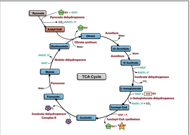
Figure 5: The TCA cycle
Schematic representation of the TCA cycle (TriCarboxcylic Acid cycle).
The cycle consists in a series of chemical reactions used to release stored energy through the oxidation of acetyl-CoA derived from carbohydrates, fats, and proteins. The
acetyl-CoA and water are used and allow the reduction of NAD+to NADH, and produces
carbon dioxide as a waste byproduct. The cycle also provides precursors of certain amino acids. The reducing agent NADH is fed into the oxidative phosphorylation (electron transport) pathway to produce usable chemical energy in the form of ATP. The TCA cycle and the oxidative phosphorylation are closely linked, as Complex II is part of the TCA cycle.
9 Aside from metabolic functions, mitochondria also play a crucial role in regulating cells life but also death. Indeed, in animals, mitochondria play a central role for a particular programmed cell-death (PCD) event, apoptosis. Calcium signaling and the release of Cytochrome c were found to be essential (Wang and Youle, 2009). As in animal cells, mitochondria also seem to be involved in PCD in plants. Upon induction of PCD, plant mitochondria aggregate and swell, a process known as the mitochondrial morphology transition, similar to what is observed during apoptosis (Van
Aken and Van Breusegem, 2015). Mitochondria are also key players in regulation of Ca2+ due to
their ability to accumulate rapidly and transiently this ion involved in cell signaling (Clapham, 2007).
Iron-sulfur cluster synthesis
Iron–sulfur (Fe/S) clusters belong to the most ancient protein cofactors in life, and fulfil many different functions, including electron transfer, redox sensing, enzyme catalysis and sulphur activation. In mitochondria, they are involved in various metabolisms, such as the TCA cycle (aconitase), the electron transfer chain (respiratory complexes I–III), fatty acid oxidation (ETF-ubiquinone oxidoreductase), and in lipoate and biotin biosynthesis (lipoate and biotin synthases) (Lill et al., 1999; Stehling and Lill, 2013). They also retain essential roles in other compartments of the cell, they are involved for example in DNA replication and repair in the nucleus (Stehling and Lill, 2013). Because of the simple nature of Fe/S clusters and their widespread distribution, they have been assigned essential roles in the evolution of life (Martin and Russell, 2003). In eukaryotes, the synthesis of Fe/S clusters and their insertion into apoproteins requires almost 30 proteins, the process being initiated in mitochondria. Interestingly, even in highly degenerated mitochondria, the synthesis of Fe/S clusters has been retained: it is found in Giardia mitosomes, Trichomonas hygrogenosomes or in the mitosome of the apicomplexan parasite C. parvum (van der Giezen, 2009). In Monocercomonoides, the only eukaryote described to date to be completely devoid of mitochondria or related organelles, a cytosolic sulfur mobilisation system, acquired through horizontal gene transfer, provides the Fe/S clusters required for protein synthesis. The regular mitochondrial Fe/S cluster synthesis pathway is considered to have been lost secondarily (Karnkowska et al., 2016).
Tricarboxylic acid (TCA) cycle
–Krebs cycle
The TCA cycle is the driver of cellular respiration. It was characterized by Hans Krebs for which he received the Nobel Prize for Physiology and Medicine in 1953, and after whom the cycle is sometimes named (Krebs cycle) (Fig 5). Taking place in the mitochondrial matrix, it uses the
two-Figure 6: The mitochondrial ETC – the oxydative phosphorylation
Schematic representation of the respiratory chain. The different components, i.e. respiratory complexes I to IV and the ATP synthase (V) as well as the full process are described in the main text.
10 carbon organic compound acetyl-CoA, produced by the oxidation of pyruvate and originally derived from catabolism of sugars (glycolysis) , fats (lipids beta-oxydation), or proteins (Fernie et al., 2004). In a series of redox reactions, the TCA cycle allow the conversion of acetyl-CoA in the
form of NADH, FADH2 and ATP. In a single turn of the cycle, two carbons enter from acetyl-CoA,
allowing the formation of three molecules of NADH and one molecule of FADH2, and one molecule
of ATP or GTP. The TCA cycle does not produce much ATP directly. However, the NADH and FADH2
generated by this process can make a lot of ATP indirectly. These reduced electron carriers will feed the electron transport chain and, through oxidative phosphorylation, drive synthesis of ATP
molecules produced in cellular respiration (Fernie et al., 2004). NADH and FADH2 are also
generated during lipids catabolism and glycolysis.
Oxydative phosphorylation and the respiratory chain
Oxidative phosphorylation (OXPHOS) is the main supply of energy in eukaryotic cells. This process is conceptually simple and mechanistically complex. It is supported by the respiratory
chain complexes sitting in the IMM. NADH and/or FADH2 act as electron donors that can be
oxidized by the Complex I and the Complex II, which constitute the entry points of the respiratory chain. The electrons flow through the different complexes, thanks to a physical phenomenon
called quantum electron tunneling, to reach the final electron acceptor O2 (Hayashi and
Stuchebrukhov, 2010). The electrons pass by four complexes, among which three are proton pumps. The whole process leads to the pumping of protons out of the mitochondrial matrix which result in an uneven distribution of protons across the IMM. This generates a pH gradient and a transmembrane electrical potential that creates a proton-motive force (PMF). The final phase of OXPHOS is carried out by Complex V, the ATP synthase, that is driven by the flow of protons back into the mitochondrial matrix (Fig 6).
As mentionned before, the respiratory chain is composed of five protein complexes. The first complex, Complex I or NADH:ubiquinone oxidoreductase constitutes the entry point of
electrons through the oxidation of NADH into NAD+, coupled with the proton translocation from
the matrix to the IMS. It is the largest enzyme of the electron transport chain, composed of more than 40 subunits (49 in Arabidopsis (Peters et al., 2013)). The hydrophilic arm mediates the transfer of electrons, via Fe/S clusters to the bound ubiquinone. Reduction of the ubiquinone induces conformational changes in the membrane arm resulting in proton translocation across the membrane via four channels. Electrons can also enter the ETC through the Complex II or succinate ubiquinone oxidoreductase. The oxidization of succinate to fumarate by the Complex II allows the
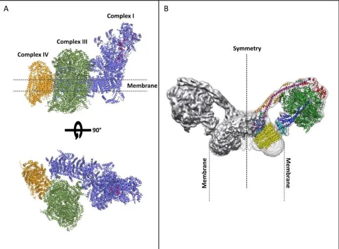
Figure 7: Supercomplexes organization
Atomic models of the A bovine respirasome and the B yeast dimeric ATPase.
A The respirasome (supercomplex I+III2+IV1) is the most prominent of the existing supercomplexes (except in plants). It contains all the components required to transfer electrons from NADH to the final acceptor, oxygen. Complex I is represented in blue, Complex III in green and Complex IV in yellow. (Sousa et al., 2016)
B The dimeric ATPase are located along the highly curved edges of the inner membrane
cristae and the angle formed by these dimers has an effect on cristae structure. Those dimers shape the inner mitochondrial membrane and mediate cristae formation. Derived from (Hahn et al., 2016)
11
transfer of electrons through the FADH2 intermediary to ubiquinone. The Complex II is the only
respiratory chain complex that is not involved in the transfer of protons to the IMS. As a result, Complex I and II both contribute to the formation of ubiquinol, which is freely diffusible in the membrane, and will shuttle electrons to Complex III. The Complex III, or cytochrome c
oxidoreductase, is usually imbedded in the membrane as a dimer (III2). The electrons from
ubiquinol are passed to the carrier cytochrome c via cytochromes b and c1. It can also oxidize ubiquinone to ubiquinol to pump two protons to the intermembrane space. Then, similar to ubiquinone, cytochrome c can travel between complex III and IV to transfer electrons. Cytochrome
c transfers its electron to the last enzyme of the ETC, the Complex IV or cytochrome c oxidase.
Once the electron is delivered, it is transferred to the final acceptor of the ETC, dioxygen, to convert it to two water molecules, leading to four protons being pumped to the intermembrane space during this process (Sousa et al., 2018).
By transporting electrons through the ETC, protons are pumped into the IMS, which lead to the establishment of an electrochemical gradient driving the proton-motive force (PMF). The ultimate step of the respiratory chain is for the protons to flow back to the matrix through the membrane-embedded ATP synthase. It is the ATP synthase, or Complex V, that converts the energy of spontaneous flow of protons into chemical energy of ATP bonds. Complex V is bipartite,
composed of a water-soluble F1-part connected to the membrane-embedded ring-like F0-part by
a central and a peripheral stalk. The protons pass first through the membrane-embedded rotor, the F0 part of the F1F0 ATP synthase, inducing a rotation of the F0 oligomeric ring that is transmitted
into conformational changes in the three nucleotide-binding pockets of the F1 head, thereby
catalyzing ATP synthesis. As mentioned above, ATP synthases are organized in rows of dimers along crista edges, suggesting that the dimers are responsible for bending the IMM and dictating cristae morphology (Lau and Rubinstein, 2012; Stock, 1999).
Beside Complex V, the respiratory components are also able to associate into homo- or heterocomplexes called supercomplexes (Fig 7). They can be divided into three main groups: I + III2, III2 + IV1-2 and I + III2 +IV1-4 (also called respirasomes, comprising different copy numbers of
Complex IV). Complex II is the only enzyme of the respiratory chain which does not associate with the other respiratory complexes. The respirasomes are the most abundant in animal mitochondria (Gu et al., 2016), contrary to plants where the majority of Complex IV (>90%) is present in monomeric state, but respirasome were identified in potato mitochondria (Eubel et al., 2003). It was also shown that respirasomes can associate into respiratory dimer, or megacomplex. These
A B
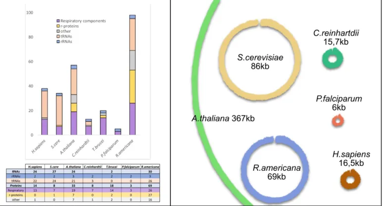
Figure 8: Mitochondrial genomes sizes and contents
A Classification of mitochondrial encoded genes from different eukaryotes. Genes
encoded in mtDNA are universally involved in oxidative phosphorylation and mitochondrial translation. Especially in Jakobids, mitochondrial genomes encode additional genes involved in processes such as transcription or RNA maturation. rRNAs are always encoded in the mt-genomes, even if they are sometimes fragmented (e.g C.
reinhardtii and P.falciparum), and a variable number of tRNA genes can be found. The
different genes present are listed in Table 1.
B The size of the different mitochondrial genomes exposed in A are represented. The
genomes are represented as master-circles even though this might not represent the in
12 results were obtained from different organisms, including porcine, potato, yeast and bacteria, suggesting the existence of a higher order arrangement of respiratory chain elements across species (Bultema et al., 2009; Davies et al., 2011; Gu et al., 2016; Heinemeyer et al., 2007; Sousa et al., 2016, 2013). The organization and complexes found between organisms vary. For example, in Saccharomyces cerevisiae, Complex I is not found but is compensated by several peripheral membrane NADH dehydrogenases (Yamashita et al., 2007).
In plants and other eukaryotes, another component is sometimes found, the alternative oxidase or AOX (McDonald and Vanlerberghe, 2006). The AOX is involved in an alternative route
for electrons passing through the electron transport chain. AOX is non-proton pumping and since
it bypasses complexes III and IV, it dramatically reduces the energy yield of respiration. It is found throughout the plant kingdom but also in the other kingdoms: it is sporadically found in protists and fungi, and also in many animal phyla, although clearly absent from vertebrates and arthropods (Vanlerberghe, 2013). The expression of the AOX gene is influenced by stresses, but the benefit conferred by this activity remains uncertain. It may enhance an organisms' ability to resist these stresses, through reducing the level of oxidative stress (Maxwell et al., 1999).
Mitochondrial genomes:
Mitochondria possess their own genome, vestige of its free-living bacterium ancestor. This genome is localized in the mitochondrial matrix. Over the course of evolution, the majority of the original genome of the endosymbiont was transferred to the nucleus or lost. As a result, only few genes remain encoded in the mitochondrial genome (Gray, 2012). Moreover, the evolutive radiation of eukaryotes most likely occurred after the acquisition of mitochondria, consequently, even if gene transfer was already an ongoing process, great differences in term of structure, size, gene content and expression mechanisms can be observed between the different groups of eukaryotes.
Sizes and gene contents
The size of mitochondrial genome greatly varies between organisms (Fig 8 B). It can be highly reduced like in Plasmodium falciparum, which is the smallest known mitochondrial genome with only 6 kb in size, harboring only three protein-coding genes, highly fragmented rRNA genes and no tRNA gene (Feagin et al., 2012). It can also be extremely large, Silene conica being the largest with an 11 Mb multichromosomal mt-genome, exceeding the size of some bacterial and even some nuclear genomes (Sloan et al., 2012). Large genomes are also a common feature of the Cucurbitaceae family (Alverson et al., 2011), but for most plants, mt-genomes have about
~300-Table 1: The mitochondrial genomes composition
Size and composition of different
mt-genomes from model
eukaryotes.
a bacterial RNApol, b putative
inner membrane ABD transporter,
c protein translocase, d unclear
function, e protein transporter, f
possible function in intron
maturation, g formed of maxi and mini-circles, h Ramrath et al., 2018
Source: H.sapiens (Taanman, 1999), S.cerevisiae (Wolters et al., 2015), A.thaliana (Marienfeld et al., 1999), C.reinhardtii (Salinas-Giegé et al., 2017), T.brucei (Kirby
and Koslowsky, 2017),
P.falciparum (Tyagi et al., 2014), R.americana (Burger et al., 2013)
13 500 kb. This variety of genome sizes in plants is a direct cause of their composition, large repeat sequences promoting recombinations (Gualberto and Newton, 2017). But larger genomes does not imply that more genes are present. In metazoan, the genome is relatively small and conserved, ranging between 15–17 kb and about 16 kb in human. The gene content is also quite stable, with 37 genes. In Arabidopsis, where the genome is 367 kb and contains 57 genes, only 20 genes more, for a genome 20 times larger than that of metazoan (Fig 8 A) (Unseld et al., 1997). The genes still encoded in the mitochondrial genome are rather conserved throughout eukaryotes, and can be classed in two groups for the protein-coding ones: “ribosomal protein” and “bioenergetics”; but additional proteins are sometimes encoded in mt-genomes (Table 1). The former are involved in ribosomal subunit synthesis and mainly occur in protist and plant mt-genomes; the later code for subunits of the respiratory chain complexes as well as cytochrome c maturation proteins. Genes coding for the mitochondrial rRNAs are also always found. They are among the few genes universally encoded by mtDNA across eukaryotes (Gray, 2012). tRNAs, required for the translation of the few proteins encoded in the mt-genome, are also found however the presence of a complete minimalist set of tRNA genes encoded by the mt-genome is more an exception than a general rule (Salinas-Giegé et al., 2015). In fungi, Saccharomyces cerevisiae encodes a complete set of tRNAs, but in trypanosomatides (e.g. Trypanosoma brucei, Leishmania tarentolae) and alveolates such as Plasmodium, the mt-genomes can be completely devoid of tRNA genes (Salinas-Giegé et al., 2015). Interestingly, the mt-genomes of angiosperms contain chloroplast-like genes,
as a replacement for tRNAHis and tRNAAsn that were lost in all investigated angiosperms (Fey et al.,
1997).
Organization and structures
The mt-genomes organization also greatly differs between eukaryotes. In metazoan, the genomes are quite reduced, with minimal to no intergenic regions, and no intron sequences in vertebrate mtDNA. All protein-coding genes and rRNAs are flanked by tRNAs (excepted for the COIII - ATP6 junction). This allows the production of large polycistrons which will be processed by pre-tRNA maturation enzymes, RNase P and Z, releasing individual processed mRNAs, rRNAs and tRNAs (Ojala et al., 1980). In plants, the genes are separated by large non-coding regions that are not conserved across species, contributing to the extensive size of plant mt-genomes. Those non-coding sequences are the result of horizontal gene transfer, most likely derived from chloroplastic, nuclear, or viral DNA (Gualberto and Newton, 2017). Several genes contain introns, mainly of group II, that are self-catalytic ribozymes (Cech, 1986). In Arabidopsis, 23 group II introns are present. Interestingly, Arabidopsis and all angiosperms possess the matR gene, encoded within
14 intron 4 of nad1. This gene encodes for the MatR maturase, a protein binding to several group II introns in vivo, but its putative roles in splicing are yet to be determined (Brown et al., 2014b; Unseld et al., 1997).
Mitochondrial genomes are usually represented as singular circular molecules. However, this is not always the case. In Cucumis sativus, the mitochondrial genome assembles into three circular chromosomes of different lengths (Alverson et al., 2011). In kinetoplastid protists such as
Trypanosoma, the mitochondrial genome is composed of ~50 so called maxicircles and thousands
of minicircles. Maxi- and mini-circles form a packed network of circular DNA that constitute the kinetoplast (called kDNA). This arrangement contributes to the specific posttranscriptional processing modification where uridines are inserted into, or deleted from, messenger RNA precursors (Aphasizhev and Aphasizheva, 2014). But mt-genomes are not always arranged into circular molecules, linear mt-genomes also exist. It has been described in the green-algae Chlamydomonas (Smith et al., 2010), Plasmodium, some fungi, and several cnidarian animals (Nosek and Tomáška, 2003). An unusual situation has been identified in a single-celled protist relative of animals, Amoebidium parasiticum, whose large mtDNA (>200 kbp) consists of several hundred linear chromosomes that share elaborate terminal-specific sequence patterns (Burger et al., 2003). In the case of linear mt-genomes, they are protected by specialized end-structures, such as covalently closed single-stranded DNA termini or protective proteins, and they also tend to have telomere-like repeats (Burger et al., 2003; Nosek and Tomáška, 2003).
Why is the genome retained?
The question remains open on why mitochondria (and chloroplast) retained a genome. It would certainly be more advantageous in term of energy if the cell would not need to have a second complete gene expression machinery in mitochondria, with a complete different DNA replication system, RNA polymerase and ribosomes, all this required to express a very small set of genes. Several models have been proposed to understand why mitochondria actually retained a genome.
One of the hypothesis is that the impossibility to transfer the remaining genes in the nucleus relies on the differences between the nuclear genetic code and the one used in mitochondria. Indeed, in animals, yeast and several protists (D.N.J. de Grey, 2005), the codon usage is different. For example, UGA is a ‘stop’ codon in the universal code and in plant mitochondria, but it codes for tryptophan in animals and fungi mitochondria. This is not the case in plants.
15 It was also proposed that the mitochondrial gene transfer is still an ongoing process and that we are only witnessing an intermediary phase of eukaryote-mitochondria evolution. Eventually all genes should/could be transferred to the nucleus – researchers are even trying, through genetic engineering, to artificially transfer mitochondrial genes to the nucleus, not without difficulties. For example, a nuclear encoded copy of cytochrome b fused to a mitochondrial targeting signal is not imported in mitochondria as it forms protein aggregates (Claros et al., 1995).
This lead to the hydrophobicity hypothesis which stipulates that the product of the few remaining genes are too hydrophobic to be synthesized in the cytoplasm and then imported into mitochondria (von Heijne, 1986). This is supported by the observation that the two large ribosomal RNAs, as well as the highly hydrophobic proteins cytochrome b and Cox1 are the ones universally still encoded in mt-genomes.
An alternative explanation is that the retention of these small but functional genomes persist because organellar gene expression must be under direct redox control (Allen, 2003). This hypothesis is termed CORR (co-location for redox regulation), and explains that genes in mitochondria and chloroplast must be under the direct regulatory control of the redox state of their gene products to allow the fine tuning of mitochondrial gene expression in response to metabolic changes. Such a mechanism has been described in yeast: Mss51, a translational activator of Cox1, is able to bind heme B. It could therefore sense oxygen levels to modulate Cox1 synthesis and its subsequent assembly into Complex IV, the major oxygen-consuming mitochondrial enzyme (Soto et al., 2012).
Mitochondrial gene expression:
Mitochondrial gene expression is a complex – patchy – mechanism, completely dependent on nuclear-encoded factors. Indeed, only a few genes involved in mitochondrial gene expression are encoded in the mt-genome, these being most frequently ribosomal RNA and ribosomal proteins required for the final step of gene expression. For their expression, mitochondrial genes must be transcribed, their RNAs then undergo a number of post-transcriptional maturations and they are translated. Thus the majority of the factors involved in these processes are encoded in the nucleus, expressed in the cytosol and imported into the mitochondria.
tRNAs, which are crucial for translation are also, in some organisms, imported from the cytosol. In human and yeast for example, tRNA import is not required as a full set of tRNAs is
16 present, but it does occur nonetheless (Salinas-Giegé et al., 2015). In contrast, Trypanosoma and Plasmodium mt-genomes have no tRNA genes, therefore they all have to be imported (Hancock and Hajduk, 1990; Salinas-Giegé et al., 2015). In plants, tRNAs have to be imported from the cytosol to ensure translation, as mt-genomes are incomplete. In Arabidopsis, 6 tRNAS must be imported (Salinas-Giegé et al., 2015; Salinas et al., 2008).
Due to the early evolutive radiation of eukaryote and the bacterial origin of mitochondria, gene expression in mitochondria combines bacterial-like features, inherited from the original endosymbiont, with eukaryote traits coming from the host cell (Adl et al., 2012; Gray, 2012). Moreover specific gene expression features evolved independently in the different groups of eukaryotes. As a result, gene expression mechanisms in mitochondria are unique and diverse between species.
Transcription
Gene expression starts with transcription to synthesize RNA. To fulfil mitochondrial transcription, most eukaryotes possess a nuclear-encoded phage-type RNA polymerase (mtRNAP or NEP for nuclear-encoded polymerase) which replaced the ancestral bacterial-type RNA polymerase (Liere and Börner, 2011). In contrast, in Jakobids, primitive protists of which
R.americana belongs, the mitochondrial genome still encode a bacterial-type RNA polymerase
(Lang et al., 1997).
In plants, several promoters are necessary for the transcription of the genome. In Arabidopsis mt-genome, among the 57 genes, five transcripts are produced in poly-cistronic form, three transcripts are trans-spliced and the others are monocistronic (Forner et al., 2007). Two nuclear genes code for two RNA polymerases, RpoTm is targeted to mitochondria and RpoTmp is targeted to both mitochondria and chloroplast, another one, RpoTp, is targeted only to chloroplast (Liere and Börner, 2011). RpoTm is involved in the transcription of the majority of the mt-genome, thus the role of RpoTmp is still poorly understood, but in vitro and in vivo studies showed that a subset of mitochondrial genes depend on RpoTmp for their expression (Kühn et al., 2009). In human, only two polycistronic transcripts are synthetized. The transcription starts from two promoters, a heavy strand promoter and a light strand promoter (Pearce et al., 2017), which will generate the two pre-transcripts that will later be processed. In yeast, several transcription start sites are found producing polycistronic precursor molecules encoding two or more coding sequences (Christianson and Rabinowitz, 1983).