HAL Id: tel-03127237
https://tel.archives-ouvertes.fr/tel-03127237
Submitted on 1 Feb 2021HAL is a multi-disciplinary open access
archive for the deposit and dissemination of sci-entific research documents, whether they are pub-lished or not. The documents may come from teaching and research institutions in France or abroad, or from public or private research centers.
L’archive ouverte pluridisciplinaire HAL, est destinée au dépôt et à la diffusion de documents scientifiques de niveau recherche, publiés ou non, émanant des établissements d’enseignement et de recherche français ou étrangers, des laboratoires publics ou privés.
anchorage of Shigella flexneri
Daniel Isui Aguilar Salvador
To cite this version:
Daniel Isui Aguilar Salvador. Characterization of the role of lpaA on the cytoskeletal anchorage of Shigella flexneri. Microbiology and Parasitology. Université de Paris, 2019. English. �NNT : 2019UNIP7144�. �tel-03127237�
1
Université de Paris
Ecole doctorale BioSPC ED562
Unité de Communications Intercellulaires et Infections
Microbiennes.
CIRB. CNRS UMR 7241/ INSERM 1050
Characterization of the role of
IpaA on the cytoskeletal
anchorage of Shigella flexneri
Par Daniel Isui AGUILAR SALVADOR
Thèse de doctorat de Microbiologie Procaryote et Eucaryote
Dirigée par Guy TRAN VAN NHIEU
Présentée et soutenue publiquement le 07/11/2019
Devant un jury composé de :
Agathe SUBTIL, DR2/CNRS, Institut Pasteur, Rapportrice
Christophe LE CLAINCHE, DR2/CNRS, Institut de Biologie Intégrative de la Cellule, Rapporteur Sandrine BOURDOULOUS, DR2/CNRS, Institut Cochin, Examinatrice, Présidente
Fabrizia STAVRU, CR/CNRS, Institut Pasteur, Examinatrice
2 Titre :
Caractérisation du rôle de la protéine IpaA sur l'ancrage cytosquelettique de Shigella flexneri
Résumé :
Shigella envahit les cellules de l'épithélium intestinal en injectant des protéines effectrices via un système de sécrétion de type III (T3SS). L’effecteur de type III IpaA se lie à la taline et à la vinculine par l’intermédiaire de trois sites de liaison à la vinculine (VBS), favorisant ainsi l’ancrage
cytosquelettique ainsi que l’internalisation des bactéries. Contrairement à d'autres bactéries invasives, Shigella ne montre pas d'activité de liaison cellulaire constitutive mais déclenche une adhésion transitoire par l'activation de T3SS. IpaA VBS3 s'est avéré être structurellement analogue à l'hélice de la taline H5, qui, avec H1-H4, forme le faisceau de taline R1. L'analyse fonctionnelle identifiée IpaA VBS3 est nécessaire au recrutement de la taline dans les foyers d'entrée de Shigella et à la formation d'adhérences naissantes et de filopodes. IpaA VBS3 stimule également la capture filopodiale des bactéries et stabilise les adhérences cellulaires dans les cellules envahies. D'autre part, les modèles structurels des interactions IpaA VBS / Vinculin indiquent un nouveau mode d'activation de la vinculine, impliquant des changements conformationnels dans le domaine de la tête de la vinculine, conduisant à la trimérisation et à la stabilisation des adhérences focales. IpaA a également été observé comme étant injecté par une bactérie "s'embrassant et courant", facilitant des événements supplémentaires de liaison bactérienne et d'invasion. Cet amorçage dépendant d'IpaA dépendait d'IpaA VBS3 mais pas d'IpaA VBS1-2. Une fois injecté, IpaA, forme des grappes apposées sur le côté apical de la cellule pour induire une grappe d'intégrines. Globalement, les résultats indiquent que l’IpaA provoque des modifications générales des propriétés adhésives des cellules et favorise un mécanisme coopératif d’invasion de Shigella.
Mots clefs :
3 Title :
Characterization of the role of IpaA on the cytoskeletal anchorage of Shigella flexneri
Abstract :
Shigella invades cells of the intestinal epithelium by injecting effector proteins through a Type III Secretion System (T3SS). The type III effector IpaA binds to talin and vinculin through three Vinculin Binding Sites (VBSs), promoting cytoskeletal anchorage to promote bacterial
internalization. As opposed to other invasive bacteria, Shigella does not show constitutive cell binding activity but triggers transient adhesion through T3SS activation. IpaA VBS3 was found to be structurally analogous to talin H5 helix, which, together with H1-H4, forms the R1 talin bundle. Functional analysis identified IpaA VBS3 is necessary for talin recruitment to Shigella entry foci and formation of nascent adhesions and filopodia. IpaA VBS3 also stimulates filopodial capture of bacteria and stabilizes cell adhesions in invaded cells. On the other hand, structural models of IpaA VBSs/Vinculin interactions indicate a novel mode of vinculin activation, involving conformational changes in vinculin head domain that leads to trimerization and stabilization of focal adhesions. IpaA was also observed to be injected by "kissing-and-running" bacteria, facilitating additional bacterial binding and invasion events. This IpaA-dependent priming was dependent on IpaA VBS3 but not on IpaA VBS1-2. Injected IpaA was observed to form clusters apposed to the cell apical side and to induce integrin clustering. Overall, the results indicate that IpaA triggers general changes in cell adhesive properties and promotes a cooperative mechanism of Shigella invasion.
Keywords :
4 Qui zuuyu’ naa gate’
Qui zuuyu’ naa gate’ qui zanda gusiaandu’ naa Naa nga jñou’
bixhozelu’
diidxa’ yooxho’ bixhozegolalu’ guira’ ni ma bisiaa ca dxi ca lii nisa ruuna ti guesa ma stale dxi bibani ti na’ yaga ni jmá nabana’ biniti lade bandaga Qui zuuyu’ naa gate’ ti naa nga ti dxumi su ra caniibi ru’ na’ bixhoze bendabua’ benda ni gudó diuxi beenda ni bichá ruaa ti lexu lexu ni gudxite gueu’ gueu’ ni gubi lidxi bizu dxiña bizu ni rindani lu xidxe’ xquipilu’ nga naa ne qui zuuyu’ gate’ Neca zacuxhou’ ma guirá tu zé qui zuuyu naa gate’ ziuu ti xuba’
ga’chi’ lade gui’xhi’ nuu lu neza ndaani’ guidxi di’ zabigueta’ ne laa gusindani guendanabani ne laa gaca gueta xquendanu ne laa gusibani stiidxanu ne qui zuuyu’ naa gate’ ti zácanu nadipa’ ti zabaninu xadxí ti riuunda stinu qui zati ti zacanu laanu ne lii ne ca xiiñi’ xiiñinu ne xu guidxilayú ni gunibidxacha nisado’ ne zacanu stale ladxidó’ naaze dxiichi’ xquenda binnizá ne qui zuuyu’ gate’
qui zuuyu’ naa gate’ qui zuuyudio’ naa gate’ No me verás morir No me verás morir no podrás olvidarme Soy tu madre tu padre
la vieja palabra de tu abuelo la costumbre de los tiempos la lágrima que brota de un anciano sauce la más triste de las ramas
perdida entre las hojas No me verás morir porque soy un cesto de carrizo
donde aún se mueven las tenazas del papá del camarón el pescado que Dios comió la serpiente que devoró un conejo el conejo que siempre se burló del coyote el coyote que tragó un panal de avispas la miel que brota de mis senos tu ombligo soy
y no me verás morir
Aunque creas que todos se han marchado no me verás morir
Habrá una semilla
escondida entre los matorrales del camino que a esta tierra ha de volver
y sembrará el futuro
y será alimento de nuestras almas y renacerá nuestra palabra y no me verás morir porque seremos fuertes porque seremos siempre vivos porque nuestro canto será eterno porque seremos nosotros y tu y los hijos de nuestros hijos y el temblor de la tierra que sacudirá el mar y seremos muchos corazones aferrados a la esencia de los binnizá y no me verás morir
no me verás morir no me verás morir
You will not see me die
You will not see me die you will not forget me I am your mother your father
the word from your grndfather the habitude of times
the tear welling from an old willow the saddest branch
lost among the leaves You will not see me die because I am a reed-woven basket where the old spiny lobster still waves his pincers the fish eaten by God the snake that gulped a rabbit the rabbit that always teased the coyote the coyote that swallowed a wasps’ nest the honey that wells from my breasts your navel I am
and you will not see me die You may think everyone has gone away but you will not see me die There will be a seed hidden in the scrub by the path that to this land will return and will seed the future and will feed our souls and our stories will be reborn and you will not see me die because we will stay strong because we will always be alive because our song will be eternal because we will be us and you and our children’s children and the earth’s quaking that will shake the sea and we will be many hearts
anchored to the essence of the binnizá and you will not see me die
you will not see me die you will not see me die
Tu ne me verras pas mourir
Tu ne me verras pas mourir tu ne pourras pas m'oublier je suis ta mère
ton père
l'ancien mot de votre grand-père la coutume des temps La larme qui jaillit d'un vieux saule la plus triste des branches perdue entre les feuilles Tu ne me verras pas mourir parce que je suis un panier de roseaux où les pinces bougent encore du papa crevette le poisson que Dieu a mangé le serpent qui a dévoré un lapin le lapin qui s'est toujours moqué du coyote le coyote qui a avalé un nid de guêpes le miel qui jaillit de mes seins ton nombril je suis
et tu ne me verras pas mourir
Même si tu penses que tout le monde est parti tu ne me verras pas mourir
Il y aura une graine
caché dans les fourrés de la route que sur cette terre reviendra et semera l'avenir et nourrira nos âmes et notre parole renaîtra et tu ne me verras pas mourir parce que nous serons forts parce que nous serons toujours vivants parce que notre chanson sera éternelle parce qu’on sera nous et vous et les enfants de nos enfants et le tremblement de terre qui secouera la mer et nous serons beaucoup de coeurs accrochés à l'essence des binnizá et tu ne me verras pas mourir tu ne me verras pas mourir tu ne me verras pas mourir
5
List of abbreviations
PAI Pathogenicity Island
LPS Lipopolysaccaride
ORF Open Reading Frame
T3SS Type III Secretion System
T3SA type III Secretion Apparatus
ADF Actin Depolymerization Factor
ABD Actin Binding Domain
VBS Vinuclin Binding Site
VD Vinculin Domain
Vt Vinculin Tail Domain
ROCK Rho Associated Protein Kinase
FAK Focal Adhesion Kinase
EPEC Enteropathogenic E. coli
EHEC Enterohemorragic E. coli
GEF Guanin Exchange Factor
6
List of Figures
FIGURE 1. SHIGELLA DIARRHEA MORTALITY RATE ... 10
FIGURE 2. SHIGELLA WHOLE-GENOME PHYLOGENETIC TREE ... 14
FIGURE 3. COMPARATIVE MAP OF THE SHIGELLA VIRULENCE PLASMID ... 17
FIGURE 4. STRUCTURE OF THE INTESTINAL MUCOSA... 19
FIGURE 5. STRUCTURE OF THE SHIGELLA T3SS ... 22
FIGURE 6. REGULATION OF THE SECOND WAVE OF EFFECTORS ... 26
FIGURE 7. DYNAMICS OF ACTIN FILAMENTS... 29
FIGURE 8. MYOSIN II AND Α-ACTININ IN ACTIN FILAMENT CONTRACTION ... 31
FIGURE 9. INTEGRINS LIGANDS AND INTRACELLULAR DYNAMICS ... 33
FIGURE 10. MATURATION OF ADHESION STRUCTURES ... 35
FIGURE 11. MODEL OF THE MOLECULAR CLUTCH HYPOTHESIS ... 36
FIGURE 12. STRUCTURAL MODEL OF TALIN ... 37
FIGURE 13. VINCULIN STRUCTURE AND ACTIVATION ... 39
FIGURE 14. FOCAL ADHESION ORGANIZATION ... 40
FIGURE 15. CATCH-BOND MECHANISM SCHEME ... 44
FIGURE 16. PEDESTALS FORMED BY EPEC AND EHEC ... 46
FIGURE 17. FILOPODIAL CAPTURE ... 48
FIGURE 18. IPAA VINCULIN BINDING SITES ... 51
FIGURE 19. MODEL OF IPAA VBSS INTERACTION WITH FOCAL ADHESION PROTEINS ... 151
FIGURE 20. MODEL FOR IPAA ACTION ON TALIN AND VINCULIN ... 155
List of Tables
TABLE 1. SHIGELLA EFFECTOR PROTEINS ... 23TABLE 2. PILI/FIMBRIAE OF PATHOGENIC GRAM-NEGATIVE BACTERIA ... 41
7
Table of contents
TABLE OF CONTENTS 7
1 INTRODUCTION 9
1.1BACILLARY DYSENTERY 9
1.1.1CLINICAL MANIFESTATIONS, COMPLICATIONS AND TRANSMISSION 9
1.1.2EPIDEMIOLOGY 10
1.2SHIGELLA SPP. 11
1.2.1SHIGELLA CLASSIFICATION 11
1.2.2EVOLUTION OF SHIGELLA 12
1.2.3SHIGELLA VIRULENCE PLASMID 15
1.3OVERVIEW OF SHIGELLA INVASION 18
1.3.1ANATOMY OF THE INTESTINAL EPITHELIUM 18
1.3.2SHIGELLA INVASION ROUTES OF THE INTESTINAL EPITHELIUM 20
1.4THE SHIGELLA TYPE IIISECRETION SYSTEM 21
1.4.1THE SHIGELLA T3SS STRUCTURE 21
1.4.2SHIGELLA T3SS EFFECTOR PROTEINS 22
1.5MAIN PLAYERS OF THE CYTOSKELETON 26
1.5.1ACTIN MONOMERS 26
1.5.2ACTIN SEQUESTERING PROTEINS 27
1.5.3SEVERING PROTEINS 27
1.5.4ACTIN NUCLEATORS 30
1.5.5CAPPING PROTEINS 30
1.5.6CROSS-LINKING PROTEINS 31
1.6ADHESION STRUCTURES 32
1.6.1INTEGRINS 32
1.6.2ADHESION FORMATION AND MATURATION 34
1.6.3TALIN AND VINCULIN MOLECULAR CLUTCH 35
1.6.4FORCE GENERATION AND MATURATION 39
1.7ACTIN CYTOSKELETON REORGANIZATION BY BACTERIAL PATHOGENS 40
1.7.1BACTERIAL ADHESION AND INTERNALIZATION STRATEGIES 41
1.7.2E. COLI FIMH CATCH-BOND ADHESION 43 1.7.3ZIPPERING:LISTERIA MONOCYTOGENES INVASION MECHANISM 44
1.7.4ATTACHING AND EFFACING LESION PATHOGENS 45
1.7.5SALMONELLA “TRIGGERED” INTERNALIZATION 46
1.7.6SHIGELLA INVASION AND CYTOSKELETON REMODELLING 47
1.8IPAA 49
1.8.1IPAAVINCULIN BINDING SITES 50
2 RATIONALE OF THE PHD PROJECT 52
3 ARTICLE 1 54
3.1OVERVIEW 54
8 4.1OVERVIEW 75 5 ARTICLE 3 124 5.1OVERVIEW 124 6 DISCUSSION 146 6.1IPAAVBS3 BINDING TO TALIN 147
6.2VINCULIN SUPRA-ACTIVATION: A NOVEL INTERACTION MODE 149
6.3FUNCTIONAL IMPLICATIONS OF VINCULIN SUPRA-ACTIVATION ON CELL ADHESION 152
6.4IPAAVBSS ROLE DURING SHIGELLA INVASION 153
6.5GENERAL CONCLUSION 155
9
1 Introduction
1.1 Bacillary dysentery
1.1.1 Clinical manifestations, complications and
transmission
Dysentery is a disease characterized by fever, abdominal pain, cramps, and diarrhea
with bloody and mucoid stools. This disease is commonly caused by Entamoeba
histolytica or by Shigella species, which are respectively termed amoebic dysentery
and bacillary dysentery (“WHO | Dysentery” 2010). Although both diseases cause a
similar set of symptoms, the underlying mechanisms are different.
Specifically, the symptoms caused by Shigella spp. reflect the pathogen ability to
invade and multiply inside the colonic mucosa, triggering an intense inflammatory
reaction, which eventually leads to destruction of infected tissue and is rarely
followed by a systemic infection. The first symptoms are usually fever, headache,
malaise, anorexia and vomiting, followed by watery diarrhea. In healthy individuals
the symptoms recede in a few days. However, in some individuals, infection can lead
to excretion of stools containing blood and mucus with abdominal cramps and
tenesmus and in young infants can also lead to necrotising enterocolitis. In rare
cases it can lead to meningitis, osteomyelitis, arthritis, splenic abscess, rectal
prolapse, intestinal obstruction, toxic megacolon and perforation (Kotloff et al. 2018).
Humans are the only known natural host for Shigella, which is predominantly
transmitted by faecal-oral contact favored by inadequate sanitation and hygiene. In
rare cases, transmission has also been correlated with houseflies presence (Farag et
al. 2013). In a study carried out in volunteers from a correctional facility, it was
determined that around 100 colony-forming units suffice to produce a symptomatic
infection (DuPont et al. 1989). Case reports of asymptomatic long-term carriage
exist, but the prevalence at the population level is unknown (Levine et al. 1973)
10
1.1.2 Epidemiology
It is estimated that Shigella causes 269 million cases yearly worldwide, leading to
~210 thousand deaths. The global distribution of Shigella is strongly correlated with
income levels, being the lower-income regions, such as sub-saharan Africa and
south Asia (Figure 1), where incidence and mortality rates are higher (Khalil et al.
2018).
Figure 1. Shigella diarrhea mortality rate per 100 000 people in 2016 (Modified from
(Khalil et al. 2018).
Figure 1. Shigella diarrhea mortality rate
In particular, infants under five years are highly vulnerable. In this population the
incidence is estimated in 116 episodes per 1000 child-years. Elderly people are also
at high risk of mortality. Shigella is the most common cause of diarrhea in adults
older than 70 years, leading to 74 thousand deaths (Khalil et al. 2018). In
high-income countries, Shigella is associated with travellers who visit regions where the
incidence is high. It is estimated that 2-9% of the cases of traveller’s diarrhea is due
to these bacterial species (Shah, Ramsey, and DuPont 2009). Other relevant and
11
recent outbreaks in Europe have occurred among refugees from the Middle East
living under poor sanitation conditions, where antibiotic-resistant strains of Shigella
have been detected (Georgakopoulou et al. 2016).
1.2 Shigella spp.
Shigella discovery is attributed to Kiyoshi Shiga in 1898, who cultured the bacillus
from a patient during an epidemic in Japan (Shiga 1901). Originally named Bacillus
dysenteriae, the strain was subsequently called Shigella dysenteriae serotype 1.
Discovered soon after E. coli, Shigella was placed in a different genus although it can
also be considered as an E. coli sub-species. Absence of flagellum-based motility
and lack of lactose fermentation were the main diagnostic criteria for its identification.
A different strain of Shigella was isolated by Simon Flexner, which is now known as
Shigella flexneri. Yet another group was observed by Boyd, who undertook a project
that resulted in several thousands cultures from different cases with different
antiserum agglutination properties. Based on this collection he proposed a
classification that forms the backbone of the current classification system (Boyd
1938), further improved by Ewing (Ewing 1949).
1.2.1 Shigella classification
Shigella is a Gram negative, non-motile, non-pigmented and facultatively anaerobic
bacteria. It belongs to the Enterobacteraceae family and it has a morphology of
straight rods of 1-3 x 0.7-1 μM (Strockbine and Maurelli 2015).
The existing classification of the Shigella genus has mainly historical reasons, and it
has been kept as a separate genus from E. coli mainly due to the seriousness of the
disease. There are 4 validated species within the Shigella genus, and within each
species several serotypes have been classified: S. dysenteriae (with 15 serotypes),
S. boydii (19 serotypes), S. flexneri (14 serotypes and subserotypes) and S. sonnei
(Strockbine and Maurelli 2015). S. dysenteriae and S. flexneri are the main cause of
bacillary dysentery epidemics.
12
The division of each species into serotypes is based on the lipopolysaccharide O
antigen. S. flexneri serotypes share a common basic lipopolysaccharide structure
and differ by the addition of glucosyl and/or O-acetyl residues (Simmons and
Romanowska 1987). S. boydii and S. dysenteriae have well differentiated serotypes
from other Shigella species, although they are highly related to E. coli O antigen (P.
R. Edwards and Ewing 1986). S. sonnei has only one serotype, which reflects its
clonal origin and low variation level (Karaolis, Lan, and Reeves 1994)
1.2.2 Evolution of Shigella
Despite being categorized as a separate genus, current evidence indicates that
Shigella species are biotypes or clones of Escherichia coli. Early studies using Multi
Locus Enzyme Electrophoresis to compare 1600 E. coli strains with 123 strains of the
four species of Shigella clustered into E. coli groups, rather than clustering in a
separate group (Ochman et al. 1983). Phylogenetic analysis of the 16S ribosomal
DNA also placed Shigella species and E. coli in the same phylogenetic group (Cilia,
Lafay, and Christen 1996).
Analysis of polymorphisms in housekeeping genes of Shigella and E.coli also
suggests that different Shigella species are the result of convergent evolution rather
than a single speciation event, with up to 8 separate divergence events happening
within a time frame of 35,000 to 270,000 years from the present (Pupo, Lan, and
Reeves 2000), although more recent analysis indicate that the number of divergence
event could be as low as four (Sahl et al. 2015). Convergent evolution is further
stressed by the fact that phenotypic characteristics such as lack of motility and
lactose fermentation have different genetic basis in different strains across the genus
(Pupo, Lan, and Reeves 2000; Al Mamun, Tominaga, and Enomoto 1997; Ito et al.
1991). Also, the high diversity of the O antigen come from different mechanisms of
acquisition; S. flexneri variations come mostly from phage-encoded enzymes
(Simmons and Romanowska 1987), while several other unique antigens are identical
to other E. coli strains (P. R. Edwards and Ewing 1986). S. sonnei O antigen
synthesis gene is encoded on a plasmid, presumably acquired by lateral transfer
(Kopecko, Washington, and Formal 1980). Furthermore, a
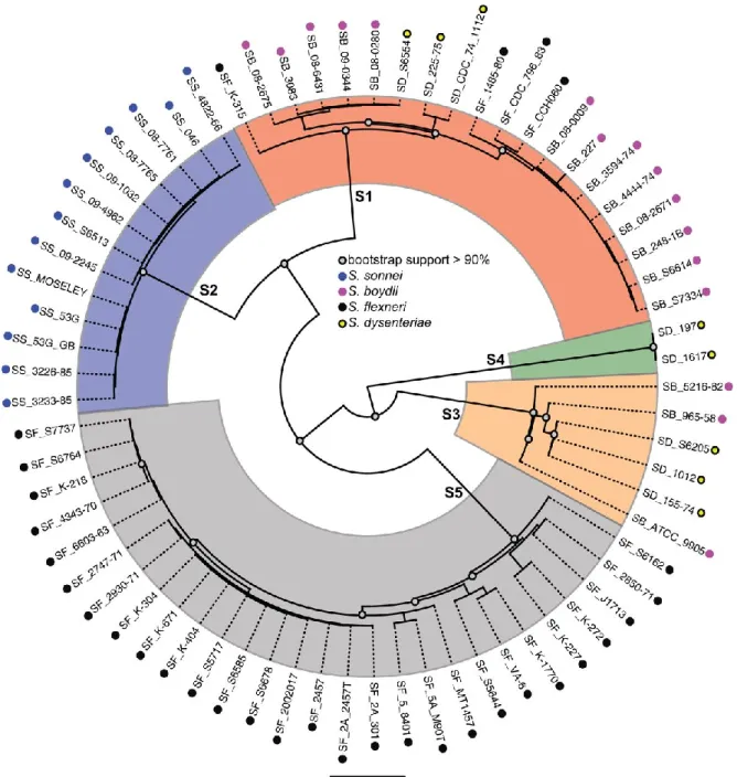
whole-genome-alignment-phylogeny classifies Shigella serotypes in 5 distinct clades, differing from their
13
encodes a Type VI Secretion System that contributes to killing of other microbiota
competitors like E. coli, which can prevent pathogenic infections (Anderson et al.
2017). This advantage may be relevant for its increasing prevalence in developed
countries, where S. sonnei is the most common Shigella serotype (Kotloff et al.
2018).
14
Figure 2. Shigella whole-genome phylogenetic tree. Strains from S. flexneri (SF), S.
sonnei (SS), S. boydii (SB) and S. dysenteriae (SD) are clustered in 5 different
monophyletic lineages (S1-S5). Reproduced from (Sahl et al. 2015).
Figure 2. Shigella whole-genome phylogenetic tree
One of the most relevant evolutionary events in the Shigella species is the
appearance of a large virulence plasmid pINV, which will be further discussed in
more detail. In addition to this event, other plasmids with different functions have also
15
been acquired by different species, including the plasmid pHS-2, which control the O
antigen chain-lengtht and the plasmids pDPT1 and pSSE3, which encode the
production of colicins (Calcuttawala et al. 2015). Most recently, acquisition of
plasmids encoding antibiotic resistance have been found, including resistance to
sulfonamide, streptomycin and tetracycline (Holt et al. 2012), fluoroquinolones (Gu et
al. 2012) and azithromycin (Baker et al. 2015).
At the genomic level, Shigella spp. has incorporated pathogenesis-associated
genomic regions, or Pathogenicity Islands (PAI), which enhance its virulence and
adaptation to its niche. Three PAIs have been identified in S. flexneri, with the role of
some encoded proteins identified. LPS modification enzymes (Allison and Verma
2000) allow the evasion of the humoral immune response, which targets mainly the O
antigen; enterotoxins ShET1 and ShET2 play a role in the watery diarrhea (Fasano et
al. 1995) Proteases (Rajakumar, Sasakawa, and Adler 1997) play different roles, Pic
plays a role in the degradation of the extracellular environment in the gut and in the
host (Henderson et al. 1999; Al-Hasani et al. 2009); iron acquisition genes in the
aerobactin operon allow Shigella to counteract the iron-deficient environment (Moss
et al. 1999; Nassif et al. 1987); colicin resistance genes may play a role in its survival
in the intestinal lumen (Vokes et al. 1999). S. dysenteriae type 1 also produces the
Shiga toxin, which inhibits host protein synthesis. This leads to fluid accumulation,
paralysis and cell death. Because the kidney highly expresses the toxin receptor
Gb3, this toxin can also lead to hemorrhagic uremic syndrome (Johannes and Römer
2010).
1.2.3 Shigella virulence plasmid
The Shigella large virulence plasmid varies in size, being as large as 220 kb (Figure
3). This plasmid is sufficient to confer invasion in eukaryotic cells by otherwise
non-invasive E. coli strains (Sansonetti et al. 1983). This plasmid contains around 280
open reading frames (ORFs), of which half are associated with insertion sequence
elements suggesting a highly polyphyletic origin. For example, Yersinia
low-calcium-response plasmid harbors a sequence homologous to a region in the ipa-mxi-spa
16
gene cluster. Other ORFs homologous to genes of M. tuberculosis, S. typhimurium,
M. bovis, E. coli, and V. cholerae have also been reported (Venkatesan et al. 2001).
Importantly, the virulence plasmid harbors a 32 kb gene cluster that encodes the
Mxi-Spa Type III Secretion System (Figure 3), which structure and role in invasion will be
further described. In the same cluster, there are also a series of effector proteins,
termed “Invasion Plasmid Antigens”, or Ipas, that mediate bacterial invasion and
manipulate the immune response. The transcriptional regulation of this region is
controlled by temperature, osmolarity and pH, with optimal conditions being 37°C,
physiological saline concentration and pH 7.4 (Maurelli, Blackmon, and Curtiss 1984;
Porter and Dorman 1994; Nakayama and Watanabe 1995). The virF regulatory gene
is normally silenced by the chromosomally encoded H-NS protein until a shift in
temperature to 37°C releases the transcriptional silencing by adjusting the DNA
structure at the virfF promoter (Maurelli and Sansonetti 1988; Stoebel, Free, and
Dorman 2008).
VirF promotes the transcription of VirB, which antagonizes H-NS repression.
Specifically, VirB activates the mxi and spa genes that encode the T3SS, chaperon
and protein effector genes and the regulator gene mxiE (Schroeder and Hilbi 2008).
17
Figure 3. A comparative gene map of the Shigella virulence plasmid. Black ring
shows the GC content of the reference pSs_046 sequence. The following purple,
pale green, teal, khaki, and blue rings show comparisons between pSs_046 and
the virulence plasmids of S. boydii str. BS512, S. boydii str. Sb227, S. dysenteriae
str. Sd197, S. flexneri F2a str. 301 (pCP301) and S. flexneri F5a (pWR501),
respectively. Outer ring represents annotations of genes or genetic clusters based
on function: known virulence factor genes (red); plasmid replication, transfer and
maintenance genes (black); transposon, phage-borne and insertion sequence
elements (orange); genes encoding hypothetical proteins (teal); S. sonnei-specific
O antigen biosynthesis cluster (blue); genes encoding proteins with other known
functions (green). ipa, invasion plasmid antigen gene; icsP, also known as sopA;
T3SS, type III secretion system. Modified from (The et al. 2016).
18
1.3 Overview of Shigella invasion
Before reaching the intestinal epithelium, Shigella needs to overcome physiological
barriers throughout the digestive tract. Shigela spp. are resistant to acidic
environments, presumably through glutamate-dependent acid-resistance pathway
which allows them to survive the passage through the stomach (Jennison and Verma
2007). Afterwards, its primary sites of infection are the terminal ileum, colon and
rectum. Another barrier Shigella has to overcome is the mucin layer that covers the
intestinal epithelium. This layer consists of a heterogeneous meshwork of proteins
including the large glycoproteins mucins (>2 x 10
6Da). All four species of Shigella
produce mucinase and neuraminidase (Haider et al. 1993), which may allow them to
degrade this barrier and get access to the intestinal epithelial cells.
1.3.1 Anatomy of the intestinal epithelium
The intestinal epithelium is a singe-cell layer that extends across the 4 regions of the
intestine (jejunum, ileum, caecum and colon). Its structure differs between regions.
The small intestine is rich in villi, protrusions that extend into the lumen and maximize
the contact area and therefore the nutrient adsorption. In opposition, villi are absent
in the colon. The epithelium also forms invaginations named “crypts of Lieberkün”.
Figure 4 depicts the spatial segregation of epithelial cell types. Stem cells are located
at the depth of the crypts and as daughter cells are produced and differentiate, they
migrate upwards. Four distinct cell types are produced from this stem cell population:
absorptive, goblet, enteroendocrine and Paneth cells. Adsorptive cells, or
enterocytes, are the most abundant population of cells and have microvilli on their
apical side; Goblet cells secrete mucus; Enteroendocrine cells secrete peptides and
catecholamines, whilst Paneth cells secrete antibacterial proteins. As the intestinal
epithelium is renewed, the old cells are extruded into the intestinal lumen at the villi
apical extremities or the crypts' mouth. (For a review on the mechanisms of cell
renewal see (Crosnier, Stamataki, and Lewis 2006)).
19
Enterocytes are polarized and form a highly tight barrier through tight junctions,
adherens junctions and desmosomes. This barrier is permeable to ions, nutrients and
water, while restricting the passage of proteins, lipids and microorganisms from the
lumen to the lamina propria (Takuya Suzuki 2013).
Figure 4. Structure of the intestinal mucosa in the jejunum (left) and in the colon
(right). IEL: Intraepithelial lymphocytes; IEC: Intraepithelial cells; AMP:
Antimicrobial peptides. Modified from (Mowat and Agace 2014)
Figure 4. Structure of the intestinal mucosa
In addition to epithelial cells, there is also an abundant population of immune cells
interspaced with epithelial cells. Tuft cells detect the presence of helminths (Gerbe et
al. 2016) and M cells perform uptake and presentation of luminal antigens to immune
cells (Ohno 2016).
M cells are associated to subepithelial lymphoid aggregates, termed gut-associated
lymphoid tissue, which prime adaptive immune cell responses in the intestine by
20
presenting antigens to dendritic cells. The best characterized of these aggregates are
the Peyer’s patches in the small intestine (Cornes 1965), which consists of B cell
lymphoid follicles and T cell areas. Also, in the large intestine, there are structures
termed cecal patches in the ileocaecal valve and colonic patches in the colon and
rectum. Both patches resemble in function and structure to Peyer patches, albeit
smaller in size (Owen, Piazza, and Ermak 1991).
In the lamina propria, there are also CD4+ and CD8+ T cells, with an effector
memory phenotype. There is also a large population of IgA and IgM producing B cells
and macrophages. These antibodies are shed into the lumen of the intestine and
prevent bacterial adhesion to the epithelium. Eosinophils and mast cells are also
present along the intestinal tract and contribute to the regulation of the inflammatory
response (for a review see (Mowat and Agace 2014)).
1.3.2 Shigella invasion routes of the intestinal
epithelium
The initial routes of invasion are not entirely clear and will be further discussed in
detail. Early observation of biopsies from Shigella-infected patients suggested the
gut-associated lymphoid tissue as one of the primary sites targeted (Mathan and
Mathan 1991). In the rabbit ileal loop model, M cells were reported as the initial route
of entry (Wassef, Keren, and Mailloux 1989). However, a guinea pig model that
recapitulates several clinical features of the human infection shows that epithelial
cells are invaded as early as 2-6 hours post-administration (Shim et al. 2007).
Using a guinea pig intra-rectal inoculation model, it was observed that enterocytes at
the colonic crypts mouths were targeted at the onset of colonization and bacteria
spread by cell-to-cell transmission into the crypts along the infection (Arena et al.
2015). In the rabbit ileal loop model, there is evidence that Shigella traverses the cell
monolayer through M cells (Sansonetti et al. 1996), a step that would allow it to infect
macrophages in the M cell pouch. According to this model, after inducing the
pyroptotic death of macrophages, Shigella would invade epithelial cells from the
basal side. Pyroptosis also leads to the release of pro-inflammatory cytokines IL-1β
21
and IL-18, and invasion of epithelial release of IL-8. This causes a strong
inflammatory response that recruits polymorphonuclear leukocytes and destabilizes
the intestinal epithelium, hence facilitating invasion from lumenal bacteria.
1.4 The Shigella Type III Secretion
System
1.4.1 The Shigella T3SS structure
A key element for the pathogenesis of Shigella is its T3SS. This apparatus functions
as a needle, translocating effector proteins from the bacterial cytoplasm to the host
cell cytosol (Figure 5). Assembly of fully functional T3SS apparatus is regulated by
temperature but the injection of T3 effectors is dependent on host membrane contact
(Veenendaal et al. 2007).
The T3SS is generally divided in three parts: the basal body, the needle and the tip
complex. The basal body anchors the apparatus to the bacterial membranes (Figure
5). MxiD and MxiM form a ring in the outer membrane. On the cytoplasmic side, a
C-ring formed by the proteins Spa33, MxiK and MxiN mediates the recognition, sorting
and secretion of effectors, where the ATPase activity of Spa47 is involved (B. Hu et
al. 2015; Burgess et al. 2016; Morita-Ishihara et al. 2006). Spa32 interacts with
Spa40 and controls the length of the needle (Botteaux et al. 2010; Tamano et al.
2002). Other proteins are also present, but their roles remain unclear.
The needle is composed of the major MxiH and the minor MxiI subunits. MxiH
assembles in a helical fashion and constitutes the needle rod with a 2-3 nm inner
diameter (Blocker et al. 2001), while MxiI is proposed to be involved, together with
MxiC, in the secretion activation upon contact of the host membrane (El Hajjami et al.
2018; Cherradi et al. 2013).
Before contact with the host membrane the needle tip complex composed of IpaD
and perhaps IpaB remains in a closed conformation (Veenendaal et al. 2007;
Cheung et al. 2015). IpaB and IpaC are translocated upon contact with host cells,
22
likely by interacting with membrane enriched in cholesterol and
sphingolipids-containing membranes (Goot et al. 2004). IpaB and IpaC form the translocon
allowing the translocation of effector proteins.
Figure 5. Structure of the Shigella T3SS. Tip components shown in yellow and
green. Needle components in blue-greens, transmembrane base dark purple, inner
membrane export apparatus light purple, C-ring and ATPase complex in light blues.
OM, outer membrane. IM, inner membrane
Figure 5. Structure of the Shigella T3SS
1.4.2 Shigella T3SS effector proteins
At 37°C the transcriptional activator VirF is expressed and activates virB expression.
VirB is also a transcriptional activator that activates the ipa (Invasion Plasmid
Antigens) operon and the mxi (Membrane Expression of Ipa) - spa (Surface
Presentation of Ipa) operon. Expression of these operons triggers bacterial entry
mediated by the T3SS (Du et al. 2016), along with other effector genes. Injection of
effector proteins is temporarily divided into two waves. After initial host cell contact,
23
the injection of the first wave of effectors happens within minutes (Enninga et al.
2005).
Shigella T3SS uses chaperons to prevent degradation and to target effector proteins
to the secretion apparatus. The IpgC chaperone also regulates the expression of the
second wave of T3S effectors. Prior to activation of T3 secretion, the IpgC chaperone
associates with IpaB and IpaC and prevents their premature interaction in the
bacterial cytosol (Parsot 2003; Pilonieta and Munson 2008). After activation of T3
secretion, IpaB and IpaC are secreted, releasing IpgC, which may then interact with
the MxiE transcriptional activator. This allows the formation of the complex
IpgC-MxiE that promotes triggering the transcription of genes harboring a IpgC-MxiE box,
(Figure 6) corresponding to genes of the second wave of effectors (Kane et al. 2002).
Genes belonging to the second wave of effectors are mainly involved in high-jacking
host signaling, apoptosis inhibition and regulation of long-term effects on the host
immune response (For review, see Mattock and Blocker 2017).
Table 1. Shigella effector proteins
Table 1. Shigella effector proteins. Modified from (Killackey, Sorbara, and
Girardin 2016)
Shigella
effector
(wave)
Host targets
Effect on host process
IpgB1
(1st)
Acts as GEF for Rac
Induces actin remodeling to facilitate
invasion
(Z. Huang et al. 2009)
IpgB2
(1st)
Acts as GEF for RhoA
Induces actin remodeling to facilitate
invasion
(Z. Huang et al. 2009)
IpgD
(Ist)
Acts as PI(4, 5)P
2-4-phosphatase to generate PI5P
Diverts host cell recycling
endosome machinery
Increases levels of PI5P leading
to p53 degradation
Induces actin remodelling to facilitate
invasion.
(Niebuhr et al. 2002)
Allows rapid escape from the entry
vacuole.
(Mellouk et al. 2014)
Induces PI3K/Akt-dependent survival
pathways.
24
IpaC
(1st)
Activates Src Kinase and Cdc42
Inserts into vacuole membrane
Mediates invasion
(T. Suzuki et al. 2000; Mounier et al.
2009)
IpaA
(1st)
Targets Host-cell focal adhesion
components vinculin and talin.
Promotes RhoA activation
Promotes anchoring at the site of
membrane ruffling
(Tran Van Nhieu, Ben-Ze’ev, and
Sansonetti 1997; Izard, Tran Van
Nhieu, and Bois 2006; C.
Valencia-Gallardo et al. 2019)
Promotes actin rearrangement
(Demali, Jue, and Burridge 2006)
IpaB
(1st)
Binds to cholesterol on the host
cell surface
Inserts into vacuole membrane
Reduces and disrupt balanced
levels of cholesterol and lipids
Promotes bacterial adhesion
(Romero et al. 2011; Lafont et al. 2002)
Interferes with proper Golgi function
(Mounier et al. 2012)
VirA
(1st)
Catalyzes GTP hydrolysis in
Rab1
Induces p53 degradation
Disrupts ER-to-Golgi trafficking and
autophagy
(Selyunin et al. 2014)
Blocks apoptosis
(Bergounioux et al. 2012)
IcsB
(1st)
Blocks autophagy targeting by
binding Atg5
Binds cholesterol
Reduces autophagy
(Kayath et al. 2010; Ogawa et al. 2005)
OspB
(2nd)
Induces mTOR signaling
through IQGAP1
Inhibits autophagy
(Lu et al. 2015)
IpaJ
(2nd)
Cleaves N-myristoylated glycine
from ARF1 to disrupt its
localization
Disrupts autophagosome maturation
and host membrane trafficking
(Dobbs et al. 2015)
IpaH9.8
(2nd)
Targets and degrade
NEMO/IKKγ
Disrupts NF-κB signaling
(Ashida et al. 2010)
IpaH4.5
(2nd)
Targets and degrades p65
subunit of NF- κB
Targets and degrades TBK1
Disrupts NF- κB signaling
(Wang et al. 2013)
(Wang et al. 2013)
(Zheng et al. 2016)
IpaH072
2 (2nd)
Targets and degrade TRAF2
Disrupts NF- κB signaling
(Ashida, Nakano, and Sasakawa 2013)
OspZ
(2nd)
Prevents nuclear translocation
of p65
Disrupts NF- κB signaling
(Newton et al. 2010)
OspI
25
OspG
(2nd)
Interferes with ubiquitin
proteasomal degradation of
IκB-α
Disrupts NF- κB signaling
(Kim et al. 2005)
OspF
(2nd)
Inactivates the ERK and p38
MAP kinases using its
phosphothreonine lyase activity
Disrupts MAPK signaling
(Reiterer et al. 2011)
OspC3
(2nd)
Interacts with caspase-4-p19
subunit and inhibits its
heterodimerization and
activation
Inhibits pyroptosis within epithelial cells
(Kobayashi et al. 2013)
IpaH1.4/
2.5
Induces proteosomal
degradation of HOIP
Supression of NF- κB activation
(de Jong et al. 2016)
26
Figure 6. Regulation of the second wave of effectors. A. Inactive T3SA. B. Active
T3SA. Prior to activation of T3 secretion, the IpgC chaperone associates with IpaB
and IpaC. After activation, IpaB and IpaC are secreted, releasing IpgC, which may
then interact with the MxiE transcriptional activator. This allows the transcription of
genes harboring a MxiE box. Modified from (Campbell-Valois and Pontier 2016)
Figure 6. Regulation of the second wave of effectors
1.5 Main players of the cytoskeleton
1.5.1 Actin monomers
The actin cytoskeleton is commonly targeted by pathogens, being highly conserved
among animals and a central element in various cellular processes. Actin monomer,
termed G-actin, can polymerize into actin filaments, or F-actin. ATP-G-actin
27
polymerizes into ATP-F-actin, which has an intrinsic ATPase activity leading to ADP
F-actin. ADP F-actin is less stable than ATP G-actin, rendering ADP F-actin filaments
prone to disassembly (Dominguez and Holmes 2011).
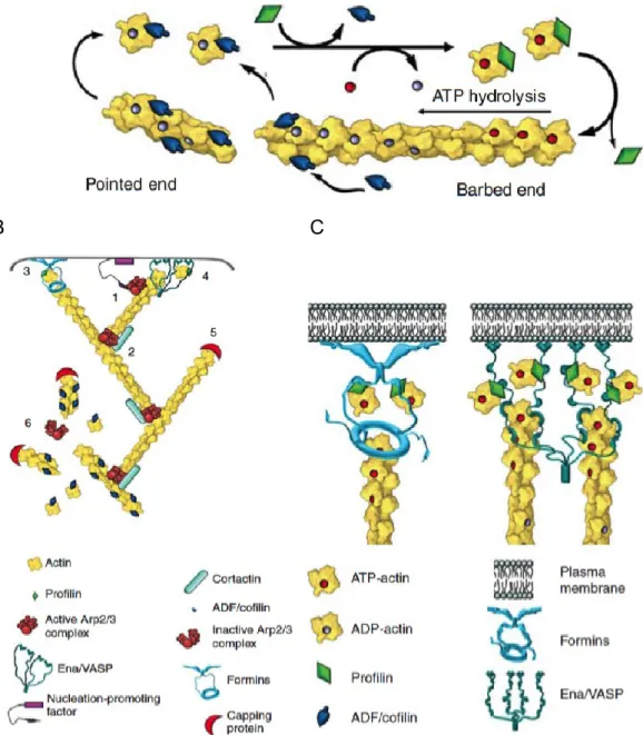
The incorporation of actin monomers is asymmetrical. The barbed end (or plus end)
of actin filaments incorporates ATP-actin monomers, while the pointed end (or minus
end) is more prone to be disassembled (Figure 7A). Simultaneous incorporation of
monomers at one end and disassembly on the other gives rise to treadmilling.
1.5.2 Actin sequestering proteins
In the intracellular milieu, actin binds to profilin. This complex represents the main
source of monomers for polymerization. Profilin catalyzes the ADP/ATP exchange in
actin monomers, while inhibiting the spontaneous filament nucleation property of
actin monomers (Dominguez and Holmes 2011). Profilin-actin also binds to other
proteins involved in actin polymerization, such as formins and Ena/VASP proteins
(Kovar et al. 2006) (Figure 7).
Thymosin-β4 is a peptide of 43 residues. In its N-terminus it has short helix that binds
the actin barbed end and the rest of the helix, forms an extended region that binds
the front surface of actin and a second helix that caps actin pointed end (Xue et al.
2014). This results in the blockage of engagement of actin monomers into
polymerization. However, profilin and thymosin-β4 are constantly exchanging actin
monomers, allowing elongation of actin filaments (Pantaloni and Carlier 1993).
1.5.3 Severing proteins
Depolymerization in cells is enhanced by proteins of the ADF/Cofilin family, which
includes Actin Depolymerization Factor (ADF), cofilin-1, cofilin-2 and twinfilin. This
family of proteins have a higher affinity for ADP-actin than for ATP-actin, and
therefore bind to ADP-actin fiber stretches and promote the severing of these
filaments (Barbara W. Bernstein 2010).
28
Gelsolin proteins also sever actin filaments and cap their barbed ends. Most of its
family members’ activity is regulated by calcium binding. Mutants of this protein have
defects in cellular motility and decreased blood clotting.
29
A
B C
Figure 7. Dynamics of actin filaments. A. Actin polymerization. B. Actin regulation.
(1) Arp2/3 recruitment (2) Actin branching (3) Formin elongation of actin fibers (4)
Ena/VASP elongation of actin fibers (5) Filament capping (6) Depolimerization of
actin filaments. C. Formin and Ena/VASP nucleation and elongation activity.
Modified from (Svitkina 2018).
30
1.5.4 Actin nucleators
Actin nucleators initiate the de novo polymerization of actin filaments. The Arp2/3
complex is one of the best characterized actin nucleators. It is composed of a core of
7 proteins. Upon the interaction of Nucleation Promoting Factors (members of the
Wiskott-Aldrich Syndrome Protein family, WAVE, among others), actin filaments and
actin monomers, Arp2/3 is recruited and activated by Arf1 and Rac1 (Koronakis et al.
2011). Through the VCA domains of the Nucleation Promoting Factors, Arp2/3 is
connected to the mother actin filament and nucleates the polymerization of a new
branched actin filament (Pollard 2007). Given the Arp2/3 structure, the daughter
filament elongates at an angle of 70° from the mother filament (Amann and Pollard
2001). At a large scale, polymerization of new branched filaments upon existing
filaments give rise to branched actin networks, which are enriched in lamellipodia
(Figure 7B).
In opposition to Arp2/3, formins nucleate and elongate unbranched actin filaments in
filopodia, stress fibers and actin cables. They are characterized by the presence of
the Formin Homology Domains 1 and 2, which mediate their interaction with profilin
and their nucleation activity, respectively. Their ATP-dependent activity results in a
highly processive actin polymerization (Romero et al. 2004). In a similar fashion,
Ena/VASP proteins also promote the nucleation and elongation of actin filaments
(Figure 7C), and works in neurons show that they are particularly important for
filopodial formation (Lebrand et al. 2004).
1.5.5 Capping proteins
Capping protein and CapZ bind to the barbed end of actin filaments, blocking both
the addition and loss of actin subunits (Isenberg, Aebi, and Pollard 1980; J. Xu,
Casella, and Pollard 1999), therefore preventing the assembly/disassembly of
filaments. In conjunction with the branching activity of Arp2/3, a network of short and
highly branched actin filaments can be formed (Figure 7B) (M. Edwards et al. 2014).
31
1.5.6 Cross-linking proteins
I
ndividual actin filaments can be bundled together through the action of actin
cross-linkers, such as α-actinin isoforms 1 and 4 or fimbrin. These proteins contain
Actin-Binding Domains (ABD) that connects two separate actin fibers. α-actinin has also
the ability to associate with other cytoskeletal, signaling and membrane molecules
(Sjöblom, Salmazo, and Djinović-Carugo 2008).
The organization into bundles by filament bundling facilitates the mechanical
contraction exerted by myosins molecular motors. In particular, Myosin II transforms
the chemical energy released by ATP hydrolysis into mechanical work that results in
pulling of actin filaments (Figure 8). The pulling activity is the result of the myosin II
polymerization into bipolar filaments, with the motor domains walking in opposing
cross-linked actin filaments (Figure 8) (Sweeney and Holzbaur 2018).
Figure 8. Myosin II and α-actinin in actin filament contraction. Myosin II polymerizes
into bipolar filaments, with the motor domains walking in opposing actin filaments
cross-linked by α-actinin. Modified from (Svitkina 2018).
32
1.6 Adhesion structures
1.6.1 Integrins
The intracellular actomyosin cytoskeleton dynamically interacts with the extracellular
environment through linker molecules anchoring transmembrane receptors that
connect with the extracellular environment. Integrins and cadherins are two of the
main receptors that connect the actomyosin cytoskeleton with the outer environment
and to respond to mechanical and biochemical cues.
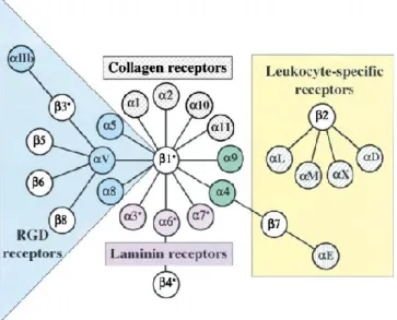
Integrins are cell adhesion receptors, and its super-family is composed of 18 types of
α subunits and 8 types of β subunits, which can associate into 24 different αβ
heterodimers, the β subunit determining the family. The α and β subunits are
non-covalently associated transmembrane proteins. Different pairs can bind to different
ligands, such as RGD motif (present in fibronectin and vitronectin), collagen, laminin,
or ligands in leukocytes (Kechagia, Ivaska, and Roca-Cusachs 2019; Hynes 2002)
(Figure 9).
Although not proven for all αβ pairs, some integrins can transition from a “bent
closed” or inactive form, to an extended closed and finally to an extended open. This
transition is associated with an increase in the affinity of the integrin for its substrate.
33
Figure 9. Integrins ligands and intracellular dynamics. Different αβ pairs and their
ligands (Modified from Hynes 2002).
Figure 9. Integrins ligands and intracellular dynamics
Integrin transition from closed bent to open extended is enhanced by the binding of
kindlin, and notably talin, which binds to the cytoplasmic tail of the β subunit, a
process termed “inside-out activation“, as opposed to "outside-in signaling"
implicating the activation of integrins through the binding of their extracellular ligand.
Talin recruitment to the membrane is presumably achieved through its interaction
with Rap1-GTP-Interacting-Adaptor-Molecule (RIAM), which links membrane
associated Rap1 with cytoplasmic talin (Lee et al. 2009) and concentrates it at the tip
of actin growing filaments in lamellipodial and filopodial protrusions (Lagarrigue et al.
2015).
Integrins display catch-slip bonds with RGD-ligands (Kong et al. 2009), ICAM (W.
Chen, Lou, and Zhu 2010) and VCAM (Choi et al. 2014). In an ideal bond between
molecules, the lifetime of the interaction would not depend on force. However, bonds
usually behave under non-ideal regimes. Under a slip regime, the bond between two
molecules will decrease its lifetime as force is applied. Counter-intuitively, catch-slip
bonds undergo first a catch regime where application of force will increase the
34
lifetime of the bond, therefore strengthening the interaction. This catch regime is
followed by a slip regime until detachment (Rakshit and Sivasankar 2014).
In order for the force to be effectively transmitted between the actomyosin
cytoskeleton and the extracellular environment, integrin molecules need to be
connected to the actin cytoskeleton. This task is performed by linker proteins such as
talin, tensin, filamin and α-actinin, which bind simultaneously to integrins and F-actin.
This force-enhanced process leads to the maturation of adhesion structures through
the recruitment of additional scaffolding and signaling molecules.
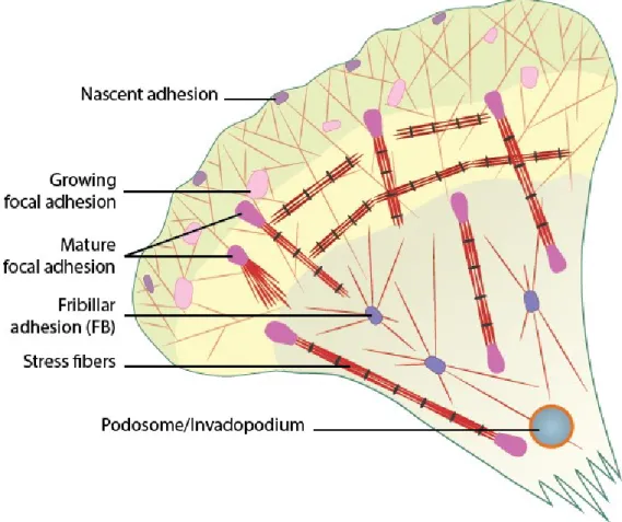
1.6.2 Adhesion formation and maturation
Filopodia are finger-like structures that protrude out of the cells’ edge. Its extension is
driven by actin polymerization mediated by formins and regulated by Rho GTPases,
capping proteins and Ena/VASP (Gupton and Gertler 2007; Lagarrigue et al. 2015;
Albuschies and Vogel 2013). Deeper into the cell edge, short-lived adhesions (~60
seconds turnover) can be found in the lamellipodium, termed “nascent adhesions”.
Larger adhesions, known as “focal complexes” with a persistence of several minutes
can be found further back from the leading edge. These focal complexes can mature
into large focal adhesions (3-10 μm long) located at the extremes of stress fibers,
with a persistence time of 30-60 min (Zimerman, Volberg, and Geiger 2004).
There is evidence that adhesions at the shaft of the filopodia and nascent adhesions
mature into focal adhesions upon the advancement of lamellipodia (Figure 10)
(Wong, Guo, and Wang 2014). Nascent adhesion formation is independent of myosin
II and contain integrins, talin, kindlin, vinculin, α-actinin, paxilin and FAK. These
adhesions can either disassemble or further mature into focal adhesions in a process
largely driven by tension and crosslinking exerted by myosin II.
35
Figure 10. Maturation of adhesion structures. Modified from
(www.mechanobio.info)
Figure 10. Maturation of adhesion structures
1.6.3 Talin and Vinculin molecular clutch
The molecular clutch hypothesis postulates that focal adhesions mediate the
interactions between the retrograde-moving actin and ligand-bounded cytoskeleton.
This interaction regulates the protrusive activity of the actin cytoskeleton. In adherent
migrating cells, the cortical actin cytoskeleton is organized into two structurally and
functionally distinct regions: the lamellipodium and the lamellum. In the lamellipodium
Arp2/3-driven actin polymerization generates pushing force. However, when the
molecular clutch is not engaged (i.e. when actin fibers are not tethered to the
extracellular matrix), actin movement result in treadmilling behaviour towards the
interior of the cell due to the resistance from the plasma membrane, generating
rearward movement. When the clutch is engaged, on the other hand, forces required
36
to counteract membrane resistance are transmitted to the matrix and protrusions at
the cell edge are generated (Figure 11).
Figure 11. Model of the molecular clutch hypothesis. Disengaged molecular clutch
results in actin treadmilling behaviour towards the interior of the cell, generating
rearward movement. Engagement, forces required to counteract membrane
resistance are transmitted to the matrix and protrusions at the cell edge are
generated.
37
Interaction between actin cytoskeleton and the extracellular matrix involves
transmembrane proteins and linker molecules. In focal adhesions, the linker integrins
bind to the extracellular matrix. In addition, the role of talin as an integrin-actin linker
molecule is remarkable, because it possesses structural characteristics of a
molecular mechanosensor regulated by force. One characteristic is its ability to
scaffold vinculin in a force-dependent manner (Figure 12). Talin is organized in two
domains, the N-terminal FERM domain can bind phospholipids and integrin,
responsible for the inside-out signaling activity through integrins (Anthis et al. 2009).
The C-terminal domain contains 13 helical bundles (designated R1-R13) that contain
two actin binding sites on the rod domain and one in the FERM domain at least 11
putative Vinculin Binding sites (VBSs), each one constituted by a single amphipathic
helix (Gingras et al. 2005). However, all of the VBSs helices are buried within
α-helix bundles until they become exposed, which happens only upon force-dependent
stretching of talin and bundle unfolding (Figure 12B) (Hytönen and Vogel 2008); (del
Rio et al. 2009).
Figure 12. A. Structural model of Talin. Depicted in blue are the Vinculin Binding
sites. B. Unfolding of helix bundle. Talin bundles unfolding leads to unveiling VBSs.
Modified from (Yao et al. 2015)
38
Talin bundles unfold in a hierarchical manner, depending on the force required for
their unfolding. It has been shown that all bundles, al least in vitro, are suceptible to
be unfolded by stretching forces ranging from 5 to 30 pN (Haining et al. 2016; Yao et
al. 2015). In its inactive form, vinculin has a globular structure consisting of two
domains: a head domain and a tail domain connected by an unstructured linker
(Figure 13). Both head and tail domains consist of α-helix bundles, the head is
organized in four subdomains (named VD1-VD4), while the tail domain contains a
single bundle (termed Vt) (Borgon et al. 2004). The vinculin intra-molecular
interaction occurs between the Vt and head subdomains VD1 and VD4 (Izard et al.
2004; Cohen et al. 2005). When these interactions are disrupted, the actin binding
site on Vt is free to interact with F-actin under a catch-bond regime (D. L. Huang et
al. 2017). Reported VBSs from talin and α-actinin bind to the VD1 domain (Izard et al.
2004). However
,whether talin VBSs exposure alone is sufficient for vinculin
activation in vivo is a matter of debate. There are observations that phosphorylation
on vinculin tyrosine residues 100 and 1065 by Src (Auernheimer et al. 2015) and
F-actin binding (H. Chen, Choudhury, and Craig 2006) facilitates vinculin activation.
Also, PI(4, 5)P
2is required for vinculin sequestration at FAs and contributes to their
39
A
B
Figure 13. A. Vinculin structure and activation. Vinculin subdomains are
indicated in colors (D1, blue; D2, cyan; D3, green; D4, yellow; Tail domain,
red). Binding sites for Talin, α-catenin, PIP
2, F-actin, Arp2/3, vinexin and
Ponsin are indicated. B. Vinculin activation by VBSs. Vinculin head domain is
colored in cyan, VD1 is indicated in red, Tail domain in green. VBS (light cyan)
interaction with VD1 disrupts heat-tail interaction and leads to interaction with
F-actin (purple). Modified from (Bakolitsa et al. 2004 and Park et al. 2014).
Figure 13. Vinculin structure and activation
1.6.4 Force generation and maturation
Initial nascent adhesion formation is dependent on actin polymerization, in which
Rac1 activation and Arp2/3 have been implicated (Wu et al. 2012).
This is in contrast to nascent adhesions that mature into focal adhesions in a process
that requires stress fibre assembly and myosin II activity (Oakes et al. 2012). Myosin
II activity is sustained by Rho through the action of Rho associated protein kinase
(ROCK). As adhesion structures mature, its composition changes and generates a
complex 3D organization (Figure 14). Signalling proteins like FAK and Paxilin localize
next to the integrin cytoplasmic tails, together with the Talin head. However, talin rod
40
protrudes inwards, colocalizing with vinculin, which is initially recruited to the plasma
membrane, but relocalizes as the adhesion matures and talin VBS are unveiled by
force. Talin tail, which binds to F-actin colocalizes with proteins like zyxin, VASP and
α-actinin (Case and Waterman 2015).
Figure 14. Focal adhesion organization. Internal organization changes with respect
to traction forces and protein composition follow a layered organization. Modified
from (Case and Waterman 2015)
Figure 14. Focal adhesion organization
1.7 Actin cytoskeleton
reorganization by bacterial
pathogens
41
1.7.1 Bacterial adhesion and internalization
strategies
Shigella, as other bacterial pathogens, has evolved different ways to target and
manipulate host cells. As a first step, pathogens must adhere to host cells. Several
pathogens express pili- or fimbriae-associated adhesins. Pili are adhesive
peritrichous, non-flagellar, filamentous organelles that stick out of the bacterial
surface. They allow the bacteria to attach to host cell receptors, targeting glycolipids,
glycoproteins, mannose residues or mucus. There are four types of pili: type I, type
IV, P pili, bundle-forming pili and Curli and they are widespread among pathogenic
Gram-negative bacteria, as described in Table 2.
Table 2. Pili/fimbriae of pathogenic Gram-negative bacteria
Table 2. Pili/fimbriae of pathogenic Gram-negative bacteria (modified from Bhunia
2018)
Type of pili
Present in
Type I
E. coli, H. influenzae, Y. pestis
Type IV
Pseudomonas spp., Vibrio spp., Enteropathogenic E. coli, N.
meningitides
P pili
E. coli, H. influenzae, Y. pestis
Bundle-forming
pili
Enteropathogenic E. coli
Curli
S. enterica, E. coli
Bacteria also express adhesion proteins at their surface that promote a tight binding
to host cells that are relevant for attachment and invasion. Theses proteins
commonly target host surface molecules or the extracellular matrix, as can be seen
in table 3.
42
Table 3. Non-exhaustive list of adhesion factors and host receptors of pathogenic bacteria
Table 3. Non-exhaustive list of adhesion factors and host receptors of
pathogenic bacteria (modified from Bhunia 2018; Solanki, Tiwari, and Tiwari 2018)
Pathogen
Adhesion factors Host receptor
L. monocytogenes
Internalin A
E-cadherin
Internalin B
c-Met, gC1q-R/p32
Virulence invasion
protein
Gp90
Listeria adhesion
protein
Hsp60
Campylobacter spp.
CadF
Fibronectin
Arcobacter
Hemagglutinin
Glycan receptor
Enteropathogenic,
enterohemorragic E.coli
Intimin
Translocated intimin receptor
Y. enterocolitica
YadA
Collagen/fibronectin/laminin/β1-integrin
S. aureus
Fibronectin-binding
protein
Fibronectin
V. cholerae
Toxin-coregulated
pili
Glycoprotein
N. meningitides
OpcA
Vitronectins, proteoglycans
E. coli
OmpA
Ecgp glycoprotein
B. pertussis
Pertactin
Not identified
Y. pseudotuberculosis
Invasin
β1-integrin
In addition to classically described adhesin-receptor interactions, some bacteria have
evolved more sophisticated forms of targeting their niche, which in some cases
include the manipulation of the cell intracellular machinery, notably the cell
cytoskeleton.
43
1.7.2 E. coli FimH catch-bond adhesion
Uropathogenic E. coli, a bacterial species that colonizes the urinary tract, expresses
a variant of the type 1 fimbriae whose affinity for its receptor is enhanced under high
flow velocity conditions (high shear-stress) (Thomas et al. 2002).
Type I fimbriae are assembled via the chaperone/usher pathway, which consists of: a
chaperon protein which interacts with the pilus and usher proteins including an usher
protein, which forms the translocation pore in the outer membrane. Around 1000
copies of the subunit FimA forms the fimbriae’s rod, while a single copy of the FimF,
FimG and FimH subunits constitute the adhesive tip. FimH, located at the fimbriae
distal part, is highly variable, which reflects different specificities for different targets
(Mortezaei et al. 2015). FimH from the uropathogenic E.coli strain J96 contains three
aminoacid substitutions relative to the gene encoded by the intestinal E. coli strain
F18 that confer its catch-bond binding properties for the mannose receptor (Thomas
et al. 2002). Conformational changes between the pilin domain and the lectin domain
mediate this process. The physical separation of these domains induced by force
removes the allosteric inhibition exerted by the pilin domain upon the binding of the
lectin domain for its target (Figure 15), increasing the affinity of the lectin for the
mannose resiude (Sauer et al. 2016).
44
Figure 15. Catch-bond mechanism scheme. On low force, FimH binds transiently to
its receptor. Upon flow application, lectin and pilin domains are separated and the
high-affinity state for the lectin domain is stabilized after release of the
allosteric-inhibitory pilin domain.
Figure 15. Catch-bond mechanism scheme