Introduction
The operative treatment of intraligamentous ruptures of the anterior cruciate ligament in patients with open growthplates is still controversially discussed. In the past decades, an increasing frequency of complete ACL-ruptures in children and adolescents is found [7,8,15]. The increasing number of young athletes in high demanding sports with respectable career prospectives has led to a better awareness of this injury by coaches and physicians [15].
Until now the natural history of intrasubstance rup-tures of the ACL is not fully understood and different options of treatment exist [22]. Non-operative treatment
and primary ligament repair has not been successful [10,
20]. Ongoing activities resulting in episodes of instability do put the menisci at risk for damage with possible early degenerative changes [16]. Even in the growing knee intra-articular reconstruction of the ACL is possible with various techniques to regain stability [4,7, 12,19,
21,24,26–29]. Standard ACL-reconstruction procedures involve drilling across the femoral and tibial growth-plates. Drilling through open physes might increase the risk of premature closure of the physes with subsequent deformities. The literature demonstrates that the po-tential risk of growth arrest exists [22,23]. The purpose of this multicenter study was to investigate four different operative methods and their outcome.
F. Gebhard A. Ellermann F. Hoffmann J.-H. Jaeger N. F. Friederich
Multicenter-study of operative treatment
of intraligamentous tears of the anterior
cruciate ligament in children and adolescents
Comparison of four different techniques
Received: 21 September 2004 Accepted: 12 October 2005 Published online: 21 April 2006 Ó Springer-Verlag 2006
Abstract Tears of the anterior cru-ciate ligament in skeletally immature patients were operated with four different methods and their outcome compared to each other. Sixty-eight patients (33 males, 35 females), mean 12.5 years, were treated in four different centers from 1984 to 2001. Twenty-eight patients under-went the ACL-reconstruction with hamstring grafts, 16 patients with bone–patella–bone autografts, 12 patients with quadriceps grafts and 12 patients with facia lata. The mean follow-up was 32 months. Postop-erative evaluation included radio-graphs, KT-1000/2000 stability measurements, Lysholm score, The Tegner activity scale and IKDC score. Neither leg length discrepancy nor angular deformities were noted. Mean KT-1000 difference was
2.1 mm, mean postoperative Lys-holm knee score 93.3, IKDC 87% normal or nearly normal. The Tegner index decreased from 6.6 to 5.7. In total, six patients developed instability due to an adequate trau-ma 1 year after the index operation. Two patients showed mild arthrotic changes. All but two patients were able to return to the same level of preoperative sports participation. None of the four methods studied showed major differences in out-come compared to the other. No growth disturbance could be noted. Keywords ACL Æ Reconstruction Æ Children Æ Sports
DOI 10.1007/s00167-006-0055-4
F. Gebhard Æ N. F. Friederich (&) Department of Orthopaedics and Traumatology, Kantonsspital Bruderholz, 4101 Bruderholz, Switzerland E-mail: [email protected] Tel.: +41-61-4362741
Fax: +41-61-4363676 A. Ellermann
Arcus Sportsclinic, Pforzheim, Germany F. Hoffmann
Department of Orthopaedic Surgery, Rosenheim, Germany
J.-H. Jaeger
Department of Orthopaedic Surgery, University-Hospital, Strasbourg, France
Materials and methods
A retrospective analysis was done in four centers. His-tory taking was used to identify the mechanism of the injury, associated injuries and the postoperative ability to return to sports. The medical record review showed the nature of the tear (acute versus chronic) and the methods of meniscal tear reconstruction. More than 8 weeks between the injury and the operation was con-sidered as a chronic injury. All ligament reconstructions were done because of consistent instability. We divided the patients into two groups concerning their maturity. Boys with Tanner [35] stages 1–3 and premenstrual girls formed the immature group and Tanner stages 4–5 for boys and postmenstrual girls were the mature group.
The physical examination consisted of clinical assessment of ligament instability, drawer sign, Lach-man and pivot shift tests. Leg length discrepancy was examined by the pelvic obliquity and standing blocks. Objective IKDC and subjective Lysholm and Tegner activity scores were utilized. Every patient was examined with the KT-1000/2000 Arthrometer [6]. The physical examination and KT-1000/2000 measurements were re-done by at least one orthopedic surgeon. Table1shows the collected data. All patients had a.p. and lateral standing radiographs at follow-up. Scanograms were taken in the center of Strasbourg. The postoperative evaluation at the Bruderholz-Hospital in Basel and in Strasbourg was personally done by the first author (FG). The other institutions used our protocol.
Techniques used Bruderholz
The anterior cruciate ligament reconstruction in the immature group (12 patients) was performed with the quadrizeps tendon as first described by Blauth (1984) [5]
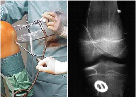
and Sta¨ubli (1990) [34]. The quadrizeps tendon had one bone block which was placed onto the femoral side in the epiphysis under the growthplate. The mature group (16 patients) received a patellar bone–tendon–bone graft where the bone block was placed at the same anatomical position in the femoral epiphysis like the quadrizeps tendon. The distal block was placed under the tibial epiphysis. The grafts were inserted through transphyseal drill holes and were secured extraphyseal with non-absorbable sutures around 3.5 mm AO screws (Fig.1). The procedure was arthroscopically assisted, meniscal injuries were addressed before harvesting the graft.
Strasbourg
A modified MacIntosh technique [27] was used with facia lata for the ACL-reconstruction (12 patients). In this technique, the graft remains attached to Gerdy’s tubercle and is driven under the lateral collateral liga-ment over the top position. In this intra-articular, ‘‘ex-traphyseal’’ (sparing the growthplate—intraphyseal) technique the graft is taken through a drill hole in the tibial epiphysis and secured with a staple below the anterior epiphysis. A medial arthrotomy is used for examining the joint, resection of the ACL stump and placing the clamp for distal fixation of the graft (Fig.2).
Pforzheim
A 4-string hamstring reconstruction (24 patients) was used in all patients for replacement of the anterior cru-ciate ligament. This arthroscopic assisted procedure used an endo-buttonÒat the femoral side and a suture diskÒ
Table 1 Data collected
History Age
Sex
Skeletal maturity Mechanism of injury
Tegner activity level prior to injury Time between accident and surgery Clinical assessment of ligament instability Operation Graft used
Fixation method
Associated injuries: menisci, ligaments Treatment of associated injuries Follow-up Tegner activity level at follow-up
Lysholm score
Physical examination with IKDC KT-1000/2000
Radiographs
Fig. 1 Post-OP X-ray after ACL replacement with quadrizeps tendon, medial and lateral meniscus repair, extra-articular fixation with 3.5 AO screws, Bruderholz technique
(Aesculap, Tuttlingen, Germany) at the tibial side to secure the graft. The transphyseal tunnels were drilled by hand avoiding thermal injury to the growthplate (Fig.3).
Rosenheim
All children (four patients, immature group) were treated with a hamstring graft for ACL-reconstruction. Two 2-string and two 3-string grafts were inserted. An endo-buttonÒ was used for fixation at the femoral side. At the tibial side the graft was secured once with a suture diskÒand three times with a staple below the tibial physis.
Meniscal injuries
The overall incidence of meniscal injuries at the time of ACL-reconstruction was 38% (26 meniscal tears, n=68) in both groups. The incidence in the immature group was 30% (12 of 40) and 50% (14 of 28) in the mature group. Table2 gives a survey of the treatment differ-ences. Eighteen times (69%) the tear was repaired and six (23%) tears were partially resected. All resections were made after a minimum of 8 weeks after the trauma. Most of the meniscal injuries were sutured using a PDSÒ (Ethicon, Johnson&Johnson International, Somerville, NJ, USA) suture. Pforzheim did use one Meniscus ArrowÒ(Bionx Implants, Tampere, Finland).
Results
No growthplate disturbances with leg length discrepan-cies or angular deformities were noted among all patients (68 patients) in both groups. Figure4 shows an X-ray series of a 2-year follow-up of a patient from the Bru-derholz hospital. Almost all patients were able to return to their desired sports. Table3demonstrates the dominating causes of an ACL-rupture in children and adolescents.
Immature group
Forty patients (21 boys and 19 girls, mean 11.9 years, range 7–14 years) were treated between 1984 and 2001 in the four centers. The mean follow-up was 33 months (range 13–204). Figure 5gives a survey of the techniques used. Lysholm score at time of follow-up was 94.3 of 100
Fig. 2 Two-year follow-up X-ray after ACL replacement with fascia lata, modified MacIntosh technique, Strasbourg
Fig. 3 Hand-drilled tunnels in Pforzheim using the Aesculap technique with hamstring ten-don, post-OP X-ray
(range 53–100). IKDC objectives were A in 19 (47%), B in 16 (40%) and C in 5 (13%) patients. Tegner activity score decreased from 6.5 pre-injury to 5.9. KT-1000 measurements showed 2.0 mm difference (range 0– 4 mm) at time of follow-up.
Re-ruptures occurred in three patients (8%, n=40) with a quadriceps-tendon autograft. All had an ade-quate trauma. Failure of the meniscal repair needed to be operated in 33% (4 of 12 repairs), one with a
re-rupture of the graft and one failure of the Meniscus ArrowÒ. Three of those were partially resected, one was repaired together with a second stabilization procedure (patellar tendon graft). Loss in range of motion were discovered in two patients (5%, n=40). One mobiliza-tion under anesthesia and one arthroscopic debride-ment. In 25% (ten patients, n=40) hardware removal was necessary. The allocation and the type of hardware showed different patterns.
The removal was undertaken at the tibial side only because of skin irritations. Mainly the 3.5-mm AO screw (Bruderholz) and the staple (Strasbourg) were a prob-lem. One staple was placed crossing the proximal epiphyseal plate of the tibia. The staple was removed 6 months after the ACL-reconstruction. No growth ar-rest occurred.
Mature group
Twenty-eight patients (14 boys, 14 girls, mean 15.3 years, range 14–18 years) were treated between 1990 and 2002. The mean follow-up was 40 months (range 14–144). Lysholm score at the time of follow-up was 93.7 of 100 (range 80–100). IKDC objectives were A in 13 (46%), B in 10 (36%) and C in 5 (18%) patients (n=32). Tegner activity score decreased from 6.5 pre-injury to 5.9. KT-1000 measurements showed 2.0 mm difference (range 0–4 mm) at time of follow-up.
The re-rupture rate was 14% (4 of 28) with one patellar tendon and three hamstring-autografts. One patient had an MRI-confirmed re-rupture of the hamstring-autograft without an explainable reason. The others had an ade-quate second trauma. Failure of the meniscus repair oc-curred in 21% (3 of 14 repairs). One combined with a re-rupture. All second meniscal tears were resected. Hard-ware removal (one suture diskÒand two 3.5 AO screws) was necessary in 11% (3 of 28 patients).
Statistical analysis
We used the SPSS 12.0 Statistic-program (SPSS Inc., Chicago, IL, USA) to calculate with our databases. A level of significance of a=0.05 was chosen. The values of IKDC rating were set as: IKDC A=1, B=2, C=3 and
Table 2 Comparison of grafts
and maturity of the patients n Immature group Adolescent group
Graft Graft
KSBH 28 12 Quadrizeps tendon 16 Patella-tendon
Strasbourg 12 12 Fascia lata 0
Pforzheim 24 12 Hamstrings 12 Hamstrings
Rosenheim 4 4 Hamstrings 0
Total 68 40 28
Fig. 4 X-ray series from Bruderholz with 2-year follow-up, removal of the tibial screw after skin irritations
D=4. The difference of Tegner activity index before the injury and at day of follow-up was taken.
No deviation from the normal distribution was ob-served in any of the treatment groups (Kolmogorov– Smirnov test with correction of significance according to Liliefors). A univariate ANOVA was calculated in be-tween the four treatment groups of the immature pa-tients and the two treatment groups of the mature patients. The F-value was not significant (p>0.05), which indicates that the groups did not differ from each other. Thereafter, each pairwise comparison was tested using Tukey’s test. Each comparison resulted in a p-value <0.5 (Tukey’s test checks the type I error). ANOVA were performed for the treatment groups (immature and mature group) and the comparison of the transplants versus the Lysholm score. Comparison of activity (Tegner index) and stability (KT-1000/2000) were not significantly different from the transplants in the immature and mature groups (p>0.05).
Discussion
This study presents a retrospective review of four hos-pitals who have a major experience in arthroscopic surgery of the knee in children and adolescents. The
shortest time for follow-up of 13 months (average time for follow-up was 2.6 years) may not allow a conclusive answer of outcome but can retrieve a growthplate dis-turbance after the index operation. Several orthopedic surgeons were involved in the follow-up process for use. Despite those facts, four different techniques were combined and showed no differences in their outcome. In none of the methods applied any growth disturbances with subsequent leg length discrepancies or angular deformities could be noted.
There are no specially adapted instrumented clinical measurements such as the IKDC, Lysholm and Tegner scores for children and adolescents available.
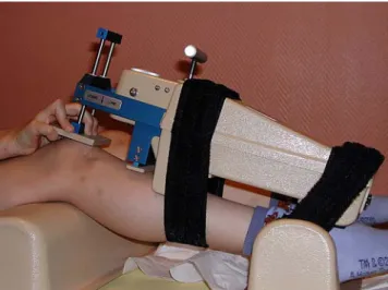
The use of these instruments in children and adoles-cents is very common in the literature [4, 14, 18, 25]. However, no adaptation to the smaller size of children has been made with the KT-1000/2000 stability device. The lever arm (Fig.6) is much shorter and measurement errors are possible.
Revision rate of the distal fixation (either screw or staple) is high. The screw or the staple often irritated the skin and needed to be taken out in 28% (Bruderholz,
Table 3 Injury to the menisci
and their treatment Associated injuries
Meniscus Treatment
Medial Lateral Both Repair Resection None
Bruderholz Acute 10 1 2 3 n=28 Chronic 18 6 2 1 3 5 1 Strasbourg Acute 3 1 1 n=12 Chronic 9 1 1 Pforzheim Acute 16 3 6 9 n=24 Chronic 8 3 2 1 6 Rosenheim Acute 3 n=4 Chronic 1 1 1 0 1 2 3 4 5 6 7 8 Soccer Ski Bike Hockey Traf fic Scho ol Basket/Hand Hor se Running others KSBH Strasbourg Pforzheim
Fig. 5 History of the injury
Fig. 6 The KT-1000 at a girl’s knee in Strasbourg, the length of the KT-1000 exceeds the lower leg
Switzerland) and 42% (Strasbourg, France) of the cases. Therefore, a better fixation method at the tibial side should be achieved in the future.
Using a patellar tendon graft for ACL-replacement in children is not uncommon [32]. Placing the femoral bone block as described of the Bruderholz-Hospital, Switzer-land, in the epiphysis of the femur under the growthplate is very difficult. Not touching the growthplate seems to be impossible [31]. There are only a few millimeters of the femoral attachment of the ACL to the growthplate, as Behr showed in his anatomical cadaver study [3]. Anyhow, no growthplate disturbances were noted among the patients in this study.
ACL-disruption in children and adolescents is a serious injury. Numerous articles were published in the past decades. Reconstructive surgery of the anterior cruciate ligament in the skeletally immature patient is performed in many centers. As long as the growthplates are open a substantial risk of growth arrest exists. The most often quoted investigations in the animal model show mixed results. Guzzanti et al. (1994) [17] were using 21 immature rabbits. They did not find any bone bar across the physeal plates. No alterations of the graft or the epiphyseal plate occurred. Stadelmaier et al. (1995) [33] used a canine model. None of the four dogs demonstrated a growth arrest with a fascia lata trans-plant. The control group without a crossing transplant showed bone bridging after drilling across the growth-plates. Their conclusion was that the graft prevents bone formation across the physis. Edwards et al. (2001) [9] were using a canine model as well. Their main alteration was a tensioned graft across the epiphyseal plates of 12 dogs. A fascia lata autograft was tensioned with 80 N. Significant valgus deformities at the femoral side and varus deformities at the tibial side were noted. None of the dogs showed a bone bridging across the physeal plates. Investigations of the tension of the anterior cru-ciate ligament in human knees versus knee stability was done by Friederich and O’Brien in 1998 [13]. The normal tension of the ACL recently showed a maximum load of 72.3 N in hyperextension [2]. The load pattern of the ACL form a U-shaped curve with a normal tension of 32 N in extension to 11.7 N in 90° flexion [2, 13]. Edwards et al. [9] tensioned the graft with 80 N in the canine-knee-model, which is much smaller than a human knee.
Another difference is that dogs are fully grown up in several months compared to human beings where it will
take many years. Animal studies [9,17,33] are essential steps in understanding the growing skeleton but should not be overinterpreted.
The survey of the ACL Study Group and the Hero-dicus Society [22] showed 15 patients (11%) with growthplate disturbances. Most of these cases were associated with surgical mistakes such as malplacement of the proximal fixation of the graft, bone plugs across the physis and staples over the apophysis of the tibial tubercle. In our study, one staple was placed across the proximal tibial epiphysis and needed to be taken out after 6 months.
No limb length discrepancy or angular deformity was noted among the patients in our series. Most reported angular deformities are caused by avoidable faults dur-ing the surgical procedure [23, 29]. In most cases a fix-ation device such as a staple was placed across the lateral aspect of the femoral physis [23,24].
Decision making in performing a reconstructive procedure in patients with open physes is difficult. A thorough history as well as a physical examination is crucial in selecting the surgical approach. A part of the physical examination should be done in the 90° flexed knee (hanging over the edge of the stretcher) to assess rotatory instabilities. Assessing the remaining growth with hand films (Strasbourg) or grading the children with the Tanner [35] classification plays a role in choosing the graft.
Despite the potential risk of growthplate distur-bances, conservative treatment does lead to early dete-rioration of the knee [1, 20]. All conservatively treated patients in Aichroth’s [1] group showed the Fairbank sign [11]. Engebretsen et al. [10], Graf et al. [16], Kannus and Ja¨rvinnen [20] and Mizuta et al. [30] are referenced as examples for poor results with a conservative, non-operative treatment. Compared with our own experi-ences we recommend to performing ACL-reconstructive surgery to avoid subsequent instability with possible early degenerative changes of the knee joint. Any ACL-rupture with instability in a young patient should be seen by an orthopedic surgeon as soon as possible before instability arises and meniscal injury occurs. The surgi-cal procedure must be done by the most experienced (arthroscopic) surgeon.
Acknowledgements This study was financially supported by the ESSKA and presented at the 10th ESSKA Congress in Rome, on 25 April 2002.
References
1. Aichroth P, Patel D, Zorilla P (2002) The natural history and treatment of rupture of the anterior cruciate ligament in children and adolescents. J Bone Joint Surg Br 84:38–41
2. Arnold M, van Kampen A (2002) Ten-sion in the normal anterior cruciate ligament—a cadaver study with clinical relevance. At ACL Group Study Meet-ing. Big Sky, MT, USA
3. Behr C, Potter H, Paletta GJ (2001) The relationship of the femoral origin of the anterior cruciate ligament and the distal femoral physeal plate in the skeletally immature knee. Am J Sports Med 29:781–787
4. Bisson L, Wickiewicz T, Levinson M, Warren R (1998) ACL reconstruction in children with open physes. Orthopedics 21:659–663
5. Blauth W (1984) Die zweizu¨gelige Er-satzplastik des vorderen Kreuzbandes aus der Quadrizepssehne. Unfallheilk-unde 87:45–51
6. Daniel D, Stone M, Sachs R, Malcom L (1985) Instrumented measurement of anterior knee laxity in patients with acute anterior cruciate ligament disrup-tion. Am J Sports Med 13:401–407 7. DeeLee J, Curtis R (1983) Anterior
cruciate ligament insufficiency in chil-dren. Clin Orthop Relat Res 172:112– 118
8. Deibert M, Aronsson D, Johnson R, Ettlinger C, Shealy J (1998) Skiing injuries in children, adolescents and adults. J Bone Joint Surg Am 80:25–32 9. Edwards T, Greene C, Baratta R,
Zieske A, Willis R (2001) The effect or placing a tensioned graft across open growth plates. J Bone Joint Surg Am 83:725–734
10. Engebretsen L, Svenningsen S, Benum P (1988) Poor results of anterior cruci-ate ligament repair in adolescence. Acta Orthop Scand 59:684–686
11. Fairbank T (1948) Knee joint changes after meniscectomie. J Bone Joint Surg Br 30:664–670
12. Fehnel D, Johnson R (2000) Anterior cruciate ligament injuries in the skele-tally immature athlete. Sports Med 29:51–63
13. Friederich N, O’Brien W (1998) Ante-rior cruciate ligament graft tensioning versus knee stability. Knee Surg Sports Traumatol Arthrosc 6:S38–S42 14. Fuchs R, Wheatly W, Uribe J,
Hecht-mann K, Zvijac J, Schurhoff M (2002) Intra-articular anterior cruciate liga-ment reconstruction using patellar ten-don allograft in the skeletally immature patient. Arthroscopy 18:824–828 15. Gaulrapp H (1997) Sportverletzungen
bei Kindern und Jugendlichen. Sport-ortho Sporttrauma 13:137–147 16. Graf B, Lange R, Fujisaki C, Landry G,
Saluja R (1992) Anterior cruciate liga-ment tears in skeletally immature pa-tients: meniscal pathology at
presentation and after attempted con-servative treatment. Arthroscopy 8:229– 233
17. Guzzanti V, Falciglia F, Gigante A, Fabbriciani C (1994) The effect of intra-articular ACL reconstruction on the growth plate of rabbits. J Bone Joint Surg Br 76:960–963
18. Janarv PM, Nystrom A, Werner S, Hirsch G (1996) Anterior cruciate liga-ment injuries in skeletally immature patients. J Pediatr Orthop 16:673–677 19. Jarvela T, Kannus P, Jarvinen M (2001)
Anterior cruciate ligament reconstruc-tion in patients with or without accompanying injuries: a re-examina-tion of subjects 5 to 9 years after reconstruction. Arthroscopy 17:818–825 20. Kannus P, Ja¨rvinnen M (1988) Knee
ligament injuries in adolescents—eight year follow-up of conservative man-agement. J Bone Joint Surg Br 70:772– 776
21. Kim S, Ha K, Ahn J, Chang D (1999) Anterior cruciate ligament reconstruc-tion in the young patient without vio-lation of the epiphyseal plate. Arthroscopy 15:792–795
22. Kocher M, Saxon B, Hovis D, Hawkins R (2002) Management and complica-tions of anterior cruciate ligament in-juiries in skeletally immature patients: survey of the Herodicus Society and the ACL Study Group. J Pediatr Orthop 22:452–457
23. Koman J, Sanders J (1999) Valgus de-formitiy after reconstruction of the anterior cruciate ligament in a skeletally immature patient. J Bone Joint Surg Am 81:711–715
24. Lipscomb A, Anderson A (1986) Tears of the anterior cruciate ligament in adolescents. J Bone Joint Surg Am 68:19–28
25. Lo I, Kirkley A, Fowler P, Miniaci A (1997) The outcome of operatively treated anterior cruciate ligament dis-ruptions in the skeletally immature child. Arthroscopy 13:627–634 26. Lo I, Bell D, Fowler P (1998) Anterior
cruciate ligament injuries in the skele-tally immature patient. Instr Course Lect 47:351–359
27. MacIntosh D (1974) The anterior cru-ciate ligament over the top repairs. J Bone Joint Surg Br 56:591–593 28. Matava M, Siegel M (1997)
Arthro-scopic reconstruction of the ACL with semitendinosus-gracilis autograft in skeletally immature adolescent patients. Am J Knee Surg 10:60–69
29. McCarroll J, Rettig A, Shelbourne K (1988) Anterior cruciate ligament inju-ries in the young athlete with open physes. Am J Sports Med 16:44–47 30. Mizuta H, Kubota K, Shiraishi M,
Ot-suka Y (1995) The conservative treat-ment of complete tears of the anterior cruciate ligament in skeletally immature patients. J Bone Joint Surg Br 77:890– 894
31. Neyret P (2004) ACL replacement in children. First annual visiting Profes-sorship in Basel (personal communica-tion, June 21, 2004)
32. Robert H, Bonnard C (1999) The pos-sibilities of using the patellar tendon in the treatment of anterior cruciate liga-ment tears in children. Arthroscopy 15:73–76
33. Stadelmaier D, Arnoczky S, Dodds J, Ross H (1995) The effect of drilling an soft tissue grafting across open growth plates. Am J Sports Med 23:431–435 34. Sta¨ubli H (1990) Technik der arthro-skopisch assistierten Substitution mit-tels autologer Quadrizepssehne. In: Jakob R, Sta¨ubli H (eds) Kniegelenk und Kreuzba¨nder. Springer, Berlin Heidelberg New York, pp 456–464 35. Tanner J, Davies P (1985) Clinical
lon-gitudinal standards for height and height velocity for North American children. J Pediatr 107:317–327