Coronary stenosis vasomotion during dynamic
exercise before and after PTCA*
T. M. SUTER, O. M. H E S S , A. BORTONE, H. NONOGI, J. GRIMM AND H. P. KRAYENBUEHL
Department of Internal Medicine, Medical Policlinic, Cardiology, University Hospital, Zurich, Switzerland
KEY WORDS: Coronary artery disease, coronary vasomotion, percutaneous transluminal coronary angio-plasty, supine bicycle exercise.
Coronary vasomotion was evaluated in eight patients (age 50 + 8 years) with coronary disease before and 3-3 ± 1 -9 months after successful percutaneous transluminal coronary angioplasty (PTCA). Luminal area of a normal and a stenotic coronary artery was determined before and after PTCA using biplane quantitative coronary arteriography. Patients were studied at rest, during supine bicycle exercise and 5 min after 1-6 mg sublingual nitroglycerin. Workloads before and after PTCA were identical.
Percentage diameter stenosis decreased from 78% to 24% (P < 0-001) after PTCA. Mean pulmonary artery pressure increased during exercise from 21 to 40 mmHg (? < 0-001) before and from 19 to 34 mmHg
(P < 0-001) after PTCA. Peak exercise pulmonary artery mean pressure was significantly (P < 0-05) lower after PTCA. Normal coronary arteries showed a minimal increase in mean luminal area before (+2%; NS) as well as after (+ 6%; NS) PTCA. Nitroglycerin produced dilation of the normal vessel segment to a similar extent pre- (+27%; P < 0-001) and post- (+31%; P < 0-001) PTCA. In contrast, stenotic vessel segments showed coronary vasoconstriction during exercise before PTCA (—28%; P < 0-01); after PTCA, exercise-induced vasoconstriction of the diseased segment was minimal (—4%; NS). Nitroglycerin was associated with vasodi-lation of the stenotic vessel segment before (+17%; NS) as well as after (+26%; P < 0-005) PTCA.
Thus, exercise-induced coronary vasoconstriction of stenotic coronary arteries is observed before as well as after PTCA, but vasoconstriction after PTCA is significantly less than before PTCA. Coronary vasomotion appears to be modified in a positive way by PTCA, but the exact mechanism remains unclear.
Introduction Methods and patients Coronary vasomotion plays an important role in STUDY POPULATION
the regulation of coronary flow at rest and during E i g h t m a l e p a t i en t s (mean age 50 years, range physical exercise'"3J. Not only normal but also steno- 3 9 H 5 1 y e a r s ) w e r e i n ciu d ed in the present study tic coronary arteries show vasomotion during exer- ( T a b l e xy A 1 ] p a t i e n t s h a d a ^ o r y o f e x e rcise-cise since approximately 70% of all coronary stenoses i n d u c ed angina pectoris and four had previous have a normal musculo-elastic wall segment at the m yocardial infarction. Upright bicycle exercise site of the stenosis' K Previous studies have indicated t e s t i n g w a s performed in all patients before and after that normal coronary arteries dilate but diseased PTCA. ST-segment depression of more than 0-1 mV coronary arteries constrict during dynamic exer- w a s f o u n d i n s i x p a tien t s (mean 0-24 ± 0 1 4 mV) cisel3'. Thus, the purpose of the present study was to b e f o r e P T C A N o ST-segment depression was evaluate the effect of percutaneus transluminal coro- p r e s e n t a f t e r p-pcA e x c e p t i n o n e p a t i e n t ( 0.2 8 m V nary angioplasty (PTCA) on coronary vasomotion at i n p a t i e n t 4 ) A n g i n a p e c t o r i s occurred in five rest and during exercise in patients with coronary p a t i e n t s b e f o r e a n d i n t h e p a t i e n t w i t h ST-segment artery disease before and after PTCA. depression after PTCA. Patients were selected for
Address for correspondence: Otto M. Hess, M.D., Medical Poli- t h e present Study on a consecutive basis when the clinic, Cardiology, University Hospital Raemistrasse 100, CH-8091 following inclusion criteria were fulfilled: (1) COrO-Zurich, Switzerland. n a r v a rte r v stenosis clearly visible on angiography for * This work was supported by a grant of the Swiss National Science . . , . , „ . trw^-lf J Foundation. quantitative evaluation; (2) successful PTCA; and 0195-668X/89/0GO058 + 06 $03.00/0 © 1989 The European Society of Cardiology
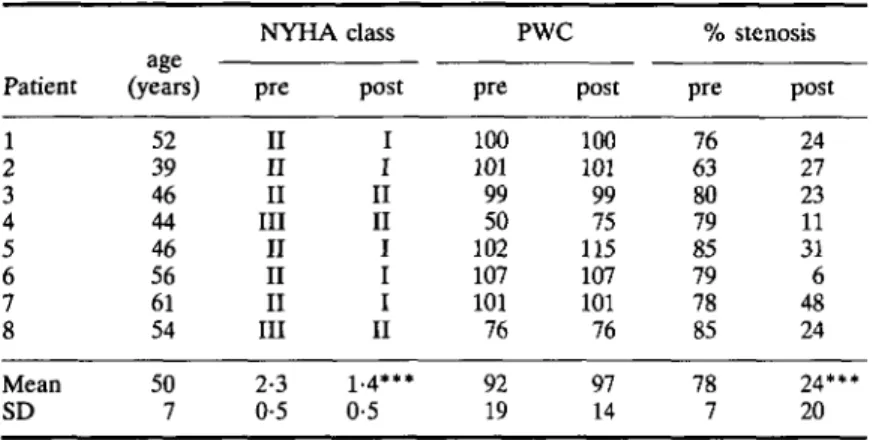
Table 1 Patient characteristics Patient 1 2 3 4 5 6 7 8 Mean SD age -(years) 52 39 46 44 46 56 61 54 50 7 NYHA pre II II II III II II II III 2-3 0-5 class post I I II II I I I II 1-4"* 0-5 pre 100 101 99 50 102 107 101 76 92 19 PWC post 100 101 99 75 115 107 101 76 97 14 % pre 76 63 80 79 85 79 78 85 78 7 stenosis post 24 27 23 11 31 6 48 24 24*** 20 NYHA: functional class according to the New York Heart Association scheme; PWC: physical working capacity in percentage of the age-, sex- and height-corrected normal value; % stenosis: percentage diameter stenosis; pre, post: before, after PTCA; *"p < 0001 (pre vs post); SD: standard deviation.
(3) coronary arteriography after PTCA (mean 3-3 months, range 2-6 months).
CARDIAC CATHETERIZATION
Patients underwent right and left heart catheteri-zation for diagnostic purposes. Informed consent was obtained from all patients. Premedication consisted of 10 mg chlordiazepoxide administered orally 1 h before the procedure. Aortic pressure was measured with a 8F Judkins-catheter introduced from the right femoral artery. Pulmonary artery pressure was meas-ured with a 6F pacing catheter with a side lumen for pressure recordings introduced from the right fem-oral vein. PTCA was performed 1-4 weeks after diagnostic coronary arteriography.
STUDY PROTOCOL
After routine coronary arteriography (Judkins technique) an interval of at least 10 min was allowed for dissipation of the effect of the contrast
medium (Iopamiro 370 R = iopamidol 755-2 mg ml"1, trometamol 1 mg ml"1) on coronary vaso-motion. Subsequently, the patient's feet were attached to the ergometer (Siemens-Elema AG, model 380 B). Aortic and pulmonary artery pressures were recorded immediately before each coronary angiogram (Table 2). Biplane coronary angiography in orthogonal projection was carried out at rest and repeated at the end of each level of exercise which was begun at 50-75 W and was increased every 2 min in increments of 25-50 W. The exercise test was terminated because of anginal pain, fatigue or ST-segment depression of more than 0-2 mV. The average workload was 115 W. The same exercise protocol was used for each patient at recatheteri-zation after PTCA. At the end of the exercise test, 1-6 mg nitroglycerin was administered sublingually and biplane coronary arteriography was repeated 3 and 5 min thereafter. There were no complications
Table 2 Haemodynamic data
Pre mean SD Post mean SD R 67 7 78 13 HR (beats min" EX 113*" 15 115"* 15 ') NTG 79* 8 85 15 R 104 15 103 15 MAP (mmHg) EX 109 23 112" 10 NTG 93 22 93 13 R 21 6 19 5 MPAP (mmHg) EX f 4 0 * " • 7 I 3 4. . . 8 NTG 19 5 15 5 HR: heart rate; MAP: mean aortic pressure; MPAP: mean pulmonary artery pressure; R: rest; Ex: peak exercise; NTG: sublingual nitroglycerin; pre, post: before, after PTCA; 'P < 0-05, "P < 0-01,
related to the procedure in the eight patients before and after PTC A.
QUANTITATIVE CORONARY ARTERIOGRAPHY
The currently used technique for quantitative coronary arteriography has been described pre-viously'3'5'. Quantitative evaluation of biplane coro-nary arteriography was carried out in a blinded fashion. Tracings were made manually from both projections during diastasis or end-diastole. Each vessel segment was analysed from six different frames and the results were averaged to reduce sampling error. A section of the catheter of known dimensions was traced as a scaling factor. The trac-ings of the coronary vessel segment were digitized manually and analysed on a PDP11/34 computer'3-51. The luminal area of a normal and a stenotic vessel
segment was calculated in each patient and expressed in absolute values and in percent of the resting value (see Table 3, Figs. 1 and 2). Quantitative evaluation of the normal and stenotic vessel segment was repeated after PTCA using certain landmarks (e.g. side-branches) for exact identification of the evalu-ated segments prior to PTCA. In one patient diffi-culties were encountered in visualization of the stenosis in the left anterior oblique projection before PTCA due to low contrast; in this patient a single (RAO) view was used before and after PTCA.
STATISTICAL ANALYSIS
Statistical comparisons of haemodynamic and angiographic data at rest and during exercise as well as after nitroglycerin were carried out by a two-way analyis of variance for repeated measures.
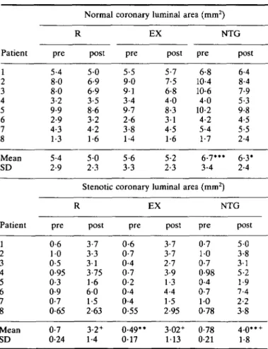
Compari-Table 3 Calculated luminal areas
Patient 1 2 3 4 5 6 7 8 Mean SD Patient 1 2 3 4 5 6 7 8 Mean SD pre 5-4 8-0 8 0 3-2 9-9 2-9 4-3 1-3 5-4 2-9 pre 0-6 10 0-5 0-95 0-3 0-9 0-7 0-65 0-7 0-24 Normal R post 5 0 6-9 6-9 3-5 8-6 3-2 4-2 1-6 5-0 2-3 Stenotic R post 3-7 3-3 3 1 3-75 1-6 6 0 1-5 2-63 3-2+ 1-4 coronary luminal EX pre 5-5 9 0 9 1 3-4 9-7 2-6 3-8 1-4 5-6 3-3 post 5-7 7-5 6-8 4-0 8-3 3-1 4-5 1-6 5-2 2-3 coronary luminal EX pre 0-6 0-7 0-4 0-7 0-2 0-4 0-4 0-55 0-49" 0-17 post 3-7 3-7 2-7 3-9 1-3 4-4 1-5 2-95 3-02" 1 1 3 area (mm pre 6-8 10-4 10-6 4-0 10-2 4-2 5-4 1-7 2) NTG post 6-4 8-4 7-9 5-3 9-8 4-5 5-5 2-4 6-7*" 6-3* 3-4 area (mm pre 0-7 1 0 0-7 0-98 0-4 0-7 1 0 0-78 h 0-78 0-21 2-4 2) NTG post 5-0 3-8 3-1 5-2 1-9 7-4 2-2 3-8 4 - 0 " + 1-8 R: rest; peak exercie; NTG: sublingual nitroglycerin; pre, post: before, after PTCA; *P < 0 0 1 (vs R); "P < 0 0 1 (vs R); '"P < 0001 (vs R); +P< 0-001 (vs pre) SD: standard deviation.
Rest Ex2 NTGI NTG2
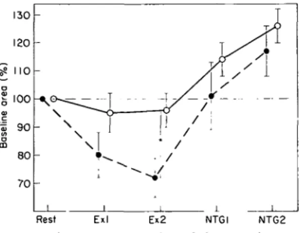
Figure 1 Coronary vasomotion of the normal vessel
segment before (closed symbols) and after (open symbols) PTCA. Ex 1: first level of exercise; Ex 2: second level of exercise; NTGI: 3 min after 1-6 mg sublingual nitroglycerin; NTG2: 5 min after 1-6 mg sublingual nitroglycerin.
130 -120 ? 110 I 100 % 90 01 <3 80 70
-Figure 2 Coronary vasomotion of the stenotic vessel
segment before (closed symbols) and after (open symbols) PTCA. Abbreviations are as in Fig. 1. * P < 001.
sons before and after PTCA were made by an paired Student's t test. In all figures, mean values ± 1 stan-dard error are reported.
Results
PATIENT CHARACTERISTICS (TABLE l )
Functional class (according to the New York Heart Association system) decreased significantly after successful PTCA. Physical working capacity during upright bicycle exercise increased slightly, although not significantly, from 92 to 97% after PTCA.
Per -\ \ \ > 1 Rest
. . _ y
^T
1 Exl 1 ' / / ^ • / Ex2 / NTGI NTG2centage diameter stenosis was 78% before and 24% after angioplasty.
HAEMODYNAMIC DATA (TABLE 2)
Heart rate and mean pulmonary artery pressure increased significantly during exercise before as well as after PTCA. Mean pulmonary artery pressure during peak exercise was, however, significantly lower post than prePTCA (P < 005). There was a slight but statistically not significant increase in mean aortic pressure before, but a significant increase after PTCA (P < 001).
ANGIOGRAPHIC DATA (TABLES 3, FIGS 1 AND 2)
Normal coronary arteries (Fig. 1) showed a mini-mal increase in mean luminal area during supine bicycle exercise before (+2%; NS) as well as after (+6%; NS) PTCA. Sublingual administration of nitroglycerin was associated with a definite increase in normal vessel area before (+ 27%; P < 0-001) and after (+31%; P < 0-001) PTCA.
Stenotic coronary arteries (Fig. 2) showed before PTCA a significant reduction in vessel area dur-ing supine bicycle exercise (—28%; P < 0-01) and an increase after sublingual administration of nitroglycerin (+17%; NS). Bicycle exercise after PTCA was associated with a slight decrease in mini-mal luminal area of the treated (formerly stenotic) vessel segment (—4%; NS) followed by a significant increase after sublingual administration of nitroglycerin (+26%; P < 0-01). The decrease in minimal luminal area of the stenotic vessel segment (Fig. 2) was significantly less {P < 0-01) after than before PTCA.
Discussion
Coronary blood flow is determined by several fac-tors such as perfusion pressure, coronary vascular resistance, metabolic demands and vasomotor tone'6'. In normal coronary arteries, blood flow increases three to five times during physical exercise, whereas in stenotic coronary arteries the increase in blood flow is strongly dependent on the severity of the stenosis. The actual regional blood flow is, however, not solely determined by the luminal area but is the result of the complex interplay of the aforementioned haemodynamic, metabolic and neu-ral factors. Seveneu-ral studies'6'71 have established the presence of both alpha- and beta-adrenergic recep-tors in animal and human coronary arteries. An
increase in alpha-adrenergic tone has been associated with coronary artery vasoconstriction, whereas an increase in beta-receptor tone is accompanied by coronary artery vasodilation'5'. The exact mech-anism responsible for the decrease in minimal lumi-nal area of the stenotic vessel segment during exercise is not clear, and might involve active vaso-constriction or a passive collapse of the disease-free vessel segment within the stenosis, due to increased coronary blood flow'3'. Active vasoconstriction seems, however, more likely to be responsible for exercise-induced stenosis narrowing than a passive collapse. The following two mechanisms could explain this phenomenon: endothelial dysfunction with insufficient production of the endothelium-derived relaxing factor (= EDRF) or platelet aggre-gation with release of thromboxane A2 and seroto-nin[3>9'. Which of these two mechanisms is responsible for exercise-induced stenosis narrowing is not clear and cannot be determined from present data. It is, however, obvious that this phenomenon can aggravate myocardial ischaemia during physical exercise and represents, therefore, an important mechanism in the pathophysiology of angina pectoris.
The purpose of the present study was to evaluate coronary vasomotion in patients with stable angina pectoris and to examine the effect of PTCA on exercise-induced stenosis narrowing. The question was if the increase in stenosis area after PTCA could influence coronary vasomotion of the stenotic vessel segment.
CORONARY VASOMOTION BEFORE AND AFTER PTCA Normal coronary arteries showed minimal
vasodi-lation during dynamic exercise with a further increase after sublingual administration of nitroglycerin (Fig. 1). Exactly the same behaviour was found before and after PTCA as one would expect, but the extent of coronary vasodilation was smaller (+2% pre-, +6% postPTCA) than that of non-stenotic vessels reported previously'3'5'9'. It might be that the wall of the so-called 'normal vessel' in this study was not truly normal and elicited a depressed responsiveness to physiologic dilatory stimuli. In patients with diseased but not visibly stenotic coro-nary arteries, no dilation during exercise has been observed previously'10'11'.
Stenotic coronary arteries showed vasoconstriction during exercise with a decrease in minimal luminal area of approximately 30% (Fig. 2). Similar data have been reported by Gage and coworkers'3'. All
120 i— 110 ^ 100 I L 90 r g 5 80 I
I I
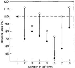
8 70 o ' m i 50 2 3 4 5 6 7 Number of patientsFigure 3 Changes in coronary luminal area of the stenotic
vessel segment during peak exercise before (closed sym-bols) and after (open symsym-bols) PTCA. Individual data (in % of the baseline area) are given for each patient. Vasoconstric-tion occurred in all except one patient before, but only in three patients after PTCA.
but one patient showed vasoconstriction during exer-cise in the present study (Fig. 3). PTCA was associ-ated with an increase in minimal luminal area from 0-7 to 3-2 mm2 and a decrease in percentage diameter stenosis from 78 to 24%. After PTCA coronary vessel segments showed significantly less vasocon-striction during peak exerise than before PTCA (pre-- 2 8 % , post(pre-- (pre-- 4 % ; P < 0(pre--01; Fig. 3). Three of the eight patients elicited vasodilation during postPTCA exercise, whereas two patients showed no vaso-motion and three had less but persisting vasoconstric-tion (Fig. 3). The patient with ST-segment depression (0-28 mV, patient 4) in the upright exer-cise test after PTCA showed minimal vasodilation (+4%; Fig. 3) during exercise. Residual stenosis was 11% in this patient (before PTCA: 79%).
The stenotic vessel segments were able to dilate both before and after PTCA, since sublingual administration of nitroglycerin was associated with an increase in minimal luminal area of 17% before and 26% after PTCA, respectively. However vasodi-lation did not occur during exercise; hence some other mechanism must have influenced coronary vasomotion. Coronary vasodilation during exercise might be dependent on an intact endothelium with an adequate production of the endothelium-derived relaxing factor. 'Healing' after PTCA might lead to re-endothelialization and improvement of endothe-lial function.
LIMITATIONS OF THE STUDY
The accuracy of quantitative coronary arterio-graphy has been established previously in our labora-toryf3'5' Brown and coworkers'121 reported that the accuracy of quantitative coronary arteriography is within 0-08 mm for measurement of known dimen-sions and 0-10 mm for minimum diameter estimates. The changes observed in our study, such as the 0-21 mm2 (0-15 mm diameter) decrease in stenosis area at peak exercise, are small but clearly larger than reported angiographic resolution. Therefore, the observed changes after several interventions can be considered representative, even with monoplane assessment.
References
[1] Brown BG, Bolson EL, Dodge HT. Dynamic mech-anisms in human coronary stenosis. Circulation 1984; 42: 917-22.
[2] Mates RE, Gupta RL, Bell AC, Klocke FJ. Fluid dynamics of coronary artery stenosis. Circ Res 1978; 42: 152-432.
[3] Gage JE, Hess OM, Murakami T, Ritter M, Grimm J, Krayenbuehl HP. Vasoconstriction of stenotic coro-nary arteries during exercise in patients with classic angina pectoris: reversibility by nitroglycerin. Circu-lation 1986; 73: 865-76.
[4] Freudenberg H, Lichtlen PR. The normal wall segment in coronary stenosis—a postmortem study. Z Kardiol 1981; 70: 863-9.
[5] Gaglione A, Hess OM, Corin WJ, Ritter M, Grimm J, Krayenbuehl HP. Is there coronary vasoconstriction after intracoronary beta-adrenergic blockade in patients with coronary artery disease? J Am Coll Cardiol 1987; 10: 299-310.
[6] Mohrman DE, Feigl EO. Competition between sym-pathetic vasoconstriction and metabolic vasodilatation in the canine coronary circulation. Circ Res 1978; 42: 79-86.
[7] Feigl EO. Sympathetic control of coronary circulation. Circ Res 1967; 20: 262-71.
[8] Saner HE, Gobel FL, Salomonowitz E, Erlien DA, Edwards JE. The disease-free wall in coronary artherosclerosis: its relation to degree of obstruction. J Am Coll Cardiol 1985; 6: 1096-9.
[9] Bortone A, Hess OM, Gaglione A, Nonogi H, Grimm J, Krayenbuehl HP. Effect of intracoronary and intravenouspropranolol on coronary vasomotionat rest and during exercise. Circulation 1988; 78 (Suppl II); 11-455 (abstr).
[10] Hess OM, Bortone A, Eid K et al. Coronary vasomo-tor tone during static and dynamic exercise. Eur Heart J 1988; 10 (Suppl F).
[11] Gordon JB, Zebede J, Wayne RR, Mudge GH, Ganz P, Selwyn AP. Coronary constriction with exercise: possible role for endothelial dysfunction and alpha tone. Circulation 1986; 74 (Suppl II): 11-481 (abstr). [12] Brown BG, Bolson E, Frimer M, Dodge HT. Quanti-tative coronary arteriography: estimation of dimen-sions, hemodynamic resistance, and atheroma mass of coronary artery lesions using arteriogram and digital computation. Circulation 1977; 55: 329-37.