639
Increased Circulating Soluble CD14 Is Associated with High Mortality in
Gram-Negative Septic Shock
Regine Landmann, Werner Zimmerli,
Sebastiano Sansano, Susanne Link, Alfred Hahn,
Michel Pierre Glauser, and Thierry Calandra*
Divisions of Infectious Diseases and Hypertension (Departments of Research and Medicine). University Hospital. Basel; Division of Infectious Diseases (Department of Internal Medicine). Centre Hiipital Universitaire, Lausanne, Switzerland The soluble glycoprotein sCD 14 binds lipopolysaccharide, a complex that activates
endothe-lial cells and that may be crucial in gram-negative sepsis, Therefore, serum sCD14 was analyzed in 54 patients with gram-negative septic shock and in 26 healthy controls, sCD14 was tested by ELISA and Western blotting, Patients had higher sCD14 concentrations than controls (median, 3.23 vs. 2.48 #!g/mL,P= .002). Increased levels were associated with high mortality (median, 4.2 #Lg/mL in nonsurvivors vs. 2.8 #!g/mL in survivors,P= .001). sCD14 was found in two isoforms
(49 and 55 kDa) in monocyte cultures. In sera only one of either form was detectable. Controls had the 49-kDa form, and patients had either the 49- or 55-kDa form, but patients with high levels of sCD14 had only the 55-kDa form. Twenty-one (53%) of 39 with the 55-kDa form and 8 (57%) of 14 with the 49-kDa form died. Thus, the level of sCD14 but not its biochemical form had a prognostic value in patients with gram-negative septic shock.
Lipopolysaccharide (LPS) plays a key role in the patho-physiology of sepsis [I]. The LPS receptor on myeloid cells is the phosphatidylinositol-Iinked membrane glycoprotein CD 14 [2]. CD 14 occurs in a membrane-associated form and as a soluble (s) molecule [3, 4]. sCD 14 is derived from the monocytic cell line Mono-Mac 6 in two forms [5]: A 48-kDa form is shed from the membrane of monocytes and Mono-Mac 6 cells [6, 7], and a 56-kDa form is detectable in meta-bolically labeled Mono-Mac 6 cells and in sCD 14 purified from urine [3, 5]. The serum concentration of sCD 14 is ,...., I millionfold higher than that of cytokines [8]. Although much is known about cytokine liberation during experimental en-dotoxemia or sepsis, little is known about sCD 14 in these conditions [9-12]. Because CD 14 is the principle LPS recep-tor, the serum concentration ofsCD 14 may be modified dur-ing endotoxemia or sepsis. The physiologic role of sCD 14 is unknown. Itcould act as a scavenger by neutralizing circu-lating LPS [13] or, alternatively, it may playa harmful role by transmitting LPS effects on endothelial cells, which do not express membrane CD 14 [I4]. Indeed, it was recently
Received 4 May 1994; revised 22 September 1994.
Presented in part: annual meeting. Infectious Diseases Society of Amer-ica. New Orleans. October 1993 (Clin Infect Dis 1993; 17:531).
Informed consent was obtained from all patients studied. The study was approved by the Ethical Committee. University Hospital. Basel. in accor-dance with the guidelines of the Swiss Academy for Medical Science.
Grant support: Swiss National Foundation (31-32310.91).
Reprints or correspondence: Dr. Regine Landrnann. Division of In-fectious Diseases. Dept. of Research. University Hospital. CH-4031 Basel. Switzerland.
*Present affiliation: Picower Institute of Medical Research. Manhasset. New York.
The Journal of Infectious Diseases 1995;171:639-44
© 1995 by The UniversityofChicago.Allrights reserved. 0022-1899/95/7103-0017$01.00
shown that complexes ofsCD 14 with LPS activate these cells [14,15].
We measured sequential serum concentrations of sCD 14 in 54 patients with gram-negative septic shock, compared these with levels in normal controls, and correlated sCD 14 levels with patient outcome. We also determined the bio-chemical form of serum sCD 14 by Western blotting and compared it with sCD 14 derived from normal monocytes.
Methods
Patients. The study population comprised 54 patients (33 males, 21 females) with documented gram-negative septic shock. Their median age was 58 years (range, 7-78). All were part of a previously described study that compared the efficacy of J5 hyperimmunoglobulin (n
=
22) with standard intrave-nously (iv) administered IgG(n=
32) (Sandoglobulin; Sandoz Pharmaceuticals, Basel, Switzerland) [16]. The etiology of septic shock was established by the isolation of gram-negative bacteria from blood in 37 patients (69%). Of these, 30 had monomicro-bial and 7 had polymicromonomicro-bial bacteremia. Seventeen patients (3t%) had documented gram-negative infections with negative blood cultures. At study entry, the median duration of shock was 10 h (range, 2-144), the median urinary output was 5 mL/h (range, 0-190), and the median arterial pH was 7.37 (range, 7.17-7.57). Blood was obtained from all patients at study entry (day 0), 2 h later (after infusion of either standard or J 5 hyper-immunoglobulin), and after 24 hand 10 days. In addition, 26 healthy controls (16 men, 10 women; median age, 48 years; range, 27-72) and 2 women with paroxysmal nocturnal hemo-globinuria (ages 30 and 38 years) were investigated. Sera were aliquoted and kept at -70°C until analyzed.Cells. Mononuclear leukocytes were isolated from heparin-ized blood by density gradient centrifugation over a ficoll-hypa-que gradient. Monocytes were purified from mononuclear leu-kocytes by centrifugation over a continuous Percoll density
640 Landmann et al. JID 1995; 171 (March)
gradient as described [17]. Cells were taken up in RPMI me-dium with 5% heat-inactivated human AB serum before being labeled with [35S]methionineor
1251.They were cultured for 48 h
in serum-free HB 101 medium (Hana Biologics, Alameda,CA) substituted with 2% human serum albumin and 10-8M
1,250'-dihydroxyvitamin D3 before the supernatant was harvested for
Western blotting.
Reagents, antibodies to C{J14. and recombinant (r) sCD14. Goat polyclonal anti-CD 14 antiserum was a gift of R. Ulevitch (Scripps Research Institute. La Jolla,CA). We used the follow-ing mouse monoclonal antibodies: MEM 18 (IgG I, which recog-nizes membrane CDI4 and both forms of sCDI4; gift of V. Bazil, Prague[3]); RoMo I (IgG2a; IBL, Hamburg, Germany) [18], 63D3 (lgG I), and 3C I0 (IgG2b; both purified by affinity chromatography from hybridoma culture supernatants; Ameri-can Type Culture Collection, Rockville, MD); and My4 (IgG I) (Coulter, Luton, UK). rsCD 14 was immunopurified on immo-bilized 3C I0 from culture supernatants of CHO cells that were transfected with an expression vector containing truncated CDI4 cDNA (unpublished data).
The endotoxin content in the stock solutions of antibodies was <100 pg/rnl., as determined by quantitative limulus assay (Chromogenix, Molndal, Sweden).
sCD14 depletion from serum. The antibody to CDI463D3 was coupled to CNBr-activated Sepharose. Human AB serum containing 3 JLg/mL sCD 14 was absorbed to antibody-CNBr-Se-pharose overnight at 4°C. After centrifugation, no sCDI4 was detected by ELISA or Western blot in the sCD 14-depleted serum. Heat inactivation of normal serum at 56°C for 30 min also destroyed sCD 14 immunoreactivity.
Assay for sCD14. Serum concentrations of sCD 14 were de-termined by sandwich ELISA. Polystyrene plates (Greiner 96-well; Microlon, Frickenhausen, Germany) were coated over-night at room temperature with 2 JLg/mL MEM 18 (50 JLL/well). After plates were washed three times with PBS and 0.05% Tween (wt/vol), nonspecific binding was blocked for I h at room tem-perature with 50 JLL/well 0.2M TRIS-HCI buffer (pH 7.5) with I% bovine serum albumin (BSA; wt/vol) and 0.05% Tween (wt/ vol). Serum samples diluted 1:150, 1:300, and 1:600 in 0.1 M phosphate buffer (pH 7.2) with 1% BSA and 0.05% Tween (wt/ vol) or standards (dilutions of sCD 14 isolated from serum by affinity chromatography [19]; 50 JLL/well) were incubated for 3 h at room temperature. Plates were washed three times with PBS and 0.05% Tween. Captured sCD 14 was measured by peroxi-dase-coupled RoMo I (2-h incubation, 50 JLL/well, in 0.1 M phosphate buffer [pH 7.2] with 1% BSA and 0.05% Tween). The signal was detected using tetramethylbenzidine and read atA450
in an ELISA reader (Molecular Devices, Palo Alto, CA). The serum concentration of sCD 14 was the weighted average of the concentrations determined for each dilutionby extrapola-tion from the linear part (3-40 ng/mL) of the standard sCD 14 curve. When 5 JLg/mL LPS was added to the sCD 14 serum stan-dard, serum sCD 14 values did not change. For detection of sCD 14 in monocyte culture supernatants, a similar ELISA with 63D3 as a primary antibody ( 100 JLL, 5 p,g/mL) and peroxidase-labeled 3C I0 (500 ng/mL) as a capturing antibody was used; purified rsCD 14 (0.3-10 ng/mL) from CD 14-transfected CHO cells (unpublished data) served as a standard.
The specificity of the assay was confirmed by the following experiments. sCD 14-depleted human serum (see above) showed no reactivity in the ELISA. The immunoreactivity was recov-ered when the eluted sCD 14 was added back to the depleted serum. In addition, the specificity of the second peroxidase-la-beled antibody was tested by adding it along with unlaperoxidase-la-beled CD 14 antibody My4 to normal and sepsis sera. My4 competi-tively inhibited the binding of RoMo I and of 3C I0 (data not shown).
Effect of standard and J5 hyperimmunoglobulin on sCD14 determination. Patients were treated with standard iv IgG (200
mg/kg)or the same dose of an anti-Lf'S hyperimmunoglobulin (15) within 2 h of diagnosis of septic shock. The sCDI4 content of the immunoglobulins given and their influence on sCD 14 serum values were evaluated. Standard IgG. at a twofold higher concentration than the serum IgG level measured after patients received therapy ( 12mg/ml.),contained 100 ng/mL sCD 14. J 5 hyperimmunoglobulin at 12 mg/ml. contained 5 ng/mL sCD 14. When either immunoglobulin preparation was added to sCD 14 standard pooled human serum or sCD 14-free serum, the optical densities of the preparations increased by the amount of sCDI4 in standard IgG or J5 hyperimmunoglobulin.
sCD 14 levels were determined in the 26 healthy controls. The median serum concentration ofsCDI4 was 2.48 JLg/mL(range, 1.75-2.90), and intra- and interindividual coefficients of varia-tion were 12% and 23%, respectively(n
=
5, repeated measure-ments at 0, 2, 24, and 240 h).SDS-PAGE and Western blotting. Serum samples were di-luted to contain 3 ng of sCD 14/ I0 JLL and were denatured by boiling for 5 min in 0.5 M TRIS-HCI containing 5% SDS, 2.5% glycerol, and 0.0 I% bromphenol blue (sample buffer). Dena-tured proteins and prestained molecular mass standards (Bio-Rad, Glattbrugg, Switzerland) were electrophoresed (200 V, 500 rnA) for 60 min in 7.5% polyacrylamide gels under nonre-ducing conditions and transferred for 60 min (100 V, 500 rnA) onto Immunlite membranes (Bio-Rad) presoaked in 25 mM TRIS and 192mMglycine transfer buffer (TBS; pH 8.3) using a wet transblot system (Bio-Rad). Membranes were incubated overnight at 4°C in TBS containing 7.5% dry milk to reduce nonspecific protein binding and washed twice with TBS and 0.05% Tween (TTBS). They were then incubated for 3 h at room temperature on a rocking platform with the monoclonal anti-body to CD 14(3C I0), diluted I:400 in TTBS containing I%dry milk or with polyclonal goat anti-CD 14 antiserum diluted I: 1000. After being washed three times in TTBS, the mem-branes were incubated for 2 h at room temperature with alkaline phosphatase-conjugated goat anti-mouse IgG (I :3000; Bio-Rad) or mouse anti-goat IgG (1:10,000; Sigma, St. Louis) in TTBS containing I% dry milk. The membranes were then washed three times with TTBS, incubated for 5 min with 5 mL of chemoluminescent substrate solution (adamantyl dioxetane phenyl phosphate; Bio-Rad) and exposed to Kodak X-Omat films for 10-30 min.
Labeling with [35S]methionine. Monocytes were incubated for 48 h in medium with 5% heat-inactivated AB serum. CDI4-transfected CHO cells were cultured in serum-free RPMI me-dium containing G418 (Geneticin; Boehringer, Mannheim, Germany). Both cell types were washed and kept for I h in
JID 1995: 171 (March) Soluble CD14 in Septic Shock 641
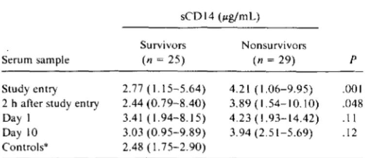
NOTE. Dataare median (range) sCDI4 determination with ELISA. *n= 26.
Table 1. Serum concentrations of soluble (s) COl4 during and
after septic shock.
~55 ~49 4 3 2 1
46
69
97
monocyte
rsCD14
sCD14
vivors showed significantly higher median sCD 14 concentra-tions than survivors. This difference was significant at study entry(P = .00 I) and 2 h later but not after 1 and 10 days. The seD 14 level remained predictive for a high mortality when age andTNF-awere included in a multivariate analy-sis(P< .01).
Relationship between sCDl4 and clinical parameters or cytokines. The earliest measure of sCD 14 correlated in-versely with mean arterial pressure at that time(P= .03) but did not correlate with duration of shock at entry(P= .90) or with urinary output(P= .86). In addition, it did not corre-late with TNF-a (P = .77), interleukin (IL)-I,8(P = .44), IL-6(P
=
.79), interferon (IFN)-')'(P=
.95), or IFN-a(P =.69). Serum levels of these cytokines have been described [II, 12].
Isoforms ofsCDl4. The monocytic cell line Mono-Mac 6 releases two sCDl4 forms [5], and in sCDl4 purified from urine two forms are identified under reducing conditions [3, 8]. However, the expression of two CD 14 isoforms has not been seen in normal monocytes, which are the major source of serum sCD 14. We therefore analyzed culture superna-tants of membrane or metabolically labeled monocytes by immunoprecipitation and compared them with immuno-blots of 48-h monocyte culture supernatants (figure 1). The smaller (49 kDa) form ofsCDl4 was derived from the mem-brane, since it was the only molecule detectable in superna-tants ofI251-labeled monocytes (figure I, lane 2). In contrast,
both the larger (55 kDa) and to a lesser extent the smaller forms were found in supernatants of metabolically labeled cells (figure I, lane 3). This suggests that the 55-kDa form has an intracellular origin without membrane attachment.
Figure 1. Biochemical analysis ofsCO 14. By lane: I, supernatant containing [35S]methionine-labeled recombinant sCO 14; 2, super-natant from 125I-labeled monocytes after overnight culture; 3, su-pernatant ofmonocytes after overnight metabolic labeling: 4, West-ern blot of 48-h serum-free monocyte culture supWest-ernatant. p .001 .048 .11 .12 Nonsurvivors (n = 29) 4.21 (1.06-9.95) 3.89 (1.54-10.10) 4.23 (1.93-14.42) 3.94 (2.51-5.69) sCDI4 (#Lg/mL) Survivors (n = 25) 2.77 ( 1.15-5.64) 2.44(0.79-8.40) 3.41 (1.94-8.15) 3.03 (0.95-9.89) 2.48 (1.75-2.90)
Results
sCDl4 concentrations in sera ofpatients with gram-negative septic shock. Serum sCD 14 concentrations were higher in the 54 patients with gram-negative septic shock than in 26 healthy controls (median, 3.23 vs. 2.48 JLg/mL;P
=
.002). Among the 54 patients with gram-negative septic shock, 29 (53.8%) died. The clinical course of the patients has been described [16]. A high sCDl41evei at the onset of shock was associated with poor outcome. As shown in table 1, nonsur-methionine-free medium supplemented with 2% dialyzed fetal calf serum before addition of 100 JLCi of[35S]methionine (Amer-sham Laboratories, Arner(Amer-sham, UK). After overnight culture, the supernatant was harvested, centrifuged at 100,000 g for 30 min at 4°C; Nonidet P-40 and phenylmethylsulfonyl fluoride (Sigma) were added at final concentrations of 0.5% and 2mM, respectively. For preclearing, the supernatant was incubated 30 min at 4°C with protein G-Sepharose (Pharmacia, Piscataway, NJ) in the presence of normal serum. After centrifugation, the supernatant was first incubated for 60 min at 4°C with 3CIO and then for 30 min at 4°C with protein G-Sepharose. The immunoprecipitate was washed three times in 50 mMTRIS, 150 mMNaCl, and 5mMEDTA buffer (pH 7.3). The sample was taken up in buffer and then denatured and electrophoresed in a 7.5% SOS-polyacrylamide gel as described above. Gels were fixed for 30 min in Amplify solution (Amersham Laboratories, Arnersharn, UK) and then washed in distilled water, dried, and autoradiographed.Labeling with/25/. Freshly isolated monocytes (2.5 X 10⁶ )
were washed three times with PBS, iodinated for 15 min at room temperature with 500 JLCi of Na, 1251, and Iodobeads (Pierce, Rockford, IL) in 6-well culture plates, washed again with PBS, and cultured overnight in RPMI medium with 5% heat-inacti-vated human AB serum. The chase medium was then harvested and processed as described for the [35S]methionine-Iabeled su-pernatant.
Statistics. Patient groups and controls were compared by Mann-Whitney test. The relationship between different sero-logic parameters was evaluated by Wilcoxon rank correlation test. The effect of sCD 14, tumor necrosis factor-a(TNF-a),and age on mortality was studied by multivariate analysis of variance (Superanova: Abacus, Berkeley, CA)
Serum sample Study entry 2hafterstudy entry Day I Day 10 Controls"
642 Landmann et al. lID 1995; 171 (March)
Figure 2. Western blots from sera. By lane: I. nonspecific (ns) binding. blot with alkaline phosphatase-conjugated second anti-body; 2. serum from healthy control; 3, serum from paroxysmal nocturnal hemoglobinuria(PNH)patient; 4-7. sera from sepsis pa-tients at study entry (lanes 4 and 5. 55-kDa sCD 14. survivor and nonsurvivor, respectively; lanes 6 and 7. 49-kDa sCD 14. survivor and nonsurvivor, respectively).
Finally. in Western blots of serum-free monocyte culture su-pernatants. the 55-kDa form was clearly seen; the smaller forms gave only a weak signal (figure 1, lane 4). To docu-ment the existence of the two CD 14 isoforms, the immuno-precipitate from metabolically labeled culture supernatant of CD 14-transfected CHO cells is shown on lane 1 in figure 1.
In this supernatant. which was very rich in sCD 14 (2 ILg/
ml.), the upper band (55 kDa) was stronger than the 49-kDA band and even a third intermediate form ofsCDl4 could be detected.
Western blot ofsera. After recognizing that rsCD 14 and monocyte culture-derived CD 14 both exist in two molecular forms, we analyzed human sera to see if it also contained both forms. For this purpose, Western blots were made with 26 sera from healthy controls and with sera from the 54 study patients. Serum from 1 patient gave no signal in Western blot; therefore, the analysis includes 53 patients. As shown in lane 2 offigure 2, the 49-kDa form ofsCDl4 was found in serum from healthy volunteers. In contrast, patients with septic shock had either the 49- or the 55-kDa form at study onset, provided that their sCD 14 level remained <3.5 ILg/
mL (figure 2 shows two examples of the 49-kDa form. lanes 6 and 7). An sCDl41evei >3.5~g/mLwas exclusively asso-ciated with the 55-kDa form ofsCDl4 (figure 2, lanes 4 and 5). The difference in molecular mass was also seen when a polyclonal instead of a monoclonal antibody was used for immunoblotting (data not shown). In addition, sera from patients with paroxysmal nocturnal hemoglobinuria (who
ns control PNH
Figure 3. Relationship between sCD 14concentration and molec-ular forms. Left:1:::., 26 healthy controls, 14 sepsis patients with 49-kDa form (6 survivors[0], 8 nonsurvivors [.]). Right: 2 pa-tients with paroxysmal nocturnal hemoglobinuria (0) and 39 sepsis patients with 55-kDa form (18 survivors[0],21 nonsurvivors [.]). Serum from I of 54 patients did not give a signal in Western blot.
10
•
sCD14
•
J.lg/ml
•
8 6<>
~.
~
<>
~
4_.~
2;0
~
049
55
sCD14 Form (kDa)
lack membrane CD 14 and therefore cannot release the smaller form of sCD 14) had the 55-kDa form exclusively (figure 2, lane 3).
Relationship between sCD14molecular form and concentra-tion. To determine clinical conditions associated with pre-dominance of either form of sCD 14, patients were grouped by sCD 14 form and serum concentrations at onset of shock. Elevated sCD 14 concentrations were associated with a 55-kDa sCDI4 (figure 3). Patients with 55-55-kDa sCDI4 had me-dian sCDl4 concentration of 3.43 ILg/mL (range, 1.41-9.95); those with the 49-kDa form had a median sCDl4 of 2.28 ~g/mL(range, 1.06-2.90; P < .00 I). The molecular form of sCD 14 by itself was not associated with high mortal-ity; 21 (53%) of 39 with 55-kDa sCDl4 and 8 (57%) of 14 with the 49-kDa form died (figure 3). All 26 healthy controls had 49-kDa sCDl4 (figure 3). Both patients with paroxysmal nocturnal hemoglobinuria had elevated levels of 55-kDa CD 14 in their serum.
Immunoblot of CD14from sera of patients during sepsis.
To detect a possible switch from one molecular form of sCD 14 to the other, sera from 4 patients were analyzed con-secutively at study entry and 2 and 24 hand 10 days later (figure 4; 2 patients with 49-kDa sCD 14 [A, C] and 2 with >3 ILg/mL 55-kDa seD 14 [B, D]). In figure 4, patients in the upper row survived; those in the lower row died. Each pa-tient had the same form of sCD 14 throughout the observa-tion period, that is, no switch in CD 14 isoforms was ob-served. . - 55 . - 49 7 6 sepsis 5 4 3 2 97 69 46 150
JID 1995: 171(March) SolubleCD14 in Septic Shock 643
Discussion
Figure 4. Western blots and sCDI4 concentrations during and after septic shock. A and B, Survivors with 49- and 55-kDa forms, respectively. C and D, Nonsurvivors with 49- and 55-kDa forms, respectively. Far right of Band D: nonspecific binding in serum with alkaline phosphatase-conjugated second antibody.
In the present study, serum concentrations and biochemi-cal forms of sCD 14 were investigated in patients with gram-negative septic shock. They had elevated serum sCD 14 con-centrations. In addition, the CD 14 derived from normal monocytes was found in 49- and 55-kDa forms that originate from the cell membrane and the intracellular space. In serum, only a single form was detectable. High levels of sCDI4 were associated with expression of the 55-kDa form. The serum concentration but not the isoform of sCD 14 was predictive for death.
Soluble CD 14 is spontaneously released by the Mono-Mac 6 cell line, by monocytes, macrophages, and granulo-cytes [5, 7. 17, 20]. After release, sCDl4 circulates in the bloodstream [8]. Whereas the serum concentration of sCD 14 is constant in normal persons (see Methods), we found increased levels in patients with gram-negative septic
shock. This could be due to a reduced sCD 14 clearance or alternatively to enhanced liberation. The former is unlikely, since there was no relationship between urinary output and sCD 14 concentration. The latter probably occurs during sep-sis. The stimulus for sCD 14 liberation may be LPS itself or a cytokine. We found that LPS enhances sCD 14 release from normal monocytes in vitro (unpublished data). In contrast, we found no effect ofTNF-a, IL-I
fl,
IL-6, or IFN-')" on sCD 14 release in vitro [I 7].The pathophysiologic significance of an increased sCD 14 concentration in septic shock is unknown, but it is associated with increased mortality. sCD 14 promotes LPS binding to endothelial cells [15]. The ensuing activation of these cells may be detrimental and thus may explain the worse progno-sis in patients with increased sCD 14. This relationship sug-gests that serum levels of sCD 14 may be crucial for the fate of endothelial cells.
The existence of two forms of sCD 14 has been docu-mented in immunoprecipitates isolated from urine [3]. We sought to find if the two isoforms are also detectable in serum. In earlier studies. only the smaller form was detected in monocytes [6, 20]. Therefore, we investigated the cellular mechanisms of release by cells that are the major source of sCD 14. For this purpose, we analyzed supernatants ofmono-cytes by Western blotting and after membrane or metabolic labeling. We detected both forms.
The larger form predominated in supernatants of normal 48-h cultured monocytes. This conclusion is based on the Western blot analysis of concentrated supernatants. Since the cultures were serum-free, a contribution of serum CD 14 could be excluded. In addition, the metabolic labeling re-vealed that in overnight cultures, the 55-kDa form was mainly synthesized. The origin of this larger form is un-known. Because it is slightly larger than the well-described membrane CDI4 (54 kDa) [5], it may be released as a pre-cursor before processing and attachment of the glycosyl phosphatidylinositol (GPI) anchor. This mechanism has been described for several other GPI-linked proteins [21). Moreover, the immunoblot of sera from our patients with paroxysmal nocturnal hemoglobinuria, which lacked mem-brane CD 14, showed the 55-kDa form exclusively. We also found that the smaller form was derived from the membrane, since it was the only isoform detected in the supernatant after membrane labeling of viable monocytes. The 49-kDa form is smaller than membrane CD 14, which confirms its proteolytic cleavage. In case of shedding by a GPI-specific phospholipase C, the molecular mass of membrane and sCD 14 would be identical. In addition, Bazil and Strominger [6] found that protease inhibitors reduced the release of membrane-derived small sCD 14.
Sera from patients with gram-negative septic shock and controls had one of the two sCD 14 isoforms. In Western blots of patients with an sCD 14 >3.5 ~g/mL,the 55-kDa
2.29 2.99 2.01 2.81 3.13 3.61 3.5 4.511 CD14 ug,ml B 2.28 2.56 2.53 2.51 6.24 5.67 4.119 4.89 CD14 ug/ml 97 C 46 69 69 A 46 97 150 150
644 Landmann et al. lID1995; 171 (March)
form was detected exclusively. In contrast, sera from healthy volunteers and from patients with sCD 14 <3.5 JLg/mL con-tained either form. This difference was not an artifact; it ap-peared with a constant load of 3 ng of sCD 14 per slot in the polyacrylamide gel. As outlined above, in healthy volunteers the smaller isoform of sCD 14 is membrane derived, and the larger form is from the intracellular space. One could argue that strong cell activation in septic patients results in the depletion of membrane CD 14. However, these cells could probably still secrete unprocessed 55-kDa CD 14. In con-trast, cells with normal surface CD 14 may serve as reservoir for the shedding of the small isoform. Under culture condi-tions, normal monocytes preferentially released the 55-kDa isoform, yet normal serum exclusively contained the 49-kDa form. This could indicate that a large pool of cells other than monocytes (e.g., macrophages or granulocytes) mainly con-tribute the 49-kDa form. In patients from whom we consecu-tively tested sCD 14 forms after septic shock, we found no changes from higher to lower molecular mass forms. Thus, the mechanisms that regulate the secretion of the isoforms of sCD 14 remain unknown.
In conclusion, the serum level of sCD 14 was a prognostic marker in gram-negative septic shock. The functional conse-quences of the elevated sCD 14 concentration should be ana-lyzed in an in vitro or in vivo model.
Acknowledgments
We thank Alois Gratwohl (Division of Hematology, Univer-sity Hospital, Basel) for collaboration in the study of patients with paroxysmal nocturnal hemoglobinuria and Harald Gallati (Hoffman-La Roche, Basel) for help in setting up the CD14
ELISA.
References
I. Glauser MP, Zanetti G, Baumgartner JD, Cohen J. Septic shock: patho-genesis. Lancet 1991;338:732-6.
2. Wright SO, Ramos RA, Tobias PS. Ulevitch RJ, Mathisonrc.CDI4, a receptor for complexes oflipopolysaccharide (LPS) and LPS binding protein. Science 1990;249: 1431-3.
3. Bazil V, Horejsi V, Baudys M, et al. Biochemical characterization ofa soluble form of the 53-kDa monocyte surface antigen. Eur J Irn-munoI1986;16:1583-9.
4. Maliszewski CR, Ball ED, Graziano RF, Fanger MW. Isolation and characterization of My23, a myeloid cell-derived antigen reactive with the monoclonal antibody AML-2-23. J Immunol 1985; 135: 1929-36.
5. Labeta MO, DurieuxJJ, Fernandez N, Herrmann R, Ferrara P. Release from a human monocyte-like cell line of two different soluble forms
of the lipopolysaccharide receptor, CD 14. Eur J Immunol 1993;23:2144-51.
6. Bazil V, Strominger JL.Shedding as a mechanism of down-modulation of CDI4 on stimulated human monocytes. J Immunol 1991;
147:1567-74.
7. Haziot A, Tsuberi BZ, Goyert SM. Neutrophil CD 14: biochemical prop-erties and role in the secretion of tumor necrosis factor-a in response to lipopolysaccharide. J Immunol 1993; 150:5556-65.
8. Bazil V, Baudys M, HilgertI,et al. Structural relationship between the soluble and membrane-bound forms of human monocyte surface glycoprotein CDI4. Mol Immunol 1989;26:657-62.
9. Spinas G, Bloesch 0, Keller U, Zimmerli W, Cammisuli S. Pretreat-ment with ibuprofen augPretreat-ments circulating tumor necrosis factor-a, interleukin-6, and elastase during endotoxemia. J Infect Dis 1991;163:89-95.
10. Billiau A, Vandekerckhove F.Cytokines and their interactions with other inflammatory mediators in the pathogenesis of sepsis and septic shock. Eur J Clin Invest 1991;21 :559-73.
II. Calandra T, Baumgartner JD, Grau GE, et al. Prognostic values of tumor necrosis factor/cachectin, interleukl , interferon-a, and in-terferon-v in the serum of patients with septic shock. J Infect Dis 1990;161:982-7.
12. Calandra T, Gerain J, Heumann 0, Baumgartner JD, Glauser MP, Swiss-Dutch J 5 Immunoglobulin Study Group. High circulating lev-els of interleukin-6 in patients with septic shock: evolution during sepsis, prognostic value, and interplay with other cytokines, Am J Med 1991 ;91 :23-9.
13. SchuttC. Schilling T, Grunwald U, Schonfeld W, Kruger C. Endo-toxin-neutralizing capacity of soluble CDI4. Res Immunol 1992;143: 1-8.
14. Frey EA, Miller OS, Jahr TG, et al. SolubleCDl4participates in the response of cells to lipopolysaccharide. J Exp Med 1992; 176:
1665-71.
15. Pugin J, Schurer-Maly
cc,
Leturcq0,Moriarty A, Ulevitch RJ, Tobias PS. Lipopolysaccharide activation of human endothelial and epithe-lial cells is mediated by lipopolysaccharide-binding protein and solu-bleCDI4.Proc Nat! Acad Sci USA 1993;90:2744-8.16.Calandra T, Glauser MP, Schellekens J, Verhoef J, Swiss-Dutch J5 Immunoglobulin Study Group. Treatment of gram-negative septic shock with human IgG antibody to Escherichia coli J5: a prospective, double-blind, randomized trial. J Infect Dis 1988; 158:312-9. 17. Landmann R, Fisscher AE. Obrecht JP. Interferon-gamma and
inter-leukin-4 downregulate soluble CD 14 release in human monocytes and macrophages. J Leukoc Bioi 1992;52:323-30.
18. SchuttC.Ringel B, Nausch M, et al. Human monocyte activation in-duced by an anti-CD 14 monoclonal antibody. Immunol Lett 1988; 19:321-8.
19. Grunwald U, KrugerC Westermann J, Lukowsky A, Ehlers M, Schlitt C. An enzyme-linked immunosorbent assay for the quantification of solubilized CD 14 in biological fluids. J Immunol Methods 1992; 155:225-32.
20. Haziot A, Chen S, Ferrero E. Low GM, Silber R, Goyert SM. The monocyte differentiation antigen. CD 14. is anchored to the cell membrane by a phosphatidylinositol linkage. J Immunol 1988; 141:547-52.
21. Kodukula K, Micanovic R. Gerber L, Tamburrini M. Brink L. Uden-friend S. Biosynthesis of phosphatidyl inositol glycan-anchored mem-brane proteins. J Bioi Chern 1991;266:4464-70.
![Figure 3. Relationship between sCD 14concentration and molec- molec-ular forms. Left: 1:::., 26 healthy controls, 14 sepsis patients with 49-kDa form (6 survivors [0], 8 nonsurvivors [.])](https://thumb-eu.123doks.com/thumbv2/123doknet/14892385.649851/4.871.446.827.651.959/figure-relationship-concentration-healthy-controls-patients-survivors-nonsurvivors.webp)

